Embed Size (px)
Citation preview
J Am Soc Nephrol 9: 1242-1248, 1998
Potassium Citrate/Citric Acid Intake Improves Renal Function
in Rats with Polycystic Kidney Disease
GEORGE A. TANNERDepartment of Physiology and Biophysics, Indiana University School of Medicine, Indianapolis, Indiana.
Abstract. Polycystic kidney disease (PKD) has been shown to
be exacerbated by acidosis or a low potassium intake, and there
is evidence that administration of alkali might have a beneficial
effect. This study determined whether ingestion of potassium
citrate and citric acid would ameliorate PKD. Healthy normal
and heterozygous littermate Han:SPRD rats with autosomal
dominant PKD were provided with either tap water or 55 mM
K3citrate/67 mM citric acid solution (KCitr) to drink starting at
the age of 1 mo. Renal clearance measurements and histologic
assessments were performed when the rats were 3 mo old.
KCitr intake did not affect body weight or urine flow, but
completely prevented the decline in GFR found in untreated
rats with PKD. In rats that drank tap water, left kidney GFR
averaged (in p.1/mm per 100 g body wt) 503 ± 78 (ii = 9) in
normal animals and 242 ± 56 (ii = 6) in rats with PKD. In rats
that drank KCitr, GFR averaged 562 ± 123 (n = 7) in normal
animals and 534 ± 103 (ii = 7) in rats with PKD. Kidneys of
rats with PKD were approximately double normal size. KCitr
treatment did not affect kidney size, but led to fewer interstitial
abnormalities and smaller cysts in cystic kidneys. KCitr inges-
tion led to a significantly lower (P < 0.001) plasma [K�] in
rats with PKD (3.3 ± 0.2 versus 4. 1 ± 0.2 mEq/L in rats on tap
water). Chronic KCitr intake in the young heterozygous Han:
SPRD rat with PKD yields a modest improvement of kidney
histology and a dramatic improvement in GFR. The mecha-
nism of action of KCitr and the long-term effects of this
treatment on renal structure and function in PKD deserve
further study.
Autosomal dominant polycystic kidney disease (PKD) is a
common genetic disease that often leads to renal failure. The
expression of this disease is quite variable, probably because of
differences in the primary gene defect, modifier genes, and
environmental factors. Practical treatments to halt or slow the
progression of PKD in patients are extremely limited ( 1).
The discovery of the Han:SPRD rat with autosomal domi-
nant PKD has provided a unique opportunity for testing various
treatments (2-5). PKD in heterozygous rats of this strain
closely mimics the disease in humans. Major advantages of this
model are that treatments can be tested in the young animal,
before advanced renal failure develops, and that the usual time
course (months) of the disease is much shorter than in people
(decades). Using these rats, Torres et al. (4) found that acido-
sis, induced by NH4CI ingestion, resulted in a diminished GFR
and greater kidney size in heterozygous rats with PKD. These
investigators also found that treatment with alkalinizing salts,
e.g.. KHCO3 and NaHCO3, diminished enlargement of cystic
kidneys, but they did not find a statistically significant im-
provement in endogenous creatinine clearance. A potential
Received November 1 1. 1997. Accepted January 13, 1998.Portions of this study were presented at the XlVth meeting of the International
Society of Nephrology. May 1997. Sydney. Australia and at the National
Institute of Diabetes and Digestive and Kidney Diseases Workshop on Poly-
cystic Kidney Disease, September 1997. Crystal City. Virginia.Correspondence to Dr. George A. Tanner. Department of Physiology and
Biophysics. Indiana University School of Medicine. 635 Barnhill Drive, Indi-
anapolis. IN 46202.
l046-6673/()907- I 242$03.00/0Journal of the American Society of Nephrology
Copyright (1) 1998 by the American Society of Nephrology
problem with the alkalizers was that large doses of bicarbonate
led to precipitation of calcium stones in the kidneys. Also, rats
ingesting KHCO1 solutions (200 or 300 mM) showed impaired
growth. Recent studies by Cowley et al. (5) demonstrate the
detrimental effects of NH4C1 ingestion or a low potassium
intake on renal cystic disease in the Han:SPRD rat.
In the present study, the effects of ingestion of a potassium
citrate/citric acid solution (KCitr) on renal structure and func-
tion in Han:SPRD rats were investigated. KCitr was chosen for
several reasons. Citrate, like bicarbonate or carbonate, is an
alkalizer. Citrate forms a soluble complex with calcium in the
urine and so prevents intrarenal calculi formation (6). The
potassium salt was selected because it would be expected to
have less of an effect on BP than the sodium salt. Also,
administration of potassium might serve to correct potassium
deficiency, a condition known to favor the development of
renal cysts (5,7). This study hypothesized that administration
of KCitr would ameliorate renal cystic disease. Chronic treat-
ment of cystic Han:SPRD rats with KCitr led to complete
normalization of GFR and less severe histologic changes in the
kidneys.
Materials and MethodsExperiments were performed on 13 male heterozygous Han:SPRD
rats with PKD and 16 normal littermates. The breeding stock was
obtained from the Polycystic Kidney Program at the University of
Kansas. courtesy of Dr. Benjamin D. Cowley Jr. All animals were
allowed free access to a diet containing 24% protein and 6% fat
(Teklad 6% mouse/rat diet 7002, Harlan, Madison, WI). Beginning atI mo of age and until 3 mo of age. animals were provided with asolution of 55 mM tripotassium citrate/67 mM citric acid (KCitr) ortap water to drink. The KCitr had an osmolality of 240 ± 3
J Am Soc Nephrol 9: 1242-1248. 1998 Citrate Treatment in PKD Rats 1243
mosmol/kg H,O (ii = 10) and a pH of 4.2 ± 0.04 (‘z = 9). In 12
3-mo-old rats, two animals per cage, daily consumption of KCitr
averaged 16.7 ± 2.5 mb/bOO g body wt (ii = 6), a normal fluid intake.
The experimental design included four groups, because both normal
rats and rats with PKD drank tap water or the KCitr. Whether the
animals were normal or had PKD was determined by gross inspection
of the kidneys after completion of renal clearance measurements. All
experiments were conducted in accordance with National Institutes of
Health Guide for the Care and Use of Laboratory Animals.Before experiments. the rats were deprived overnight of food but
had free access to tap water or KCitr. They were anesthetized with the
thiobarbiturate Inactin (Byk Gulden. Konstanz, Germany), 130 mg/kg
body wt, intraperitoneally. Each animal was placed on a heated table,
and rectal temperature (monitored with a probe) was kept at 37#{176}C.The
trachea was cannulated, and a slow flow of moistened 35% 02/65%
N, was passed over the opening of the cannula. A femoral vein was
cannulated for infusions. One milliliter of 6 g/dl fraction V bovine
serum albumin (Sigma, St. Louis, MO) in 0.9% NaCl was adminis-
tered intravenously during the surgical preparation. To measure GFR,
a prime dose of a solution of polyfructosan (PFS), a synthetic inulin
(Laevosan Co., Linz, Austria), was injected intravenously, followed
by a sustaining infusion of 3% PFS in 0.9% NaCI at 3 mb/h. A femoral
artery was cannulated for periodic blood sampling (0.25 ml) and for
measuring BP with a pressure transducer (Gould-Statham. Hato Rey,
Puerto Rico). The abdomen was opened by a midline incision, a
cannula was inserted into the bladder to drain urine from the right
kidney. and the left ureter was cannulated for urine collection. After
2 h for equilibration, urine was collected for three 20-mm periods,
with mid-period blood sampling. Urine and plasma samples were
analyzed for PFS with an anthrone method (8) and for potassium with
an atomic absorption spectrophotometer (Instrumentation Laboratory,
Wilmington, MA). GFR was calculated from the rate of excretion of
PFS divided by the plasma PFS concentration. All data for a single
animal (three clearance periods) were averaged.
After completion of the clearance measurements, in a few experi-
ments an arterial blood sample was collected for anaerobic measure-
ment of blood pH. Subsequently, a solution of 5 mg of bromode-
oxyuridine (BrdU)/ml in 0.9% NaCI was infused intravenously over
several minutes at a dose of 1 mlJlOO g body wt, to assess DNA
synthesis (cell proliferation rate). After 1 h, the kidneys were fixed
with I % glutaraldehyde in Tyrode’s solution by retrograde aortic
perfusion at an applied pressure of 150 to 170 mmHg for 20 mm. The
kidneys were removed and placed in fixative solution for several days
in the refrigerator and were then weighed, sliced with a razor blade,
immersed in 0. 1 M cacodylate solution, pH 7.25, and embedded in
paraffin for routine light microscopy. Some sections were stained with
hematoxylin and eosin. Others were stained for BrdU (BrdU staining
kit, Zymed Laboratories, South San Francisco, CA): standard proce-
dures were followed, except that kidney sections were exposed to
0.17% trypsin for 60 mm at 37#{176}Cto improve BrdU detection. Slides
were coded, so that all evaluations were done in a blinded manner.
Histologic evaluation of kidney sections included the following.
First, all cystic kidneys were examined for interstitial changes (wid-
ening of internephron spaces, fibrosis, presence of inflammatory
cells), using an arbitrary scale of 0 to 3, where 0 represents the normal
condition and 3 represents severe changes. On two separate occasions,
scoring of cystic kidneys was in agreement 92% of the time. Second,
individual tubule/cyst lumen areas were measured in five to eight
random regions of outer cortex using an Image I measuring program(Universal Imaging, West Chester, PA). Each region had an area of
I . 1 mm2, and up to 50 individual tubule/cyst lumen areas were
measured per region, for a total of 250 measurements per kidney.
Mean and median values were calculated. Third, at a low magnifica-
tion (X 12.5), four random regions of the cortex (each region had an
area of 6.6 mm2 ) of 13 cystic kidneys were videotaped, and the
number of cysts with lumen areas greater than 50,000 p.m2 was
measured. Fourth, the number of BrdU-positive nuclei in 60 nonover-
lapping areas of cortex was determined at a magnification of X400 in
sections from all kidneys. The total area evaluated per kidney was
2.54 mm2. BrdU-positive nuclei in cysts or tubule epithelial cells and
in the interstitium were counted separately.
Statistical Analyses
Data presented are means ± SD. They were analyzed by an
ANOVA, after a preliminary test for homogeneity of variances. Indi-
vidual groups were compared with the Bonferroni method. If vari-
ances were heterogeneous, the Welch F’ test or Welch-Satterthwaite
1’test was used to compare means.
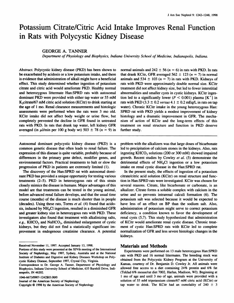
ResultsTable 1 and Figure 1 summarize the functional data in
normal rats and rats with PKD. There were no statistically
significant differences in rat age, body weight, or urine flow
rate (V) among the four groups of rats. The rats with PKD had
mean arterial BP significantly higher than the normal rats (P <
0.01), and these pressures were not affected by KCitr ingestion.
Rats with PKD that drank tap water had significantly lower
hematocrit levels than normal rats drinking tap water (P <
0.001); KCitr ingestion yielded a higher average hematocrit
level in rats with PKD, but the difference was not significant.
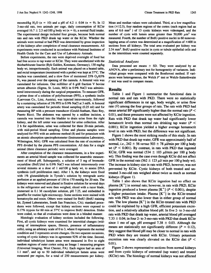
Figure 1 shows the most striking results of this study. In rats
with PKD that drank tap water, GFR was approximately half of
normal, i.e., 242 ± 56 versus 503 ± 78 pA/mm per 100 g body
wt (P < 0.001). By contrast, in rats with PKD that ingested
KCitr, GFR was normal (534 ± 103 j.tl/min per 100 g body
wt). This finding was the case even though KCitr did not affect
GFR in the normal rats (562 ± 123 jd/ mm per 100 g body wt).
The increase in kidney size in rats with PKD, however, was not
prevented by KCitr. Cystic kidneys of both treated and un-
treated 3-mo-old rats weighed about twice as much as normal
kidneys (Figure 1).
Table 1 also shows that KCitr ingestion had no effect on
plasma [K�] in normal rats; however, in rats with PKD, KCitr
ingestion produced a lower plasma [K�] (P < 0.001), despite
a higher potassium intake. Plasma [K�] in the KCitr-treated
rats with PKD was also lower than in either group of normal
rats. The low plasma [K�] in the KCitr-treated rats with PKD
could be explained by a high GFR, efficient potassium excre-
tion, and a relatively alkaline blood pH. In five 2- to 3-mo-old
rats with PKD that drank tap water, arterial blood pH averaged
7.33 ± 0.04; in four 2- to 3-mo-rats with PKD that drank KCitr
since 1 mo of age, pH averaged 7.38 ± 0.03. Although the
means are statistically not significantly different (P = 0. 12),
they suggest that blood pH may be closer to normal in rats with
PKD when they are treated with KCitr. Urinary potassium
excretion rate was clearly elevated on the KCitr diet (P <
0.01).
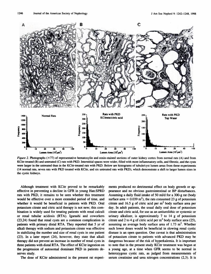
Figure 2 shows representative sections from normal kidneys
and from cystic kidneys of untreated (tap water) and treated
(KCitr) rats. The histology of normal kidneys was not affected
1244 Journal of the American Society of Nephrology J Am Soc Nephrol 9: 1242-1248, 1998
Table I. Functions in normal rats and rats with PKD�
Variable
Norm al Rats Rats w ith PKD
Tap Water KCitr Tap Water KCitr(;z=9) (ti=7) (n=6) (,i=7)
Ratage(days) 96±5 100±9 98±5 94±3
Body weight (g) 392 ± 15 384 ± 22 388 ± 23 363 ± 56
MABP(mmHg) 105±5 103±7 ll9±8h ll8±8’�
Hematocrit (% cells) 48 ± I 46 ± 1 43 ± IC 45 ± 2
V (p1/mm per 100 g body wt) 8.0 ± 3.8 6.0 ± 3.4 9.5 ± 2.4 10.1 ± 2.7
Plasma [K�] (mEqIL) 3.95 ± 0.19 3.71 ± 0.14 4.1 1 ± 0.20 3.32 ± 024h.d
K� excretion (�Eq/min) 1.25 ± 0.27 2.21 ± 0.71C 1.29 ± 0.23 2.30 ± 0.42e
a Values are means ± SD. Kidney data are for the left kidney. PKD, polycystic kidney disease: KCitr, potassium citrate/citric acid
solution; MABP, mean arterial BP; V. urine flow rate.h p < 0.01 compared with normal rats with same treatment.
C p < 0.001 compared with normal rats with same treatment.
(I p < 0.001 compared with untreated rats with PKD.,, P < 0.01 compared with rats on tap water.
by KCitr ingestion. In the rats with PKD, KCitr ingestion was
associated with a less abnormal interstitium and smaller cysts.
The histograms in Figure 2 show the distribution of tubule/cyst
lumen sizes in normal kidneys (pooled data), and in treated and
untreated rats with PKD. The histograms reveal a clear ten-
dency for lumen sizes to be larger in the cystic animals,
especially in the untreated (tap water) group. In the normal
kidneys, only 0.3% of the lumen areas were greater than 6000
�.tm2, whereas in the cystic kidneys, 15% (KCitr-treated rats)
and 24% (untreated rats) of lumen areas were greater than 6000
�tm.
Table 2 summarizes the histologic assessment of the kidneys
in animals with PKD. Interstitial changes were significantly
fewer in the KCitr-treated animals than in the untreated ani-
mals. Both the median and mean lumen areas were signifi-
cantly smaller in the rats treated with KCitr than in untreated
rats. The untreated rats also had a significantly greater number
of very large cysts compared with the KCitr-treated rats. Fi-
nally, the number of BrdU-positive interstitial cells was sig-
nificantly higher in the untreated rats than in the KCitr-treated
rats with PKD. This last result is consistent with the finding
that interstitial changes are more prominent in the untreated
rats and suggests increased proliferation of interstitial fibro-
blasts.
DiscussionThis study demonstrates that early treatment of Han:SPRD
rats with KCitr prevents the fall in GFR that normally accom-
panics PKD. This prevention is an important finding, because
it is the first treatment that preserves GFR in an animal model
of PKD without untoward side effects. This treatment also
lowers the plasma [K�] in rats with PKD, but does not prevent
hypertension or enlargement of the kidneys in 3-mo-old rats.
Unlike effects produced by ingesting 200 or 300 mM KHCO3
(4), KCitr ingestion has no detrimental effect on body weight.
Histologic evaluation of cystic kidneys reveals fewer intersti-
tial changes and smaller cysts in rats with PKD treated with
KCitr.
The remarkable beneficial effect of KCitr ingestion on renal
function in rats with PKD raises several important issues: What
is the mechanism of action of KCitr? How can we explain the
changes in GFR? Would KCitr have beneficial effects with
longer treatments, with lower doses, and in other species?
The mechanism of action of KCitr ingestion remains to be
defined. Potassium, citrate, and citric acid could be beneficial
for many reasons. First, citric acid is a metabolic substrate in
the kidneys (9) and so could increase energy production by
kidney cells. Second, citrate is a base and is converted, at least
in part, to bicarbonate in the body. It has the effect of alkalin-
izing the body fluids, including the urine. The alkalinizing
effect of citrate might counteract the metabolic acidosis that
frequently accompanies renal failure. Third, citrate/citric acid
administration results in decreased renal ammonia synthesis by
two mechanisms: a direct metabolic effect of citric acid (10)
and the alkalinizing effect of citrate. Increased ammonia syn-
thesis per nephron is thought to result in interstitial fibrosis in
a variety of renal diseases, possibly by activating the alterna-
tive complement pathway ( 1 1) or by favoring the formation of
long-lived oxidants such as chloramines ( I 2). Interstitial
changes are a prominent feature of the cystic kidney, and cyst
fluid samples from patients with PKD have elevated ammonia
levels ( 1 3). Ammonia increases the growth rate of cultured
cells ( 14). Alterations of metabolism associated with renal
ammoniagenesis could also have an impact on PKD (4).
Fourth, reduced citrate excretion is commonly seen in patients
with PKD. This reduction could contribute to the increased
incidence of renal stone disease in these patients ( 1 ) and to
development of obstruction, a factor that contributes to cyst
enlargement (15). In Han:SPRD rats, renal citrate excretion is,
surprisingly, higher in rats with PKD than in normal animals
( I 6). Therapy with citrate, by complexing calcium in the urine
or in renal tissue, could diminish renal stone formation and
r-P<O.001 I
I P<O.OO1
1.5�
1.0
0.5
NORMAL
J Am Soc Nephrol 9: 1242-1248. 1998 Citrate Treatment in PKD Rats 1245
2
CC
I
r-P<O.OO1 -1
PKDFigure 1. GFR and left kidney weight ± body weight in 3-mo-old
normal rats and in rats with polycystic kidney disease (PKD). Chronic
ingestion of a solution of potassium citrate/citric acid (KCitr) had no
effect on GFR in the normal animals, but resulted in a normal GFR in
the rats with PKD. Kidney weights were about doubled in the rats with
PKD, compared with normal rats. KCitr ingestion had no effect on
kidney weight.
deposition of insoluble calcium salts in the kidneys. Fifth,
citrate is an antioxidant; oxidant injury may play a role in the
progression of PKD ( 17). Sixth, ingestion of citrate influences
intestinal absorption of iron and trace elements (e.g. , alumi-
num, zinc) ( 18); whether this could affect the expression of
PKD is not known. Seventh, administration of a potassium salt
might alleviate intracellular acidosis and the associated in-
crease in renal ammonia synthesis.
Shohl ( 19) first demonstrated, more than 60 yr ago, that
administration of citrates to rats on a rachitogenic diet could
prevent rickets. Interestingly, sodium citrate alone or citric acid
alone did not cure the rickets, but a combination of citrate and
citric acid in the diet did. He concluded that the beneficial
effects of citrates were related to their specific properties and
not to acid-base effects alone. Although the study of Shohl was
done on a different disease, it seems reasonable to suggest that
the beneficial effects of KCitr in the present study, likewise,
are not only due to acid-base effects but also reflect the many
other actions of citrates in the body.
The maintenance of a normal GFR in the KCitr-treated rats
with PKD may seem puzzling, because these animals clearly
had prominent changes in renal structure. Both treated and
untreated rats with PKD had kidneys that were about twice the
normal size. There was less cystic enlargement and fewer
interstitial changes in the KCitr-treated animals than in the
untreated rats with PKD. It should be noted that GFR may be
normal, despite markedly enlarged cystic kidneys in both rats
( I 5,20) and people (2 1 ). The reason for the decline in GFR in
the untreated rats with PKD is most likely related to the severe
anatomical changes observed. These rats had kidneys with
marked interstitial damage and greatly enlarged cysts. With
widening of the interstitial spaces and more large cystic
nephrons per unit mass of kidney, normal nephrons must be
lost, explaining the reduced GFR. Indeed, when tubule/cyst
lumen areas were measured (Figure 2), more regions had to be
surveyed to count 250 lumens per kidney. Thus, there is clearly
a loss of patent nephrons in untreated cystic kidneys.
There are several important differences between this study
and an earlier study by Torres ci a!. (4) on the effects of alkali
therapy in Han:SPRD rats. This study used animals at 3 mo of
age, whereas Torres et a!. examined 2-mo-old animals. The
onset of treatment was about the same in both studies (3 to 4
wk of age). The different ages at which measurements were
made may explain why in the present work the treatment did
not result in a difference in kidney size (Figure 1 ). Studies in
this laboratory on five 2-mo-old rats with PKD show that KCitr
treatment is indeed associated with a smaller kidney size (un-
published data). This finding is consistent with the findings of
Torres et al. with other alkalinizing agents. Cowley et al. (2)
reported that kidney size in heterozygous male Han:SPRD rats
with PKD reaches a peak value at 8 wk (2 mo) of age and then
declines. Thus, the effect of a treatment on kidney size may
depend on rat age. Moreover, overall kidney size does not
indicate the functional state of cystic kidneys. Also, in the
present study, the histology of treated and untreated cystic
kidneys was different despite their being the same size.
Other differences from the study by Torres ci a!. (4) should
be noted. First, in the present study, KCitr treatment produced
a highly significant improvement, in fact, a normalization of
GFR. By contrast, bicarbonate therapy did not yield any sta-
tistically significant improvement in creatinine clearance (4).
Second, Torres et a!. used metabolic cages to collect urine and
calculated GFR from the endogenous creatinine clearance. In
the present study, GFR was determined by direct collection of
ureteral urine and by use of a synthetic inulin, polyfructosan,
allowing this determination to be more accurate (22). Third, the
absence of data on normal animals in the article by Torres et a!.
precludes judging how sick the animals with PKD were and
how much of an improvement the treatments produced. Fi-
nally, the KCitr treatment, unlike some treatments with bicar-
bonate salts (4), produced no untoward effects on the animals,
such as impaired growth or precipitation of calcium salts in the
kidneys.
B
1400 350
1200 300
;���IOO0 250
�8OO �200�. 600 ��I50
a.400 100
200 50
0 � � 2’O 3’O 4�l >50 0 10 20 30 40 >50 0 10 20 30 40 >50
Lumen Area (I03�m2) Lumen Area (1OMm2) Lumen Area ( I0’�un2)
Figure 2. Photographs ( X77) of representative hematoxylin and eosin-stained sections of outer kidney cortex from normal rats (A) and from
KCitr-treated (B) and untreated (C) rats with PKD. Interstitial spaces were wider, filled with more inflammatory cells, and fibrotic, and the cysts
were larger in the untreated than in the KCitr-treated rats with PKD. Below are histograms of tubule/cyst lumen areas from these experiments( 14 normal rats. seven rats with PKD treated with KCitr, and six untreated rats with PKD). which demonstrate a shift to larger lumen sizes in
the cystic kidneys.
Normal RatsRats with PKD
KCitrate/citric acid
L
Rats with PKD
Tap Water
L
250
200>�
� 150
�. 100
a.
50 L
1246 Journal of the American Society of Nephrology J Am Soc Nephrol 9: 1242-1248, 1998
Although treatment with KCitr proved to be remarkably
effective in preventing a decline in GFR in young Han:SPRD
rats with PKD, it remains to be seen whether this treatment
would be effective over a more extended period of time, and
whether it would be beneficial in patients with PKD. Oral
potassium citrate and citric acid therapy is not new; this com-
bination is widely used for treating patients with renal calculi
or renal tubular acidosis (RTA). Igarashi and coworkers
(23,24) found that renal cysts are a common complication in
patients with primary distal RTA. They reported that 5 yr of
alkali therapy with sodium and potassium citrate was effective
in stabilizing the number and size of renal cysts in one patient
(23). In a later report (24), however, they state that alkali
therapy did not prevent an increase in number of renal cysts in
three patients with distal RTA. The effect of KCitr ingestion on
the progression of autosomal dominant PKD in patients de-
serves study.
The dose of KCitr administered in the present rat experi-
ments produced no detrimental effect on body growth or ap-
pearance and no obvious gastrointestinal or BP disturbances.
Assuming a daily fluid intake of 50 ml/d for a 300-g rat (body
surface area 0.039 m2), the rats consumed 23 g of potassium
citrate and 16.5 g of citric acid per m2 body surface area per
day. In adult patients, the usual daily oral dose of potassium
citrate and citric acid, for use as an antiurolithic or systemic or
urinary alkalizer, is approximately 7 to 14 g of potassium
citrate and 2 to 4 g of citric acid per m2 body surface area (25),
assuming an average body surface area of 1 .73 m2. Whether
such lower doses would be beneficial in slowing renal cystic
disease is an open question. One caveat is that administration
of potassium citrate to patients with advanced PKD may be
dangerous because of the risk of hyperkalemia. It is important
to note that in the present study KCitr treatment was begun at
a young age, at a time when GFR seems to be normal in
heterozygous cystic rats, as judged from measurements of
serum creatinine and urea nitrogen concentrations (2,3). It is
J Am Soc Nephrol 9: 1242-1248, 1998 Citrate Treatment in PKD Rats 1247
Table 2. Histologic evaluation of kidneys from KCitr-treated and untreated rats with PKD�
ParameterTap Water
(no)KCitr
(n=7)P Value
Interstitial damage score 3.0 ± 0 2.3 ± 0.49 <0.01
Cyst/tubule lumen area (�.tm2)”
median 2340 ± 530 1734 ± 220 <0.02
mean 6529 ± 892 3534 ± 527 <0.001
No. of large cysts per 26-mm2 cortex’ 15 ± 6.6 6 ± 2.2 <0.01
BrdU-positive nuclei per 2.54-mm2 cortex’�
cyst/tubule cells 9 ± 12 2 ± 2.5 NS
interstitial cells 51 ± 30 13 ± 8.6 <0.001
a BrdU. bromodeoxyuridine. Other abbreviations as in Table I.1� In kidney sections from 14 normal rats, median tubule lumen area averaged 887 ± 170 �m2, and mean tubule lumen area averaged
I I 70 ± 227 �m2; no effect of KCitr ingestion was seen.
C Large cysts were defined as having a lumen cross section greater than 50,000 �m2.
d In kidney sections from 14 normal rats, the number of BrdU-positive nuclei per 2.54-mm2 cortex averaged I ± 2.1 for tubule
epithelial cells and 1 ± 2.0 for interstitial cells. No effect of KCitr ingestion was seen in the normal rats.
not known whether KCitr treatment of animals with more
advanced disease would be beneficial.
In conclusion, chronic intake of KCitr in young Han:SPRD
rats with PKD prevented the fall in GFR that is usually seen in
these animals, thus helping to maintain good renal function.
The kidneys, although enlarged, had less severe interstitial
damage and smaller cysts. If such striking effects on GFR were
to be seen in other species, this might prove to be a valuable
treatment for patients, because it is the decline in GFR that puts
PKD patients at peril. The mechanisms involved in the action
of KCitr remain to be elucidated.
AcknowledgmentsI am indebted to Dr. Judith A. Tanner for her encouragement and
help in analyzing the data. to Dr. James A. McAteer for preparing thehistologic slides and critiquing the manuscript, to Dr. Lynn R. Willis
for use of his atomic absorption spectrophotometer, to Dr. H. Glenn
Bohlen for use of his Image I system, and to the Polycystic Kidney
Research Foundation for grant support.
References1 . Martinez JR. Grantham II: Polycystic kidney disease: Etiology,
pathogenesis, and treatment. Dis Mon 41 : 695-765, 1995
2. Cowley BD Jr. Gudapaty S. Kraybill AL, Barash BD, Harding
MA, Calvet IP. Gattone VH II: Autosomal dominant polycystic
kidney disease in the rat. Kidney In! 43: 522-534, 1993
3. Schafer K, Gretz N, Bader M, Oberb#{228}umer I. Eckardt K-U, Kriz
W, Bachmann 5: Characterization of the Han:SPRD rat modelfor hereditary polycystic kidney disease. Kidney hit 46: 134-
152, 1994
4. Torres VE, Mujwid DK. Wilson DM, Holley KH: Renal cystic
disease and ammoniagenesis in Han:SPRD rats. J A�n Soc Neph-
rol 5: 1 193-1200, 1994
5. Cowley BD Jr. Grantham II. Muessel MI, Kraybill AL, Gattone
VH II: Modification of disease progression in rats with inheritedpolycystic kidney disease. Am J Kidney Dis 27: 865-879, 1996
6. Pak CYC: Citrate and renal calculi: An update. Miner Elect rol vte
Metab 20: 371-377, 1994
7. Torres VE, Young WF Jr. Offord KP, Hattery RR: Association of
hypokalemia, aldosteronism, and renal cysts. N EngI J Med 322:
345-351, 1990
8. Davidson WD, Sackner MA: Simplification of the anthrone
method for the determination of inulin in clearance studies. J Lab
CliiiMed 62: 351-356, 1963
9. Elhamri M, Martin M, Ferrier B, Baverel G: Substrate uptake and
utilization by the kidney of fed and starved rats in vivo. Rena!
Physiol Biochem 16: 31 1-324, 199310. Welbourne TC: Influence ofcitric acid administration upon renal
ammoniagenesis in the acidotic dog and rat. Can J Physiol
Phar,nacol 50: 883-889, 1972
I I . Nath KA, Hostetter MK, Hostetter TH: Pathophysiology of
chronic tubulo-interstitial disease in rats: Interactions of dietary
acid load, ammonia, and complement component C3. J Clin
Invest 76: 667-675, 1985
I 2. Nath KA, Fischereder M, Hostetter TH: The role of oxidants in
progressive renal injury. Kidney hit 45[Suppl 45]: 51 1 I-SI 15,
I 994
13. Torres yE, Keith DS, Offord KP, Kon SP, Wilson DM: Renal
ammonia in autosomal dominant polycystic kidney disease. Kid-
,iev hit 45: 1745-1753, 1994
14. Golchini K, Norman I, Bohman R, Kurtz I: Induction of hyper-
trophy in cultured proximal tubule cells by extracellular NH4C1.
J Clin lnvest 84: 1767-1779, 1989
15. Tanner GA, Gretz N, Connors BA, Evan AP, Steinhausen M:
Role of obstruction in autosomal dominant polycystic kidney
disease in rats. Kidney Int 50: 873-886, 1996
16. Ogborn MR. Sareen 5, Prychitko I. Buist R, Peeling I: Alteredorganic anion and osmolyte content and excretion in rat poly-
cystic kidney disease: An NMR study. Am J Pkvsiol 272: F63-
F69, 1997
17. Torres VE, Bengal Ri, Litwiller RD. Wilson DM: Aggravation
of polycystic kidney disease in Han:SPRD rats by buthionine
sulfoximine. J Am Soc Nephrol 8: 1283-1291, 1997
18. Ecelbarger CA, Greger IL: Dietary citrate and kidney function
affect aluminum, zinc and iron utilization in rats. J Nutr 121:
1755-1762, 1991
19. Shohl AT: The effect of the acid-base content ofthe diet upon theproduction and cure of rickets with special reference to citrates.
J Nutr 14: 69-83, 1937

1248 Journal of the American Society of Nephrology
20. Tanner GA, Gretz N, Shao Y, Evan AP, Steinhausen M: Organic
anion secretion in polycystic kidney disease. J Am Soc Nephrol
8: 1222-1231, 1997
21. Franz KA, Reubi FC: Rate of functional deterioration in poly-
cystic kidney disease. Kidney mt 23: 526-529, 1983
22. Namnum P. Insogna K, Baggish D, Hayslett IP: Evidence for
bidirectional net movement of creatinine in the rat kidney. Am J
Physiol 244: F719-F723, 1983
I Am Soc Nephrol 9: 1242-1248, 1998
23. Igarashi T, Shibuya K, Kamoshita 5, Higashihara E, Kawato H,
Hagishima K, Kosugi T: Renal cyst formation as a complication
ofprimary distal renal tubular acidosis. Nephron 59: 75-79, 1991
24. Igarashi T, Kosugi T: The incidence of renal cyst formation in
patients with primary distal renal tubular acidosis [Letter].
Nephron 66: 474, 1994
25. Physicians Desk Reference, 52nd Ed., Montvale, NJ, Medical
Economics, 1998, p 595


![Effect of synthesis conditions on the properties of citric ...ppong/manuscripts/Effect of...dently after nanoparticle synthesis [23]. Since the citrate ions are known to interfere](https://img.pdfslide.net/doc/110x75/60a60c90332852759401034c/effect-of-synthesis-conditions-on-the-properties-of-citric-ppongmanuscriptseffect.jpg)