Embed Size (px)
Citation preview
Practice microscope - toy or tool?
IN this article, Donald Mactaggart argues that the microscope ispossibly the most important tool in a veterinary practice. If usedto its full potential it is also one of the most cost-efficient. Treatit with the respect that it deserves and the practice will be amplyrepaid.
DONALD MACTAGGART
REQUIREMENTS OF APRACTICE MICROSCOPE
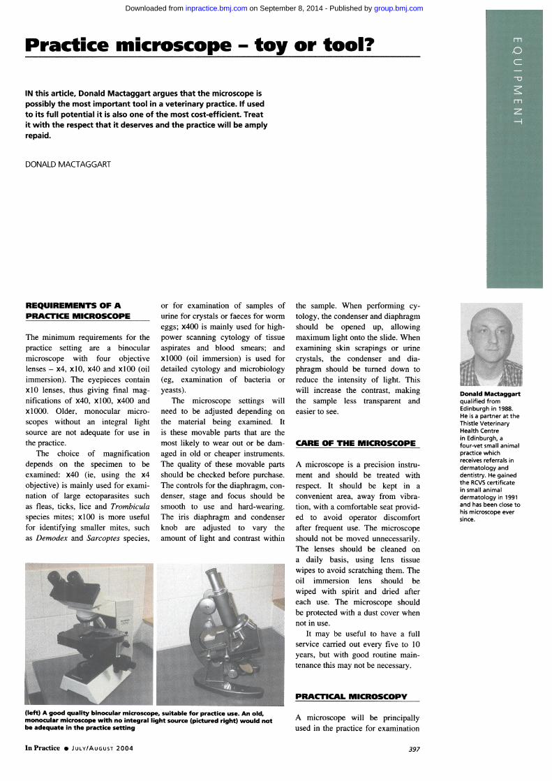
The minimum requirements for thepractice setting are a binocularmicroscope with four objectivelenses - x4, x lO, x40 and xlO0 (oilimmersion). The eyepieces containxIO lenses, thus giving final mag-nifications of x40, xlO0, x400 andxIO00. Older, monocular micro-scopes without an integral lightsource are not adequate for use inthe practice.
The choice of magnificationdepends on the specimen to beexamined: x40 (ie, using the x4objective) is mainly used for exami-nation of large ectoparasites suchas fleas, ticks, lice and Trombiculaspecies mites; xlO0 is more usefulfor identifying smaller mites, suchas Demodex and Sarcoptes species,
or for examination of samples ofurine for crystals or faeces for wormeggs; x400 is mainly used for high-power scanning cytology of tissueaspirates and blood smears; andx00 (oil immersion) is used fordetailed cytology and microbiology(eg, examination of bacteria oryeasts).
The microscope settings willneed to be adjusted depending onthe material being examined. Itis these movable parts that are themost likely to wear out or be dam-aged in old or cheaper instruments.The quality of these movable partsshould be checked before purchase.The controls for the diaphragm, con-denser, stage and focus should besmooth to use and hard-wearing.The iris diaphragm and condenserknob are adjusted to vary theamount of light and contrast within
the sample. When performing cy-tology, the condenser and diaphragmshould be opened up, allowingmaximum light onto the slide. Whenexamining skin scrapings or urinecrystals, the condenser and dia-phragm should be turned down toreduce the intensity of light. Thiswill increase the contrast, makingthe sample less transparent andeasier to see.
CARE OF THE MICROSCOPE
A microscope is a precision instru-ment and should be treated withrespect. It should be kept in aconvenient area, away from vibra-tion, with a comfortable seat provid-ed to avoid operator discomfortafter frequent use. The microscopeshould not be moved unnecessarily.The lenses should be cleaned ona daily basis, using lens tissuewipes to avoid scratching them. Theoil immersion lens should bewiped with spirit and dried aftereach use. The microscope shouldbe protected with a dust cover whennot in use.
It may be useful to have a fullservice carried out every five to 10years, but with good routine main-tenance this may not be necessary.
Donald Mactaggartqualified fromEdinburgh in 1988.He is a partner at theThistle VeterinaryHealth Centrein Edinburgh, afour-vet small animalpractice whichreceives referrals indermatology anddentistry. He gainedthe RCVS certificatein small animaldermatology in 1991and has been close tohis microscope eversince.
PRACTICAL MICROSCOPY
(left) A good quality binocular microscope, suitable for practice use. An old,monocular microscope with no integral light source (pictured right) would notbe adequate in the practice setting
A microscope will be principallyused in the practice for examination
In Practice S JULY/AUGUST 2004 397
group.bmj.com on September 8, 2014 - Published by inpractice.bmj.comDownloaded from
Successfulmicroscopy
Successful microscopy dependsupon good sample collection,a good quality microscope andgood interpretation/diagnosticskills.
One of the main advantagesof examining samples micro-scopically yourself is the rapidresult. Another advantage isthat one knows if the samplecollected is of good qualitystraight away and, if it isnot, it is simple to collect an-
other one. The main limitingfactor is your own diagnos-tic skills, but these will im-prove with experience. If indoubt, send the sample off to
a more experienced clinicianfor assessment.
of skini scrlpings. loi cytology, atid
foi exa miniIIIngl- salmliples ol urillne.
faeces an1d blood.
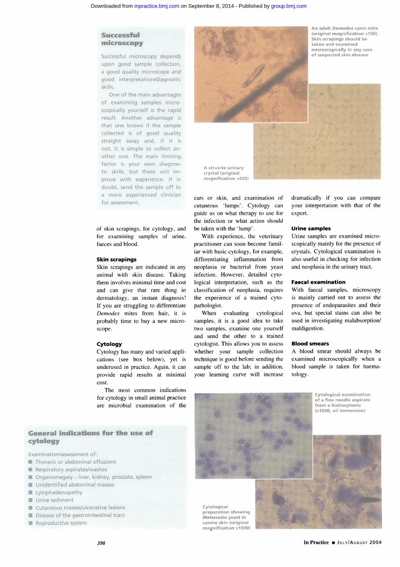
Skin scrapingsSkin scraping2-s ai e indicated in anx
anliml ith skiin disease. TaLkinel
theminilolxcs minimiial time anid cost
Ltnd c tln ix C thiat iare thine, in
deI -imaItologx ati inistaiit diae1nosis.
If Nxou are stitlLuclinel to dilfererntiatet)coiodex. mites Iro mii hail-. it is
probably timc to bux a ne1x micio
scoipe.
CytologyCvtologx has manvx Cand xvaried appli-cattioins (see box belo\x ). v et is
unldel-lsed in practice. Again. it can
proxide rapid results at inimililalcost.
The iriost COtiltilOil iiidicattionS
tor cvtologv in smaill animal priactice
are imicrobial examiiiatiion of the
General indications for the use ofcytology
Examination/assessment of:* Thoracic or abdominal effusions* Respiratory aspirates/washes* Organomegaly - liver, kidney, prostate, spleen
c.ars or skinl. 'and examination olCutaieouS 'lLmsCMtIC lo g0 x CanenLlidIe uS on1 wxhat thei lp> to LISC forthe linlcctioll or Wxllhlt aCtioli shouIldbe tlaken xxith the luMp
WN!ith CXpCIricnce, the x eterlina V
practitionlel cAll soon becomile tamlill-lar w ith basic cv tologx br cxamlplC,ditfei entiatin,e inlammationi fromilncoplasil 01' hacterial l'Iroml veCastinitcetiotn. Howex Cvr. dletailed cy tologicall intelrpretatioll. sLiChi as tileclassification of neoplasia. rIeCluii esthe cxperience otf a trained cx to-
pathologist.When C\caluatin,eT cvtolooical
samples. it is a tJood idceL to taketxNo s.aLmlples.examiiinle o(le yLoursellfaind send the other to a trainedcytologist. This aIlloxxs on to assessxxhethei ' oIur samplC collectiontechlliqueI Is good befor-e sending thesample oIlf to the labb; in addition.our learninLg Icuie V ill increatse
diramiiaticallv if NrouL Calnl COm11parleVonLI intelprettationll with thalt of thecxpei t.
Urine samplesUi-inic saamples aie examui-ined micio-scopically mainlixl for the presencce ofcixvstails. Cy tological exam-itia-tioll isaIlso iseltSil In checkitie fOr infeCtionaniid n-eoplasica inl the IL-itiiary tr-act.
Faecal examinationWith tfaecal saMiples. mici oscopyis iminixI carried out to assess thepiCescicc of endopairasites aLncl theiloxVa, but special stainis canilalso beulsed inl ilnx'stigaltir I labsoi-ptioni/-ialdilestioll.
Blood smearsA blood simieai- shouldc alxaxs beexamiiniedt microscopiczillx xxwhen a
blood samilple is takenl foi hcaemca-toloov.
* Unidentified abdominal masses
Lymphadenopathy* Urine sediment* Cutaneous masses/ulcerative lesions* Disease of the gastrointestinal tract* Reproductive system
In Practice 0 JULY/AUGUST 2004398
group.bmj.com on September 8, 2014 - Published by inpractice.bmj.comDownloaded from
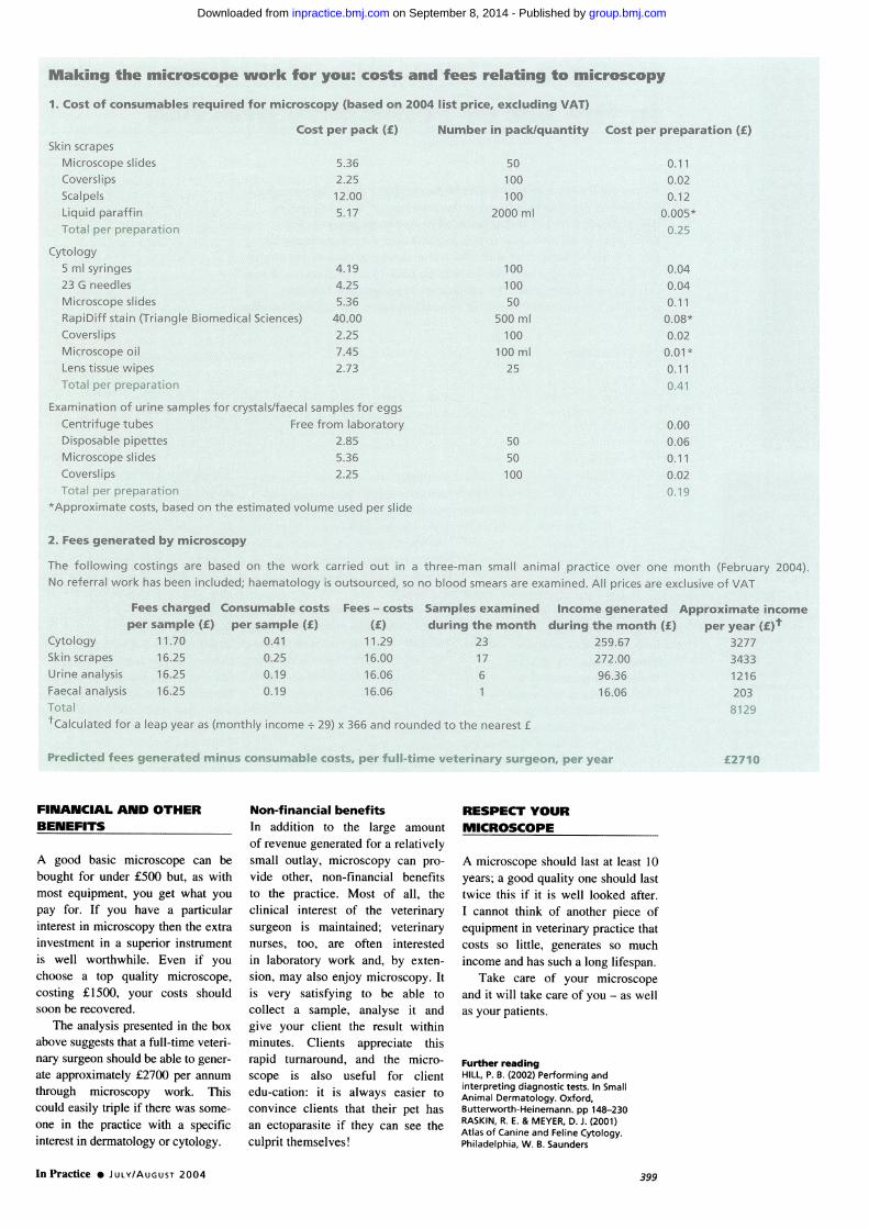
Making the microscope work for you: costs and fees relating to microscopy1. Cost of consumables required for microscopy (based on 2004 list price, excluding VAT)
Skin scrapesMicroscope slidesCoverslipsScalpelsLiquid paraffinTotal per preparation
Cost per pack (£)
5.362.2512.005.17
Number in pack/quantity Cost per preparation (£)
50100100
2000 ml
0.110.020.12
0.005*0.25
Cytology5 ml syringes23 G needlesMicroscope slidesRapiDiff stain (Triangle Biomedical Sciences)CoverslipsMicroscope oilLens tissue wipesTotal per preparation
4.194.255.3640.002.257.452.73
Examination of urine samples for crystals/faecal samples for eggsCentrifuge tubes Free from laboratoryDisposable pipettes 2.85Microscope slides 5.36Coverslips 2.25Total per preparation
*Approximate costs, based on the estimated volume used per slide
10010050
500 ml100
100 ml25
5050100
0.040.040.110.08*0.020.01*0.110.41
0.000.060.110.020.19
2. Fees generated by microscopy
The following costings are based on the work carried out in a three-man small animal practice over one month (February 2004).No referral work has been included; haematology is outsourced, so no blood smears are examined. All prices are exclusive of VAT
Fees charged Consumable costs Fees - costs Samples examined Income generated Approximate incomeper sample (£) per sample (£) (f) during the month during the month (£) per year (£)t
Cytology 11.70 0.41 11.29 23 259.67 3277Skin scrapes 16.25 0.25 16.00 17 272.00 3433Urine analysis 16.25 0.19 16.06 6 96.36 1216Faecal analysis 16.25 0.19 16.06 1 16.06 203Total 8129tCalculated for a leap year as (monthly income + 29) x 366 and rounded to the nearest f
Predicted fees generated minus consumable costs, per full-time veterinary surgeon, per year £2710
FINANCIAL AND OTHERBENEFITS
A good basic microscope can bebought for under £500 but, as withmost equipment, you get what youpay for. If you have a particularinterest in microscopy then the extrainvestment in a superior instrumentis well worthwhile. Even if youchoose a top quality microscope,costing £1500, your costs shouldsoon be recovered.
The analysis presented in the boxabove suggests that a full-time veteri-nary surgeon should be able to gener-ate approximately £2700 per annumthrough microscopy work. Thiscould easily triple if there was some-one in the practice with a specificinterest in dermatology or cytology.
Non-financial benefitsIn addition to the large amountof revenue generated for a relativelysmall outlay, microscopy can pro-vide other, non-financial benefitsto the practice. Most of all, theclinical interest of the veterinarysurgeon is maintained; veterinarynurses, too, are often interestedin laboratory work and, by exten-sion, may also enjoy microscopy. Itis very satisfying to be able tocollect a sample, analyse it andgive your client the result withinminutes. Clients appreciate thisrapid turnaround, and the micro-scope is also useful for clientedu-cation: it is always easier toconvince clients that their pet hasan ectoparasite if they can see theculprit themselves!
RESPECT YOURMICROSCOPE
A microscope should last at least 10years; a good quality one should lasttwice this if it is well looked after.I cannot think of another piece ofequipment in veterinary practice thatcosts so little, generates so muchincome and has such a long lifespan.
Take care of your microscopeand it will take care of you - as wellas your patients.
Further readingHILL, P. B. (2002) Performing andinterpreting diagnostic tests. In SmallAnimal Dermatology. Oxford,Butterworth-Heinemann. pp 148-230RASKIN, R. E. & MEYER, D. J. (2001)Atlas of Canine and Feline Cytology.Philadelphia, W. B. Saunders
In Practice * JULY/AUGUST 2004 399
group.bmj.com on September 8, 2014 - Published by inpractice.bmj.comDownloaded from
doi: 10.1136/inpract.26.7.397 2004 26: 397-399In Practice
Donald Mactaggart Practice microscope - toy or tool?
http://inpractice.bmj.com/content/26/7/397Updated information and services can be found at:
These include:
serviceEmail alerting
the box at the top right corner of the online article.Receive free email alerts when new articles cite this article. Sign up in
Notes
http://group.bmj.com/group/rights-licensing/permissionsTo request permissions go to:
http://journals.bmj.com/cgi/reprintformTo order reprints go to:
http://group.bmj.com/subscribe/To subscribe to BMJ go to:
group.bmj.com on September 8, 2014 - Published by inpractice.bmj.comDownloaded from