Embed Size (px)
Citation preview
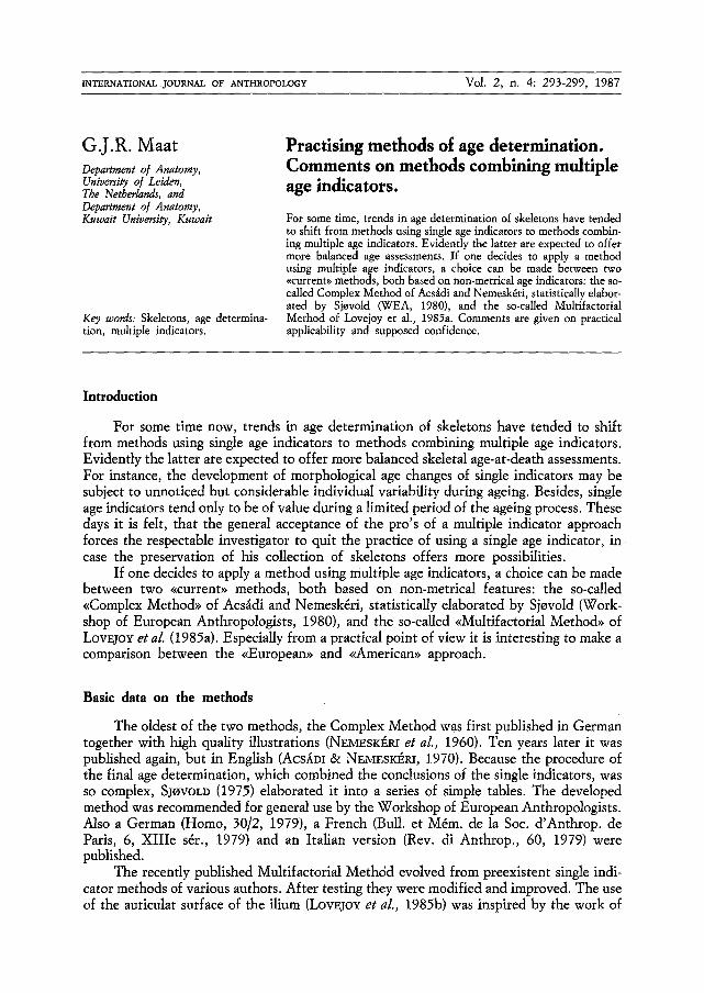
INTERNATIONAL JOURNAL OF ANTHROPOLOGY Vol. 2, n. 4: 293-299, 1987
G.J.R. Maat Department o/ Anatomy, University of Leiden, The Netherlands, and Department o[ Anatomy, Kuwait University, Kuwait
Key words: Skeletons, age determina- tion, multiple indicators.
Practising methods of age determination. Comments on methods combining multiple age indicators.
For some time, trends in age determination of skeletons have tended to shift from methods using single age indicators to methods combin- ing multiple age indicators. Evidently the latter are expected to offer more balanced age assessments. If one decides to apply a method using multiple age indicators, a choice can be made between two ~<currenb> methods, both based on non-metrical age indicators: the so- called Complex Method of Acsfidi and Nemesk6ri, statistically elabor- ated by Sjovold (WEA, 1980), and the so-called Multifactorial Method of Lovejoy et al., 1985a. Comments are given on practical applicability and supposed confidence.
Introduction
For some time now, trends in age determination of skeletons have tended to shift from methods using single age indicators to methods combining multiple age indicators. Evidently the latter are expected to offer more balanced skeletal age-at-death assessments. For instance, the development of morphological age changes of single indicators may be subject to unnoticed but considerable individual variability during ageing. Besides, single age indicators tend only to be of value during a limited period of the ageing process. These days it is felt, that the general acceptance of the pro's of a multiple indicator approach forces the respectable investigator to quit the practice of using a single age indicator, in case the preservation of his collection of skeletons offers more possibilities.
If one decides to apply a method using multiple age indicators, a choice can be made between two <~current>> methods, both based on non-metrical features: the so-called <<Complex Method>> of Acsfidi and Nemesk6ri, statistically elaborated by Sjovold (Work- shop of European Anthropologists, 1980), and the so-called <<Multifactorial Method>> of LOVEJOu et al. (1985a). Especially from a practical point of view it is interesting to make a comparison between the <~European>> and <<American>> approach.
Basic data on the methods
The oldest of the two methods, the Complex Method was first published in German together with high quality illustrations (NEMESK~RI et al., 1960). Ten years later it was published again, but in English (Acs~m & NEMESK~RI, 1970). Because the procedure of the final age determination, which combined the conclusions of the single indicators, was so complex, SJOVOLD (1975) elaborated it into a series of simple tables. The developed method was recommended for general use by the Workshop of European Anthropologists. Also a German (Homo, 30/2, 1979), a French (Bull. et M6m. de la Soc. d'Anthrop~ de Paris, 6, XIIIe s6r., 1979) and an Italian version (Rev. di Anthrop., 60, 1979) were published.
The recently published Mnltifactorial Meth6d evolved from preexistent single indi- cator methods of various authors. After testing they were modified and improved. The use of the auricular surface of the ilium (LOVEJOY et al., 1985b) was inspired by the work of
294 MAAT
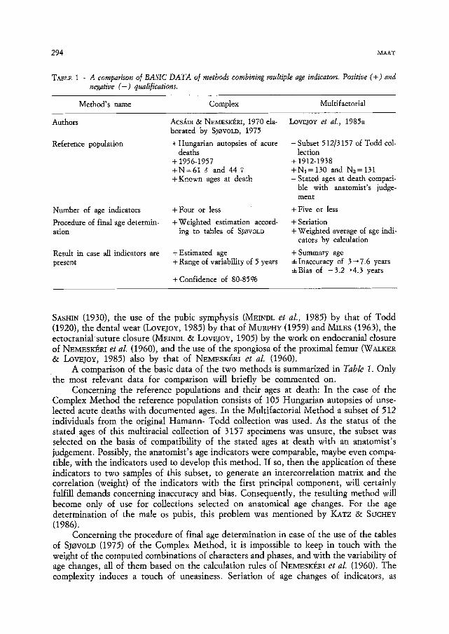
TABLE 1 - A comparison of BASIC DATA of methods combining multiple age indicators. Positive (+) and negative ( - ) qualifications.
Method's name Complex Multifactorial
AcsltDi & NEMESKI~RI, 1970 ela- LovEJoY et al., 1985a borated by SJ~VOLD, 1975
+ Hungarian autopsies of acute deaths
+ 1956-1957 +N=618 and 44 + Known ages at death
Authors
Reference population
Number of age indicators
Procedure of final age determin- ation
Result in case all indicators are present
+ Four or less +Weighted estimation accord-
ing to tables of SJOVOLD
+ Estimated age + Range of variability of 5 years
+Confidence of 80-85%
-Subset 512/3157 of Todd col- lection
+ 1912-1938 +N1=130 and Nz=131 - Stated ages at death compati-
ble with anatomist's judge- ment
+ Five or less
+ Seriation + Weighted average of age indi-
cators by calculation
+ Summary age +Inaccuracy of 3-*7.6 years • of -3.2-.4.3 years
SASHIN (1930), the use of the pubic symphysis (MEINDL et al., 1985) by that of Todd (1920), the dental wear (LovEjoY, 1985) by that of MURVHY (1959) and MINES (1963), the ectocranial suture closure (MEINDL & LOVEJOY, 1905) by the work on endocranial closure of NEMESK~RI et al. (1960), and the use of the spongiosa of the proximal femur (WALKER & LOVEJOY, 1985) also by that of NEMESK~RI et al. (1960).
A comparison of the basic data of the two methods is summarized in Table 1. Only the most relevant data for comparison will briefly be commented on.
Concerning the reference populations and their ages at death: In the case of the Complex Method the reference population consists of 105 Hungarian autopsies of unse- lected acute deaths with documented ages. In the Multifactorial Method a subset of 512 individuals from the original Hamann- Todd collection was used. As the status of the stated ages of this multiracial collection of 3157 specimens was unsure, the subset was selected on the basis of compatibility of the stated ages at death with an anatomist's judgement. Possibly, the anatomist's age indicators were comparable, maybe even compa- tible, with the indicators used to develop this method. If so, then the application of these indicators to two samples of this subset, to generate an intercorrelation matrix and the correlation (weight) of the indicators with the first principal component, will certainly fulfill demands concerning inaccuracy and bias. Consequently, the resulting method will become only of use for collections selected on anatomical age changes. For the age determination of the male os pubis, this problem was mentioned by KATZ & SUCHEY (1986).
Concerning the procedure of final age determination in case of the use of the tables of SJOVOLD (1975) of the Complex Method, it is impossible to keep in touch with the weight of the computed combinations of characters and phases, and with the variability of age changes, all of them based on the calculation rules of NEMESK~RI et al. (1960). The complexity induces a touch of uneasiness. Seriation of age changes of indicators, as
PRACTISING METHODS OF AGE DETERMINATION 295
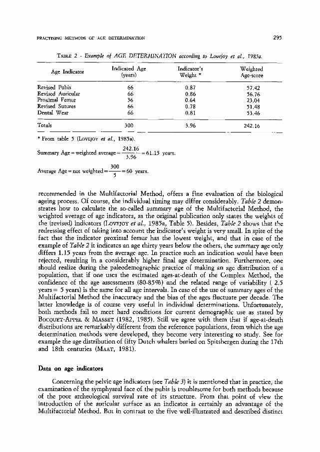
TABLE 2 - Example o[ AGE DETERMINATION according to Love]oy et aL, 1985a.
Indicated Age Indicator's Weighted Age Indicator (years) Weight * Age-score
Revised Pubis 66 0.87 57.42 Revised Auricular 66 0.86 56.76 Proximal Femur 36 0.64 23.04 Revised Sutures 66 0.78 51.48 Dental Wear 66 0.81 53.46
Totals 300 3.96 242.16
* From table 5 (LovEjoY et aL, 1985a).
Summary Age = weighted average = 242.16
, =61.15 years. 3.96
300 Average Age = not weighted = = 60 years.
5
recommended in the Multifactorial Method, offers a fine evaluation of the biological ageing process. Of course, the individual timing may differ considerably. Table 2 demon- strates how to calculate the so-called summary age of the Multifactorial Method, the weighted average of age indicators, as the original publication only states the weights of the (revised) indicators (LovEjoY et al., 1985a, Table 5). Besides, Table 2 shows that the redressing effect of taking into account the indicator's weight is very small. In spite of the fact that the indicator proximal femur has the lowest weight, and that in Case of the example of Table 2 it indicates an age thirty years below the others, the summary age only differs 1.15 years from the average age. In practice such an indication would have been rejected, resulting in a considerably higher final age determination. Furthermore, one should realize during the paleodemographic practice of making an age distribution of a population, that if one uses the estimated ages-at-death of the Complex Method, the confidence of the age assessments (80-85%) and the related range of variability ( 2.5 years = 5 years) is the same for all age intervals. In case of the use of summary ages of the Multifactorial Method the inaccuracy and the bias of the ages fluctuate per decade. The latter knowledge is of course very useful in individual determinations. Unfortunately, both methods fail to meet hard conditions for current demographic use as stated by BOCQUET-APPEL &; MASSET (1982, 1985). Still we agree with them that if age-at-death distributions are remarkably different from the reference populations, from which the age determination methods were developed, they become very interesting to study. See for example the age distribution of fifty Dutch whalers buried on Spitsbergen during the 17th and 18th centuries (MAAT, 1981).
Data on age indicators
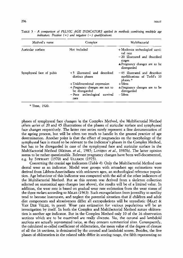
Concerning the pelvic age indicators (see Table 3) it is mentioned that in practice, the examination of the symphyseal face of the pubis is troublesome for both methods because of the poor archeological survival rate of its structure. From that point of view the introduction of the auricular surface as an indicator is certainly an advantage of the Multifactorial Method. But in contrast to the five well-illustrated and described distinct
296 MAAT
TABLE 3 - A comparison of PELVIC AGE INDICATORS applied in methods combining multiple age indicators. Positive (+) and negative ( - ) qualifications.
Method's name Complex Multifactorial
Auricular surface Not included + Moderate archeological survi- val rate
-20 illustrated and described stages
*Pregnancy changes are to be disregarded
Symphyseal face of pubis + 5 illustrated and described -45 ilXustrated and describes distinct phases modifications of Todd's 10
phases * + Unidimensional expression + Idem + Pregnancy changes are not to .4-Pregnancy changes are to be
be disregarded disregarded - Poor archeological survival - Idem
rate
* TODD, 1920.
phases of symphyseal face changes in the Complex Method, the Multifactorial Method offers series of 20 and 45 illustrations of the phases of auricular surface and symphyseal face changes respectively. The latter two series surely represent a fine documentation of the ageing process, hut will be often too much to handle in the general practice of age determination. Another point is that the effect of pregnancies on the morphology of the symphyseal face is stated to be relevant to the indicator's phases in the Complex Method, but has to be disregarded in case of the symphyseal face and auricular surface in the Multifactorial Method (MEII~DL et al., 1985; LovEjoY et al., 1985b). The latter opinion seems to be rather questionable. Relevant pregnancy changes have been well-documented, e.g. by STEWART (1970) and ULLV, ICH (1975).
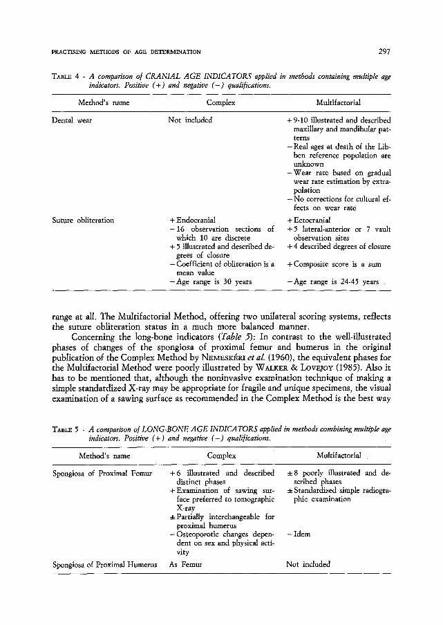
Concerning the cranial age indicators (Table 4): Only the Multifactorial Method uses dental wear as an indicator. Modal wear groups with attendant age estimations were derived from Libben-Amerindians with unknown ages, an archeological reference popula- tion. Age behaviour of this indicator was computed with the aid of the other indicators of the Multifactorial Method. But as this system was derived from a skeleton collection selected on anatomical ages changes (see above), the results will be of a limited value. In addition, the wear rate is based on gradual wear rate estimation from the wear status of the three molars according to MILES (1963). Such extrapolations from juvenility to senility tend to become inaccurate, and neglect the potential situation that if children and adult diet components and abrasiveness differ all extrapolations will be unrealistic (MAAT & VAN DER VELDE, in press). Wear rate estimation for various populations will be an investigation by itself. In both the Complex and Multifactorial Method suture oblitera- tion is another age indicator. But in the Complex Method only 10 of the 16 observation sections which are to be examined are really discrete. Six, the coronal and lambdoid sections are actually represented twice, as they concern symmetrical sites. Consequently, the calculated so-called coefficient of obliteration, the mean value of the degree of closure of all the 16 sections, is dominated by the coronal and lambdoid scores. Besides, the five phases of obliteration in the related table differ in scoring range, the fifth representing no
PRACTISING METHODS OF AGE DETERMINATION 297
TABLE 4 - A comparison o/ CRANIAL AGE INDICATORS applied in methods containing multiple age indicators. Positive (+) and negative ( - ) qualifications.
Method's name Complex Multifactorial
Dental wear Not included + 9-10 illustrated and described
Suture obliteration + Endocranial - 1 6 observation sections of
which 10 are discrete + 5 illustrated and described de-
grees of closure - Coefficient of obliteration is a
mean value -Age range is 30 years
maxillary and mandibular pat- terns
-Real ages at death of the Lib- ben reference population are unknown
-Wear rate based on gradual wear rate estimation by extra- polation
- N o corrections for cultural ef- fects on wear rate
+ Ectocranial +5 lateral-anterior or 7 Vault
observation sites + 4 described degrees of closure
+ Composite score is a sum
-Age range is 24-45 years
range at all. The Multifactorial Method, offering two unilateral scoring systems, reflects the suture obliteration status in a much more balanced manner.
Concerning the long-bone indicators (Table 5): In contrast to the well-illustrated phases of changes of the spongiosa of proximal femur and humerus in the original publication of the Complex Method by NV_~ESK~ et al. (1960), the equivalent phases for the Multifactorial Method were poorly illustrated by WALKER & LOVEJOY (1985). Also it has to be mentioned that, although the noninvasive examination technique of making a simple standardized X-ray may be appropriate for fragile and unique specimens, the visual examination of a sawing surface as recommended in the Complex Method is the best way
TABLE 5 - A comparison o/LONG-BONE AGE INDICATORS applied in methods combining multiple age indicators. Positive (+) and negative ( - ) qualifications.
Method's name Complex Multifactorial
Spongiosa of Proximal Femur + 6 illustrated and described • 8 poorly illustrated and de- distinct phases scribed phases
+Examination of sawing sur- q-Standardized simple radiogra- face preferred to tomographic phic examination X-ray
4-Partially interchangeable for proximal humerus
- Osteoporotic changes depen- - Idem dent on sex and physical acti- vity
Spongiosa of Proximal Humerus As Femur Not included
298 MAAT
to assess age. Both methods are affected by sex dependent (postmenopausal) and physical activity dependent osteoporotic changes. Such processes are well-described in the paleo- pathological literature (e.g. STEINBOCK, 1976; ORTNER & PUTSCHAR, 1981). Still, reports on sex and locomotor effects remain contradictory. Finally, in the Complex Method both the femur and humerus are applied, resulting in a considerable overlap in diagnostic power.
Concluding remarks
Although the application of the age indicators as proposed in both the Complex and Multifactorial Method has certain disadvantages, it is without doubt that methods combining multiple indicators will appear to be superior to single indicator systems. From a practical point of view the 26 year old, but updated Complex Method seems preferable, as its reference population represents unselected individual variability in a population, and as its age indicators are simpler to apply. In most cases the stated degrees of confidence of the age determinations of bo th methods will be acceptable for individual age assessments; they seem to be inadequate for paleodemographic use. Seriation, of limited value in reconstructing chronological calendar ages, is of extreme paleopathologic interest as an aid to reconstruct sequences of biological ageing.
References
ACS.~DI G. & NEMSK~RI J., 1970. History of human life span and mortality. Budapest. BOCQUET-APPEL J.-P. & MASSET C., 1982. Farewell to Paleodemography. J. of Hum. Evol., 11: 321-333. BOCQUET-APPEL J.-P. & MASSET C., 1985. Matters of moment. J. of Hum. Evol., 14: 107-111. KATZ D. & MEYERS SUCHEY J., 1986. Age determination of the male os pubis. Am. J. Phys. Anthrop., 69:
427-435. LovEJo~c C. O., 1985. Dental wear in the Libben population: its functional pattern and role in the
determination of adult skeletal age at death. Am. J. Phys. Anthrop., 68: 47-56. LovEjoY C. O., Meindl R. S., Mensforth R. P. & Barton Th. J., 1985a. Multi[actorial determination of
skeletal age at death: a method and blind tests of its accuracy. Am. J. Phys. Anthrop., 68: 1-14. LOVEJOY C. O., MEIm)L R. S., PaVZUECK TH. R. & MENSFORTH R. P., 1985b. Chronological metamorphosis
of the auricular surtace of the ilium: a new method/or the determination of adult skeletal age at death. Am. J. Phys. Anthrop., 68: 15-28.
MAAT G. J. R., 1981. Human remains at the Dutch whaling stations on Spitsbergen. A physical anthropological study. In: Van Holk, A. G. F., 's-Jacob, H. K. and Temmingh, A. A. H. J. (eds.) - Early European exploitation of the Northern Atlantic 800-1700. Groningen.
MAAT G. J. R. & VAN DER VELDE E.A. The caries attrition competition. Int. J. of Anthropol., In press. MEIm)L R. S. & LowJoY C. O., 1985. Ectocranial suture closure: a revised method for the determination of
skeletal age at death based on the lateral-anterior sutures. Am. J. Phys. Anthrop., 68: 57-66. MEINDL R. S., LOVEJOY C. O., MENSFOaTH R. P. & WAH:ER R. A., 1985. A revised method of age
determination using the os pubis, with a review and tests of accuracy of other current methods of pubic symphyseal aging. Am. J. Phys. Anthrop., 68: 2%45.
MILES A. E. W., 1963. Dentition in the assessment of individual age in skeletal material. In: Brothwell, D. R. (ed.). Dental anthropology, Oxford.
MURPHY, T., 1959. The changing pattern of dentine exposure in human tooth attrition. Am. J. Phys. Anthrop., 17: 167-178.
NEMESK~RI J., HARS~NYI L. & ACS,(DI G., 1960. Methoden zur Diagnose des Lebensalters yon Skelet[unden. Anthrop. Anz., 24: 70-95.
OaTNEa D. J. & PUTSCHAR W. G. J., 1981. Identification of pathological conditions in human skeletal remains. Washington.
SASHIN D., 1930. A critical analysis of the anatomy and pathologic changes of sacro-iliac joints. J. Bone Jt. Surg., 33A: 119-130.
PRACTISING METHODS OF AGE DETERMINATION 299
SJOVOLD T., 1975. Tables of the combined method for determination of age at death given by Nemesk&i, Harsdnyi and Acsddi. Anthrop. K6zl., 19: 9-22.
STEINBOCK R. T., 1976. Paleopathological diagnosis and interpretation. Springfield. STEWART T. D., 1970. Identification of the scars of parturition in the skeletal remains of females. In: Stewart
T. D. (ed.). Personal identification in mass disasters. Washington. TODD T. W., 1920. Age changes in the pubic bone: I the white male pubis. Am~ J. Phys. Anthrop., 3: 285-
334. ULLRICH H., 1975. Estimation of fertility by means of pregnancy and childbirth alterations at the pubis, the
ilium and the sacrum. Ossa, 2: 23-39. WALKER R. A. & LovEJoY C. O., 1985. Radiographic changes in the clavicle and proximal femur and their use
in the determination of skeletal age at death. Am. J. Phys. Anthrop., 68: 67-78. WORKSHOP OF EUROPEAN ANTHROPOLOCISTS, 1980. Recommendations for age and sex diagnoses of skeletons.
J. Hum. Evol., 9: 517-549.
Received: Nov. 15 th, 1986. Accepted: May 20 th, 1987.