Embed Size (px)
Citation preview
356
TRANSACTIONS OF THE ROYAL Socran OF TROPICAL MEDICINE AND HYGIENE (1994) 88, 356
Presence of annular rings on Mansonella ozzardimicrofilariae
Dave D. Chadeel, Samuel C. Rawlins*, R. Doonl and S. Baboolal* ‘Insect Vector Control Division, Ministry of Health, 3 Queen Street, St Joseph, Trinidad; Xaribbean l?&demiology Centre, P.O. Box 164, Port of Spain, Trini-
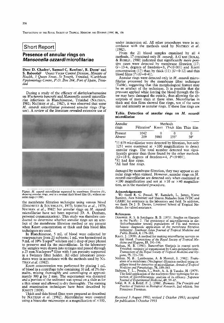
During a study of the efficacy of diethylcarbamazine on Wuchereria bancrofti and Mansonella ozzardi microfila- riae infections in Blanchisseuse, Trinidad (NATHAN, 1981; NATHAN et al., 1982), it was observed that some M. ozzardi microfilariae possessed annular rings (Fig- ure). A review of the literature revealed extensive use of
Figure. M. ozzardi microfilariae separated by membrane filtration (A), showing annular rings, and in a normal thick blood film (B), without an- nular rings (X 100).
the membrane filtration technique using venous blood (DESOWITZ & SOUTHGATE, 1973; SHIBUYA et al., 1979; NATHAN et al., 1982) but annular rings on M. ozzardi microtilariae have not been reported (D. A. Denham, personal communication). This study was therefore con- ducted to determine whether annular rings are an arte- fact of the membrane filtration method or are present when Knott concentration or thick and thin blood film techniques are used.
In Blanchisseuse, 5 mL of blood were collected by venepuncture from 2 1 subjects; 1 mL was haemolysed in 9 mL of 10% TeepoP solution and 1 drop of pure phenol to preserve and fix the microfilariae. In the laboratory the samples were drawn into syringes and passed through a 25 mm Nuclepore@ filter with 3 urn porosity supported in a Swinney filter holder. All other laboratory proce- dures were in accordance with the methods used by NA- THAN et al. (1982).
Knott concentration was performed by placing 1 mL of blood in a centrifuge tube containing 10 mL of 2% for- malin, mixing thoroughly and centrifuging at approxi- mately 300 g for 2 min. The supernatant was decanted and a portion of the sediment was spread on a slide as for a thin smear and allowed to dry thoroughly. The staining and examination techniques have been described by KNOTT (1939).
Thick and thin blood films were prepared as described by NATHAN et al. (1982). Microtilariae were counted using a binocular microscope at a magnification of x 100,
under immersion oil. All other procedures were in ac- cordance with the methods used by NATHAN et al. (1982).
From the 21 blood samples examined by all 4 methods, 17 contained only M. ozzardi. A G test (SOKAL & ROHLF, 1980) indicated that significantly more posi- tive cases were detected by membrane filtration (17; G=13.4, degrees of freedom=3, P<O.OOl) and Knott concentration (17) than by thick (11) (G=0+12) and thin blood films (7) (G=0.41).
Annular rings were detected only in M. ozzardi micro- filariae processed by the membrane filter technique (Table), suggesting that this morphological feature may be an artefact of the technique. It is possible that the pressure applied while forcing the blood through the fil- ter may have damaged the cuticle, thus allowing the ab- sorption of more stain at these sites. Microfilariae in thick and thin films showed fine rings, not of the same size and intensity as annular rings. If these fine rings are
Table. Detection of annular rings on M. ozzardi microfilariae
Annular Methods rings Filtration” Knott Thick film Thin film
Present 1042 Absent 209 598: 13% 5:’
“11 619 microfilariae were detected by filtration, but only 1251 were examined at x100 magnification to detect annular rings. The total number detected was signi- ficantly greater than those found by the other methods (G=18.8, degrees of freedom=4, P<O.OOl).
b31 had fine rings. ‘All had fine rings.
damaged by membrane filtration, they may appear as an- nular rings when stained. However, annular rings on M. ozzardi microfilariae are detected only when examined at x 100 magnification and not at X 10 or x40 magnifica- tion, as in the standard procedure.
Acknowledgements We thank R. C. Persad, W. Ramdath, L. James, Nurse
Ramirez, Alex Nimblett and Sharon Mahadeo from IVDC and CAREC for assistance in the laboratory and field. In addition, we thank Dr J. B. Davies, Liverpool School of Tropical Me- dicine, for valued assistance.
References Desowitz, R. S. & Southgate, B. B. (1973). Studies on filariasis
in the Pacific: 2. The persistence of microtilaremia in die- thylcarbamazine treated populations of Fiji and western Samoa: diagnostic application of the membrane filtration technique. Southeast Asian Journal of Tropical Medicine and Public Health, 4, 179-183.
Knott, J. (1939). A method for making microfilariae surveys on day blood. Transactions of the Royal Society of Tropical Me- dicine and Hygiene, 33, 191-196.
Nathan, M. B. (1981). Bancroftian filariasis in coastal north Trinidad: intensity of transmission by Culexquinquefasciatus. Transactions of the Royal Society of Tropical Medicine and Hy- giene, 75721-730.
Nathan, M. B., Lambourne, A. & Monteil, S. (1982). Evalu- ation of a membrane (Nuclepore) filtration method using ca- pillary blood for detection of microfilariae. Annals of Tropical Medicine and Parasitology, 76, 339-345,
Shibuya, T. L., Penaia, L., Buck, A. A. &Tanaka, H. (1979). The field application of the nuclepore filter technique for de- tection of microlilaraemia: analysis of its efficacy. Japanese Journal of Experimental Medicine, 49, 59-65.
Sokal, R. R. & Rohlf, F. J. (1980). Biometry. The Principle and Practice of Statistics in Biological Research, 2nd edition. San Francisco: Freeman.
Received 3 August 1993; revised 1 October 1993; accepted for publication 5 October 1993