Embed Size (px)
Citation preview
98 THE JOURNAL OF UROLOGY® Vol. 179, No. 4, Supplement, Sunday, May 18, 2008
were used to calculate crude and adjusted odds ratios (OR) controlling for estimated gestational age (EGA).
RESULTS: The median detectable maternal PBB level at enrollment was 3.0 parts per billion (ppb) among younger sons (ages 5-18) and 3.5 ppb among older sons (ages 18-30). Of sons with available maternal PBB levels (n=464), 35 reported any GU condition (13 hernias, 10 hydroceles, 9 cryptorchidism, 5 hypospadias, 2 phimosis and 1 varicocele). Four reported both hernia & hydrocele and one both hernia & cryptorchidism. 9.3% of younger sons and 5.6% of older sons reported any GU condition. Sons born to mothers with the highest PBB levels (>5 ppb) were more likely to report any GU condition than those born to mothers with the lowest levels (12.2% vs. 5.5%). In GEE models, sons with estimated maternal PBB at conception > 5 ppb were nearly four times as likely to report any GU condition compared with the least exposed (<1 ppb) (OR=3.53, 95% CI: 1.27 - 9.84). Individuals with estimated maternal PBB at conception 1-5 ppb were not at a statistically
4.34). Birth weight and EGA neither differed by maternal PBB level nor confounded our analyses.
such study to document an increased incidence of male GU conditions associated with PBB exposure, provides support for the hypothesis that in utero EDC exposure may play a role in the secular increase in male GU conditions.
Source of Funding: US Environmental Protection Agency(R 825300), National Institute of Environmental Health Sciences (RO1 ES08341, R01 ES12014) and Centers for Disease Control and Prevention cooperative agreement U37/CCU500392.
278IDENTIFICATION OF THE FIRST FUNCTIONALLY SIGNIFICANT MUTATION IN THE GENE FOR THE INSULIN-3 RECEPTOR, LGR8, WHICH CAUSES FAMILIAL CRYPTORCHIDISMYi Wang*, Maggy Fina, Shaohua Zhang, Agneta Nordenskjold, Ronald Taussig, Linda A Baker. Dallas, TX, and Stockholm, Sweden.
INTRODUCTION AND OBJECTIVE: Cryptorchidism is familial in 14% of cases, suggesting a genetic basis. The testis hormone INSL3 and its receptor, LGR8, are key mediators in murine and human testicular descent. Knockout mice for Insl-3 or the Insl-3 receptor (LGR8) demonstrate bilateral intraabdominal cryptorchidism. While several mutations in INSL3 causing cryptorchidism have been reported
population of cryptorchid males with a positive family history (+FHx) of cryptorchidism for LGR8 mutations.
METHODS: Lymphocyte genomic DNA from 113 cryptorchid
and coding regions of the human LGR8 genes and products were sequenced. Control data were obtained from the NCBI SNPs database.
mutations were tested following transfection of the mutant receptors into HEK293 cells, and subsequent assessment of cAMP production in response to stimulation with INSL3.
RESULTS: Four novel missense mutations (D76G, R532G,I604V, and A620T) were found in 1, 2, 12, and 2 patients, respectively. In addition, 4 silent SNPs (A87A, Q210Q, E319E, and L331L) and one
(I604V) were detected. Most notably, the D76G mutant is functionally inactive, whereas the R532G, I604V, and A620T LGR8 receptors, as
are activated by INSL3 to stimulate cAMP indistinguishable from wild type receptor.
LGR8 mutation in a cryptorchid child, D76G, located in the LDL-A module of the protein. This location is absolutely conserved across species. Our work refutes that the previously reported T222P mutation is functionally
caused by alterations in the Insl3/LGR8 signaling pathway.Source of Funding: NIH R01 HD48838 (Baker, LA).
279THE MANAGEMENT OF UNILATERAL NONPALPABLE TESTIS: HOW TO MANAGE THE BOYS WITH BLIND ENDING VESSELS ON INITIAL LAPAROSCOPYYuichiro Yamazaki*, Mari Suzuki, Yoshiyuki Shiroyanagi, Daisuke Matsuno, Yukichi Tanaka. Yokohama, Japan.
INTRODUCTION AND OBJECTIVE: Initial laparoscopic assessment has been widely accepted in the management of unilateral nonpalpable testis (UNT). It is commonly believed that the testis is truly absent when the testicular vessels disappear above the closed internal ring and subsequent inguinal exploration is not indicated. However, it is not clear the absent testis means absent of any kind of viable testicular tissue. The aim of this study is to verify the anatomy of UNT with blind ending vessels.
METHODS: We retrospectively reviewed 80 consecutive cases with UNT in the last 4-year period. Laparoscopy was initially performed and we checked the testis position, patency of the processus vaginalis (PV), character of the vas deferens and presence/absence
subsequent inguinal exploration was basically performed except when both vessels and a vas disappeared above the internal ring. In case of inguinal exploration, any kind of remnant or nubbin was removed and evaluated histologically. According to the character of PV and
ending vessels into 4 categories and investigated presence/absence of testicular remnant.
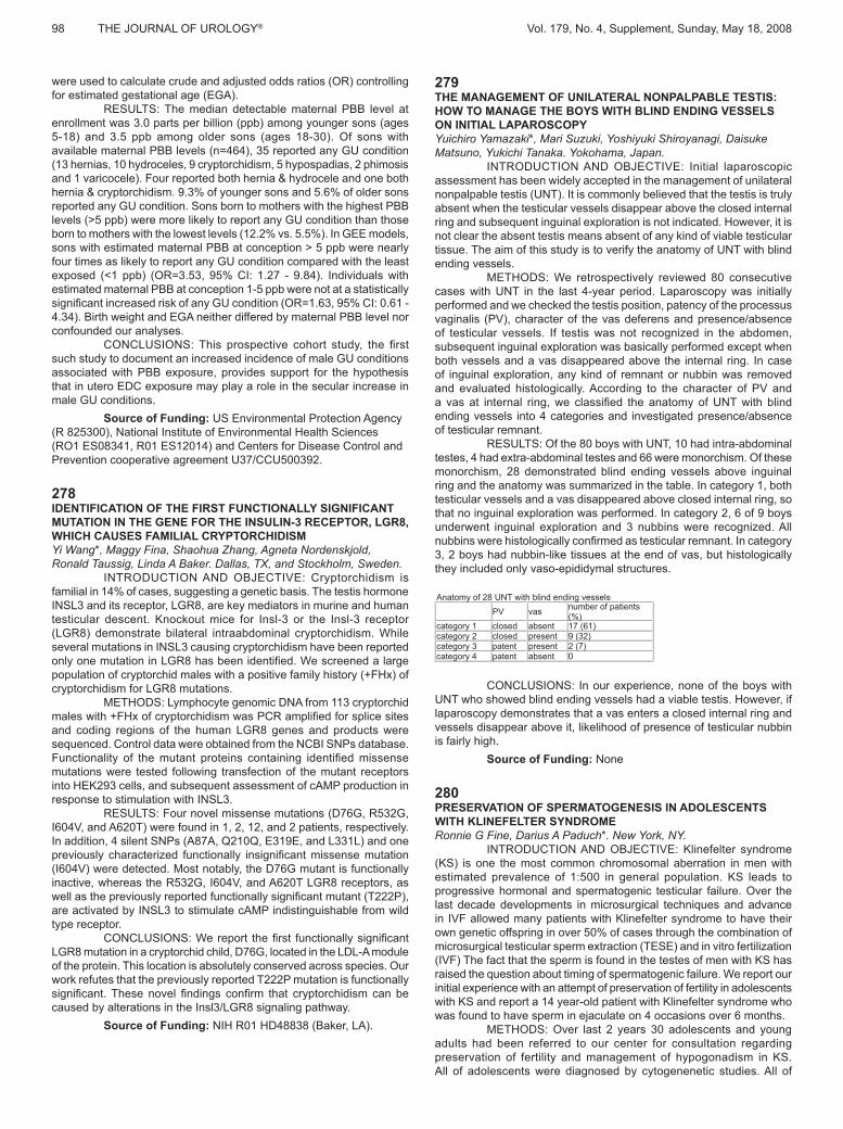
RESULTS: Of the 80 boys with UNT, 10 had intra-abdominal testes, 4 had extra-abdominal testes and 66 were monorchism. Of these monorchism, 28 demonstrated blind ending vessels above inguinal
testicular vessels and a vas disappeared above closed internal ring, so that no inguinal exploration was performed. In category 2, 6 of 9 boys
3, 2 boys had nubbin-like tissues at the end of vas, but histologically they included only vaso-epididymal structures.
Anatomy of 28 UNT with blind ending vesselsPV vas number of patients
(%)category 1 closed absent 17 (61)category 2 closed present 9 (32)category 3 patent present 2 (7)category 4 patent absent 0
CONCLUSIONS: In our experience, none of the boys with UNT who showed blind ending vessels had a viable testis. However, if laparoscopy demonstrates that a vas enters a closed internal ring and vessels disappear above it, likelihood of presence of testicular nubbin is fairly high.
Source of Funding: None
280PRESERVATION OF SPERMATOGENESIS IN ADOLESCENTS WITH KLINEFELTER SYNDROMERonnie G Fine, Darius A Paduch*. New York, NY.
INTRODUCTION AND OBJECTIVE: Klinefelter syndrome (KS) is one the most common chromosomal aberration in men with estimated prevalence of 1:500 in general population. KS leads to progressive hormonal and spermatogenic testicular failure. Over the last decade developments in microsurgical techniques and advance in IVF allowed many patients with Klinefelter syndrome to have their own genetic offspring in over 50% of cases through the combination of
(IVF) The fact that the sperm is found in the testes of men with KS has raised the question about timing of spermatogenic failure. We report our initial experience with an attempt of preservation of fertility in adolescents with KS and report a 14 year-old patient with Klinefelter syndrome who was found to have sperm in ejaculate on 4 occasions over 6 months.
METHODS: Over last 2 years 30 adolescents and young adults had been referred to our center for consultation regarding preservation of fertility and management of hypogonadism in KS. All of adolescents were diagnosed by cytogenenetic studies. All of
Vol. 179, No. 4, Supplement, Sunday, May 18, 2008 THE JOURNAL OF UROLOGY® 99
adolescents and parents were aware of the fertility preservation program and requested to have semen analysis performed as a part of the initial consultation. Each patient underwent full physical examination with anthropometric measurements, Tanner stage (TS) assessment, and full hormonal work-up.
RESULTS: Out of 30 patients only one had sperm in to ejaculate. The 14 year old patient who had sperm in ejaculate was TS stage II/III, testosterone naive, and with natural production of testosterone
months he preserved total of 6 vials with concentration of 1 mil/ml each and post-thaw motility of 2 %. None of the other patients had sperm in
sperm in ejaculate was the level of FSH <20 and TS III or less. This
by one of patient who was given oral testosterone in outside institution around the age of 10. This patient initiated puberty much earlier than his older brother, and his FSH and LH 45 and 30 and although he was only 12 he was fully matured.
CONCLUSIONS: Our report seems to support existing testicular biopsy data indicating that progressive loss of spermatogenesis occurs during early puberty. We believe that further research will help us to establish optimal timing and methods for preservation of fertility in adolescents with KS.
Source of Funding: Unrestricted research support to Dr. Paduch was provided by KS&A.
281CORRECTION OF PENILE TORSION ASSOCIATED WITH HYPOSPADIAS BY MOBILIZATION OF URETHRAL PLATE AND URETHRA: A NEW TECHNIQUEAmilal Bhat*. Bikaner, India.
INTRODUCTION AND OBJECTIVE: Severe penile torsion is managed by penile de-gloving & re-attaching of skin, incising the base
counter torque. The objective of the study was to assess the feasibility of correction of torsion by urethral plate with corpus spongiosum and
METHODS: We managed 31 cases of congenital penile torsion managed from 1999 to June 2007. Among these 23 had hypospadias (distal penile14, mid penile 6, and proximal penile 3), and 8 cases were of chordee without hypospadias. Age of patients varied from 1 year to twenty-three years with the average of 6 years and 8.5 months. Correction of torsion was done step-by-step by:
1.Penile skin de-gloving.
to corona.
glans.
RESULTS: The torsion varied from 30o-110o with an average of 48.07o (67.74% left & 32.26 % right). In distal penile hypospadias ( 14 cases ) the torsion varied from 30o to 110o( average 51.43o) , 80 % of them had mild chordee and 20 % had severe chordee, and left : right was 62.48 % : 37.52%. In the mid penile (6 cases) torsion varied from 30o to 90o (average 46.66o), two third had left side and one third had right side and all of them had mild chordee. In proximal hypospadias (3 cases) two third had severe chordee and left side torsion of 30o and one third had mild chordee with 30 o torsion of right side. In chordee without hypospadias (8 cases) torsion varied from 30o to 70o with average of 44o,80% of them had moderate chordee and 20 % had mild chordee and left to right ratio was 3:1. Correction of torsion was possible in 93.13%
spongiosum (25.80%), proximal urethra (25.80%), and urethral plate / hypoplastic urethra with spongiosum in to glans (25.80%).
urethra up to bulb and distally urethral plate /hypoplastic urethra with corpus spongiosum into glans) corrects the torsion in most of the cases. The technique is as per anatomical principles of surgery and is
simple, safe and effective technique for correction of both torsion and chordee.
Source of Funding: None
282ECONOMIC ANALYSIS OF INFANT VERSUS POST-PUBERTAL ORCHIDOPEXY TO PREVENT TESTIS CANCERMichael H Hsieh*, David R Roth, Maxwell V Meng. Houston, TX, and San Francisco, CA.
INTRODUCTION AND OBJECTIVE: Recent studies suggest prepubertal orchidopexy may confer additional protection from development of testis cancer compared to post-pubertal orchidopexy. Infant surgery is often performed by pediatric subspecialists, with a potential for increased costs compared to later orchidopexy. Testis
versus late orchidopexy with regards to development and treatment of testis cancer.
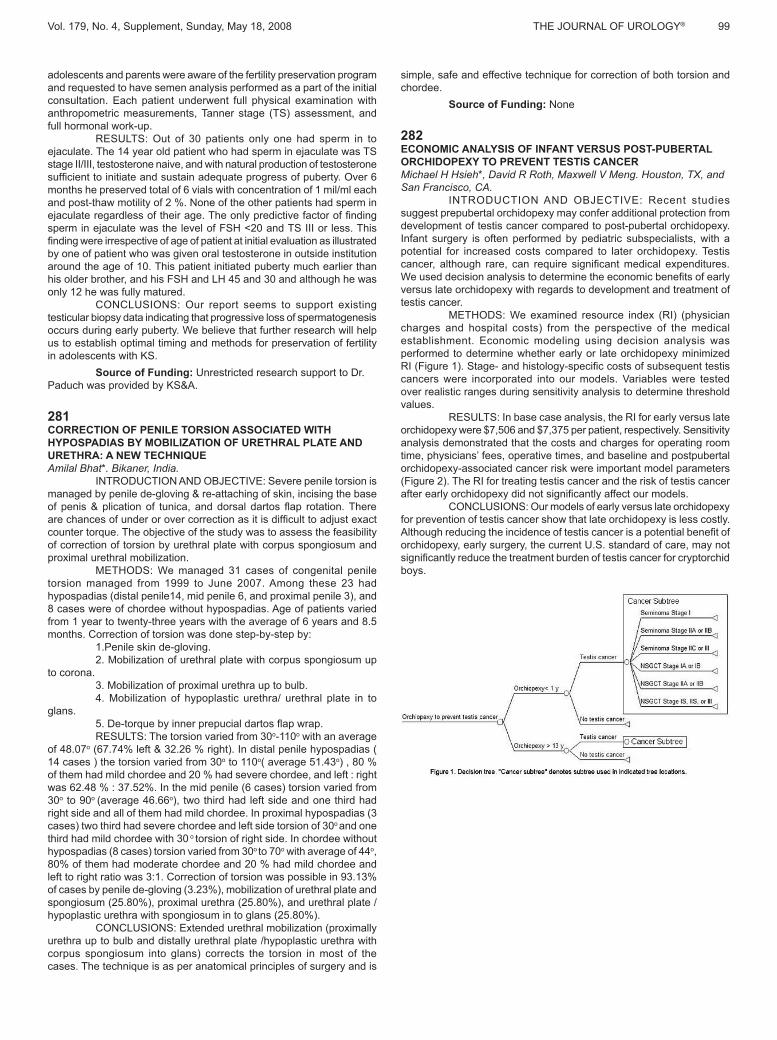
METHODS: We examined resource index (RI) (physician charges and hospital costs) from the perspective of the medical establishment. Economic modeling using decision analysis was
cancers were incorporated into our models. Variables were tested over realistic ranges during sensitivity analysis to determine threshold values.
RESULTS: In base case analysis, the RI for early versus late
analysis demonstrated that the costs and charges for operating room time, physicians’ fees, operative times, and baseline and postpubertal orchidopexy-associated cancer risk were important model parameters (Figure 2). The RI for treating testis cancer and the risk of testis cancer
CONCLUSIONS: Our models of early versus late orchidopexy for prevention of testis cancer show that late orchidopexy is less costly.
orchidopexy, early surgery, the current U.S. standard of care, may not
boys.