Embed Size (px)
Citation preview
PREVALENCE OF PNEUMOCYSTIS CARIN'11 PM l MOM A INHUMAN IMMUNODEFFICIENCY VIRUS SEROPOSITIVEINFANTS AND YOUNG CHILDREN ADMITTED WITH SEVEREPNEUMONIA AT THE KENYA I I A NATIONAL HOSPITAL.
MEDICAL LIBRARY 1SIVERSITY OF NAIROBI
iity of NAIROBI Library
A dissertation presented in part fulfillment for the
Degree o f Masters o f Medicine (Paediatrics)
In the University o f Nairobi
MEDICAL LIBRARY W1IVERSITY OF NAIROBI
By:
Dr. Judith Z S Rose M.B.Ch.B (NBI) Department of Paediatrics University of Nairobi
II

DECLARATION
I declare that this dissertation is my original work and has not been published elsewhere or presented for a degree in any otherUniversity. Signed: Date: °l§L>ts4-
Dr. Judith Z. Kose . MBChB (NBI)This dissertation has been submitted to the University of Nairobiwith our approval as University Supervisors.
Signed: Date: I T 1 T w t . 3 a o tDr. D. Mbori - Ngacha M.B.Ch.B., M.Med (Paed), MPH Senior Lecturer, Dept, of Paediatrics.University of Nairobi,
Sighed: ( L * ( * 1 ! ■ D.te:Dr. Chakaya J Muhwa.M.B.Ch.B., M.Med, DThM.
Chest physician/ Principal Research Officer, CRDR, KEMRI.
Ill
DEDICATION
TO ALL THE CHILDREN AFFECTED AND AFFLICTED BY HIV/AIDS
IV

a c k n o w l e d g e m e n t s
I would like to express my sincere appreciation to the following: -1. My supervisors Dr. D. Mbori-Ngacha and Dr. J.M. Chakaya for their
guidance, patience and support from the start to the end of the study.2. Ms. C. Bii, Mr. T. Ouko and the other staff at the mycology laboratory
at the CRDR, KEMRI for the assistance with part of the laboratory work and their moral support.
3. Statisticians Janet Musia, Francis and Ben Muchoki for the statistical support.
4. Ms. Carolyn Maringa of the Regional Aids Training Network (RATN) for her support with literature search and subsequent reviews.
5. All the children and their mothers who participated in this study.6. Finally to my family, especially my husband James and the cY
for all the support, especially Financially and morally, that complete the study. *
To all of you, Asante and God’s abundant blessings.
V

a c k n o w l e d g e m e n t s
I would like to express my sincere appreciation to the following: -1. My supervisors Dr. D. Mbori-Ngacha and Dr. J.M. Chakaya for their
guidance, patience and support from the start to the end of the study.2. Ms. C. Bii, Mr. T. Ouko and the other staff at the mycology laboratory
at the CRDR, KEMRI for the assistance with part of the laboratory work and their moral support.
3. Statisticians Janet Musia, Francis and Ben Muchoki for the statistical support.
4. Ms. Carolyn Maringa of the Regional Aids Training Network (RATN) for her support with literature search and subsequent reviews.
5. All the children and their mothers who participated in this study.6. Finally to my family, especially my husband James and the children
for all the support, especially financially and morally, that enabled me complete the study. ‘
To all of you, Asante and God’s abundant blessings.
V
TabIc of Contents PageTitle .................................................................... IDeclaration.......................................................... IllDedication ...................................................................IVAcknowledgements................................................ VTable of Contents.................................................... VIList of abbreviations................................................ VIIList of figures and tables......................................... VIIIAbstract.................................................................... IXIntroduction and Literature review............................ 1Study objectives and justification............................... 17Methodology................................................................ 18Results......................................................................... 26Discussion.................................................................... 39Conclusion......................................................................47Recommendations............ :...........................................47References......................................................................48Appendix 1 .....................................................................58
VI
I ,ist of AbbreviationsKNH Kenyatla National 1 lospilalBAL. B ro n c h o a i v co 1 a r 1 a v ageNPA Nasopharyngeal aspirateIS Induced sputumHIV Human Immunodeficiency VirusCMV CytomegalovirusAIDS Acquired Immune Deficiency Syndrome.PCR Polymerase chain reactionPCP Pneumocystis carinii pneumoniaIF I m m un o 11 u o re see neeCYT - Cytological staining techniqueKEMRI - Kenya Medical Research InstituteCRDR Center for Respiratory Diseases ResearchWHO - World I lealth Organization.ICU Intensive Care UnitTMP-SMX - Trimelhoprim/Sulpamelhoxa/.ole combi
Ml
UST OF FIGURES AND TABLES PACE
Figure 1: The age distribution of the HIV seropositive childrenadmitted with severe pneumonia........................................... 26
Figure 2a: Arterial oxygen saturation distribution for the HIV seropositivechildren admitted with severe pneumonia.............................................33Figure 2b: Comparison of median and the spread of oxygen saturation and distribution between the HIV seropositive children admitted with severepneumonia with and without PCP.........................................................34Table 1: Characteristics of HIV seropositive children with PCP admittedwith severe pneumonia.............................................................................28Table 2: Mortality in HIV seropositive children admitted with severepneumonia by PCP status........................................................................35Table 3: I Tnivariate analysis of the correlates of death in the HIVseropositive children with PCP, admitted with severe pneumonia.........36
VIII
ABSTRACT
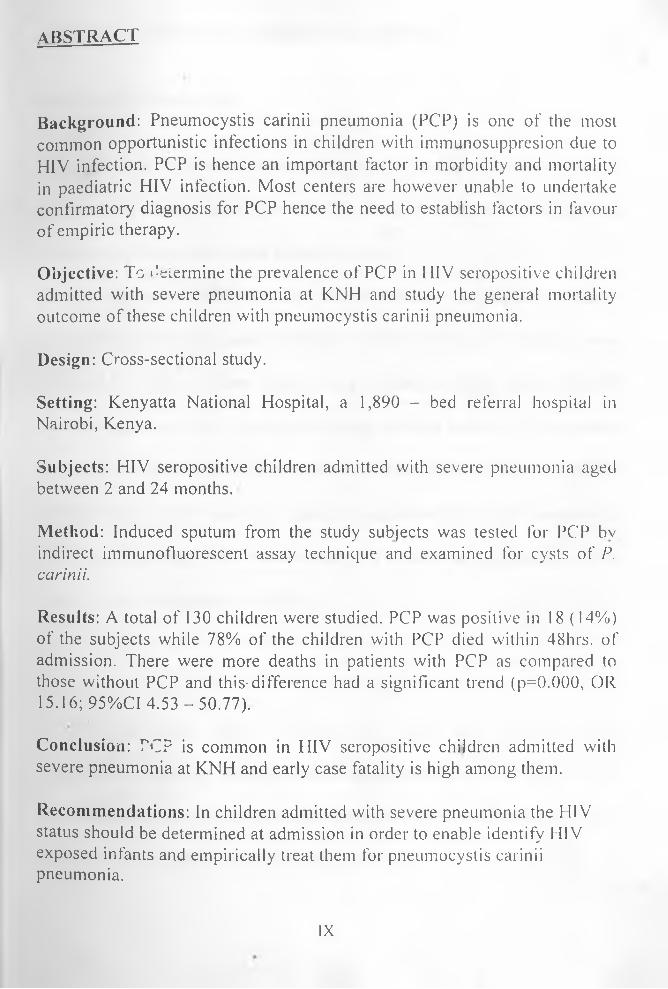
Background: Pneumocystis carinii pneumonia (PCP) is one of the most common opportunistic infections in children with immunosuppresion due to HIV infection. PCP is hence an important factor in morbidity and mortality in paediatric HIV infection. Most centers are however unable to undertake confirmatory diagnosis for PCP hence the need to establish factors in favour of empiric therapy.Objective: To determine the prevalence of PCP in HIV seropositive children admitted with severe pneumonia at KNH and study the general mortality outcome of these children with pneumocystis carinii pneumonia.Design: Cross-sectional study.Setting: Kenyatta National Hospital, a 1,890 - bed referral hospital in Nairobi, Kenya.Subjects: HIV seropositive children admitted with severe pneumonia aged between 2 and 24 months.Method: Induced sputum from the study subjects was tested for PCP bv indirect immunofluorescent assay technique and examined for cysts of P. carinii.Results: A total of 130 children were studied. PCP was positive in 18 (14%) of the subjects while 78% of the children with PCP died within 48hrs. of admission. There were more deaths in patients with PCP as compared to those without PCP and this-difference had a significant trend (p=0.000, OR 15.16; 95%CI 4.53 -50.77).Conclusion: PC? is common in HIV seropositive children admitted with severe pneumonia at KNH and early case fatality is high among them.Recommendations: In children admitted with severe pneumonia the HIV status should be determined at admission in order to enable identify HIV exposed infants and empirically treat them for pneumocystis carinii pneumonia.
IX
INTRODUCTION AND LITERATURE REVIEW
HIV/AIDS Epidemiology
Infection with the Human Immunodeficiency Virus (HIV) and the Acquired Immune Deficiency Syndrome (AIDS) that it causes is a fast growing epidemic in Kenya and indeed the whole world.Global estimates of the HIV/AIDS epidemic indicates that in 2002, a total of 5.0 million people were newly infected with HIV, of which 4.2 million were adults, 2.0 million of these adults being women. In 2002, about 800,000 children aged less than 15 years were newly infected with HIV. The number of people living with HIV/AIDS by the end of 2002 was 42 million with 3.2 million being children aged less than 15 years. The HIV/AIDS mortality in 2002 was 3.1 million with women being 1.2 million and children aged less than 15 years being about 610,000^ 2j.
In the major urban areas of Kenya like Nairobi and Mombasa, HIV seroprevalence among antenatal women increased from 2% in 1985 to 19% in 1995 but the prevalence showed a decrease to 13% in 2002 and 1 1% in 2003 in Nairobi and 16% in Mombasa in 2003 [3J.
The HIV seroprevalence among antenatal women in Kenya outside the major urban areas increased from 1% in 1988 to 13% in 1999 but this declined to less than 10% in most rural areas in 2003 |3).
I he majority of childhood HIV-1 infection are acquired from the child's mother, whether before, during or after birth. Low maternal CD4+ cell count, infant oral thrush before 6 months of age, breastfeeding for more than 15 months and maternal mastitis or breast lesions are significant!} linked to postnatal HIV transmission risk |4j Magnitude of risk of HIV transmission has not been precisely defined for all modes of transmission and cumulative rates of mother to child HIV transmission reflect transmission in the intrauterine, intrapartum or postnatally through breast milk. In a randomized clinical trial of breast feeding and formula feeding among infants of i IIV-1 infected women in Nairobi, Kenya the cumulative probability of HIV-1 infection at 2 years among breastfeeding group was 36.7%. In this same study, the rate of breast milk transmission of HIV-1 was 16.2% with breastfeeding accounting for 44% of all transmitted infections in the breastfeeding group. I he majority of infections occur earlv during breastfeeding i<;
In pediatric HIV, 90% of children acquire the infection from their mothers and approximately one third of the infants born to HIV-1 infected women acquire HIV infection The estimated number of children aged less than 15 years living with HIV/AIDS in Kenya by the year 2002 was 220,000 compared to an estimate of 78,000 children by the end of 1999 showing an increase in the number of children infected through mother to child transmission of HIV j2|. The number of current living orphans by 2002 was 890,000 while by the end of 1999 there were about 550,000 living orphans
[2J*
Pulmonary disease in HIV-infected individuals
Pulmonary disease including opportunistic and bacterial infections and HIV related lymphoproliferative disorders and neoplasms substantially contribute to mortality and morbidity in paediatric HIV infection and are present in more than 80% of cases. From clinical case series in industrial countries, the most common causes of pulmonary disease in paediatric HIV in lection are bacterial pneumonia, pneumocystis carinii pneumonia [PCP], lymphocytic interstitial pneumonitis (LIP), recurrent bacterial infections and tuberculosis |6.71* A dramatic decrease in the frequency of most opportunistic infections.
including pneumocystis carinii pneumonia, and other severe manifestations of HIV infection in children have been noted in the U.S.A. and this was said to be primarily because of lower rates of mother-to-child HIV transmission, development and implementation of guidelines for PCP prophylaxis, and availability of highly active antiretroviral therapy |8). In the tropics the overlap of HIV infection with many pulmonary pathogens has made pulmonary disease a common manifestation of HIV infection. Acute and chronic respiratory diseases are major causes of morbidity and mortality in HIV-infected children and represent a significant added burden in a region where diagnostic capabilities are limited and management decisions are made on clinical guidelines. In a review article on pulmonary disease in HIV infected children, it was concluded that PCP is now a recognized important cause of acute severe pneumonia and death in HIV- infected children but there are few data on incidence and aetiology for more treatable conditions like bacterial pneumonia and other HIV- associated conditions including tuberculosis and lymphoid interstitial pneumonitis (y|. In a large study in adults admitted at a public hospital in Nairobi, Kenya pulmonary disorders accounted for 28.4% of admissions in HIV-1 seropositive patients compared to 16.5% in HIV seronegative patients ||0|. In a descriptive necropsy study on
9children dying from respiratory illness in Zambia, the presence ol multiple
4
disease was common and acute pyogenic pneumonia (39.1%), PCP (27.5%), tuberculosis (18.8%) and cytomegalovirus pneumonia (27.5%) were the four most common findings overall [nj. In a necropsy study in Abidjan, Cote d’Ivoire to document the range of disease of African children infected with HIV, of the 78 HIV positive children, PCP was found in 31% of them but the study concluded tuberculosis and lymphocytic interstitial pneumonitis to be rare, (1/78) and (1/78) respectively |12|. In another necropsy study in Zimbabwe on pulmonary manifestations in HIV seropositivie children with malnutrition, PCP was present in 16%, cytomegalovirus pneumonia in 7% and lymphoid interstitial pneumonitis in 9% of the 184 children studied i^j .
Biology of pr,e«ii;iocystis carinii pneumoniaPneumocystis carinii is an opportunistic pathogen whose natural habitat is the lung. Pneumocystis carinii was until recently classified as a protozoan despite lack of ultra structural features consistent with protozoa. However a recently published evolutionary tree that compared rat-derived P. carinii with 38 fungal sequences placed P. carinii within the fungal kingdom, on its own branch between astomycetes and the baidiomycetes |N|. Pneumocystis carinii is an ubiquitous organism found in every region of the world and it is the most common infectious pulmonary complication of HIV/AIDS in the
5
United States of America. Animal model experiments demonstrated that P. carinii is transmitted by the airborne route; person-to-person transmission was suggested by the occurrence of outbreaks of pneumocvtosis among institutionalized debilitated infants, and in hospitals caring for immunosuppressed patients [|5|. Pneumocystosis can occur in mammals that lack a strong immune defence and each species of pneumocystis appears to be specific for the mammal in which it is found. The species that infects humans is Pneumocystis jiroverci (|(,|.Impaired cellular immunity is a major host factor that predisposes to pneumocystosis, although defects in B cell function also play a role. The importance of CD4+ cells in protection from P. carinii infection has been shown in studies correlating the risk of pneumocystosis in HIV infected patients with CD4+ cell counts [|5. |7] Within the lung, P. carinii attaches firmly to the alveolar type 1 pneumocyte. This process is mediated by several factors including extra cellular proteins [fibronectin] and the mannose receptor ^ . As the host immunity becomes compromised, P carinii organisms propagate slowly and gradually fill alveoli leading to damage of the type 1 cell. Host response usually consists of hypertrophy of alveolar type 2 cells, and mild mononuclear cell infiltration ||X|.
6
The epidemiology of PCPPCP develops in 78% of HIV infected patients at some time during the course of the disease in North America and Europe jiyj. Pneumocystis carinii pneumonia is the most common serious HIV associated opportunistic infection among children. PCP was diagnosed for 1,080 (39%) of the 2,786 paediatric AIDS patients reported to CDC through 1990. In medical centers caring for large numbers of children with perinatally acquired HIV infection, PCP has been the initial HIV-related illness for 8%-12% of all children and for greater than 50% of those children who progress to AIDS within the first year of life [20-24].
The frequency of PCP is quite different in the tropical countries. Pneumocystis carinii pneumonia remains a potentially fatal infection among infants in whom maternal HIV status is not recognized in pregnancy, in Africa [25j. Breast-feeding increases the risk of combined PCP and cytomegalovirus [CMV] infection, which is associated with severe disease [231. Pneumocystis carinii pneumonia is an important AIDS defining infection in 20% of children with HIV but bacterial pathogens occur commonly and with similar prevalence in HIV- positive and HIV-negative children hospitalised with pneumonia t26]- Other risk factors for PCP include a CD4+
7
T-lymphocyte percentage of < 15% or a CD4+ T-lymphocyte count of < 500/uL, unexplained fever of more than 37.7(,C for more than two weeks, history of oropharyngeal candidiasis, previous episode of PCP and other AIDS defining illnesses [27]- Although no data from prospective studies are available to define the predictors of PCP among HIV-infected children, information is available regarding correlates of PCP. The strongest of these appear to be age less than 1 year, HIV-related symptoms, and reduction of CD4+ lymphocyte number and function (23. 28. 29|* Pneumocystis carinii pneumonia is believed to occur in part from activation of latent organisms acquired in early life but recent molecular evidence suggest that at least some episodes of the disease represent new bouts of infection [|5|. Even in the most severe cases, with rare exception, the organism and the disease remain localised to the lungs [|5j.
Most studies on PCP have been done on adults.The first cases of PCP noted in African patients with AIDS were diagnosed in Europe [30]. In a study of AIDS in Belgium , five of 23 AIDS patients from central Africahad PCP, 3 of who were diagnosed at autopsy |30|- In a study done in the U.S.A and Europe in 1994, PCP was identified in 14%-24% of African patients treated in Europe and in 34% of patients of African origin
8
with AIDS in the U.S.A [|9j. Whether exposure to P. carinii occurred in Africa or after leaving is not known ||9| Recently investigators in Zimbabwe reported that of 64 HIV infected adult patients with acute diffuse pneumonia unresponsive to penicillin and sputum smear-negative for acid-fast bacilli who underwent fibreoptic bronchoscopy, 33% had PCP and 39% had tuberculosis |3||. Studies from other areas of Africa have reported lower prevalence rates of PCP ranging from 0% to 1 1 %|32. 33. 3-13 hi a study at Mulago Hospital, Kampala, conducted between September 1987 and February 1988 40 HIV seropositive adult patients with pulmonary infiltrates on chest radiograph were evaluated with fibreoptic bronchoscopy. Pneumocystis carinii was not identified |33| nor was PCP noted in a prospective study from Dar-es-Salaam, Tanzania, in which over 100 HIV infected adult patients with significant symptoms of pulmonary disease were evaluated j|9| Pneumocystis carinii pneumonia was diagnosed in 5( I 1%) of 45 Congolese adult AIDS patients who were smear negative for tuberculosis and evaluated with bronchoscopy. Bronchoalveolar lavage demonstrated PCP in 5 [1 1%] cases ||9|, whereas in Bujumbura, Burundi, 5% of 222 HIV infected adult patients were found to have PCP |32| Studies from Haiti indicated rates of PCP similar to those in Sub-Saharan Africa j3s|. Pneumocystis carinii pneumonia was seen in 7% of patients with AIDS
9
compared to 71% of the first 80 AIDS patients seen at New York hospital in New York p9|. Data from Latin America have suggested PCP at a rate between African rates and those of the U.S.A. In one study from Brazil PCP occurred in 20% of cases and another 12% had PCP plus another infection I3*j. The lower prevalence rates seen in Africa and Haiti could be related to a number of reasons including absence of organism in the environment, less exposure to the organism, difference in host susceptibility, earlier deaths in patients with AIDS or lack of diagnostic facilities |m(.
There is limited data on studies of PCP carried out in children. However as early as 1970 cases of neonatal PCP were reported from Congo |^| and Uganda ^ and this was in HIV uninfected infants. In a Malawian study. 150 children admitted with pneumonia were evaluated for PCP and 14 cases were identified. All were HIV seropositive and were younger than 6 months. Ten of the children with PCP died In a study of 151 South African children with HIV, PCP occurred in 15 children [10%J hospitalized with pneumonia |30j. Another Malawian study investigated 60 children aged 24 months and less with acute respiratory tract infection [ARTI] for PC'P b\ immunofluorescence on nasopharyngeal secretions. Pneumocystis carinii was found in 5 [1 1%] of the 60 children. Three PCP cases had AIDS |,|„|.
10
Post mortem studies have demonstrated PCP prevalence similar to those reported in clinical studies; over a 6-month period in 1989, all deaths in a pulmonary medical ward in Abidjan Cote d' Ivore, West Africa underwent necropsy. Of 473 patients admitted, 100 patients (21%) died. The pathologs of 78 necropsies showed that the predominant cause of death in IIIV positive patients (40%) was disseminated tuberculosis; pyogenic bronchopneumonia was the second leading cause ot death. Pneumocystis carinii pneumonia was found in only 9% of the HIV infected patients j4| j. Postmortem studies of 24 HIV seropositive infants who died of pneumonia in Zimbabwe detected /J carinii in 67% of the children |42|. In a study of children aged between I month and younger than 16 years who died from respirator) illness in Zambia, necropsy found PCP in 27.5% of these children MM. In this same study, HIV positive children more frequenll) had PCP (OK 5.28. 95%CI 2.12-15.68, p<0.001)than did the HIV negative children |M|.
Clinical presentation and diagnosis of PCPPneumocystis carinii pneumonia in children with immunodeficiencs has abrupt onset usually with lever, tachypnea, dyspnea and cough. This progresses to nasal Haring and cyanosis. Rales are usuall\ absent.Chest radiographs generally produce diffuse bilateral infiltrates but ma\ be normal or show atypical changes such as nodular pattern, consolidation, upper or lower lobe changes or even cavities (l>|.Histopathological staining makes a definitive diagnosis. Traditional stains include methenamine silver, toluidine blue and cresyl violet that stain the wall of P. carinii cyst. Wright-Giemsa stains the nuclei of all de\ elopmental stages. Immunofluorescence with monoclonal antibodies is more sensitive than staining methods but more expensive. Successful diagnosis of pneumocystosis requires an aggressive approach to collection of specimens. Sputum induction is a non-invasive technique for diagnosing PC P. Fibreoptic bronchoscopy with bronchoaKeolar lavage [MAI | is more sensitive than sputum induction but has the disad\antage of being in\asi\e.
In a study done to compare four methods of rapid detection Pnciminc\sii\ carinii in respiratory specimens, 50 induced sputum and 50 MAI samples were tested for P. carinii using 1FA (indirect immunofluorescence). 1)1 A
(Direct immunofluorescence), modified Wright Giemsa stain and silver stain. Imunolluorescence staining was done using an immunofluoresence test kit for the detection of P. carinii in human clinical specimens made by Shield Diagnostics of the United Kingdom called DETECT IF. A positive sample was defined as any smear that was positive by two or more methods. Using this definition, the sensitivity of DETECT IF was 97% with BAL and 90.5% with induced sputum while its specificity was 100% with both BAL and induced sputum |43].BAL provides information on organism burden, host inflammatory response
and presence of other opportunistic infections. Transbronchial biopsy and open lung biopsy, which are most invasive procedures, are reserved for situations when diagnosis cannot be made by lavage. The complexity of BAL has motivated the search for noninvasive methodology to retrieve specimens for detecting the presence of various pulmonary diseases. Induced sputum (IS) has been shown to be a reliable tool in terms of sensitivity and specificity comparable to BAL. Studies have demonstrated the sensitivity and specificity of IS in diagnosing pneumocystis carinii pneumonia inpatients with AIDS ]44|.
In a study on PCP, 60 Malawian children aged between 1 month and 23 months admitted with acute lower respiratory infection were investigated for PCP by Immunofluoresence assay on nasopharyngeal aspirate (NPA). Pneumocystis carinii pneumonia was found in 5 out of the 60 children and Kamiya Y et al recommended that immunofluorescence [IF] assay on nasopharyngeal secretions could be used for first line diagnosis of PCP in Africa |45|. In another study in South Africa to evaluate the burden of PCP and the use of induced sputum and nasopharyngeal aspirate in diagnosis of PCP in African children in whom the use of BAL is unavailable, using post mortem lung biopsy as a reference, the sensitivity and specificity for IS and NPA in diagnosis of PCP were 75% and 80% respectively and the study concluded that PCP can be successfully diagnosed using NPA and IS (46|. In a study in New York to compare polymerase chain reaction [PCR] with standard cytological staining techniques (CYT) for the detection of P. carinii, out of 284 clinical respiratory specimens, 80 specimens were positive by PCR and 69 were positive by CYT. PCR was particularly more sensitive than CYT in detecting P. carinii in expectorated sputum (12 versus 4 samples). Of the 19 patients whose respiratory specimens were positive for P. carinii by PCR but negative by CYT, 5 had P. carinii confirmed by subsequent BAL and 9 had a clinical course highly suggestive of acute PCP.
14
The ability of PCR to detect a low parasite load in this study led to the conclusion that PCR could become an important additional tool along with current cytological methods for detection of P. carinii |47|.
Treatment of PCPA wide range of antimicrobial agents are effective against P. carinii. High dose cotrimoxazole remains the agent of first choice for pneumoevstis carinii pneumonia and is given for three weeks; the treatment is given intravenous in most patients until clinical improvement occurs followed by oral therap; *.o complete the twenty-one day course (t4|. Pentamidine has to be given intravenously and is more toxic than cotrimoxazole. Dapsone and trimethoprim appear to be as effective as cotrimoxazole for mild to moderate pneumocystis pneumonia and have fewer side effects ||4j.The combination of clindamycin and primaquine is now widely used in major Acquired Immunodeficiency Syndrome |A1DS] centers but there are few data on its effectiveness in comparison to other drugs such as cotrimoxazole. Anemia is a common complication of this therapy but is rarely due to hemolysis. Rash may be severe and is more common than rash related to trimethoprim-sulphamethoxazole ||4|. The role of high dose corticosteroids as adjuvant therapy in patients with respirator) failure
#
associated with pneumocystosis pneumonia is now clearly established with reduction in both morbidity and mortality.
PCP ProphylaxisAlthough no studies of chemo prophylactic regimens for PCP among HIV- infected children have been performed, extrapolations mav be made from experience with drugs used for PCP prophylaxis for children with other diseases, from clinical trials of PCP prophylaxis among HIV-infected adults, and from pediatric dosage and safety information regarding prophylactic drugs that have been used for children who have diseases other than PCP. Anti-pneumocystosis prophylaxis is effectiv e, the first drug of choice being trimethoprim-sulphamethoxazole. Other options include aerosolized pentamidine, dapsone alone or dapsone in combination with weekly pyrimethamine, which protects against PCP and toxoplasmosis. In a South African study to evaluate the burden of PCP, receipt of trimethoprim- sulphamethoxazole (TMP-SMX) prophylaxis was not associated with significant reduction (36%; 95%CI, 15.4%-64.5%) in isolation of C car ini i among children considered to have received adequate proph\ taxis (37.7% of the children), compared with children who had never received any prophylaxis (48.5% of them) |4S|.
STUDY JUSTIFICATIONPrevious well-conducted studies of pulmonary disease in patients infected with HIV in several African countries showed that bacterial pathogens and tuberculosis predominated. Pneumocystis carinii was uncommon and consequently PCP is not an early target of empirical treatment.
Since untreated PCP is lethal and effective yet inexpensive therapies are available, determining the prevalence of PCP among children hospitalized with pneumonia has important implications.
Previous studies carried out 5 to 10 years ago may not accurately reflect current spectrum of pathogens and especially in pediatric HIV infection. Therefore additional studies are required to identify the prevalent pathogens. Epidemiological studies will be required to guide PCP prophylaxis strategies that may prove to be a cost-effective management approach.
17
STUDY OBJECTIVESThis study was undertaken to:1. Determine the prevalence of Pneumocystis carinii pneumonia in HIV seropositive children admitted with severe pneumonia at the KNH.
2. Study the general mortality outcome of children with Pneumocystis carinii pneumonia.
METHODOLOGY
STUDY SITEThe study was conducted in the paediatric wards of Kenyatta National Hospital (KNH). Kenyatta National Hospital is a public national referral hospital in Nairobi, Kenya and has a bed capacity of 1,890 beds of which 342 beds are general paediatric beds. Kenyatta National Hospital also serves as a teaching hospital for the University of Nairobi. It caters for referred patients from all over the country and serves as a primary health facility for those who live in Nairobi and its environs. In KNH, about 50 children are
18
STUDY OBJECTIVESThis study was undertaken to:1. Determine the prevalence of Pneumocystis carinii pneumonia in HIV seropositive children admitted with severe pneumonia at the KNH.
2. Study the general mortality outcome of children with Pneumocystis carinii pneumonia.
METHODOLOGY
STUDY SITEThe study was conducted in the paediatric wards of Kenyatta National Hospital (KNH). Kenyatta National Hospital is a public national referral hospital in Nairobi, Kenya and has a bed capacity of 1,890 beds of which 342 beds are general paediatric beds. Kenyatta National Hospital also serves as a teaching hospital for the University of Nairobi. It caters for referred patients from all over the country and serves as a primary health facility for those who live in Nairobi and its environs. In KNH, about 50 children are
IX
admitted every day and of these 20% are admitted with acute respiratory infection.
STUDY POPULATIONSevere pneumonia was defined by World Health Organization (WHO) criteria as cough or difficulty in breathing with chest indrawing. The study population comprised of children aged between 2 months and 24 months admitted to the pediatric wards with a diagnosis of severe pneumonia using these WHO criteria
STUDY DESIGNThis was a cross-sectional study.
STUDY PERIOD
The study was carried out from November 2002 to June 2003 hence it lasted for 8 months.
19
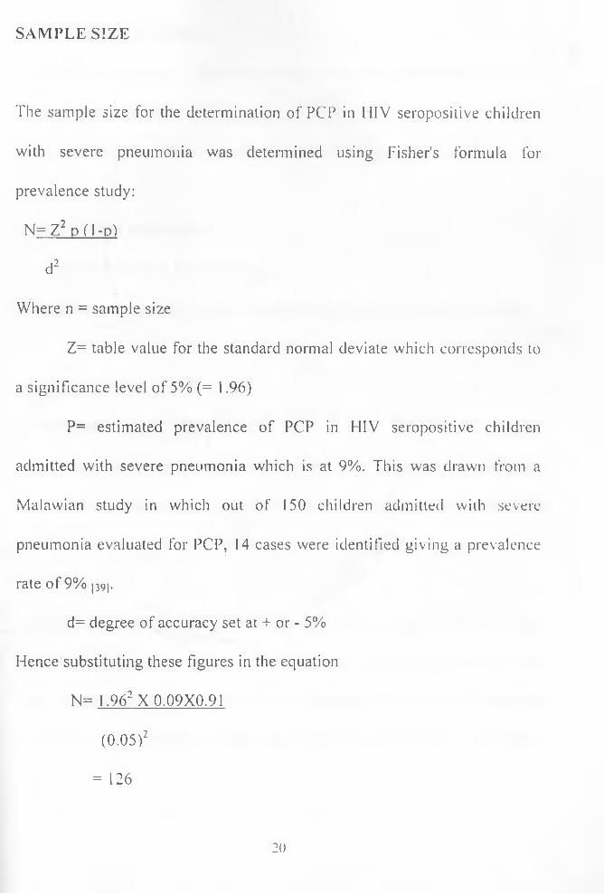
SAMPLE SIZE
The sample size for the determination of PCP in HIV seropositive children with severe pneumonia was determined using Fisher's formula for prevalence study:N= Z2p(l-p)
d2Where n = sample size
Z= table value for the standard normal deviate which corresponds to a significance level of 5% (= 1.96)
P= estimated prevalence of PCP in HIV seropositive children admitted with severe pneumonia which is at 9%. This was drawn from a Malawian study in which out of 150 children admitted with severe pneumonia evaluated for PCP, 14 cases were identified giving a prevalence rate of 9% (39).
d= degree of accuracy set at + or - 5%Hence substituting these figures in the equation
N= 1.962 X 0.09X0.91 (0.05)2
= 126
20
INCLUSION CRITERIA1. All HIV seropositive children admitted with severe pneumonia as defined by the WHO criteria.2. Consent by the parent or guardian to be included in the study.
EXCLUSION CRITERIA1. Children who were HIV seronegative.2. Children who were too ill to undergo the procedure of sputum collection.
PROCEDURESa. PatientsChildren aged 2 months to 24 months who were admitted to the Pediatric wards with severe pneumonia were recruited by the researcher between 9.00a. m. and 4.00 p.m. of every weekday over the study period.b. ClinicalClinical and demographic data. Each child had a medical history taken, which included history of cough, and it's duration, history of hotness of the body, history of difficulty in breathing or inability to breastfeed. History of recent use of antibiotics especially use of cotrimoxazole was taken,
including how long prior to admission the medication was given and duration of treatment.Anthropometries measurements were taken including age and weight. I he nutritional status was also assessed. The respirator) rate and the temperature were noted. The information was recorded in a coded case record form.
c. LaboratoryAnonymous and unlinked IIIV testing was carried out on the children.'The parallel testing method with rapid kits of Inimunocomh (Orgenics) and Determine (Abbot laboratories), both kits of which are for the detection o f
antibodies to HIV-1 and I IIV-2 in human whole blood, were used to assess HIV serostatus. I'sing the parallel testing strategy, the patient was
considered to be HIV seropositive i f both tests were positive or was considered negative when both tests were negative. However i f the two kits showed discordant results then blood was taken for a conf i rma io iy test using Enzyme Linked Immunosorbent Assa\ (ELISA) from Dade Helming Inc.
Peripheral venous blood was drawn from the child using aseptic techniques. One and a half mis (1.5 mis) of venous blood was put in each of 2 plainbottles for HIV serologv testing at the hnmunologv l.aboralorv at KM I
using both Determine and Immunocomb kits for detection ol antibodies to HIV-1 and 1IIV-2 in whole blood. Arterial oxygen saturation was measured using 1.0 ml of arterial blood taken to the intensive care unit laboratory for oxygen saturation tests.
SPUTUM COLLECTIONSputum was collected by induced sputum collection using hypertonic saline, nebuliser, a suction pump, sputum bottle trap and a suction tube at admission to the ward. The sputum bottle trap (sputum bottle) was attached to both the suction machine and the suction tube. Before sputum collection, the child was nebulised with hyper tonic saline solution for 3 minutes and then with the assistant holding the child's head firmly in anterior position, the auction tube was inserted into the patient's mouth and into the oropharynx and the suction machine was switched on to aspirate 1.0ml. of the induced sputum. The specimen obtained was sealed and sent to the mycology laboratory at the Centre for Respiratory Diseases Research (CRDR) at Kenya \ledicai Research Institute. (KHMRI). The sputum samples from IIIV seropositive children were then analyzed for presence of !J. cumin with an indirect immunoflourescent assay technique using D If I IT' I immtinollourcsccnce /'
carinii lest kit made by Shield Diagnostics of Dundee of l .K. A positive result was defined by the presence of 5( live) or more oocysts.
DATA ANALYSISI he clinical and laboratory data collected was entered in a computer iisine the SPSS program. Descriptive statistics including rates and percentages were determined during the analysis. Children were categori/ed as being PCP positive or negative. Differences between the two groups were assessed using the chi-squared test and odds ratio (for categorical variables) and Mann-Whitney IJ or Independent t-test (for continuous variables namely age, cough duration and arterial oxygen saturation). I nivariale correlates of the presence of PCP were determined using the chi-squared test and fisher's exact test. I he results were presented in descriptive form using frequency tables, graphs and cross tabulation.
24
ETHICAL CONSIDERATIONS
1. The nature of the study was explained to the parent [s] or guardian before recruitment.2. Anonymous and unlinked HIV testing was done hence there was no need for any one on one counselling.3. A written and signed consent was obtained from the parent or guardian.4. All the study samples did not bear any personal identifiers of the study subject.5. The study protocol was approved by KNH - Ethical and Research Committee.
25
RESULTS
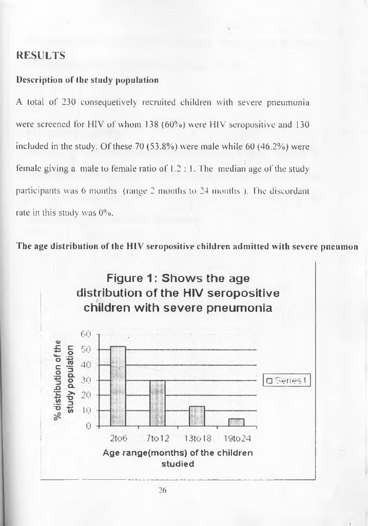
Description of the study populationA total of 230 consequetively recruited children with severe pneumonia were screened for HIV of whom 138 (60%) were HIV seropositive and 130 included in the study. Of these 70 (53.8%) were male while 60 (46.2%) were female giving a male to female ratio of 1.2 : 1. The median age of the study participants was 6 months (range 2 months to 24 months ). fhe discordant rate in this study was 0%.
The age distribution of the HIV seropositive children admitted with severe pneuinon
Figure 1: Shows the age distribution of the HIV seropositive
children with severe pneumonia
a>JCoc o-Q ' l . 00 T3
n Series i
Age range(m onths) of the children studied
26
The majority (52%) of the children in the study were aged between 2 and 6 months, while only 5% were aged greater than 18 months.
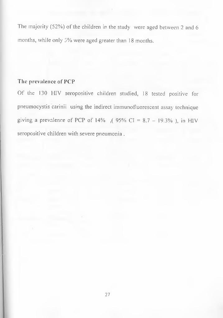
The prevalence of PCPOf the 130 HIV seropositive children studied, 18 tested positive for pneumocystis carinii using the indirect immunofluorescent assay technique giving a prevalence of PCP of 14% ,( 95% Cl = 8.7 - 19.3% ), in HIV seropositive children with severe pneumonia .
27
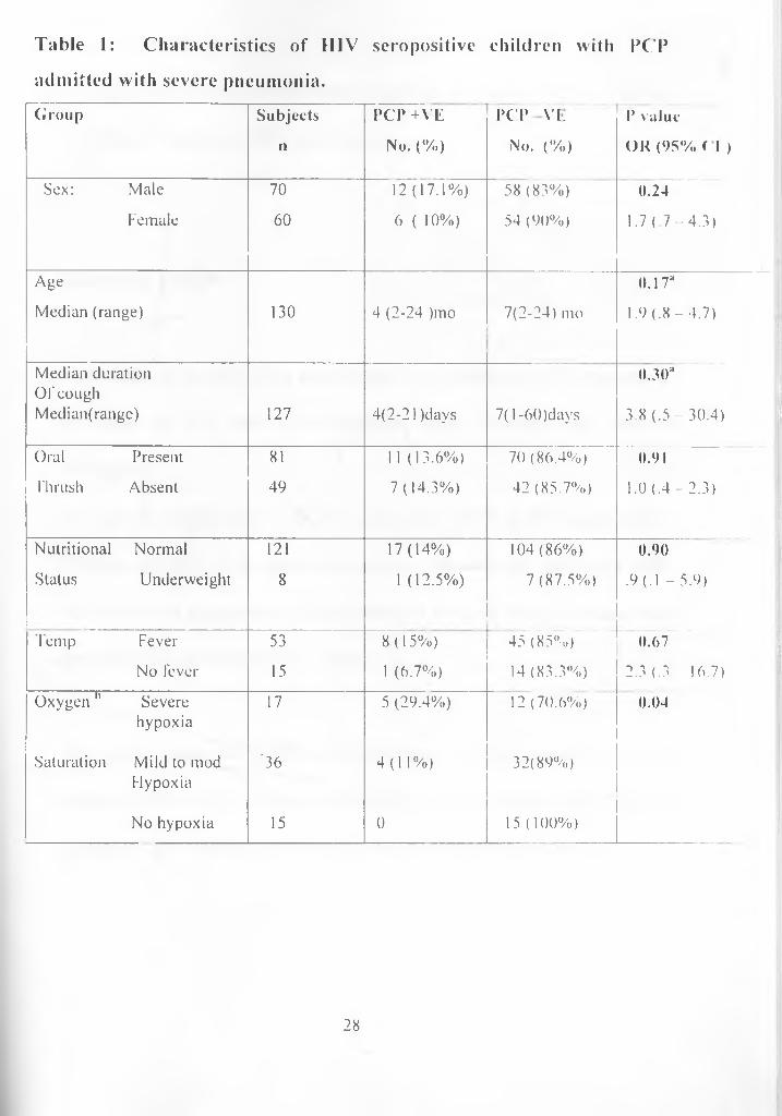
Table 1: Characteristics of HIV seropositive children with PCPadmitted with severe pneumonia.Group Subjects
n
P C P + V E
No. ( % )
P C P v i .
No. ( % )
P value
O K ( 9 5 % C l )
Sex: Male 70 1 2 ( 1 7 . 1 % ) 58 (83%) 0.24
Female 60 6 ( 10 % ) 54 (90%) 1 . 7 ( 7 - 4 . 3 )
Age
Median (range) 130 4 (2-24 )mo 7(2-24) mo
0.1 T
1 .9 ( .8 - 4 . 7 )
Median duration Ofcough Median(range) 127 4(2-21 )days 7( 1 -60)days
0.30a
3.8 ( .5 -3 0 .4 )
Oral Present 81 11 (13 .6 % ) 70(86.4%) 0.91
Thrush Absent 49 7 ( 1 4 .3 % ) 42 (85.7%) 1.0 (.4 - 2.3)
Nutritional Normal 121 1 7 ( 1 4 % ) 104(86%) 0.90
Status Underweight 8 1 ( 12 .5 % ) 7 (87.5%) .9 (.1 -5 .9 )
Temp Fever 53 8 ( 1 5 % ) 45 (85%) 0.67
No fever 15 1 (6.7%) 14 (83.3%) 2.3 (.3 16.7)
Oxygen h Severe 17 5 (29.4%) 12 (70.6%) 0.04
Saturation
hypoxia
Mild to mod '36 4 ( 1 1 % ) 32(89%)Fly pox ia
No hypoxia 15 0 15 (10 0 % )
28
a : student’s t-test.b: Oxygen saturation:- < 75% = severe hypoxia, > 75% to < 92% = mild to
moderate hypoxia, > 92% = no hypoxia.
Correlates of PCP
We recognize that the study sample size was underpowered to determine correlates of PCP hence the inferences made hereafter may not be conclusive.In a univariate analysis of clinical correlates of PCP in HIV seropositive children admitted with severe pneumonia, sex was not associated with PCP with the prevalence of PCP being 17% and 10% in males and females respectively (P=0.24). (Table 1)
The median age of the children with PCP was 4 months (range of 2 to 24 months) while that of children without PCP was 7 months (range 2 to 24 months). Age was not significantly associated with PCP (P=0.35).
29
I he median cough duration in children with PCP was 4 days (range 2 to 21 days) while that of children without PCP was 7 days (range of 1 to 60 days). Cough duration was not significantly associated with PCP (P=0.3).
In our study 81(62%) of the children had oral thrush. The prevalence of PCP in children with oral thrush was 13.6% (11/81) while in those children without oral thrush it was 14.3% (7/40). The presence of oral thrush was not significantly associated with PCP (P=0.9). (Table I )
Using the National Center for Health Statistics as a reference, the Z scores for weight for age for the study population was determined using SPSS and grouped as follows; patients whose Z score was < or = to -3SD were grouped as severe underweight; those whose Z score was between >-3SD to -2SD were grouped as mild underweight and those whose Z score was between >-2SD to 2SD were grouped as Normal weight. None of the study subjects had severe malnutrition with 94% of them having normal weight for age. The prevalence of PCP in children with normal weight for age was 14% (17/121) while 12.5% of the patients with mild underweight (1/8) had PCP. Nutritional status was not significantly associated with PCP (P= 0.9). (Table 1)
30
In this study, fever was considered as a surface temperature of 37.7 °C and above. The prevalence of PCP among children with fever was 1 5% (8/53) while it was 6.7% in children without fever (1/15). Fever was not significantly associated with PCP (P= 0.67). (Table 1)
Of the 130 children studied, only 68 (52%) of them had results for the oxygen saturation levels. Arterial oxygenation levels from only 68 subjects were included in the analysis and this was due to irregular functioning of the calibration machine for oxygen saturation calculation in the ICU laboratory of KNH. As illustrated in Figure 2, the mean arterial oxygen saturation was 83.4% (95% Cl 81.0 - 85.5). The distribution was skewed to the right hence we used the interquartile range to get a cut off. The value at 75th percentiles was 92% oxygen saturation and this value and above was classified as no hypoxia; mild to moderate hypoxia was any value less than the oxygen saturation value at 75,h percentile but more than the oxygen saturation value at the 25lh percentile. This correlated to the oxygen saturation value of more than 75% but less than 92% oxygen saturation. Oxygen saturation values that were within the 25th percentile range and less were grouped as severe
31
hypoxia and this correlated to saturation value of 75% oxygenation and less.
The prevalence of PCP in children with severe hypoxia was 29.4% while it was 11% in children with mild to moderate hypoxia. None of the children with no hypoxia had PCP. PCP was significantly associated with hypoxia (P=0.04) (Table lj. The borderline association found between hypoxia and PCP may have been stronger with larger numbers of patients.
32
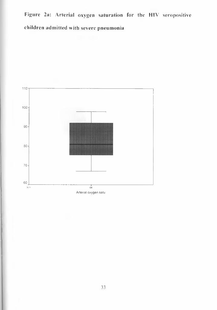
Figure 2a: Arterial oxygen saturation for the HIV seropositive children admitted with severe pneumonia
110
100-
90 -
80 -
70 .
60 -----------------------------------------------------------------------■---------------N = 68
Arterial oxygen satu
33
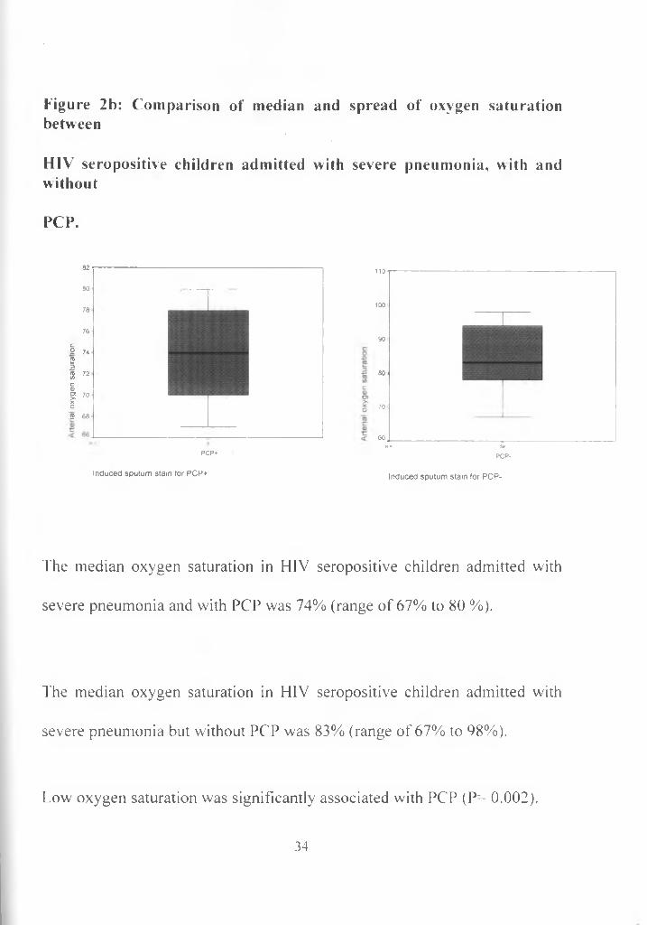
Figure 2b: Comparison of median and spread of oxygen saturation betweenHIV seropositive children admitted with severe pneumonia, with and withoutPCP.
82
80
78
76
c9 74 21 72C0)g 70
O2 68
PCP*
Induced sputum stain for PCP+
110
100
90
80
70
60N ■ 59
PCP-
Induced sputum stain for PCP-
The median oxygen saturation in HIV seropositive children admitted with severe pneumonia and with PCP was 74% (range of 67% to 80 %).
The median oxygen saturation in HIV seropositive children admitted with severe pneumonia but without PCP was 83% (range of 67% to 98%).
Low oxygen saturation was significantly associated with PCP (P= 0.002).
34
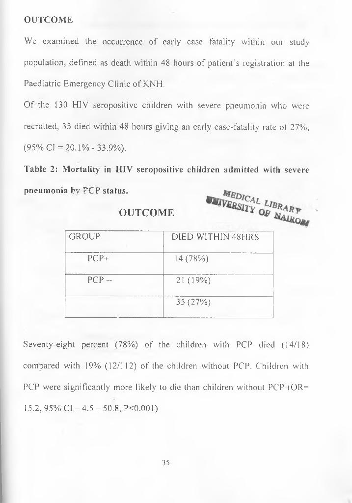
OUTCOMEWe examined the occurrence of early case fatality within our study population, defined as death within 48 hours of patient's registration at the Paediatric Emergency Clinic of KNH.Of the 130 HIV seropositive children with severe pneumonia who were recruited, 35 died within 48 hours giving an early case-fatality rate of 27%, (95% Cl = 20.1%-33.9%).Table 2: Mortality in HIV seropositive children admitted with severe pneumonia by TCP status.
OUTCOME
GROUP DIED WITHIN 48HRSPCP-i- 14(78%)P C P - 21 (19%)
35 (27%)
Seventy-eight percent (78%) of the children with PCP died (14/18) compared with 19% (12/112) of the children without PCP. Children with PCP were significantly more likely to die than children without PCP (OR= 15.2, 95% Cl - 4.5 - 50.8, PO.OOl)
35
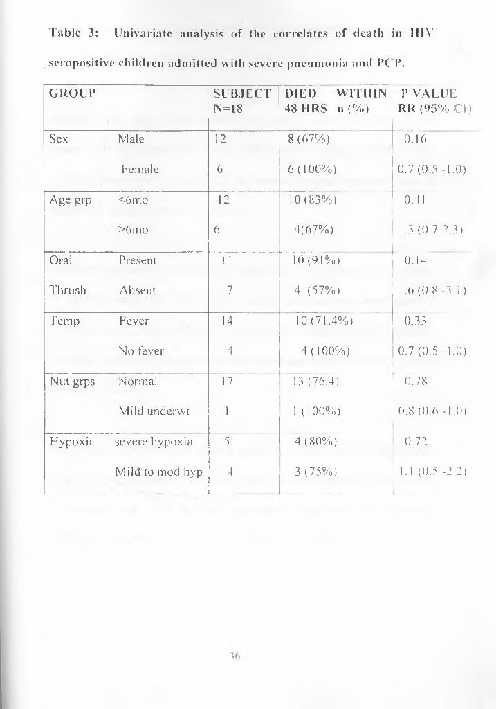
Iablc 3: Univariate analysis of the correlates of death in IIIVseropositive children admitted with severe pneumonia and PC P.GROUP SUBJECT
N=18DIED WITHIN48 HRS n (%)
P VALUERR (95% Cl)
Sex Male 12 8 (67%) 0.16Female 6 6(100%) 0.7 (0.5 -1.0)
Age grp <6 mo 12 10 (83%) 0.41>6mo 6 4(67%) 1.3 (0.7-2.3)
Oral Present 1 1 10 (91%) 0.14Thrush Absent 7 4 (57%) 1.6 (0.8 -3.1)Temp Fever 14 10 (71.4%) 0.33
No fever 4 4 (100%) 0.7 (0.5 -1.0)Nut grps Normal 17 13 (76.4) 0.78
Mild underwt 1 1 (100%) 0.8 (0.6 -1.0)Hypoxia severe hypoxia 5 4 (80%) 0.72
Mild to mod hyp 4 3 (75%) 1.1 (0.5 -2.2)L . ____
The study sample was underpowered to determine correlates of mortality among the 18 PCP cases; hence no firm inferences can be made.
Mortality was high in both males and females with PCP; 67% of the males with PCP died (8/12) while 100% of the females with PCP died (6/6) but there was no significant difference between mortality rates in males and females (P=0.16). (Table 3)
Likewise age was not found to be significantly associated with mortality in children with PCP; 83% (10/12) of the children who were aged <6 months with PCP died while 67% (4/6) of the children aged >6 months with PCP died with no significant difference between the two age groups (P=0.41). (Table 3)
Oral thrush was not found to be a significant association to death from PCP; 91% of the patients with PCP who had oral thrush died while 57% of patients with PCP but no oral thrush died with no significant difference between the two groups. (P=0.14). (Table 3)
37
Due to low rates of malnutrition in our study, it was not possible to evaluate the role of malnutrition as a predictor of poor outcome in children with PCP.
None of the children with no hypoxia had PCP so the general outcome was analyzed for those with severe hypoxia and mild to moderate hypoxia.Eighty per cent (80%) (4/5) of patients with PCP and severe hypoxia died while 75% (3/4) of patients with PCP and mild to moderate hypoxia died P=0.78. (Table 3)
38
DISCUSSIONPCP has been and continues to be an important factor in the morbidity and mortality of paediatric HIV/AIDS infection. The prevalence of PCP in this study population was found to be 14%. This is about the same as the prevalence that was found in other studies done in Africa. Our study focused only on severe pneumonia and therefore does not give us the magnitude of the problem of PCP in all children admitted with pneumonia. Furthermore exclusion of the children who were too ill to undergo sputum induction may have underestimated the magnitude of the problem.Studies done in Malawi to determine the prevalence of PCP in HIV seropositive children was reported as 9.3% and 18% of these children had been on cotrimoxazole therapy |3V|. In our study, we excluded the children who had been on cotrimoxazole therapy for at least five (5) days prior to admission to the hospital with severe pneumonia. Other studies done in Zimbabwe to determine the prevalence of PCP in HIV seropositixe children had a prevalence of 1 1% when sputum was collected using induced sputum and a prevalence of 13% when sputum was collected using bronchoalveolar lavage [32|. Similar studies done in South Africa to determine the prevalence of PCP in HIV seropositive children with
39
severe pneumonia had a prevalence of 1 1% poj. Higher percentages have been found among HIV-1-infected children. In a study in South Africa to evaluate the burden of PCP in HIV-infected children hospitalised with pneumonia, PCP was identified in 48.6% children using induced sputum. [46)- Due to lack of laboratory facilities, we were unable to establish the HIV infection status of our study subjects. This was due to lack of Polymerase Chain Reaction (PCR) technology on site.
In other African studies, higher prevalence rates of PCP have been found from postmortem studies. In Cote D’ Voire, the investigators found a PCP prevalence of 31% in HIV seropositive children (4|| while in Zimbabwe one study reported a prevalence of 67% at necropsy |42| Nathoo K J et al concluded that necropsy findings could be used as a gold standard in the diagnosis of PCP [42|. This however cannot be a useful clinical tool .In another necropsy study of children dying of respiratory illness in Zambia, Pneumocystis carinii was isolated in 27.5% of the cases. In the same study, HIV-positive children had PCP more frequently (OR 5.3, 95% Cl 2.12-15.68, p<0.001), than did the HIV-1 negative children However in a retrospective review of autopsy findings of 33 HIV infected children at a home for AIDS orphans in Nairobi, Kenya,
40
Rana et al did not isolate PCP |49|. This was thought to be due to cotrimoxazole prophylaxis, the adherence of which was reported to be 100% in the home. It was also postulated that the absence of PCP in the autopsy reviews could have been due to selection bias as rapid progressors could have died earlier on in the disease.
Pneumocystis carinii pneumonia has been described to be more common in the children aged 6 months or less. This was also the median age for our study population. Age less than 1 year has also been documented as a strong correlate of PCP in children who are HIV-infected (23| In this study however age was not found to be of significant association with PCP (P=0.17).
Pneumocystis carinii pneumonia is often the initial clinical sign of HIV infection, particularly among infants. Of the children described in literature, at least half who developed PCP were not recognized as HIV- infected before they were diagnosed as having PCP, although some had earlier on presented with HIV-associated symptoms. 121.22.23.28.501- Most of the children in this study were admitted for the first time during the study period and had not had any major illnesses prior to admission
41
despite being HIV exposed. We were not able to evaluate CD4 levels in the children found to be HIV seropositive and this was mainly as a result of the cost of the test. Establishing the CD4 levels in these children would have been an added advantage as we would have been able to
fdifferentiate between the children who were only HIV exposed and those who were infected with HIV. However most of the public health facilities in Kenya, especially in the periphery, are unable to do CD4 levels and in the least are only able to detect antibodies, so studies interpreted using CD4 levels may not be applicable directly to these centers in the periphery. This study can be used as reference in these centers because the parameters used, which are HIV exposure in relation to presence of PCP, will be the same as those available in these peripheral centers.
Hypoxia was found to be a significant indicator of the presence of PCP in our study. Of the 130 children studied, only 68 (52%) of them had results for the oxygen saturation levels. Arterial oxygenation levels from only 68
subjects were included in the analysis and this was due to irregular functioning of the calibration machine for oxygen saturation calculation in the ICU laboratory of KNH.
42
The children with PCP had lower oxygen saturations that were either severe hypoxia or mild to moderate hypoxia. Of the children with severe hypoxia (oxygen saturation of 75% or less), 29.4% had PCP while 1 1 %iof children with mild to moderate hypoxia (oxygen saturation of between 75% to less than 92%) had PCP. None of the children with no hypoxia had PCP.These differences were statistically significant (Pr 0.04). This is comparable to other studies done in Africa. In a study on PCP in Malawian children, PCP was identified in 5 of the 60 children with severe pneumonia and the arterial oxygen saturation was significantly lower in the PCP cases [4oj. In this same Malawian study, of the 5 children with PCP, 4 died indicating that the marked hypoxemia was a significant factor in their mortality [40]. In a study on Malawian children aged 2 months to 5 years with severe pneumonia aimed to describe the rate, clinical presentation and outcome of PCP in children with acute severe pneumonia, children with PCP had a lower oxygen saturation at admission than children with bacterial pneumonia and hence oxygen
v
requirements were much greater in these children with PCP than those with bacterial pneumonias (96 of 105 hospital days vs. 15 of 94, p<0.001) [39j. Therefore in a child with severe pneumonia who presents with marked hypoxia, PCP should be considered with a high index of
43
suspicion. Many health facilities however are unable to determine oxygen saturation levels. The health facilities should be equipped with simple ways to measure oxygen saturation levels like the portable pulse oximeter that is easy to use and not very expensive. The determination of oxygen saturation levels will aid in categorizing patients with pneumonia and in guiding oxygen therapy.
Other clinical factors including age groups, oral thrush, fever and underweight were not found to be significantly associated with PCP in this study. This is similar to a study carried out in South Africa where it was found that neither clinical nor laboratory tests were useful in distinguishing between presence and absence of PCP in the 111\ infected 1511. Although PCP can occur at any age, among children it is mostly diagnosed between 3-6 months of age. A population based study in Massachusetts found the minimum incidence of PCP during the first year of life to be 2.3% among all infants born to HIV seropositive mothers, or an estimated 7.7% among HIV infected infants |2i -24.28.501- From literature on the predictors of PCP in HIV-infected children, the strongest correlates have been postulated as age less than 1 \ear, I 1IV- related symptoms and the reduction of CD4+ lymphocyte number and function
44
[23. 28. 2i>|. In a study carried out in New York, ITS.A on HIV infected adults; predictors of PCP were found to be oropharyngeal thrush, night sweats and recurrent fevers j27|
This study has demonstrated that PCP has a high case fatality rate; 78% of the children with PCP died within 48hours of being seen in the hospital’s outpatient facilities as compared to 21% of the children without PCP (p<0.001). The sample size did not have enough power to evaluate the correlates of death in children with PCP. 1 he high early ease fatality rates in cur study suggests that HIV testing of all children with severe pneumonia and treating those found to be positive for PCP maybe useful in reducing mortality in these children. The wide availability of rapid HIV tests in most Kenyan hospitals makes this a feasible approach to management.
Mortality can also be addressed by giving PCP prophylaxis to the children who are HIV exposed but not yet ill with pneumonia. In a study to define the burden of PCP in African children carried out in South Africa among HIV infected children, death among children with PCP who had been taking TMP-SMX prophylaxis were markedly reduced
45
(98.6%; 95%CI, 89.l%-99.8%) compared with children who were not taking prophylaxis [48j. However routine prophylaxis and treatment is difficult to deliver in low resource settings and could also increase resistance to the drug.
Our study demonstrates the high prevalence of PCP in HIV seropositive infants with severe pneumonia in our setup and therefore early intervention is necessary especially in those presenting with severe pneumonia.
46
CONCLUSIONS1. The prevalence of PCP in HIV seropositive children with severe
pneumonia at KNH is 14%. Pneumocystis carinii is therefore an important cause of morbidity and mortality in children admitted at the KNH with severe pneumonia who are also HIV seropositive.
2. Early case fatality was 78% among the HIV seropositive children admitted with severe pneumonia with PCP at KNII.
3. Hypoxia is significantly associated with PCP among 1IIV seropositive children with severe pneumonia admitted at KNH.
RECOMMENDATIONS1. In children admitted with severe pneumonia, the HIV status should
be determined at admission in order to idemiI\ HIV exposed infants and empirically treat them for pneumoevstis carinii pneumonia.
2. In HIV seropositive children with severe pneumonia, severe hypoxia is an indication to treat empiricall\ for PCP
47
REFERENCES1. UN AIDS (2003) Report on the global HI l' AIDS epidemic. Joinl United Nations Programe on HIV AIDS.
2. UNAIDS (June 2003). Epidemiological Fact Sheets of Kenya on HIV/AIDS and Sexually transmitted infections, 2003 l Jpdate. Joint I ’nited Nations Programe on 11IV/AIDS.
3. Ministry of Health, 2003. AIDS in Kenya: Interim report on National HIV Surveillance Results.
4. Broehert A. Characteristics of mother, child linked to postnatal HIV transmission risk. IMERSAEIOSAL l-A Mil.) RI.AS A l.\ O PERSPECTIVES. 2001 Jun; 27(2); 103-4.
5. Nduati R, John (i, Mbori-Ngaeha I), el al. I*fleet of hreasU'eeding and formula feeding on transmission of 111 \ -1: a randomi/ed clinical trial JAMA 2000; 238: I 167-74.
4«S
6. Center-for Disease Control. HIV'AIDS surveillance report. Atlanta: C DC 1993: 16.
7. Gibb D.M. Davison et al. Pneumocystis carinii pneumonia in \ erlicallx acquired HIV infection in the British Isles. Arch Dis-('hi/J 1994: 70: 241-244
8. Perez MS, Van Dyke RB. Pulmonary infections in children with HIV infection. Semin Rcspir Infect 2002: Mar I7( I ): 33-40.
9. Graham SM, Coulter JB, CiiIks CIV Pulmonarv disease in 111\ - infected African children, hit J Tnhcrc /.imp D/.s. 2001 Jan: 5(1): I 2-2 v
10. Gilks ( \ Otieno L.S., Brindle R. et al, I he presentation and outcome of HIV related diseases in Nairobi; O.J. Med 1992: 82: 25-32.
1 1. Chintu C\ Mudenda V, Lucas S. el al. Lung diseases at necropsv in African children dying from respirator) illnesses: a descriptive necropsv study. Lcmcet.2002 Sep 28: 360(9338): 985-90.
12. Lucas SB, Peacock CS, Hounnou A, et al. Disease in children infected with HIV in Abidjan, Cote d'Ivoire. BMJ. 1996 Feb 10; 312 (7027): 335-8.
13. Ikeogu MO, Wolf B, Mathe S. Pulmonary manifectations in HIV seropositivity and malnutrition in Zimbabwe. Arch Dis Child. 1997 Feb; 76(2): 124-8.
14. Robert F.Miller, David M. Mitchell. Pneumocystis carinii pneumonia. AIDS and the lung; Update 1995-1. In: Thorax 1995; 50:191-200
15. Walzer P.D. Pnemocystis carinii infection.In: Harrison's Principles of Internal
Medicine, 14th Edition, McGraw-Hill, U.S.A., 1998: 1 161-2.
16. Stringer JR. Pneumocystis. Int J Med Microbiol. 2002 Oct; 292(5-6): 391-404.
50
17. Antony S. Fauci and H.Clifford Lane. Opportunistic Infections in HIV disease: In: Harrison's Principles of Internal Medicine, 14th Edition.McGraw-Hill, U.S.A., 1998: 1824-5.
18. Hughes W, Pneumocystis carinii pneumonitis; In: Nelson'sTextbook of Pediatrics’, 15th Edition, W.B.Saunders, Philadelphia, Pennsylvania; 1996:951-2.
19. Daley C L. Pulmonary infections in the tropics: impact of HIV infection. Thorax. 1994 Apr; 49(4): 370-8.
20. Oxtoby MJ. Perinataly acquired HIV infection. Pediatr Infect Dis J 1990; 9: 609-19.
21. Scott GB, Hutto C, Makuch RW, et al. Survival in children with perinatally acquired HIV type 1 infection. N Engl J Med 1989; 321: 1791-6.
51
22. Connor E, Bagarrazi M, Mcsherry Cj, et al. Clinical and Laboratory correlates of Pneumocystis carinii pneumonia in children infected with HIW.JAMA. 1991 Apr 3; 265(13): 1693-7.
23. Krasinski K, Borkowsky W, Holzman RS. Prognosis of HIV infection in children and adolescents. Pediatr Infect Dis J. 1989; 8:216- 20 .
24. Blanche S, Tardieu M, Diliege A-M, et al. Longitudinal study of 94 symptomatic infants with perinatall\ acquired HIV infection. Am ./ Dis Child 1990; 144:1210-5.
25. Williams A.J, Duong T, McNally L.M. et al. Pneumocystis Carinii Pneumonia and cytomegalovirus infection in children with \erticall\ acquired HIV infectionAIDS; 15(3): 335 -339, 2001 Feb 16.
26. Zar IIJ, Hanslo D, Tannenbaim E, et al. Aetiology and outcome of pneumonia in HIV infected Children hospitalized in South Africa. Pediatr Crit Care Med. 2001; April 2(2): 108-112.
27. Wikin A and Feinberg J: PCP: A clinical review; Am Fam Physician.
1999 Oct15; 60(6): 1699-708, 1713-4.
28. Kovacs A, Church J, Mascola L. «. al. CD4 counts as predictors of Pneumocystis carinii pneumonia in infants and children with HIV infection. JAMA. 199 1 Apr 3; 265(13). 1678 703.
29. Roilides E, Cleric i M- DePalma L, et al. T Helpei cell tesponses in children infected with HIV-1. J Pediatr. 1991 May; 118(5): 724-30.
30. Clumeck N, S o n n e t J, Taelman H, et al. Acquired immunodeficiency Syndrome in African patients. N Engl J Med. 1984 Feb 23; 3.0(8): 492-
7.
31 Malta AS, Gwratanzur. LK, Klein S, e. al. Pneumocystis carinii pneumonia in Zimb.b Lcmcel.mS No» 11; 346(8985): 1258-61.
53
32. Kamanfu G, Mlika-Cabanne N, Girard PM et ai; Pulmonary complications of HIV infection in Bujumbura, Burundi. Am Rev Respir Dis 1993 Mar; 147(3): 658-63.
33. Serwadda D, Goodgame R, Lucas S, et al. Absence of pneumocytosis in Ugandan AIDS patients. AIDS 1989 Jan; 3(1): 47-8.
34. Carme B. Mboussa J, Andzin M, et al. Pneumocystis carinii is rare in AIDS in Central Africa .Trans R Sue Trap Med Hyg. 1991 Jan-Feb; 85(1): 80.
35. Pape J, Liautaud B, Thomas F, et al. The acquired immunodeficiency syndrome in Haiti. Ann Intern Med. 1985 Nov; 103(5): 674-8.
36. Chequer P, Hearst N, Hudes ES, et al. Determinants of survival in adult Brazilian AIDS patients, 1982 - 1989 The Brazilian State AIDS Program Co-ordinators. AIDS. 1992 May; 6(5): 483-7.
37. Thijs A, Janssen P. Pneumocytosis in Congolese infants. Tropical Gengr. Med 1963; 15: 158-172.
54
38. Bvvibo N, Owor R. PCP in Ugandan African children. If Afr Med J. 1970; 19: 184-5
39. Graham SM., Mtitimila E, Kamanga SU., et al. Clinical presentation and outcome of PCP in Malawian children. The Lance! 2000 Jan 29; 355(9201): 369-73.
40. Kamivn Y, Mtitimila K. Graham SV1. et al. Pneumoewis carinii pneumonia in Malawian children. Ann.Trap Paediatr. 1907 Jun; 17(2): 121- 6 .
41. I .ucas S, Sewakambo N, Nambuya G.A. et al. The morbid anatom\ of African AIDS: In; AIDS and associated cancers in Africa: 1998; 1 1: 124-33.
42. Nathoo K.J, Gondo M, Gwanzura L, et al. fatal pneumoexstis carinii pneumonia in HIV seropositive infants in Harare, Zimbabwe. Trans RSoc Trap Med Hvg. 2001 Jan-Peb; 95(1): 37-9.
43. Cregan P, Yamamoto A, Lum A, et al, Comparison of Four Methods for the rapid detection of Pneumocystis carinii in Respiratory Specimens J Clin. Microb; 1990; 28 (2): 2432-36.
44. Turner D, Schwarz Y, Yust I. Induced sputum for diagnosing Pneumocytsis carinii pneumonia in HIV patients: new data, new issues. Eur Respir J. 2003 Feb; 21(2): 204-8.
45. Kamiya Y, Mtitimila E, Graham SM et al. Pneumocystis carinii pneumonia in Malawian children. Ann. o f Trap. Pucci 1997; 17 (2): 12 |_ 126.
46. Ruffini DD, Madhi SA. The high burden of Pneumocystis carinii pneumonia in African HIV-infected children hospitalized for severe pneumonia. AIDS [AIDS] 2002 Jan 4; 16(1): 105-112.
47. Leibovitz E, Pollack H, Moore I et al. Comparison of PC R aiK| standard cytological staining for detection of P. carinii from respiratory specimens from patients at high risk of HIV infection. J ClmMicrobiol. 1995 Nov; 33( 11): 3004-7.
48. Madhi SA, Cutland C, Ismail K. et al. Ineffectiveness ol trimethoprim-sulphaethoxazole prophylaxis and the importance of bacterial and viral co infections in African children with Pneumocystis carinii pneumonia. Clin Infect Dis. 2002 Nov 1; 35(9): 1 120-6.
t
49.Chakraborty R, Pulver A, pathology of HIV-1-infected paediatrics. 2002; 22:125-31.
Musoke R, et al. 1 he post-mortem African children. Annals of Tropica!
•D iversity o f nairoj#
50. Hsu A, Kunches L, Ng I\ et al. Pneumocystis carinii pneumonia in infants with HIV infection. (Abstract 417) 30lh Interscience Conference on Antimicrobial Agents and Chemotherapy, Atlanta, GA, 1900.
51. Dechaboon A, Hanslo D, Apolles P et al. Pneumoc\stis carinii in South African children infected with HIV. Pediatr Infect Dis J. 2000 July; 19(7): 603-7.
4
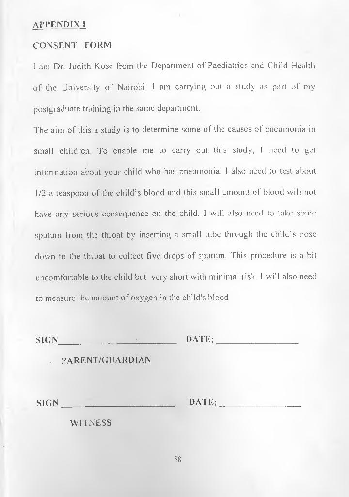
APPENDIX ICONSENT FORMI am Dr. Judith Kose from the Department of Paediatrics and Child Health of the University of Nairobi. I am carrying out a study as part of my postgraduate training in the same department.The aim of this a study is to determine some of the causes of pneumonia in small children. To enable me to carry out this study, 1 need to get information about your child who has pneumonia. I also need to test about 1/2 a teaspoon of the child’s blood and this small amount of blood will not have any serious consequence on the child. I will also need to take some sputum from the throat by inserting a small tube through the child’s nose down to the throat to collect five drops of sputum. This procedure is a bit uncomfortable to the child but very short with minimal risk. I will also need to measure the amount of oxygen in the child's blood
SIGN_________________ :________ DATE;__________________. PARENT/GUARDIAN
SIGN_________________________ DATE;__________________WITNESS
^8