Embed Size (px)
Citation preview
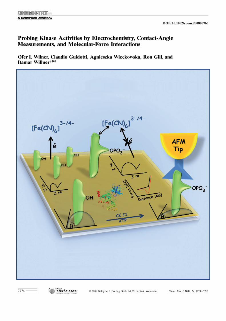
Probing Kinase Activities by Electrochemistry, Contact-AngleMeasurements, and Molecular-Force Interactions
Ofer I. Wilner, Claudio Guidotti, Agnieszka Wieckowska, Ron Gill, andItamar Willner*[a]
� 2008 Wiley-VCH Verlag GmbH&Co. KGaA, Weinheim Chem. Eur. J. 2008, 14, 7774 – 77817774
DOI: 10.1002/chem.200800765
Introduction
The phosphorylation of proteins by protein kinase is a keyprocess in signal transduction and the regulation of intracel-lular processes.[1]The over-expression of protein kinases hasbeen reported to be involved in different diseases, such ascancer[2] or Alzheimer.s disease.[3] Hence, the rapid detec-tion of the activity of protein kinases is of immediate signifi-cance for clinical diagnostics. Different methods for the de-tection of protein kinases have been developed, such as im-munoassays that use fluorescence-labeled antibodies (Fab)against sequence-specific phosphorylated peptides,[4] and theprotein kinase-induced radioactive labeling of the phos-phorylated product with radioactive ATP.[5] Other methodsto monitor the activities of protein kinases include the fluo-rescence polarization assay,[6] the amplified electrochemicaldetection of kinase with Au nanoparticles,[7] a proteinkinase-induced aggregation of Au nanoparticles,[8] and de-tection of the depletion of ATP as a result of a biocatalyzedphosphorylation process.[9] Furthermore, a label-free detec-tion of protein kinase was achieved by monitoring the phos-phorylation reaction on a field-effect transistor device.[10] Inthe present study, we probed the activity of casein kinase,CK2, which phosphorylates serine/threonine residues of pro-teins. At least 160 different proteins are phosphorylated by
CK2, and many of these proteins are active in intracellularsignal transduction, DNA replication, the synthesis of pro-teins, and cell division and proliferation. The CK2 is local-ized in the nuclei of the cells and exists also in the cytosol.[11]
The enzyme is a heterotetramer composed of a, a’ and b
subunits. The a and a’ subunits are catalytically active andthe b subunits recognize the substrates and synergisticallystimulate the catalytic activity of the a/a’ subunits.[12]
Here, we introduced different methods to monitor the ac-tivities of kinases (specifically, casein kinase), namely, elec-trochemical impedance spectroscopy, contact angle, andchemical force measurements.
Results and Discussion
Electrochemical impedance spectroscopy is a versatile toolto follow biocatalytic reactions and biomolecular recognitionevents at electrode surfaces.[13] The electron-transfer resis-tances at modified electrodes, in the presence of a redoxlabel solubilized in the bulk electrolyte solution, are stronglydependent on electrostatic interactions between the redoxlabel in the electrolyte solution and the charged electrodesurface or changes in the dielectric properties of the surfacecaused by the biocatalytic or biorecognition events.[14] Forexample, DNA hybridization on electrode surfaces was ana-lyzed in the presence of [Fe(CN)6]
3�/4� as redox label bymeans of electrochemical impedance spectroscopy (EIS).The hybridization of the analyte DNA with the capturingnucleic acid linked to the electrode increased the interfacialelectron-transfer resistances as a result of the electrostaticrepulsion of the redox label.[15] Similarly, EIS was applied tofollow the formation of an aptamer–adenosine complex by
Abstract: Three different methods toinvestigate the activity of a proteinkinase (casein kinase, CK2) are de-scribed. The phosphorylation of the se-quence-specific peptide (1) by CK2was monitored by electrochemical im-pedance spectroscopy (EIS). Phosphor-ylation of the peptide monolayer as-sembled on a Au electrode yields anegatively charged surface that electro-statically repels the negatively chargedredox label [Fe(CN)6]
3�/4�, thus increas-ing the interfacial electron-transfer re-sistance. The phosphorylation processby CK2 is further amplified by the as-sociation of the anti-phosphorylatedpeptide antibody to the monolayer.Binding of the antibody insulates theelectrode surface, thus increasing theinterfacial electron-transfer resistance
in the presence of the redox label. Thismethod enabled the quantitative analy-sis of the concentration of CK2 with adetection limit of ten units. The secondmethod employed involved contact-angle measurements. Although thepeptide 1-functionalized electrode re-vealed a contact angle of 67.58, phos-phorylation of the peptide yielded asurface with enhanced hydrophilicity,36.88. The biocatalyzed cleavage of thephosphate units with alkaline phospha-tase regenerates the hydrophobic pep-tide monolayer, contact angle 55.38.The third method to characterize the
CK2 system involved chemical forcemeasurements between the phosphory-lated peptide monolayer associatedwith the Au surface and a Au tip func-tionalized with the anti-phosphorylatedpeptide antibody. Although no signifi-cant rupture forces existed between themodified tip and the 1-functionalizedsurface (6�2 pN), significant ruptureforces (multiples of 120�20 pN) wereobserved between the phosphorylatedmonolayer-modified surface and theantibody-functionalized tip. This rup-ture force is attributed to the dissocia-tion of a simple binding event betweenthe phosphorylated peptide and thefluorescent antibody (Fab) bindingregion.
Keywords: antibodies · biosensors ·contact angles · electrochemistry ·protein kinases
[a] O. I. Wilner, Dr. C. Guidotti, Dr. A. Wieckowska, R. Gill,Prof. Dr. I. WillnerInstitute of Chemistry, The Hebrew University of JerusalemJerusalem 91904 (Israel)Fax: (+972)2-652-7715E-mail : [email protected]
Supporting information for this article is available on the WWWunder http://dx.doi.org/10.1002/chem.200800765.
Chem. Eur. J. 2008, 14, 7774 – 7781 � 2008 Wiley-VCH Verlag GmbH&Co. KGaA, Weinheim www.chemeurj.org 7775
FULL PAPER
the separation of an aptamer–nucleic acid duplex associatedwith the electrode, and the subsequent decrease of the inter-facial electron-transfer resistances.[16] The association of pro-teins to electrode surfaces insulates these surfaces and thusincreases the interfacial electron-transfer resistances in thepresence of a solubilized redox label. This has been used todetect the association of proteins to electrodes and, specifi-cally, to monitor the formation of immunocomplexes onelectrodes,[17] and to develop EIS-based immunosensors.Furthermore, biocatalytic transformations at electrode surfa-ces, such as enzyme cleavage of duplex DNA[18] or the bio-catalytic deposition of insoluble products on electrodes[19]
were investigated by EIS.In the present study, EIS measurements were applied to
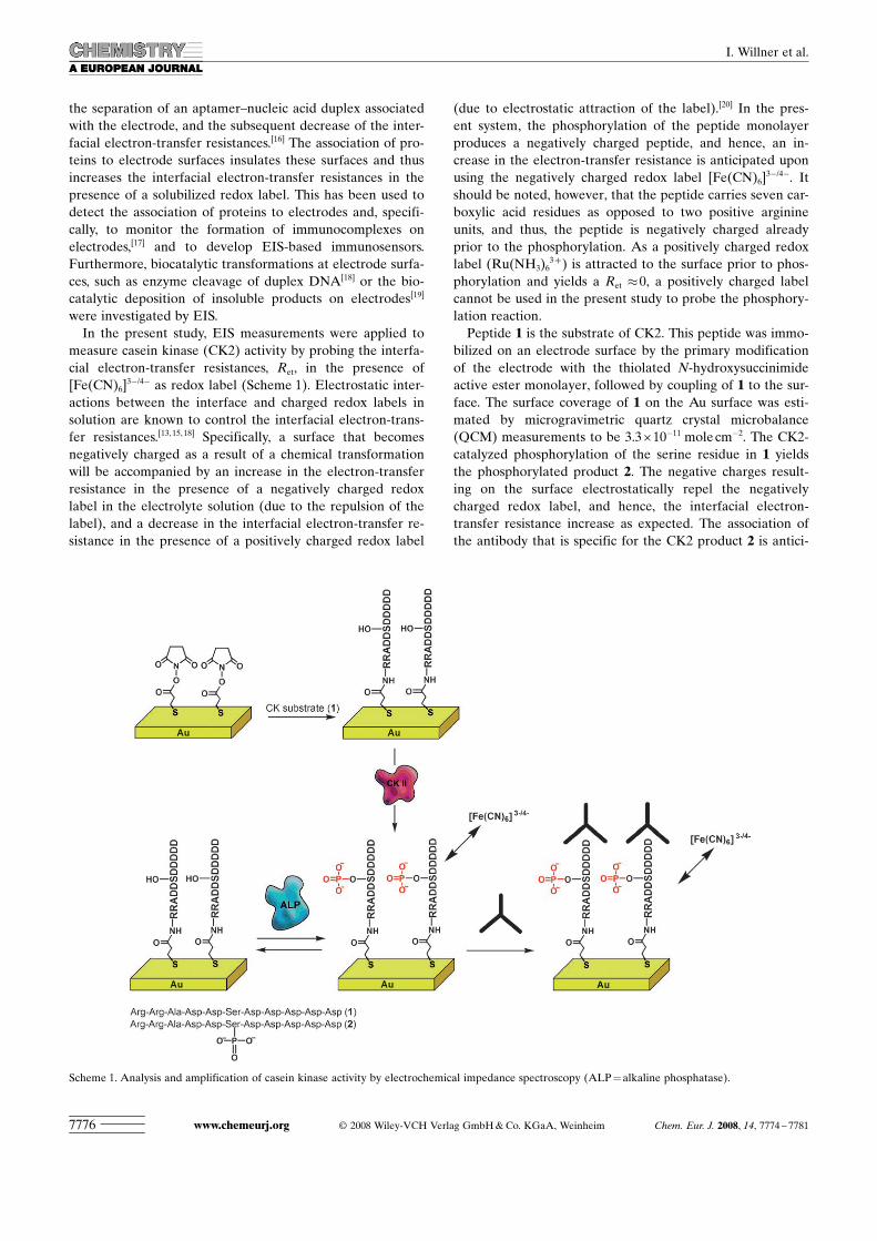
measure casein kinase (CK2) activity by probing the interfa-cial electron-transfer resistances, Ret, in the presence of[Fe(CN)6]
3�/4� as redox label (Scheme 1). Electrostatic inter-actions between the interface and charged redox labels insolution are known to control the interfacial electron-trans-fer resistances.[13,15,18] Specifically, a surface that becomesnegatively charged as a result of a chemical transformationwill be accompanied by an increase in the electron-transferresistance in the presence of a negatively charged redoxlabel in the electrolyte solution (due to the repulsion of thelabel), and a decrease in the interfacial electron-transfer re-sistance in the presence of a positively charged redox label
(due to electrostatic attraction of the label).[20] In the pres-ent system, the phosphorylation of the peptide monolayerproduces a negatively charged peptide, and hence, an in-crease in the electron-transfer resistance is anticipated uponusing the negatively charged redox label [Fe(CN)6]
3�/4�. Itshould be noted, however, that the peptide carries seven car-boxylic acid residues as opposed to two positive arginineunits, and thus, the peptide is negatively charged alreadyprior to the phosphorylation. As a positively charged redoxlabel (Ru ACHTUNGTRENNUNG(NH3)6
3+) is attracted to the surface prior to phos-phorylation and yields a Ret �0, a positively charged labelcannot be used in the present study to probe the phosphory-lation reaction.Peptide 1 is the substrate of CK2. This peptide was immo-
bilized on an electrode surface by the primary modificationof the electrode with the thiolated N-hydroxysuccinimideactive ester monolayer, followed by coupling of 1 to the sur-face. The surface coverage of 1 on the Au surface was esti-mated by microgravimetric quartz crystal microbalance(QCM) measurements to be 3.3I10�11 molecm�2. The CK2-catalyzed phosphorylation of the serine residue in 1 yieldsthe phosphorylated product 2. The negative charges result-ing on the surface electrostatically repel the negativelycharged redox label, and hence, the interfacial electron-transfer resistance increase as expected. The association ofthe antibody that is specific for the CK2 product 2 is antici-
Scheme 1. Analysis and amplification of casein kinase activity by electrochemical impedance spectroscopy (ALP=alkaline phosphatase).
www.chemeurj.org � 2008 Wiley-VCH Verlag GmbH&Co. KGaA, Weinheim Chem. Eur. J. 2008, 14, 7774 – 77817776
I. Willner et al.
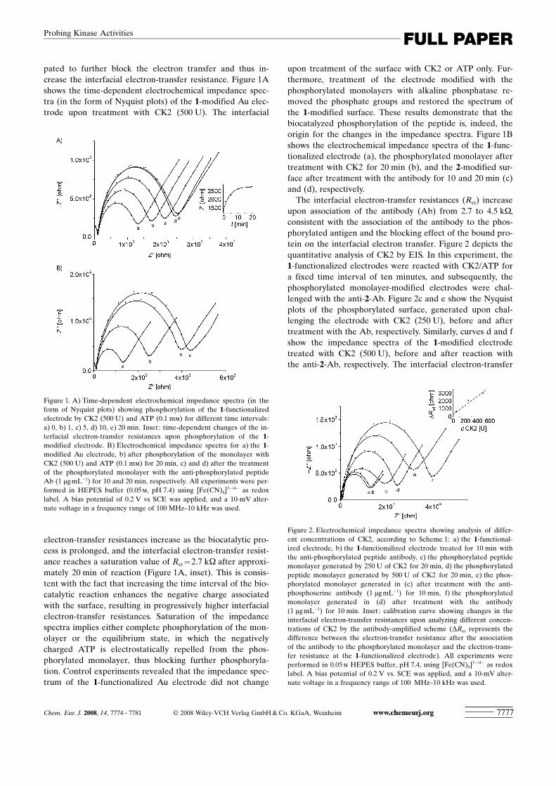
pated to further block the electron transfer and thus in-crease the interfacial electron-transfer resistance. Figure 1Ashows the time-dependent electrochemical impedance spec-tra (in the form of Nyquist plots) of the 1-modified Au elec-trode upon treatment with CK2 (500 U). The interfacial
electron-transfer resistances increase as the biocatalytic pro-cess is prolonged, and the interfacial electron-transfer resist-ance reaches a saturation value of Ret=2.7 kW after approxi-mately 20 min of reaction (Figure 1A, inset). This is consis-tent with the fact that increasing the time interval of the bio-catalytic reaction enhances the negative charge associatedwith the surface, resulting in progressively higher interfacialelectron-transfer resistances. Saturation of the impedancespectra implies either complete phosphorylation of the mon-olayer or the equilibrium state, in which the negativelycharged ATP is electrostatically repelled from the phos-phorylated monolayer, thus blocking further phosphoryla-tion. Control experiments revealed that the impedance spec-trum of the 1-functionalized Au electrode did not change
upon treatment of the surface with CK2 or ATP only. Fur-thermore, treatment of the electrode modified with thephosphorylated monolayers with alkaline phosphatase re-moved the phosphate groups and restored the spectrum ofthe 1-modified surface. These results demonstrate that thebiocatalyzed phosphorylation of the peptide is, indeed, theorigin for the changes in the impedance spectra. Figure 1Bshows the electrochemical impedance spectra of the 1-func-tionalized electrode (a), the phosphorylated monolayer aftertreatment with CK2 for 20 min (b), and the 2-modified sur-face after treatment with the antibody for 10 and 20 min (c)and (d), respectively.The interfacial electron-transfer resistances (Ret) increase
upon association of the antibody (Ab) from 2.7 to 4.5 kW,consistent with the association of the antibody to the phos-phorylated antigen and the blocking effect of the bound pro-tein on the interfacial electron transfer. Figure 2 depicts thequantitative analysis of CK2 by EIS. In this experiment, the1-functionalized electrodes were reacted with CK2/ATP fora fixed time interval of ten minutes, and subsequently, thephosphorylated monolayer-modified electrodes were chal-lenged with the anti-2-Ab. Figure 2c and e show the Nyquistplots of the phosphorylated surface, generated upon chal-lenging the electrode with CK2 (250 U), before and aftertreatment with the Ab, respectively. Similarly, curves d and fshow the impedance spectra of the 1-modified electrodetreated with CK2 (500 U), before and after reaction withthe anti-2-Ab, respectively. The interfacial electron-transfer
Figure 1. A) Time-dependent electrochemical impedance spectra (in theform of Nyquist plots) showing phosphorylation of the 1-functionalizedelectrode by CK2 (500 U) and ATP (0.1 mm) for different time intervals:a) 0, b) 1, c) 5, d) 10, e) 20 min. Inset: time-dependent changes of the in-terfacial electron-transfer resistances upon phosphorylation of the 1-modified electrode. B) Electrochemical impedance spectra for a) the 1-modified Au electrode, b) after phosphorylation of the monolayer withCK2 (500 U) and ATP (0.1 mm) for 20 min, c) and d) after the treatmentof the phosphorylated monolayer with the anti-phosphorylated peptideAb (1 mgmL�1) for 10 and 20 min, respectively. All experiments were per-formed in HEPES buffer (0.05m, pH 7.4) using [Fe(CN)6]
3�/4� as redoxlabel. A bias potential of 0.2 V vs SCE was applied, and a 10-mV alter-nate voltage in a frequency range of 100 MHz–10 kHz was used.
Figure 2. Electrochemical impedance spectra showing analysis of differ-ent concentrations of CK2, according to Scheme 1: a) the 1-functional-ized electrode, b) the 1-functionalized electrode treated for 10 min withthe anti-phosphorylated peptide antibody, c) the phosphorylated peptidemonolayer generated by 250 U of CK2 for 20 min, d) the phosphorylatedpeptide monolayer generated by 500 U of CK2 for 20 min, e) the phos-phorylated monolayer generated in (c) after treatment with the anti-phosphoserine antibody (1 mgmL�1) for 10 min, f) the phosphorylatedmonolayer generated in (d) after treatment with the antibody(1 mgmL�1) for 10 min. Inset: calibration curve showing changes in theinterfacial electron-transfer resistances upon analyzing different concen-trations of CK2 by the antibody-amplified scheme (DRet represents thedifference between the electron-transfer resistance after the associationof the antibody to the phosphorylated monolayer and the electron-trans-fer resistance at the 1-functionalized electrode). All experiments wereperformed in 0.05m HEPES buffer, pH 7.4, using [Fe(CN)6]
3�/4� as redoxlabel. A bias potential of 0.2 V vs. SCE was applied, and a 10-mV alter-nate voltage in a frequency range of 100 MHz–10 kHz was used.
Chem. Eur. J. 2008, 14, 7774 – 7781 � 2008 Wiley-VCH Verlag GmbH&Co. KGaA, Weinheim www.chemeurj.org 7777
FULL PAPERProbing Kinase Activities
resistances of the two electrodes after phosphorylation havevalues of 1.9 and 2.5 kW, respectively, and after associationof antibodies the interfacial electron-transfer resistances in-crease to 3.4 and 4.3 kW, respectively. The extent of phos-phorylation of the 1-modified peptide monolayer is con-trolled by the concentration of CK2. Hence, the increase inthe interfacial electron-transfer resistance is controlled bythe concentration of CK2, and the electron-transfer resistan-ces increase as the concentration of CK2 is elevated. For ex-ample, increasing the concentration of CK2 from 250 to500 U induces changes in the interfacial electron-transfer re-sistances of 600 and 900 W, respectively (Figure 2c and d, re-spectively). These interfacial changes are, however, less ac-curate upon analyzing lower concentrations of CK2 thatresult in lower coverage of the phosphorylated product (thereduced accuracy is due mainly to slight differences in theelectrode surface area and the coverage of the surface by 1for different electrodes). The greater differences in the inter-facial electron-transfer resistances as a result of associationof the Ab to the phosphorylated surface allow us to probewith good accuracy low concentrations of CK2. Figure 2,inset, shows the derived calibration curve. The CK2 couldbe analyzed with a sensitivity of 10 U.A series of control experiments demonstrated the specific-
ity of phosphorylation of the 1-functionalized monolayerelectrode by CK2, and revealed the specificity of the 2-modified electrode towards the anti-2-Ab (see SupportingInformation). In one set of experiments (Figure S1), the 1-functionalized monolayer was interacted with the foreign ty-rosine kinase (Src kinase) and ATP. Only a minute changein the interfacial electron-transfer resistance, DRet=100 W,was observed, implying that the foreign kinase does notphosphorylate the peptide. Treatment of the same electrodewith CK2 and ATP induced a large change in the interfacialelectron-transfer resistance, DRet=900 W, consistent with theeffective phosphorylation of the surface. Treatment of theresulting phosphorylated 2-monolayer electrode with theforeign anti-BSA Ab resulted in a minute change in the in-terfacial electron-transfer resistance, DRet=70 W, indicatingthat the anti-2-Ab binds specifically to the surface. In a setof further control experiments (Figure S2), the specificity ofthe phosphorylation of peptides by different kinases was ex-amined. In these experiments, the Au electrode was func-tionalized with the Src-kinase-specific peptide substrate. TheCK2 and ATP did not induce any significant change in theinterfacial electron-transfer resistance, but treatment of thiselectrode with the tyrosine kinase (Src kinase) and ATPyielded a DRet of approximately 1200 W. These results indi-cate that electrochemical impedance spectroscopy might beused as a versatile method to monitor different kinases.Control of the hydrophilic/hydrophobic properties of sur-
faces is a subject of extensive research.[21] Particularly inter-esting are systems in which the surface properties are rever-sibly controlled by means of external signals such as electro-chemical,[22] photochemical,[23] or chemical[24] stimuli. Theuse of enzymes to reversibly control surface properties is,however, rare.[25] The phosphorylation of peptides by kinases
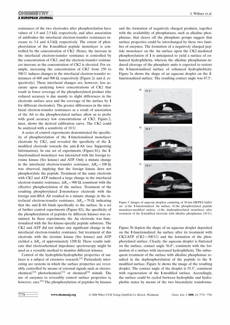
and the formation of negatively charged products, togetherwith the availability of phosphatases, such as alkaline phos-phatase, that cleave off the phosphate groups suggest thatsurface properties could be interchanged by these two fami-lies of enzymes. The formation of a negatively charged pep-tide monolayer on the Au surface upon the CK2-mediatedphosphorylation of 1 is anticipated to yield a surface of en-hanced hydrophilicity, whereas the alkaline phosphatase-in-duced cleavage of the phosphate units is expected to restorethe 1-functionalized surface of enhanced hydrophobicity.Figure 3a shows the shape of an aqueous droplet on the 1-functionalized surface. The resulting contact angle was 67.58.
Figure 3b depicts the shape of an aqueous droplet depositedon the 1-functionalized Au surface after its treatment withCK2/ATP (CK2=500 U) and the formation of the phos-phorylated surface. Clearly, the aqueous droplet is flattenedon the surface, contact angle 36.88, consistent with the for-mation of a surface with increased hydrophilicity. The subse-quent treatment of the surface with alkaline phosphatase re-sulted in the dephosphorylation of the peptide to the 1-modified surface. Figure 3c shows the image of the resultingdroplet. The contact angle of the droplet is 55.38, consistentwith regeneration of the 1-modified surface. Accordingly,the surface could be cycled between hydrophilic and hydro-phobic states by means of the two biocatalytic transforma-
Figure 3. Images of aqueous droplets consisting of 50-mm HEPES bufferon: a) the 1-functionalized Au surface, b) the phosphorylated peptidemonolayer-modified surface, c) the dephosphorylated monolayer aftertreatment of the 2-modified electrode with alkaline phosphatase (20 U).
www.chemeurj.org � 2008 Wiley-VCH Verlag GmbH&Co. KGaA, Weinheim Chem. Eur. J. 2008, 14, 7774 – 77817778
I. Willner et al.
tions. Control experiments re-vealed that treatment of the 1-modified surface with eitherCK2 or ATP alone did notaffect the contact angle of the1-modified surface. This im-plies that the phosphorylationand dephosphorylation pro-cesses are, indeed, responsiblefor the observed changes inthe contact angles. The biocat-alytic control over the hydro-philicity of the surface is at-tributed to the fact that phos-phorylation of the serine unitin the 1-modified surfaceyields an anionic site that en-hances the hydrophilic natureof the monolayer-modified sur-face. The dephosphorylation ofthe phosphate group by phos-phatase regenerated the serineunit, resulting in a surface ofhydrophobicity comparable tothe original 1-modified surface.The characterization of bio-
molecular interactions bymeans of chemical-force meas-urements has been addressedin numerous studies.[26] For ex-ample, DNA hybridization,[27]
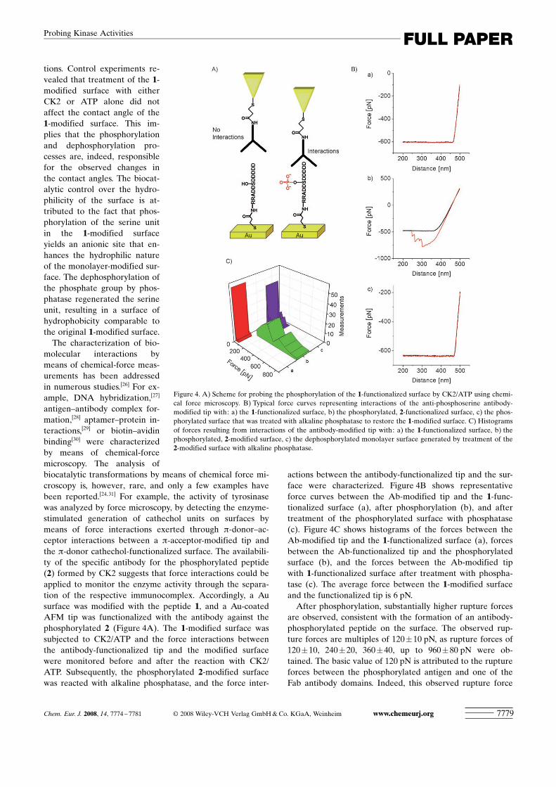
antigen–antibody complex for-mation,[28] aptamer–protein in-teractions,[29] or biotin–avidinbinding[30] were characterizedby means of chemical-forcemicroscopy. The analysis ofbiocatalytic transformations by means of chemical force mi-croscopy is, however, rare, and only a few examples havebeen reported.[24,31] For example, the activity of tyrosinasewas analyzed by force microscopy, by detecting the enzyme-stimulated generation of cathechol units on surfaces bymeans of force interactions exerted through p-donor–ac-ceptor interactions between a p-acceptor-modified tip andthe p-donor cathechol-functionalized surface. The availabili-ty of the specific antibody for the phosphorylated peptide(2) formed by CK2 suggests that force interactions could beapplied to monitor the enzyme activity through the separa-tion of the respective immunocomplex. Accordingly, a Ausurface was modified with the peptide 1, and a Au-coatedAFM tip was functionalized with the antibody against thephosphorylated 2 (Figure 4A). The 1-modified surface wassubjected to CK2/ATP and the force interactions betweenthe antibody-functionalized tip and the modified surfacewere monitored before and after the reaction with CK2/ATP. Subsequently, the phosphorylated 2-modified surfacewas reacted with alkaline phosphatase, and the force inter-
actions between the antibody-functionalized tip and the sur-face were characterized. Figure 4B shows representativeforce curves between the Ab-modified tip and the 1-func-tionalized surface (a), after phosphorylation (b), and aftertreatment of the phosphorylated surface with phosphatase(c). Figure 4C shows histograms of the forces between theAb-modified tip and the 1-functionalized surface (a), forcesbetween the Ab-functionalized tip and the phosphorylatedsurface (b), and the forces between the Ab-modified tipwith 1-functionalized surface after treatment with phospha-tase (c). The average force between the 1-modified surfaceand the functionalized tip is 6 pN.After phosphorylation, substantially higher rupture forces
are observed, consistent with the formation of an antibody-phosphorylated peptide on the surface. The observed rup-ture forces are multiples of 120�10 pN, as rupture forces of120�10, 240�20, 360�40, up to 960�80 pN were ob-tained. The basic value of 120 pN is attributed to the ruptureforces between the phosphorylated antigen and one of theFab antibody domains. Indeed, this observed rupture force
Figure 4. A) Scheme for probing the phosphorylation of the 1-functionalized surface by CK2/ATP using chemi-cal force microscopy. B) Typical force curves representing interactions of the anti-phosphoserine antibody-modified tip with: a) the 1-functionalized surface, b) the phosphorylated, 2-functionalized surface, c) the phos-phorylated surface that was treated with alkaline phosphatase to restore the 1-modified surface. C) Histogramsof forces resulting from interactions of the antibody-modified tip with: a) the 1-functionalized surface, b) thephosphorylated, 2-modified surface, c) the dephosphorylated monolayer surface generated by treatment of the2-modified surface with alkaline phosphatase.
Chem. Eur. J. 2008, 14, 7774 – 7781 � 2008 Wiley-VCH Verlag GmbH&Co. KGaA, Weinheim www.chemeurj.org 7779
FULL PAPERProbing Kinase Activities
value is in the range of rupture forces of other antigen–anti-body immunocomplexes originating from a single Fabdomain.[32] Treatment of the phosphorylated surface with al-kaline phosphatase resulted in a surface that revealed verylow rupture forces with the antibody-modified tip, 35�4 pN.This is consistent with alkaline phosphatase removing thephosphate sites, hence depleting the affinity for interactionbetween the tip and the surface.In conclusion, the present study has introduced three dif-
ferent methods to probe the biocatalytic activity of CK2 onthe 1-functionalized surface. Electrochemical impedancespectroscopy may be used as quantitative method to analyzethe activity of the kinase and to follow the phosphorylationprocess by amplification of the phosphorylated productthrough the generation of the respective antigen–antibodycomplex. Chemical force microscopy enabled us to charac-terize the specific molecular interactions between the phos-phorylated product and the antibody, and contact-anglemeasurements allowed us to monitor the biocatalytic perfor-mance of the kinase by probing the hydrophilicity of thephosphorylated/dephosphorylated peptide-modified surface.We applied these methods to analyze “pure” kinase samples,however, a future goal is their application to actual biologi-cal samples.
Experimental Section
All chemicals were purchased from Aldrich and used without further pu-rification. Ultrapure water from a NANOpure Diamond (Barnstead)source was used throughout all experiments. Casein kinase 2 was pur-chased from New England BioLabs. The substrate of the enzyme (1), ad-enosine triphosphate (ATP), monoclonal anti-phosphoserine antibody,and alkaline phosphatase (E.C. 3.1.3.1) were purchased from Sigma. Ty-rosine kinase-Src kinase was purchased from Cell Signaling Technologies.The Src kinase substrate was synthesized by Biosight, Israel.
Modification of electrodes : Au-coated (50 nm Au layer) glass plates (22I22 mm2) (Analytical m-Systems, Germany) were sonicated with acetoneand isopropanol for 30 min and then washed with ultrapure water. Theslides were then dried under a nitrogen stream and immersed for 20 minin a solution of 2 mg of dithio-diNHS (di(N-succinimidyl)-3,3’-dithiodi-propionate) in 1 mL of DMSO. After multiple rinsing cycles with DMSOand subsequent rinsing with HEPES buffer (0.05m, pH 7.4) the slideswere introduced into a solution of CK2 substrate (peptide 1, 0.01 mg) inHEPES buffer (0.05m, pH 7.4) for 1 h in the presence of 0.1 mm of ATP.After rinsing the surfaces with HEPES buffer (0.05m) the slides weremodified with a mercaptohexanol solution (1 mm) for 30 sec to fill anypinhole defects, and to prevent nonspecific adsorption. The resultingmodified slides were then reacted with different concentrations of CK2in Tris buffer, supplied with the enzyme (20 mm, pH 7.5), that includedKCl (50 mm) and MgCl2 (10 mm). One unit of enzyme (U) is the amountof enzyme that transfers 1 pmole of phosphate to peptide 1 (100 mm) inone minute at 308C in a reaction volume of 25 mL. Alkaline phosphatase(ALP) was treated in borate buffer (pH 9.3) and MgCl2 (1 mm).
Electrochemical surface characterization and contact-angle measure-ments : Electrochemical impedance spectroscopy (EIS) measurements onthe modified electrodes were performed using an Autolab electrochemi-cal analyzer (EcoChemie, The Netherlands) connected to a PC (FRAversion4.9 software). Samples were in a [Fe(CN)6]
3�/4� solution (5 mm) inHEPES buffer (0.05m) with a graphite counter-electrode and a saturatedcalomel reference electrode (SCE). The electrochemical impedance spec-tra were recorded by applying a bias potential of 0.2 V vs. SCE, and ap-
plying a 5-mV alternate voltage, using 30 equally spaced frequencies, inthe frequency range of 100 mHz–10 kHz as perturbation voltage. Electro-chemical impedance spectra were plotted as Nyquist diagrams in theform of complex plane diagrams (real impedance Z’ plotted vs compleximpedance Z’’). The experimental impedance spectra were fitted by usingelectric equivalent circuits. For this purpose commercial software (Zview,version2.1b, Scribner Associates, Inc.) was employed.
Static contact-angle measurements were performed on the modified Auby using a CAM 2000 Optical-Angle Analyzer (KSV Instruments, Fin-land). A droplet of the 0.05-m HEPES buffer solution, approximately 20-mL with diameter of roughly 0.5 cm, was deposited on the surface byusing a syringe. The images of the droplets were recorded and each con-tact-angle measurement was repeated at least three times. The reportedvalue represents the average of these results.
AFM : Au-coated AFM cantilevers (Csc 38 Cr–Au MikroMasch) weretreated with acetone and isopropanol for 30 min and then washed withultrapure water. The cantilevers were then immersed for 20 min in a solu-tion of 2 mg of dithio-diNHS (di(N-succinimidyl)-3,3’-dithiodipropionate)in 1 mL of DMSO. After multiple rinsing cycles with DMSO and subse-quent rinsing with HEPES buffer (0.05m, pH 7.4), the cantilevers wereintroduced into a solution of 0.01 mg of anti-phosphoserine Ab (Sigma) inHEPES buffer (0.05m, pH 7.4) for 1 h.
Force measurements were carried out at RT by using a Multimode scan-ning probe microscope with a Nanoscope 3A controller and a Pico Forcemodule (Digital Instruments, Veeco Probes, Santa Barbara, CA). Thespring constants of the Au-coated cantilevers (MikroMasch, Germany)were determined in air by using the thermal-noise method to give anaverage spring constant of 0.012 Nm�1. All experiments were conductedin the Tris buffer/kinase solution or the alkaline phosphatase/boratebuffer in a liquid cell. To measure the force interactions, the probe tipwas lowered to the surface and immediately retracted at a rate of0.1 mms�1, and data points were analyzed with their associated springconstants. Histograms were prepared by using Origin software. Each his-togram was the result of at least 70 separate force measurements.
Acknowledgement
This research was supported by the Israel Science Foundation within theConverging Technologies Program.
[1] G. Manning, D. B. Whyte, R. Martinez, T. Hunter, S. Sudarsanam,Science 2002, 298, 1912–1934.
[2] P. Cohen, Nat. Rev. Drug Discovery 2002, 6, 481–490.[3] a) M. Flajolet, G. He, M. Heiman, A. Lin, A. C. Narin, P. Green-
gard, Proc. Natl. Acad. Sci. USA 2007, 104, 4159–4164; b) D. P.Hanger, H. L. Byers, S. Wray, K.-Y. Leung, M. J. Saxton, A. Seer-eeam, C. H. Reynolds, M. A. Ward, B. H. Anderton, J. Biol. Chem.2007, 282, 23645–23654.
[4] J. H. Till, R. S. Annan, S. A. Carr, W. T. Miller, J. Biol. Chem. 1994,269, 7423–7428.
[5] C. Lehel, S. Daniel-Issakani, M. Brasseur, B. Strulovici, Anal. Bio-chem. 1997, 244, 340–346.
[6] a) A. Flower, D. Swift, E. Longman, A. Acornley, P. Hemsley, D.Murray, J. Unitt, I. Dale, E. Sullivan, M. Coldwell, Anal. Biochem.2002, 308, 223–231; b) T. C. Turek-Etienne, M. Lei, J. S. Terracciano,E. F. Langsdorf, R. W. Bryant, R. F. Hart, A. C. Horan, J. Biomol.Screening 2004, 9, 53–61.
[7] K. Kerman, H.-B. Kraatz, Chem. Commun. 2007, 519–521.[8] J. Oishi, Y. Asami, T. Mori, J.-H. Kang, M. Tanabe, T. Niidome, Y.
Katayama, ChemBioChem 2007, 5, 875–879.[9] K. Kupcho, R. Somberg, B. Bulleit, S. A. Goueli, Anal. Biochem.2003, 317, 210–217.
[10] R. Freeman, R. Gill, I. Willner, Chem. Commun. 2007, 3450–3452.
www.chemeurj.org � 2008 Wiley-VCH Verlag GmbH&Co. KGaA, Weinheim Chem. Eur. J. 2008, 14, 7774 – 77817780
I. Willner et al.
[11] a) B. Guerra, O.-G. Issinger, Electrophoresis 1999, 20, 391–408;b) L. A. Pinna, Biochim. Biophys. Acta 1990, 1054, 267–284; c) J. E.Allende, C. C. Allende, FASEB J. 1995, 9, 313–323.
[12] O.-G. Issinger, Pharmacol. Ther. 1993, 59, 1–30.[13] E. Katz, I. Willner, Electroanalysis 2003, 15, 913–947.[14] F. Patolsky, A. Lichtenstein, I. Willner, Angew. Chem. 2000, 112,
970–973; Angew. Chem. Int. Ed. 2000, 39, 940–943.[15] a) F. Patolsky, A. Lichtenstein, I. Willner, J. Am. Chem. Soc. 2001,
123, 5194–5205; b) T. H. Degefa, J. Kwak, J. Electroanal. Chem.2008, 612, 37–41.
[16] M. Zayats, Y. Huang, R. Gill, C.-A. Ma, I. Willner, J. Am. Chem.Soc. 2006, 128, 13666–13667.
[17] a) A. B. Kharitonov, L. Alfonta, E. Katz, I. Willner, J. Electroanal.Chem. 2000, 487, 133–141; b) R. Pei, Z. Cheng, E. Wang, X. Yang,Biosens. Bioelectron. 2001, 16, 355–361.
[18] L. Alfonta, I. Willner, Chem. Commun. 2001, 1492–1493.[19] F. Patolsky, A. Lichtenstein, I. Willner, Chem. Eur. J. 2003, 9, 1137–
1145.[20] V. Pardo-Yissar, E. Katz, O. Lioubashevski, I. Willner, Langmuir
2001, 17, 1110–1118.[21] Y. Liu, L. Mu, B. Liu, J. Kong, Chem. Eur. J. 2005, 11, 2622–2631.[22] a) J. Lahann, S. Mitragotri, T.-N. Tran, H. Kaido, J. Sundaram, I. S.
Choi, S. Hoffer, G. A. Somorjai R. Langer, Science 2003, 299, 371–374; b) X. Wang, A. B. Kharitonov, E. Katz, I. Willner, Chem.Commun. 2003, 1542–1543; c) E. Katz, O. Lioubashevsky, I. Will-ner, J. Am. Chem. Soc. 2004, 126, 15520–15532; d) M. Riskin, B.Basnar, V. I. Chegel, E. Katz, I. Willner, F. Shi, X. Zhang, J. Am.Chem. Soc. 2006, 128, 1253–1260; e) M. Riskin, B. Basnar, E. Katz,I. Willner, Chem. Eur. J. 2006, 12, 8549–8557.
[23] a) X. Wang, S. Zeevi, A. B. Kharitonov, E. Katz, I. Willner, Phys.Chem. Chem. Phys. 2003, 5, 4236–4241; b) R. Rosario, D. Gust,A. A. Garcia, M. Hayes, J. L. Taraci, T. Clement, J. W. Dailey, S. T.Picraux, J. Phys. Chem. B 2004, 108, 12640–12642; c) W. H. Jiang,G. J. Wang, Y. N. He, X. G. Wang, Y. L. An, Y. L. Song, L. Jiang,Chem. Commun. 2005, 3550–3552.
[24] A. Wieckowska, A. B. Braunschweig, I. Willner, Chem. Commun.2007, 3918–3920.
[25] A. B. Braunschweig, R. Elnathan, I. Willner, Nano Lett. 2007, 7,2030–2036.
[26] a) A. Janshoff, M. Neitzert, Y. Oberdçrfer, H. Fuchs, Angew. Chem.2000, 112, 3346–3374; Angew. Chem. Int. Ed. 2000, 39, 3212–3237;b) H. Beyer, M. K. Clausen-Schaumann, Chem. Rev. 2005, 105,2921–2948.
[27] a) G. U. Lee, L. A. Chrisey, R. J. Colton, Science 1994, 266, 771–773; b) T. Strunz, K. Oroszlan, R. SchRfer, H. J. GSnterodt, Proc.Natl. Acad. Sci. USA 1999, 96, 11277–11282; c) R. Krautbauer, M.Rief, H. E. Gaub, Nano Lett. 2003, 3, 493–496.
[28] R. Ros, F. Schwesinger, D. Anselmetti, M. Kubon, R. SchRfer, A.Plueckthun, L. Tiefenauer, Proc. Natl. Acad. Sci. USA 1998, 95,7402–7405.
[29] B. Basnar, R. Elnathan, I. Willner, Anal. Chem. 2006, 78, 3638–3642.
[30] a) V. T. Moy, E.-L. Florin, H. E. Gaub, Science 1994, 266, 257–259;b) E.-L. Florin, V. T. Moy, H. E. Gaub, Science 1994, 264, 415–417;c) G. U. Lee, D. A. Kidwell, R. J. Colton, Langmuir 1994, 10, 354–357.
[31] a) T. Suzuki, Y.-W. Zhang, T. Koyama, D. Y. Sasaki, K. Kurihara, J.Am. Chem. Soc. 2006, 128, 15209–15214; b) M. Sletmoen, G. SkjTk-Braek, B. T. Stokke, Carbohydr. Res. 2005, 340, 2782–2795.
[32] a) U. Dammer, M. Hegner, D. Anselmetti, P. Wagner, M. Dreier, W.Huber, H.-J. GSntherodt, Biophys. J. 1996, 70, 2437–2441; b) S.Allen, X. Chen, M. C. Davies, A. C. Dawkes, J. C. Edwards, C. J.Roberts, J. Sefton, S. J. B. Tendler, P. M. Williams, Biochemistry1997, 36, 7457–7463; c) S. Allen, J. Davies, M. C. Davies, A. C.Dawkes, C. J. Roberts, S. J. B. Tendler, P. M. Williams, Biochem. J.1999, 341, 173–178.
Received: April 22, 2008Published online: August 12, 2008
Chem. Eur. J. 2008, 14, 7774 – 7781 � 2008 Wiley-VCH Verlag GmbH&Co. KGaA, Weinheim www.chemeurj.org 7781
FULL PAPERProbing Kinase Activities