Embed Size (px)
Citation preview
Progenitor Cell Maturation and Initiation of Neurogenesis in the Developing Vertebrate Neural Retina
A Dissertation Submitted to the Faculty of the Graduate School
Of the University of Minnesota by
Hyun-Jin Yang
In Partial Fulfillment of the Requirements for the Degree of
Doctor of Philosophy
Advisor Steven C McLoon
October 2009
copy Hyun-Jin Yang October 2009
i
Abstract
The mature vertebrate central nervous system is composed of an enormous number
of neuronal and glial cells A relatively small number of progenitor cells generate
these cells during a finite period of time of development Progenitor cells during early
stages of central nervous system development divide so that each division produces
two progeny that divide again This lsquopreneurogenicrsquo mode of division is essential for
the exponential increase of number of progenitor cells Later progenitor cells change
their mode of division to lsquoneurogenicrsquo where one or both daughter cells produced by
a division withdraw from the mitotic cycle and differentiate This more mature
neurogenic division is critical for generation of a functional nervous system The aim
of the project described in this thesis was to understand 1) the molecular differences
that dictate the two modes of progenitor cell division namely preneurogenic and
neurogenic 2) the mechanism that regulates the switch in the mode of division and
3) the molecular trigger that initiates differentiation
Molecular differences between preneurogenic and neurogenic progenitor
cells were identified and are described in more detail in chapter II The early
preneurogenic progenitor cells express the transcription factor Sox2 and a ligand for
the Notch receptor Delta1 The more mature neurogenic progenitor cells express
Sox2 and the bHLH transcription factor E2A and do not express Delta1
Perturbation of Notch signaling resulted in conversion of progenitor cells from
preneurogenic to neurogenic and in premature neurogenesis Furthermore Sonic
hedgehog was found to be expressed by a subset of newly differentiating cells
Misexpression of Sonic hedgehog led to premature maturation of preneurogenic
progenitor cells and neurogenesis These results suggest that Notch signaling
maintains progenitor cells in the preneurogenic state and that Sonic hedgehog
ii
initiates progenitor cell maturation
Certain proneural bHLH transcription factors were found to initiate neurogenesis and
are described in more detail in chapter III Expression of a number of proneural bHLH
factors comes up in a stereotypic temporal sequence prior to the onset of ganglion
cell differentiation Ascl1 and Neurog2 were expressed first which was followed by
expression of Neurod1 and Neurod4 Finally Atoh7 was expressed which preceded
the appearance of ganglion cells Individual progenitor cells expressed
heterogeneous combinations of proneural genes prior to ganglion cell genesis
Misexpression of Ascl1 or Neurog2 in preneurogenic retina was sufficient to initiate
ganglion cell genesis Misexpression of Neurog2 initiated the stereotypic sequence of
proneural gene expression that normally preceded ganglion cell genesis Ascl1
expression was also sufficient to initiate ganglion cell genesis However it functioned
by a mechanism distinct from that of Neurog2 These results suggest that ganglion
cell genesis may be initiated by two different mechanisms
iii
Table of Contents
COVER
COPYRIGHT
ABSTRACThelliphelliphelliphelliphelliphelliphelliphelliphelliphelliphelliphelliphelliphelliphelliphelliphelliphelliphelliphelliphelliphelliphelliphelliphelliphelliphelliphelliphellip i
TABLE OF CONTENTShelliphelliphelliphelliphelliphelliphelliphelliphelliphelliphelliphelliphelliphelliphelliphelliphelliphelliphelliphelliphelliphelliphellip iii
LIST OF TABLEShelliphelliphelliphelliphelliphelliphelliphelliphelliphelliphelliphelliphelliphelliphelliphelliphelliphelliphelliphelliphelliphelliphelliphelliphelliphellip vii
LIST OF FIGUREShelliphelliphelliphelliphelliphelliphelliphelliphelliphelliphelliphelliphelliphelliphelliphelliphelliphelliphelliphelliphelliphelliphelliphelliphellip viii
CHAPTER I INTRODUCTIONhelliphelliphelliphelliphelliphelliphelliphelliphelliphelliphelliphelliphelliphelliphelliphelliphelliphelliphelliphellip 1
IA Overview of the Thesishelliphelliphelliphelliphelliphelliphelliphelliphelliphelliphelliphelliphelliphelliphelliphelliphelliphelliphelliphellip 2
IB Embryology of the Vertebrate Retinahelliphelliphelliphelliphelliphelliphelliphelliphelliphelliphelliphelliphelliphellip 3
IC Molecular Differences between Preneurogenic and Neurogenic Progenitor Cellshelliphelliphelliphelliphelliphelliphelliphelliphelliphelliphelliphelliphelliphelliphelliphelliphelliphelliphelliphelliphelliphelliphelliphelliphellip
8
ICi Issuehelliphelliphelliphelliphelliphelliphelliphelliphelliphelliphelliphelliphelliphelliphelliphelliphelliphelliphelliphelliphelliphelliphelliphelliphellip 8
ICii Current Model from Studies in Other Tissues of the Central Nervous Systemhelliphelliphelliphelliphelliphelliphelliphelliphelliphelliphelliphelliphelliphelliphelliphelliphelliphelliphelliphelliphelliphellip
8
ICiii E Proteins Class I HLH Family Transcription Factorshelliphelliphellip 11
ICiv Delta-Notch signalinghelliphelliphelliphelliphelliphelliphelliphelliphelliphelliphelliphelliphelliphelliphelliphelliphellip 15
ICv Hypothesishelliphelliphelliphelliphelliphelliphelliphelliphelliphelliphelliphelliphelliphelliphelliphelliphelliphelliphelliphelliphelliphellip 18
ID Regulation of Progenitor Cell Maturationhelliphelliphelliphelliphelliphelliphelliphelliphelliphelliphelliphelliphellip 19
IDi Issuehelliphelliphelliphelliphelliphelliphelliphelliphelliphelliphelliphelliphelliphelliphelliphelliphelliphelliphelliphelliphelliphelliphelliphelliphellip 19
IDii Candidate Factors for Regulator of Progenitor Cell Maturation 19
IDiii Intrinsic Timer Mechanismhelliphelliphelliphelliphelliphelliphelliphelliphelliphelliphelliphelliphelliphelliphellip 20
IDiv Hedgehog (Hh) Signalinghelliphelliphelliphelliphelliphelliphelliphelliphelliphelliphelliphelliphelliphelliphellip 22
IDv Fibroblast Growth Factor (FGF) Signalinghelliphelliphelliphelliphelliphelliphelliphelliphellip 27
IDvi Delta-Notch Signalinghelliphelliphelliphelliphelliphelliphelliphelliphelliphelliphelliphelliphelliphelliphelliphelliphellip 30
iv
IDvii Wnt Signalinghelliphelliphelliphelliphelliphelliphelliphelliphelliphelliphelliphelliphelliphelliphelliphelliphelliphelliphelliphellip 31
IDviii Hypothesishelliphelliphelliphelliphelliphelliphelliphelliphelliphelliphelliphelliphelliphelliphelliphelliphelliphelliphelliphelliphellip 35
IE Initiation of Neurogenesishelliphelliphelliphelliphelliphelliphelliphelliphelliphelliphelliphelliphelliphelliphelliphelliphelliphelliphellip 36
IEi Issuehelliphelliphelliphelliphelliphelliphelliphelliphelliphelliphelliphelliphelliphelliphelliphelliphelliphelliphelliphelliphelliphelliphelliphelliphellip 36
IEii Initiation of Neurogenesis in Drosophila Eye Imaginal Dischellip 36
IEiii Initiation of Neurogenesis in the Vertebrate Retinahelliphelliphelliphelliphellip 37
IEiv Proneural Genes Class II HLH Family Transcription Factors 38
IEv Hypothesishelliphelliphelliphelliphelliphelliphelliphelliphelliphelliphelliphelliphelliphelliphelliphelliphelliphelliphelliphelliphelliphellip 44
CHAPTER II PROGENITOR CELL MATURATION IN THE DEVELOPING VERTEBRATE RETINAhelliphelliphelliphelliphelliphelliphelliphelliphelliphelliphelliphelliphelliphelliphelliphelliphelliphelliphelliphelliphelliphelliphellip
45
IIA Introductionhelliphelliphelliphelliphelliphelliphelliphelliphelliphelliphelliphelliphelliphelliphelliphelliphelliphelliphelliphelliphelliphelliphelliphelliphellip 46
IIB Experimental Procedureshelliphelliphelliphelliphelliphelliphelliphelliphelliphelliphelliphelliphelliphelliphelliphelliphelliphelliphellip 48
IIC Resultshelliphelliphelliphelliphelliphelliphelliphelliphelliphelliphelliphelliphelliphelliphelliphelliphelliphelliphelliphelliphelliphelliphelliphelliphelliphelliphellip 52
IICi Differentiation Markers Distinguish Distinct Preneurogenic and Neurogenic Zones in the Developing Chick Retinahelliphelliphelliphelliphellip
52
IICii Sox2 Is Expressed by Neural Retinal Progenitor Cellshelliphellip 53
IICiii Delta1 Is Expressed by Preneurogenic Progenitor Cells and Not by Neurogenic Progenitor Cellshelliphelliphelliphelliphelliphelliphelliphelliphelliphelliphelliphelliphelliphellip
55
IICiv E2A Expression Identifies Neurogenic Progenitor Cellshelliphellip 57
IICv Interrupting Delta-Notch Signaling Converted Preneurogenic to Neurogenic Progenitor Cellshelliphelliphelliphelliphelliphelliphelliphelliphelliphelliphelliphelliphelliphelliphelliphellip
58
IICvi Shh Drives Maturation of Preneurogenic to Neurogenic Progenitor Cellshelliphelliphelliphelliphelliphelliphelliphelliphelliphelliphelliphelliphelliphelliphelliphelliphelliphelliphelliphelliphelliphellip
61
IID Discussionhelliphelliphelliphelliphelliphelliphelliphelliphelliphelliphelliphelliphelliphelliphelliphelliphelliphelliphelliphelliphelliphelliphelliphelliphellip 63
IIDi Molecular Distinction between Preneurogenic and Neurogenic Progenitor Cellshelliphelliphelliphelliphelliphelliphelliphelliphelliphelliphelliphelliphelliphelliphelliphelliphellip
63
IIDii Maintenance of the Preneurogenic Mode of Division by Delta-Notch Signlainghelliphelliphelliphelliphelliphelliphelliphelliphelliphelliphelliphelliphelliphelliphelliphelliphelliphelliphelliphellip
65
v
IIDiii Control of Progenitor Cell Maturationhelliphelliphelliphelliphelliphelliphelliphelliphelliphellip 66
CHAPTER III SEQUENTIAL CHANGES IN EXPRESSION OF PRONEURAL bHLH TRANSCRIPTION FACOTRS INITIATING NEUROGENESIS IN THE DEVELOPING RETINAhelliphelliphelliphelliphelliphelliphelliphelliphelliphelliphelliphelliphelliphelliphelliphelliphelliphelliphelliphelliphelliphelliphelliphellip
91
IIIA Introductionhelliphelliphelliphelliphelliphelliphelliphelliphelliphelliphelliphelliphelliphelliphelliphelliphelliphelliphelliphelliphelliphelliphelliphelliphellip 92
IIIB Experimental Procedureshelliphelliphelliphelliphelliphelliphelliphelliphelliphelliphelliphelliphelliphelliphelliphelliphelliphelliphellip 95
IIIC Resultshelliphelliphelliphelliphelliphelliphelliphelliphelliphelliphelliphelliphelliphelliphelliphelliphelliphelliphelliphelliphelliphelliphelliphelliphelliphellip 99
IIICi Expression of Multiple Proneural Genes Precedes the Generation of Retinal Ganglion Cellshelliphelliphelliphelliphelliphelliphelliphelliphelliphelliphelliphelliphellip
99
IIICii Cells Expressing a Variety of Proneural Genes Differentiate as Retinal Ganglion Cellshelliphelliphelliphelliphelliphelliphelliphelliphelliphelliphelliphelliphelliphelliphelliphelliphelliphellip
100
IIICiii The Onset of Proneural Gene Expression Prior to Ganglion Cell Genesis Is in a Stereotypic Sequencehelliphelliphelliphelliphelliphelliphelliphelliphelliphelliphellip
101
IIICiv Retinal Progenitor Cells Are Heterogeneous Prior to Neurogenesishelliphelliphelliphelliphelliphelliphelliphelliphelliphelliphelliphelliphelliphelliphelliphelliphelliphelliphelliphelliphelliphelliphellip
103
IIICv Misexpression of Ascl1 or Neurog2 Can Initiate Premature Ganglion Cell Genesishelliphelliphelliphelliphelliphelliphelliphelliphelliphelliphelliphelliphelliphelliphelliphelliphelliphelliphelliphellip
105
IIICvi Neurog2 and Ascl1 Initiate Neurogenesis via Different Mechanismhelliphelliphelliphelliphelliphelliphelliphelliphelliphelliphelliphelliphelliphelliphelliphelliphelliphelliphelliphelliphelliphelliphelliphellip
107
IIID Discussionhelliphelliphelliphelliphelliphelliphelliphelliphelliphelliphelliphelliphelliphelliphelliphelliphelliphelliphelliphelliphelliphelliphelliphellip 109
IIIDi Initiation of Neurogenesis in the Developing Retinahelliphelliphelliphellip 109
IIDii Stereotypic Sequence of Proneural Gene Expression Prior to Ganglion Cell Genesishelliphelliphelliphelliphelliphelliphelliphelliphelliphelliphelliphelliphelliphelliphelliphelliphelliphelliphelliphellip
110
IIDiii Heterogeneity of Progenitor Cells Prior to the Onset of Neurogenesishelliphelliphelliphelliphelliphelliphelliphelliphelliphelliphelliphelliphelliphelliphelliphelliphelliphelliphelliphelliphelliphelliphellip
112
IIIDiv Relationship between Proneural bHLH Transcription Factors and Cell Fatehelliphelliphelliphelliphelliphelliphelliphelliphelliphelliphelliphelliphelliphelliphelliphelliphelliphelliphelliphelliphelliphelliphelliphellip
113
CHAPTER IV CONCLUSIONS AND FUTURE DIRECTIONShelliphelliphelliphelliphelliphelliphellip
134
IVA Conclusionshelliphelliphelliphelliphelliphelliphelliphelliphelliphelliphelliphelliphelliphelliphelliphelliphelliphelliphelliphelliphelliphelliphelliphellip 135
IVAi Molecular Distinction between Preneurogenic and Neurogenic Progenitor Cellshelliphelliphelliphelliphelliphelliphelliphelliphelliphelliphelliphelliphelliphelliphelliphelliphellip
135
vi
IVAii Control of Progenitor Cell Maturationhelliphelliphelliphelliphelliphelliphelliphelliphelliphellip 136
IVAiii Initiation of Neurogenesis by Proneural bHLH Transcription Factors in the Developing Vertebrate Retinahelliphelliphelliphelliphelliphelliphelliphelliphelliphellip
137
IVB Future Directionshelliphelliphelliphelliphelliphelliphelliphelliphelliphelliphelliphelliphelliphelliphelliphelliphelliphelliphelliphelliphelliphellip 139
IVBi Differential Function of Notch Signaling in Two Progenitor Cell Stageshelliphelliphelliphelliphelliphelliphelliphelliphelliphelliphelliphelliphelliphelliphelliphelliphelliphelliphelliphelliphelliphelliphelliphellip
139
IVBii Ascl1 Initiated Gene Expression Cascade and Retinal Ganglion Cell Subtype Specificationhelliphelliphelliphelliphelliphelliphelliphelliphelliphelliphelliphelliphelliphellip
140
BIBLIOGRAPHYhelliphelliphelliphelliphelliphelliphelliphelliphelliphelliphelliphelliphelliphelliphelliphelliphelliphelliphelliphelliphelliphelliphelliphelliphelliphelliphellip 142
vii
List of Table
CHAPTER III SEQUENTIAL CHANGES IN EXPRESSION OF PRONEURAL bHLH TRANSCRIPTION FACOTRS INITIATING NEUROGENESIS IN THE DEVELOPING RETINA
Table III-1 Sequential Induction of Proneural Gene Expression Following Neurog2 Misexpressionhelliphelliphelliphelliphelliphelliphelliphelliphelliphelliphelliphelliphelliphelliphelliphelliphelliphelliphelliphelliphellip
133
viii
List of Figures
CHAPTER II PROGENITOR CELL MATURATION IN THE DEVELOPING VERTEBRATE RETINA
Figure II-1Distinct Preneurogenic and Neurogenic Zone in the Developing Chick Neural Retinahelliphelliphelliphelliphelliphelliphelliphelliphelliphelliphelliphelliphelliphelliphelliphelliphelliphellip
70
Figure II-2 Delta1 Expression Relative to Cell Differentiation in Developing Retina helliphelliphelliphelliphelliphelliphelliphelliphelliphelliphelliphelliphelliphelliphelliphelliphelliphelliphelliphelliphelliphelliphelliphellip
72
Figure II-3 Differentiation of Dissociated Preneurogenic Progenitor Cells
74
Figure II-4 Absence of Sox2 Expression by Differentiating Cellshelliphelliphelliphellip 76
Figure II-5 Delta1 Expression in Preneurogenic and Neurogenic Zones 78
Figure II-6 Delta1 Expression Relative to Cell Division in Neurogenic and Preneurogenic Regions of the Retinahelliphelliphelliphelliphelliphelliphelliphelliphelliphelliphelliphelliphelliphelliphelliphellip
80
Figure II-7 E2A Expression in the Neurogenic Zonehelliphelliphelliphelliphelliphelliphelliphelliphellip 82
Figure II-8 Conversion of Preneurogenic to Neurogenic Progenitor Cells Following Reduced Notch Signalinghelliphelliphelliphelliphelliphelliphelliphelliphelliphelliphelliphelliphelliphelliphelliphellip
84
Figure II-9 Expression of Shh by a Subset of Newly Differentiating Ganglion Cellshelliphelliphelliphelliphelliphelliphelliphelliphelliphelliphelliphelliphelliphelliphelliphelliphelliphelliphelliphelliphelliphelliphelliphelliphelliphellip
86
Figure II-10 Progenitor Cell Maturation Induced by Shh Misexpressionhellip 88
Figure II-11 Summary of Changes in Gene Expression during Retinal Progenitor Cell Maturation
90
CHAPTER III SEQUENTIAL CHANGES IN EXPRESSION OF PRONEURAL bHLH TRANSCRIPTION FACOTRS INITIATING NEUROGENESIS IN THE DEVELOPING RETINA
Figure III-1Expression of Proneural Genes Precedes the Generation of Retinal Ganglion Cellshelliphelliphelliphelliphelliphelliphelliphelliphelliphelliphelliphelliphelliphelliphelliphelliphelliphelliphelliphelliphelliphellip
117
Figure III-2 Newly Generated Retinal Ganglion Cells Express Multiple Proneural Geneshelliphelliphelliphelliphelliphelliphelliphelliphelliphelliphelliphelliphelliphelliphelliphelliphelliphelliphelliphelliphelliphelliphelliphellip
119
Figure III-3 Stereotypic Temporal Sequence of Proneural Gene Expression Prior to Ganglion Cell Genesishelliphelliphellip
121
Figure III-4 Progenitor Cell Heterogeneity Prior to Onset of Neurogenesis 123
ix
Figure III-5 Proneural Genes Expressed by RA4 or Atoh7 Positive Cells 125
Figure III-6 Premature Neurogenesis Induced by Ascl1 Misexpressionhellip 127
Figure III-7 Premature Neurogenesis Induced by Neurog2 Misexpressionhelliphelliphelliphelliphelliphelliphelliphelliphelliphelliphelliphelliphelliphelliphelliphelliphelliphelliphelliphelliphelliphelliphelliphelliphellip
129
Figure III-8 Differential Induction of Proneural Gene Expression by Ascl1 or Neurog2 Misexpression
131
1
CHAPTER I
INTRODUCTION
2
IA Overview of the Thesis
The mature central nervous system of the vertebrate species such as human is
composed of thousands of neuronal and glial cell types with the total cell count reaching
over 100 billion (Williams and Herrup 1988 Pakkenberg and Gundersen 1997) These
enormous numbers of cells are generated from a relatively small number of progenitor
cells the dividing cells of the developing nervous system Initially in the developing
central nervous system the number of progenitor cells expands exponentially without
generating postmitotic cells This is through a series of lsquopreneurogenicrsquo cell divisions in
which both progeny of each division divide again Later in development progenitor cells
switch their mode of division to a lsquoneurogenicrsquo mode In this mode of division one or both
progeny of a division withdraw from the mitotic cycle and differentiate as neurons or glia
This later neurogenic mode of progenitor cell division is essential for generation of the
functional nervous system Despite considerable progress having made in
understanding important developmental processes the following fundamental questions
remain unanswered 1) What molecular differences dictate the differential modes of
progenitor cell division namely preneurogenic and neurogenic 2) what is the
mechanism that regulates the switch in the mode of division and 3) what factor is
responsible for initiation of differentiation This thesis addresses these questions using
the embryonic chick retina as a model Chapter I provides background and rationale of
this thesis Chapter II describes the molecular differences between preneurogenic and
neurogenic progenitor cells and the mechanism underlying progenitor cell maturation
Chapter III investigates the role of proneural bHLH genes in initiating neurogenesis in
the retina
3
IB Embryology of the Vertebrate Retina
The vertebrate neural retina is a region of central nervous system which lines the inner
surface of the back of the eye It contains photoreceptor cells that sense the light coming
through the lens and convert it into the neuro-electrical impulses The converted signal is
transmitted through the interneurons of the retina and eventually to ganglion cells
Ganglion cells send the information to various visual centers of the brain for further
processing
The vertebrate neural retina originates developmentally from the anterior region
of the neural tube (Zuber and Harris 2006) The region specified to become retina
known as the eye field is initially a single field and later separates into two eye primordia
one on each side of the forebrain Subsequently each of the eye primordia evaginates
out of the diencephalon to form an optic vesicle The optic vesicle invaginates to form a
two-layered optic cup and the inner layer develops into the neural retina in response to
an inductive signal from the overlying head ectoderm The outer layer of the optic cup is
a single layer of epithelial cells and it eventually becomes the retinal pigmented
epithelium The optic cup remains attached to the brain through the hollow optic stalk
Retinal ganglion cells grow long projection axons through the inferior wall of the optic
stalk which later becomes the optic nerve While the optic cup is forming other
structures of the eye are also induced The contact between optic vesicle and the
overlying ectoderm induces the formation of the lens placode which invaginates to
develop into the lens and in turn induce cornea from the overlying ectoderm The optic
cup also contributes cells to some of the peripheral structures of eye Cells located at the
peripheral rim of the optic cup contribute to the iris and ciliary body The remaining eye
4
structure including sclera and extra ocular muscles are developed from neural crest cells
and head mesoderm
Retinal progenitor cells undergo multiple rounds of cell division and ultimately
generate six neuronal and one glial cell type Initially retinal progenitor cells at the optic
vesicle and early optic cup stages divide so that each division produces two cells that
divide again This mode of cell division results in an exponential increase in the number
of retinal progenitor cells Therefore this early mode of progenitor cell division is
essential for the rapid growth of the tissue within the limited time of normal development
Hereafter progenitor cell divisions producing two progenitor cells without producing any
postmitotic cells will be designated as lsquopreneurogenicrsquo division Later progenitor cells
change their mode of division and begin to generate postmitotic neurons Once the
switch of cell division modes has occurred progenitor cells undergo asymmetric
divisions in which one of the daughter cells produced by a division withdraws from the
mitotic cycle and differentiates whereas the other daughter cell remains as a progenitor
cell Following a few rounds of asymmetric division progenitor cells undergo terminal
division where both daughter cells begin to differentiate These later modes of progenitor
cell division in which one or both progeny become postmitotic will be designated as
lsquoneurogenicrsquo division This later neurogenic mode of divisions is critical for generation of
a functional nervous system
Progenitor cell maturation that is the switch in the cell division mode of
progenitor cells first occurs in a small number of progenitor cells located in the center of
the retina (Prada et al 1991 Reese and Colello 1992 McCabe et al 1999) It will form
a lsquoneurogenic zonersquo in the center with neurogenic progenitor cells and their postmitotic
progeny A sharp boundary or the lsquoneurogenic frontrsquo can be marked by the most
peripherally located postmitotic cells and separates the neurogenic zone in the center
5
from the preneurogenic zone in the periphery Progenitor cells located peripheral to the
neurogenic front undergo preneurogenic divisions (Dutting et al 1983) The
preneurogenic progenitor cells just peripheral to the neurogenic front soon switch their
mode of division to neurogenic and thereby the neurogenic front advances peripherally
The advance of the neurogenic front continues until the neurogenic front reaches the
peripheral margin of the optic cup and the entire neural retina become neurogenic In
fish and amphibia however dividing cells are retained in the periphery of the mature
retina the ciliary marginal zone (CMZ) These retinal progenitor cells continuously
produce new neurons in the retina throughout the life of an animal (Straznicky and Gaze
1971 Johns 1977 Wetts et al 1989)
Once neurogenesis commences in the developing neural retina a seemingly
homogenous pool of retinal progenitor cells gives rise to six types of retinal neurons and
one type of glial cell (Turner and Cepko 1987 Holt et al 1988 Wetts et al 1989
Turner et al 1990 Fekete et al 1994) These seven major cell types of the retina are
generated in a stereotypic sequence which is conserved across vertebrate species
(Carter-Dawson and LaVail 1979 Young 1985 La Vail et al 1991 Stiemke and
Hollyfield 1995 Cepko et al 1996 Hu and Easter 1999) Retinal ganglion cells are
generated first and are followed by generation of amacrine cells horizontal cells and
cone photoreceptor cells Rod photoreceptor cells bipolar cells and Mϋller glial cells are
produced later in the development The time during which each of the retinal cell types is
generated however exhibits considerable overlap (Young 1985 Stiemke and Hollyfield
1995)
Similar to the other regions of the brain the neural retina forms a highly
laminated structure The retinal cell types are organized into layers Three layers of cell
bodies are separated by two layers of axons and dendrites Retinal progenitor cells
6
undergo mitosis near the ventricular surface (ie towards pigmented epithelium) and the
postmitotic cells migrate to the proper layer of the retina Ganglion cells the first neurons
generated in the retina migrate to the innermost layer of the retina (ie towards vitreous)
and form the ganglion cell layer (GCL) Just outside of the GCL is the inner nuclear layer
(INL) Interneurons of the retina both excitatory and inhibitory migrate to and
differentiate in this layer These interneurons include horizontal cells bipolar cells and
amacrine cells A subset of amacrine cells the displaced amacrine cells resides in the
GCL Cone and rod photoreceptors form the outer nuclear layer (ONL) the outermost
cell layer Interposed between these three cell layers are the inner and outer plexiform
layers (IPL and OPL) These layers are composed of processes of the retinal cells and
synaptic connections between retinal neurons are formed in these layers Muller glia
span the entire thickness of the retina This architecture allows serial processing of
visual information Light is first detected by photoreceptors Photoreceptor cells convert
light stimulation into nerve impulses which is transmitted through interneurons to the
ganglion cells Ganglion cells have long projecting axons that send the signal to the
brain where the information is processed and interpreted as visual perception
The vertebrate neural retina is a region of the central nervous system with easy
accessibility and a relatively simple structure Understanding of many fundamental
developmental events comes from studies of the neural retina as a model The
embryonic neural retina is also a very suitable system to study the mechanism for
progenitor cell maturation and initiation of neurogenesis As described above
neurogenesis of the developing neural retina is first apparent in the center and expands
towards the periphery as development progresses Thus for a period of development a
cross section of the embryonic retina contains an array of cells with various maturation
stages along the central-to-peripheral axis Studies described in this thesis use the
7
developing retina to study the mechanisms underlying progenitor cell maturation and
initiation of neurogenesis
8
IC Molecular Differences
between Preneurogenic and Neurogenic Progenitor Cells
ICi Issue
The entire neural retina is derived from a relatively small number of progenitor cells All
cells of the optic vesicle and the early optic cup are dividing progenitor cells and no
postmitotic cells are present These cells undergo preneurogenic division in which both
resulting daughter cells divide again This series of preneurogenic divisions generates a
large pool of progenitor cells which is essential for growth of the tissue during the finite
period of normal retinal development Later in development retinal progenitor cells
switch their mode of division so that one or both daughter cells exit the mitotic cycle and
begin to differentiate Several rounds of neurogenic division of progenitor cells generate
an array of retinal neurons and glial cells which is essential for generation of the
functional neural retina A fundamental question that remains to be answered is what are
the molecular differences between preneurogenic and neurogenic progenitor cells
ICii Current Model from Studies in Other Tissues of the Central Nervous
System
The nature of preneurogenic and neurogenic progenitor cell divisions in the retina is
poorly understood Most studies addressing the molecular differences between the two
progenitor cell stages focused on other regions of the developing vertebrate central
nervous system including the developing cerebral cortex As retinal progenitor cells have
the same origin as progenitor cells of other tissues of the central nervous system it is
likely that preneurogenic and neurogenic progenitor cells of other parts of the central
9
nervous system share common molecular characteristics with their counterparts in the
retina
Orientation of Cell Cleavage and Differential Distribution of Numb
The current model suggests that the orientation of the cell cleavage plain during
progenitor cell division and the differential distribution of the Numb protein to the two
daughter cells can influence the mode of progenitor cell division in the developing cortex
Studies suggested that progenitor cells with a cleavage plane perpendicular to the
ventricular surface divide symmetrically generating daughter cells that divide again
whereas progenitor cells with a cleavage plane parallel to the ventricular surface divide
asymmetrically generating one daughter cell close to the ventricular surface that divides
again and the other daughter cell away from the ventricular surface differentiates (Martin
1967 Chenn and McConnell 1995) The unequal inheritance of fate-determining
molecule such as Numb has also been implicated in influencing the mode of progenitor
cell division (Wakamatsu et al 1999)
These findings led to the investigation of the role of cell cleavage plane during
progenitor cell division and the distribution of Numb protein to the resulting daughter
cells in the division mode of retinal progenitor cells In contrast to the findings in the
developing cortex however neither the plane of cell cleavage nor the distribution of the
Numb protein correlated to the mode of progenitor cell division in the developing retina
(Silva et al 2002) No difference was found in the frequency of the orientation of the
cleavage planes between preneurogenic or neurogenic zone of the developing retina
Furthermore the Numb protein was present both in the dividing and differentiating
daughter cells unlike what has been suggested in the developing cortex
10
Tis21 and Mnb Expression by Neurogenic Progenitor Cells in Cortex
Several classes of molecules have been linked to the neurogenic division of progenitor
cells in other regions of central nervous system Tis21 (also known as Btg2 or PC3) is
one such example Initially it was described as an immediate early gene whose
expression was induced during neuronal differentiation by growth factors and tumor
promoters in PC12 cells and Swiss 3T3 cells respectively (Lim et al 1987 Bradbury et
al 1991 Fletcher et al 1991 Rouault et al 1996) Tis21 mRNA which is expressed
transiently during G1 phase of the cell cycle was shown to label the neurogenic
progenitor cells in the neural tube (Iacopetti et al 1994 Iacopetti et al 1999
Haubensak et al 2004) The Tis21 protein on the other hand persists through mitosis
and is present in postmitotic cells (Iacopetti et al 1999)
Mnb (also known as Dyrk1A) also has been linked to the neurogenic mode of
division Characterization of Mnb expression in the developing chick neural tube
predicted that expression of Mnb precedes the switch of the mode of division from
preneurogenic to neurogenic and that asymmetric inheritance of Mnb to one of the
daughter cells produced by preneurogenic division may lead to the switch of cell division
mode to neurogenic (Haubensak et al 2004)
Previous characterization of Tis21 and Mnb expression raises the possibility that
they may be a common molecular marker for neurogenic progenitor cells in various
tissues of the central nervous system Expression of both Tis21 (Haubensak et al 2004)
and Mnb (Song et al 1996) was found in the developing mouse retina Since
preneurogenic progenitor cells are maintained only for a short period of development in
mouse retina their expression was not characterized in relation to the mode of
progenitor cell division Thus it will be worth testing whether their expression is specific
to retinal progenitor cells undergoing neurogenic division Due to the limited availability
11
of the reagent expression of Tis21 and Mnb in retinal progenitor cells was not
investigated in this thesis
E Proteins and Components of Delta-Notch Signaling
E proteins and components of Delta-Notch signaling were also considered as candidate
molecules whose expression can distinguish preneurogenic and neurogenic progenitor
cells from each other E proteins are binding partners of proneural bHLH transcription
factors (Lee et al 1995 Naya et al 1995 Gradwohl et al 1996) Although expression
of E proteins is thought to be ubiquitous (Roberts et al 1993) the role of E proteins has
been implicated in several aspects of neuronal differentiation (reviewed in Ik Tsen Heng
and Tan 2003) This raises the possibility that expression of E protein in the central
nervous system may be specific to neurogenic progenitor cells On the other hand
components of Delta-Notch signaling may mark preneurogenic progenitor cells Delta-
Notch signaling has been linked to inhibition of neurogenesis (Dorsky et al 1995 1997
Henrique et al 1997 Scheer et al 2001 Jadhav et al 2006) In addition cells in the
caudal stem zone of the developing neural tube which is analogous to the
preneurogenic progenitor cells are characterized with the uniform expression of Delta
the ligand for Notch (Henrique et al 1995 Caprioli et al 2002 Akai et al 2005)
E proteins and Delta-Notch signaling will be discussed further in the following
section (ICiii)
ICiii E Proteins Class I HLH Family Transcription Factors
Introduction to E Proteins
E proteins include E2A which encodes two splice variants E12 and E47 (Murre et al
1989) HEB which encodes two splice variants ME1a and ME1b (Hu et al 1992) and
12
E2-2 (Henthorn et al 1990 Soosaar et al 1994) Together these proteins compose
the class I HLH family a subclass of the helix-loop-helix (HLH) transcription factor family
They share highly conserved sequences such as a basic domain and a HLH domain
The HLH domain common to all HLH family transcription factors is composed of two
amphipathic helices interrupted by a loop (Murre et al 1989) It is responsible for
formation of a homo- or heterodimer the functional unit for transcription regulation E
proteins also contain a conserved basic domain The basic domain allows binding to
specific DNA sequences referred to as a hexanucleotide E-box sequence (Murre et al
1989)
Function of E Proteins
E proteins are expressed widely in most embryonic and adult tissues (Roberts et al
1993) In nervous system the expression of E2A an E protein family member is found
in the proliferative layer of the neuroepithelium throughout embryonic stages (Roberts e
al 1993) In addition it is also found in the subventricular zone of the mature nervous
system a region of countinuous neurogenesis (Roberts et al 1993) This broad
expression of E2A in neural progenitor cells suggests an important role of E2A in
neuronal development Expression of other members of E protein family such as HEB
and E2-2 is also found in regions of the nervous system with ongoing progenitor cell
division (Soosaar et al 1994 Chiaramello et al 1995)
Despite the ubiquitous expression in the nervous system throughout the period of
neurogenesis the function of E proteins is largely unknown Targeted deletion of E2A
did not result in an apparent defect in neurogenesis (Zhuang et al 1992 1996 Bain et
al 1994) Likewise overexpression of E2A in uncommitted P19 cells failed to stimulate
neuronal differentiation in contrast to overexpression of Ascl1 a class II HLH protein
13
(Farah et al 2000) Nevertheless studies of daughterless the class I HLH gene in
Drosophila provided an important insight into the role of E proteins Loss of
daughterless resulted in severe defects in neuronal precursor differentiation (Vaessin et
al 1994 Hassan and Vaessin 1997) These studies suggest that daughterless plays a
key role in neurogenesis It is important to note that daughterless is the only member of
E protein family in Drosophila The lack of an obvious phenotype in E2A loss of function
studies in vertebrates suggests functional redundancy among E protein family members
Significantly experiments showed that the function of E2A can be replaced with HEB
during B cell development (Zhuang et al 1998) Furthermore all three E proteins have
a comparable ability to promote neuronal differentiation when introduced into P19 cells
together with Neurod2 (Ravanpay and Olson 2008) Collectively these observations
suggest that E proteins in the vertebrate species have redundant function and that the
importance of E protein function in the vertebrate nervous system remains to be
determined
Consistent with the function of the Drosophila counterpart studies in the nervous
system and other tissues suggests the role of E proteins in the neuronal differentiation
First E proteins are binding partners of proneural bHLH transcription factors In the
nervous system proneural genes members of the class II HLH family play a key role in
neuronal differentiation and cell fate specification (reviewed in Bertrand et al 2002)
Although expression of specific proneural genes is restricted to certain neuronal lineages
they form dimers with ubiquitously present E proteins Significantly the dimerization
between proneural bHLH proteins and E proteins is important in forming a functional
transcription activator Homodimers of class II proteins have only limited transcription
activity and dimerization with E proteins is essential for efficient function of proneural
genes (Akazawa et al 1992 Johnson et al 1992 Shimizu et al 1995 Gradwohl et al
14
1996 Peyton et al 1996) Second E2A can negatively regulate the cell cycle
independent of class II HLH transcription factors Although its function in suppression of
cell cycle was not directly tested in the context of neuronal differentiation the results
from in vitro studies are consistent with its role in promoting neuronal differentiation
Analysis of E2A expression in synchronized fibroblasts showed that E2A expression
level is regulated according to the proliferative status of the cell with its expression being
high during cell cycle arrest and low during re-entry to the cell cycle (Loveys et al 1996)
In addition transient transfection of E2A in NIH3T3 cells showed that E2A suppresses
cell division presumably by direct transcriptional activation of cyclin-dependent kinase
inhibitors such as p21 p15 and p16 (Peverali et al 1994 Prabhu et al 1997 Pagliuca
et al 2000) Collectively these studies raise the possibility that E proteins may promote
neurogenesis by suppressing cell cycle progression and later by binding to proneural
bHLH transcription factors and activating the neurogenic machinery
E Proteins as a Putative Molecular Marker of Neurogenic Progenitor Cells
It is believed that expression of E proteins is ubiquitous throughout development The
proposed function of E proteins in the neuronal differentiation however raises the
possibility that they are active only as neurogenesis commences Initially progenitor
cells of the developing central nervous system divide without neuronal differentiation
Previous observations of E proteins have implicated them in neuronal differentiation
Therefore it seems likely that expression of E proteins comes up as progenitor cells
switch their mode of division to neurogenic and begin to generate postmitotic neurons
Consistent with this notion northern blot analysis of mouse brain at various
embryonic ages suggests that the expression of murine E proteins is initiated as early as
E7 (Ravanpay and Olson 2008) This stage coincides with the time of the first
15
appearance of postmitotic neurons in the developing mouse brain Therefore I propose
that E proteins are expressed specifically by progenitor cells that are generating neurons
and not by preneurogenic progenitor cells
ICiv Delta-Notch Signaling
Introduction to Delta-Notch Signaling
Notch signaling (Reviewed in Artavanis-Tsakonas et al 1999) regulates a large array of
cellular processes including differentiation proliferation and apoptotic programs in
invertebrates and vertebrates In the developing nervous system including retina Notch
signaling is known to negatively regulate neurogenesis Thereby it maintains progenitor
cells and later regulates the switch from neurogenesis to gliogenesis (Austin et al 1995
Dorsky et al 1995 Bao and Cepko 1997 Dorsky et al 1997 Henrique et al 1997
Furukawa et al 2000 Gaiano et al 2000 Scheer et al 2001) Based on these known
functions of Notch signaling in the developing retina it is possible that Notch signaling is
correlated with the preneurogenic mode of progenitor cell division
The Notch gene first characterized in Drosophila encodes a 300kD single
spanning transmembrane receptor The large extracellular domain contains 36 tandem
epideral growth factor (EGF)-like repeats and three membrane proximal cysteine-rich
Lin12NotchGlp-1 (LNG) repeats The RAM domain six ankyrin repeats a
transcriptional activator domain (TAD) and a proline- glutamine- serine- threonine-rich
(PEST) sequence are found in the intracellular domain (Wharton et al 1985 Kidd et al
1986) Notch activation involves proteolytic cleavages at three sites S1 S2 and S3 S1
cleavage occurs within the secretory pathway so that a processed heterodimeric form is
transported to the cell surface S2 and S3 cleavages occur following ligand binding Four
16
members of Notch receptors Notch1 to Notch4 are expressed in vertebrates Ligands
for Notch signaling include Delta and Jagged which are also transmembrane proteins
(Reviewed in Artavanis-Tsakonas et al 1999) Other important components of the
pathway include DNA binding protein CSL (CBF-1 or C promoter binding factor-1 Su(H)
or Suppressor of Hairless or LAG-1) (Reviewed in Artavanis-Tsakonas et al 1999)
Activation of Notch signaling requires direct cell-cell interaction as both the ligand
and receptor are transmembrane proteins (reviewed in Artavanis-Tsakonas et al 1999)
Upon activation the Notch receptor is cleaved initially at the S2 site by TACE (TNF-α
converting enzyme) and at the S3 site by the γ-secretase complex which includes the
transmembrane proteins presenilin and nicastrin S2 cleavage releases a membrane
tethered form of the Notch intracellular domain (NICD) The subsequent S3 cleavage
releases the soluble intracellular domain of Notch NICD binds to CSL and together they
interact with a transcriptional activation complex including Mastermind the histone
acetyltransferase p300 and PCAF (p300CBP-associated factor) This transcription
activation complex induces expression of members of Hes and HRPHERPHey families
These genes encode transcriptional repressors that repress expression of proneural
bHLH transcription factors whose expression would result in neurogenesis In the
absence of NICD CSL represses transcription of Hes and HRPHERPHey genes
through interactions with a co-repressor complex containing HDAC
Function of Notch Signaling in the Developing Vertebrate Retina
The ligand Delta and the receptor Notch are expressed in the developing retina of
various species (Ahmad et al 1995 1997 Austin et al 1995 Lindsell et al 1996 Bao
and Cepko 1997 Dorsky et al 1997 Henrique et al 1997) Delta1 and Delta4 ligands
for Notch are expressed in scattered isolated cells in the developing retina both in chick
17
and mouse (Ahmad et al 1997 Henrique et al 1997 Nelson and Reh 2008 2009)
The cells positive for Delta1 and Delta4 are newly differentiating postmitotic cells
(Henrique et al 1997 Nelson and Reh 2008) Notch the receptor of the pathway is
uniformly expressed by retinal progenitor cells (Austin et al 1995 Ahmad et al 1997)
Notch signaling is known to negatively regulate neuronal differentiation and thereby
maintain the pool of retinal progenitor cells Constitutive activation of Notch in the early
developing retina blocked differentiation and caused all cells to continue to divide
(Dorsky et al 1995 1997 Henrique et al 1997 Scheer et al 2001) Conversely
blocking Notch activity resulted in premature differentiation and reduced cell proliferation
(Jadhav et al 2006 Yaron et al 2006 Nelson et al 2007) The inhibition of neuronal
differentiation by Notch signaling takes place largely by the repression of proneural gene
expression via Hes1 and Hes5 (Nishimura et al 1998 Kageyama et al 2007) Notch
signaling is also implicated in the cell fate specification in the retina which will not be
discussed in this thesis
Components of Notch Signaling as a Putative Molecular Marker of Preneurogenic
Progenitor Cells
The known function of Notch signaling in inhibition of neurogenesis suggests the
possibilities that Notch signaling plays a role in maintenance of preneurogenic progenitor
cells and that expression of key players of Notch signaling is specific to preneurogenic
progenitor cells However expression of components of Notch signaling in the
preneurogenic retina was not characterized Studies from caudal stem zone of the
neural tube support the possibility that components of Notch signaling may be specific
molecular markers of preneurogenic progenitor cells Analogous to preneurogenic
progenitor cells in the retina cells located in the caudal stem zone continuously divide
18
without generation of differentiating cells and add new progenitor cells to the developing
neural tube (Mathis et al 2001) Studies report the uniform expression of components of
Notch signaling including Delta1 and Notch1 in caudal stem zone (Henrique et al 1995
Caprioli et al 2002 Akai et al 2005) Thus it is worth testing whether the similar
uniform expression of Delta1 also marks the preneurogenic progenitor cells in the early
developing retina
ICv Hypothesis
A fundamental question of how preneurogenic and neurogenic progenitor cells are
molecularly distinct from each other was addressed in this thesis Several factors that
distinguish the two stages of progenitor cells have been suggested based on work in
other tissues of the developing vertebrate central nervous system However some of the
proposed factors including the orientation of cell cleavage plane and the differential
inheritance of Numb protein failed to explain the differences between the two stages of
progenitor cells in the retina as discussed above Other molecular components such as
E proteins and the components of Delta-Notch signaling were considered instead
Although their expression was not directly assessed relative to preneurogenic and
neurogenic progenitor cells stages previous observation raised the possible correlation
Thus it was hypothesized that components of Delta-Notch signaling such as Delta1 can
specifically identify the preneurogenic progenitor cells in the developing retina whereas
expression of E proteins can identify the neurogenic progenitor cells This hypothesis
was tested in this thesis and the results are presented in chapter II
19
ID Regulation of Progenitor Cell Maturation
IDi Issue
A large number of cells that comprise the mature central nervous system are generated
from a relatively small number of progenitor cells The initial preneurogenic mode of
division accounts for the rapid increase in progenitor cell number during the limited
period of time of the normal development As development progresses progenitor cells
switch their mode of division from preneurogenic to neurogenic in which one or both
daughter cells produced by a division withdraw from the cell cycle and begin to
differentiate There is only limited increase in the number of cells with neurogenic mode
of division Rather this later neurogenic mode of progenitor cell division is critical for
generating functional neurons and glia The underlying mechanisms for maintenance of
the preneurogenic mode of cell division and the later transition of the mode from
preneurogenic to neurogenic are poorly understood
IDii Candidate Factors for Regulator of Progenitor Cell Maturation
Generation of postmitotic differentiating neurons in the retina is a readily identifiable
sign of the progenitor cell maturation In the retina the generation of postmitotic cells
begins in the center of the retina and progressively expands to its peripheral margin
Multiple factors have been implicated in this process in the retina Secreted signaling
molecules such as Hedgehog (Hh) and Fibroblast Growth Factor (FGF) have been
shown to positively regulate the central-to-peripheral expansion of the region with
neurogenesis whereas Wnt another secreted signaling molecule appears to maintain
progenitor cell proliferation in the peripheral region of the retina In addition observations
in other tissues of the developing central nervous system implicated a cell intrinsic timer
20
mechanism and Notch signaling in positive and negative regulation of the onset of
generation of postmitotic cells respectively In the following sections relevant studies
implicating each of these factors in controlling the onset of postmitotic neuron production
will be further examined
I-D-iii Intrinsic Timer Mechanism
Introduction to the Intrinsic Timer Mechanism
Several studies have demonstrated an intrinsic timer program that operates in cells of
developing tissues including the central nervous system This mechanism regulates the
sequence of certain developmental events These findings are largely based on studies
of the behavior of oligodendrocyte precursor cells (OPCs) in culture When individual
cells were isolated from embryonic rat brain or early postnatal rat optic nerve and
cultured in vitro they were able to correctly determine when to stop dividing and to begin
differentiation (Abney et al 1981 Raff et al 1985 Temple and Raff 1986)
Furthermore the isolated OPCs generated different glial cell types in the same time
schedule as they would have in vivo (Abney et al 1981 Raff et al 1985 Temple and
Raff 1986)
It has been suggested that cells can count the number of cell divisions The
progeny of an individual OPC isolated from rat optic nerve underwent the same number
of divisions for about the same period of time before they stopped dividing and
differentiated (Temple and Raff 1986) Some proteins have been identified as
components of the intracellular timer It turned out that the progressive increase in the
level of p27 and p18 cyclin dependent protein kinase (Cdk) inhibitor and the gradual
decrease in the level of Id4 an inhibitor of bHLH transcription factor family proteins
21
account at least in part for the intrinsic timer mechanism (de Nooij et al 1996 Fero et
al 1996 Kiyokawa et al 1996 Lane et al 1996 Nakayama et al 1996 Durand et al
1997 1998 Franklin et al 1998 Hong et al 1998 Kondo and Raff 2000 Tokumoto et
al 2002 Marin-Husstege et al 2006) Each of these proteins appears to account only
for a part of the molecular nature of the intrinsic timer For instance loss of function or
gain of function experiments for p27 only delayed or accelerated the timer respectively
without a complete impairment (Durand et al 1998 Tokumoto et al 2002) This
suggests that additional components participate in the cell intrinsic timer mechanism in
addition to previously identified molecules such as p27 p18 and Id4 Thus despite
some progress the molecular nature of the cell intrinsic timer mechanism still remains to
be elucidated Further work will be necessary to completely understand the molecular
nature of the cell intrinsic timer mechanism
Intrinsic Timer Mechanism in the Vertebrate Neural Retina
There is some evidence for a cell intrinsic timer mechanism in the developing retina
Studies from a heterochronic culture system (ie a culture system in which cells from
different developmental ages are mixed and cultured together) showed that an intrinsic
timer mechanism exists in retinal cells Cells isolated from E15 rat retina began to
produce rod photoreceptors only after five days in a pellet culture as they would normally
do in situ Significantly they also produced rod cells on schedule when cultured together
with a 50-fold excess of retinal cells from an older retina which generate mainly rod
photoreceptors at the beginning of the culture period (Watanabe and Raff 1990) This
along with other findings (Cayouette et al 2003) suggests that an intrinsic timer
mechanism rather than some extrinsic factor determines the temporal sequence of the
developmental events in the retina Furthermore this indicates that the intrinsic timer
22
mechanism may regulate progenitor cell maturation a rather earlier developmental
event in retina compared to cell fate determination Consistent with this ganglion cell
development was initiated and propagated normally when small pieces of the peripheral
region of chick retina were cultured without preexisting ganglion cells (McCabe et al
1999) We have not pursued study of the intrinsic timer mechanism in the work
described here Although we can not eliminate this mechanism the mechanisms for
which we do have evidence do not require an intrinsic timer mechanism
I-Div Hedgehog (Hh) signaling
Introduction to Hh Signaling
The Hedgehog (Hh) signaling pathway regulates a wide range of developmental
processes including proliferation cell survival patterning and cell fate specification in
many regions of developing embryos of invertebrate and vertebrate species Hh is a
secreted signaling molecule (Lee et al 1992 Mohler and Vani 1992 Tabata et al
1992 Tashiro et al 1993) which was first identified by a large-scale screen for
mutations that impair Drosophila development (Nusslein-Volhard and Wieschaus 1980)
Vertebrate species have three orthologs of Hh genes Desert Hedgehog (Dhh) Indian
Hedgehog (Ihh) and Sonic Hedgehog (Shh) (Echelard et al 1993 Krauss et al 1993
Riddle et al 1993 Roelink et al 1994 Marigo et al 1995) except zebrafish which
have three additional Hh orthologs tiggywinkle hedgehog (TwHh) (Ekker et al 1995)
echidna hedgehog (Ehh) (Currie and Ingham 1996) and qiqihar hedgehog (Qhh)
(Ingham and McMahon 2001) due to a whole-genome duplication occurred in the
teleost fish lineage (Jaillon et al 2004) Other components of the Hh signaling pathway
include Patched (Ptc) the transmembrane receptor of the pathway Smoothened (Smo)
23
another transmembrane protein important in the transduction of the Hh signaling and a
zinc-finger transcription factor (Ci in Drosophila GLI1-3 in vertebrates) (Lee et al 1992
Alcedo et al 1996 Alexandre et al 1996 Marigo et al 1996 van den Heuvel and
Ingham 1996)
Generation and release of the active ligand involves multiple post-translational
processing steps of the Hh protein (Burke et al 1999 Chamoun et al 2001 Ingham
and McMahon 2001 Caspary et al 2002 Dai et al 2002 Ma et al 2002) Once the
signal sequence is removed its own C-terminal domain cleaves the Hh molecule
between conserved glycine and cysteine residues generating two molecules a C-
terminal domain with no known signaling activity and a cholesterol-modified N-terminal
Hh signaling molecule (HhN) with size of approximately 19 kDa (Lee et al 1994 Porter
et al 1996) Subsequently a palmitic acid moiety (Pepinsky et al 1998) is added to the
N-terminus by the acyltransferase Skinny hedgehog (Ski HHAT in humans) (Chamoun
et al 2001 Lee and Treisman 2001 Buglino and Resh 2008) The cholesterol
modification of HhN molecule leads to tight association with the plasma membrane in the
synthesizing cells Therefore secretion of Hh-N and its transport for long-range action
require the function of additional proteins such as Dispatched (Disp) a 12-span
transmembrane transport protein (Burke et al 1999 Ma et al 2002) and Heparan
sulfate proteoglycans such as Dally and Dally-like (Bellaiche et al 1998 Lin et al 2000
Bornemann et al 2004 Han et al 2004 Koziel et al 2004) Dally and Dally-like also
affects Hh signaling by facilitating binding of Hh to cell surface (Nakato et al 1995 Lum
et al 2003 Han et al 2004)
Activation of the Hh signaling pathway involves interaction between Hh and
Patched (Ptc) the transmembrane receptor of Hh This interaction eventually leads to
the activation of a zinc-finger transcription factor Ci in Drosophila or GLI1-3 in the
24
vertebrate (Chen et al 1999a 1999b) The interaction between Hh and Ptc is facilitated
by several accessory receptor such as Dally and Dally-like (Nakato et al 1995 Lum et
al 2003 Han et al 2004) which also function in Hh transport and Cdo and Boc (iHog
and boi in Drosophila) (Tenzen et al 2006 Yao et al 2006) In the absence of Hh Ptc
inhibits the function of Smo (Taipale et al 2002) In this condition the transcription
factor CiGLI is cleaved by the ubiquitin E3 ligase slimb (beta-TrCP in vertebrates)
generating the truncated transcription repressor form of CiGLI (Jiang and Struhl 1998
Price and Kalder 1999 Wang et al 1999 Price and Kalderon 2002 Jia et al 2005)
and the nuclear translocation of CiGLI is prevented (Chen et al 1999a Wang et al
2000) Among GLI1-3 the vertebrate orthologs of Ci GLI1 and GLI2 are responsible for
activator functions (Bai and Joyner 2001) whereas GLI3 functions as transcription
repressor (Wang et al 2000 Litingtung et al 2002) GLI1 expression is induced by
active Hh signaling primarily to provide positive feedback and to prolong the cellular
responses to Hh The mechanism by which CiGLI activity is regulated in the absence or
the presence of Hh appears to be highly divergent between the Drosophila and the
vertebrate species (Huangfu and Anderson 2006 Varjosalo and Taipale 2007) The
differences in the signal transduction downstream of Smo will not be discussed in detail
here One of the major differences lies in the finding that the primary cilium acts as a
lsquosignaling centerrsquo (Simpson et al 2009)
Function of Hh Signaling in Drosophila Eye Development
Despite the structural divergence many aspects of invertebrate eye development are
conserved in vertebrates Thus to understand the mechanism of propagation of the
onset of neurogenesis in the vertebrate retina the factor that drives the neuronal
differentiation in Drosophila eye imaginal disc needs to be considered In Drosophila eye
25
disc Hh induces the initiation and the propagation of retinal differentiation The
differentiation of individual ommatidia in the Drosophila compound eye is initiated in the
posterior margin of the eye imaginal disc and propagates in a wave progressing
anteriorly (Tomlinson and Ready 1987 Treisman and Heberlein 1998) The front of
differentiation known as the morphogenetic furrow (MF) can be marked both by its
morphology and by the changes in gene expression (Tomlinson and Ready 1987 Wolff
and Ready 1991) The differentiation process is characterized by progressive changes
in the expression pattern of atonal (ato) a Drosophila proneural gene Initially ato is
expressed by ectodermal cells in a dorsoventral stripe across the disc in and just
anterior to the MF Subsequently Ato expression becomes restricted first to clusters of a
small number of cells and later to regularly spaced individual cells (Jarman et al 1994
1995 Dokucu et al 1996) Individual ato expressing cells differentiate into R8
photoreceptors (Jarman et al 1994) and orchestrate ormmatidial formation by inducing
neighboring uncommitted cells to take the fate of other photoreceptor cell types
(Freeman 1994 Tio et al 1994 Tio and Moses 1997) Hh plays in pivotal role in the
initiation and progression of ommaitidial differentiation by up-regulating ato expression
(Heberlein et al 1993 Ma et al 1993 Heberlein and Moses 1995 Borod and
Heberlein 1998 Dominguez 1999 Greenwood and Struhl 1999)
Drosophila compound eye and the vertebrate retina share similarities in key
events of development Analogous to the posterior-to-anterior propagation of ommatidial
differentiation of Drosophila eye neuronal differentiation is initiated in the center and
then the area with differentiating cells expands progressively towards peripheral margin
in the developing vertebrate retina (Dutting et al 1983 Prada et al 1991 Reese and
Colello 1992 McCabe et al 1999) In addition several key molecules or signaling
pathways are also conserved among invertebrate and vertebrate species For example
26
the vertebrate orthologues of ato required for R8 photoreceptor differentiation are also
implicated in neuronal differentiation in the vertebrate retina Furthermore beyond the
front of neurogenesis in the vertebrate retina Notch signaling inhibits neuronal
differentiation in the cells neighboring the newly differentiating cells (Dorsky et al 1995
Dorsky et al 1997 Henrique et al 1997 Scheer et al 2001 Jadhav et al 2006
Nelson et al 2007) similar to the Notch-mediated lateral inhibition in the Drosophila eye
disc These similarities raise the possibility that Hh also plays a key role in driving the
propagation of retinal differentiation in the vertebrate species Several studies
investigated the function of Sonic hedgehog (Shh) one of the vertebrate homologues of
Hh in the developing vertebrate retina The following section will examine those studies
in more detail
Sonic Hedgehog (Shh) in the Developing Vertebrate Retina
Studies in the zebrafish retina first demonstrated that sonic hedgehog (Shh) is also
required for normal progress of differentiation across the retina much as its Drosophila
counterpart promotes the wave of differentiation in the Drosophila eye disc (Neumann
and Nuesslein-Volhard 2000 Stenkamp et al 2000 Stenkamp and Frey 2003 Masai
et al 2005) Shh is expressed by a subset of newly differentiating neurons in the
zebrafish retina and Shh expression itself propagates towards the periphery following
the wave of ganglion cell differentiation (Neumann and Nuesslein-Volhard 2000)
Mutation in the Shh gene resulted in a delay in ganglion cell differentiation Shh is also
expressed by a subset of ganglion cells in other species including chick and mouse
retina (Jensen and Wallace 1997 Neumann and Nuesslein-Volhard 2000 Zhang and
Yang 2001) Thus it is likely that the function of Shh is common in other vertebrate
species
27
The mechanism by which Shh drives the front of ganglion cell development is yet
to be understood Ganglion cells are the first neuronal cell type generated in the retina
(Rubinson and Cain 1989 Snow and Robson 1994 Belecky-Adams et al 1996)
Ganglion cells have been considered as a lsquodefaultrsquo fate and the presence of ganglion
cells influences the environment so that the next cells to differentiate take a non-
ganglion cell fate (Reh and Tully 1986 Belliveau and Cepko 1999 Waid and McLoon
1998) Thus the following model is possible Shh expressed by newly differentiating
ganglion cells influence neighboring preneurogenic progenitor cells to switch their mode
of division to neurogenic Subsequently progenitor cells begin to generate postmitotic
cells which differentiate as ganglion cells the first cell type to develop in a region of
retina This possibility was tested in this thesis and the results are presented in chapter
II
IDv Fibroblast Growth Factor (FGF) Signaling
Introduction to FGF Signaling
Fibroblast growth factor (FGF) was initially identified as a substance that stimulates
fibroblast to proliferate (Gospodarowicz 1974) Later FGFs were identified as a family
of growth factors In mouse there are a total of 22 FGFs that can be classified into
seven subfamily (Ornitz and Itoh 2001 Itoh and Ornitz 2004 Popovici et al 2005)
FGFs regulate multiple biological activities including many fundamental developmental
events In the vertebrate nervous system FGFs participate in diverse cellular processes
during development including neural induction specification of regional identity cell
proliferation and differentiation (reviewed in Mason 2007)
28
FGF signaling is activated by specific ligand-receptor binding Four genes
(FGFR1-4) encode receptor proteins for FGFs the transmembrane receptor tyrosine
kinases (RTKs) (Itoh and Ornitz 2004) and they can be further diversified through
alternative splicing The interaction between FGFs and their specific receptors requires
the presence of Heparan sulphate proteoglycans (HSPGs) as a co-factor (Ornitz and
Itoh 2001) Much as signaling pathways induced by other RTKs FGFRs upon
activation trigger the sequential activation of the multiple kinases including Raf MEK
(MAPKERK kinase) and mitogen-activated protein kinase [MAPK or extracellular
regulated kinase (ERK)] (reviewed in Powers et al 2000) This pathway mediates
majority of the known functions of FGFs during development In addition two other
major branches of FGF signaling transduction exist (reviewed in Mason 2007) FGF
stimulation also activates phosphatidylinositol 3 (PI3) kinase which in turn activates the
Akt pathway This mode of FGF signaling appears to mediate the anti-apoptotic effects
of FGFs in the developing nervous system FGFs can also mediate yet another signaling
pathway involving phospholipase Cγ (PLCγ) and subsequent calcium release
FGF Signaling in the Vertebrate Retina
FGF signaling also takes place in the developing retina Studies have implicated FGF
signaling in multiple events during the vertebrate retinal development They include the
specification of neural retinal fate induction of neuronal differentiation and cell fate
specification (reviewed in Yang 2004) One of the earliest functions of FGF signaling in
retinal development is the patterning of the early optic vesicle Active FGF signaling
specifies neural retina and represses retinal pigmented epithelium (RPE) fate FGF1
(previously known as acidic FGF or aFGF) and FGF2 (previously known as basic FGF or
bFGF) are expressed in the surface ectoderm overlying the optic vesicle (de Iongh and
29
McAvoy 1993 Pittack et al 1997 Desire et al 1998 Nguyen and Arnheiter 2000) at
a close proximity to the distal optic vesicle The receptors FGFR1 and FGFR2 are
expressed in the developing optic vesicle (Wanaka et al 1991 Tcheng et al 1994)
Gain- or loss of function studies showed that active FGF signaling in the distal optic
vesicle induced specification of neural retina (Park and Hollenberg 1989 Pittack et al
1991 Guillemot and Cepko 1992 Zhao et al 1995 Pittack et al 1997 Hyer et al
1998 Nguyen and Arnheiter 2000) and repressed RPE fate This role is mediated at
least in part by negative regulation of Mitf expression (Mochii et al 1998b Nguyen and
Arnheiter 2000) whose expression is required for RPE differentiation (Mochii et al
1998a 1998b) This is mediated by RTK signaling (Galy et al 2002) In addition to
FGF1 and 2 another FGF FGF9 is expressed in the distal optic vesicle and appears to
demarcate the boundary between the retina and the RPE (Zhao and Overbeek 1999
Zhao et al 2001)
Data suggest an additional function of FGF signaling in promoting the wave of
ganglion cell differentiation FGFs have been previously shown to be both necessary
(Desire et al 1998) and sufficient (Pittack et al 1991 Guillemot and Cepko 1992
Zhao et al 1995) for ganglion cell differentiation Significantly inhibition of FGF
signaling by blocking FGFRs retarded the progression of the front of ganglion cell
differentiation whereas treatment with FGF1 resulted in the premature differentiation of
ganglion cells in chick retinal explant culture (McCabe et al 1999) Another independent
study reported that FGF3 and FGF8 are expressed in the center of the retina prior to the
first appearance of ganglion cells and are necessary for initiation of ganglion cell
development both in chick and in zebrafish (Martinez-Morales et al 2005) As ganglion
cells are the first neuronal type generated in the retina the data reported above raise the
possibility that FGF signaling drives progression of neuronal differentiation Therefore
30
FGF signaling was considered as a candidate factor that induces progenitor cell
maturation However previous studies do not agree on the specific members of FGF
involved in initiation of ganglion cell genesis and on the sources of FGF signaling More
careful characterization of FGF signaling molecules and their receptor in the developing
retina must precede testing the role of FGF signaling in the progenitor cell maturation
Thus role of FGF signaling was not studied in this thesis
FGF signaling also has been implicated in other aspects of retinal development
such as cell fate specification (McFarlane et al 1998 Patel and McFarlane 2000
Zhang et al 2003) and maintenance of retinal stem cells (Tropepe et al 2000 Fischer
et al 2002a 2002b) Although these functions of FGF signaling are significant they will
not be further discussed as they are less relevant to this thesis
IDvi Delta-Notch Signaling
As discussed above (ICiv) the uniform expression of components of the Notch
signaling system may account for the molecular characteristics that distinguish
preneurogenic progenitor cells from neurogenic progenitor cells Observations that the
caudal stem zone of the spinal cord analogous to the preneurogenic progenitor cells of
the retina has uniform expression of Delta1 and Notch1 support this view (Henrique et
al 1995 Caprioli et al 2002 Akai et al 2005) Consistent with the expression pattern
of these genes Delta-Notch signaling appears to play a role in maintaining the
proliferation of caudal stem zone cells Blocking Notch signaling by introducing dominant
negative Delta1 resulted in reduced proliferation (Akai et al 2005) A similar mechanism
may account for the maintenance of preneurogenic mode of division in the developing
retina Therefore the following hypothesis can be proposed It is possible that Notch
31
signaling in the peripheral retina negatively regulates progenitor cell maturation and
maintains the preneurogenic mode of division
IDvii Wnt Signaling
Introduction to Wnt Signaling
Signaling mediated by Wnt family proteins participate in diverse cellular processes
during development including early embryonic patterning cell proliferation and cell fate
specification The Wnt family consists of a number of secreted proteins that signal
through the cell surface receptor Frizzled (Fz) There are multiple Wnt proteins and Fz
For instance mouse has 19 Wnt proteins and 9 Fz In the signal-sending cells Wnt
molecules undergo palmitoylation at a conserved cystein (Willert et al 2003)
Palmitoylation is critical for activation of the signaling pathway although the mechanism
is not completely understood (Willert et al 2003)
Studies have characterized three main branches of the Wnt signal transduction
pathway In the canonical pathway a secreted Wnt protein interacts with a receptor
complex present on the surface of the signal-receiving cells comprising Fz a seven-pass
transmembrane protein (Bhanot et al 1996) and its co-receptors low density
lipoprotein (LDL) receptor related protein 5 (LRP5) and LRP6 a single-pass
transmembrane protein (Pinson et al 2000 Tamai et al 2000 Wehrli et al 2000) Fz
signals through heterotrimeric G protein activation (Slusarski et al 1997 Liu et al
2001 Katanaev et al 2005) The interaction between the ligand and the receptor
complex results in activation of Dishevelled (DVL) a cytoplasmic scaffold protein
(Wharton 2003) Without Wnt stimulation a complex consisting of Axin adenomatosis
polysis coli (APC) glycogen synthase kinase 3β (GSK3 β) induces phosphorylation and
32
degradation of β-catenin Activated DVL however disassembles this complex which
results in an accumulation of β-catenin in the cytoplasm β-catenin then is translocated
into the nucleus forms a complex with T-cell specific transcription factor (TCF) and
activate the transcription of Wnt-responsive genes (reviewed in Logan and Nusse 2004)
Wnt mediates other signal transduction pathways distinct from the canonical
pathway (reviewed in Ciani and Salinas 2005) As Wnt2b a major Wnt molecule
expressed in the developing retina signals through the canonical pathway to exert its
function the branches of Wnt pathway other than the canonical pathway will be
discussed only briefly here In the planar cell polarity (PCP) pathway activated DVL
signals to Rho GTPases This pathway is responsible mainly for controlling the polarity
of cells and tissues WntCalcium pathway also involves activation of DVL but signals
through the release of intracellular calcium and the subsequent activation of
calciumcalmodulin dependent kinase II (CaMKII) and the activation of protein kinase C
(PKC) WntCalcium pathway is implicated in cell fate and cell movement
Wnt Signaling in the Vertebrate Retina
Signaling mediated by Wnt2b may negatively regulate progenitor cell maturation by
maintaining the preneurogenic mode of division in the peripheral retina Previous
characterization of expression and function of Wnt2b supports this idea Wnt2b is
expressed in a small region at the peripheral margin of the optic cup (Jasoni et al 1999
Kubo et al 2003 Liu et al 2003) Active canonical Wnt signaling was monitored by the
activation of a reporter gene under the control of Wnt responsive elements or by the
expression of Lef1 one of the target genes of canonical Wnt signaling Canonical Wnt
signaling was active in the periphery of the early embryonic retina located next to the
source of Wnt2b but it was inactive in the center of the retina where cells were
33
differentiating (Kubo et al 2003 Liu et al 2003) Other Wnt molecules are also
expressed in the embryonic vertebrate retina (Liu et al 2003) However Wnt2b appears
to be the only Wnt that mediates activation of the canonical pathway in the peripheral
region of the developing retina as the other Wnt molecules are expressed in the center
of the retina (Liu et al 2003) Thus the region of the active canonical Wnt signaling is
consistent with the possibility that signaling mediated by Wnt2b maintains the
preneurogenic progenitor cells
Studies have reported the mitogenic effect of some Wnt molecules in other
regions of the central nervous system including spinal cord (Megason and McMahon
2002) and cerebral cortex (Chenn and Walsh 2002) Wnt2b signaling also appears to
have a similar mitogenic function in the developing retina (Kubo et al 2003 2005 Kubo
and Nakagawa 2009) Prolonged expression of Wnt2b in the embryonic chick retina
inhibited neuronal differentiation whereas blocking canonical Wnt signaling by
expressing a dominant negative form of Lef1 resulted in premature differentiation (Kubo
et al 2003) Furthermore cell proliferation was facilitated when cells dissociated from
peripheral region of the early embryonic chick retina were cultured in Wnt2b conditioned
medium (Kubo et al 2003) Collectively these findings suggest that Wnt2b signaling
promotes cell proliferation in the peripheral retina They also raise the possibility that
Wnt2b signaling maintains the preneurogenic mode of division
Another study however suggested a different function of Wnt2b signaling
Manipulating Wn2b signaling using a retroviral vector in chick retina showed that
canonical Wnt signaling mediated by Wnt2b and β-catenin specifies the peripheral
structures of the eye including iris and ciliary body (Cho and Cepko 2006) The
premature neuronal differentiation induced by expression of dominant negative form of
Lef1 (Kubo et al 2003) was previously interpreted that canonical Wnt signaling normally
34
maintains cell proliferation and inhibits neuronal differentiation However another
interpretation is also possible The region which normally would be specified as iris and
ciliary epithelium may have been reprogrammed towards the neural retina as a result of
inhibition of canonical Wnt signaling
To resolve the inconsistency among the suggested roles of Wnt2b preliminary
studies were done The results were most consistent with Wnt2b specifying peripheral
eye structures which are not presented here It has been suggested that the fate of the
peripheral structures of the eye is specified early during the development (Dhawan and
Beebe 1994 Kubota et al 2004) However the boundary between presumptive ciliary
epithelium and neural retina may not be fixed until later stages Several genes such as
collagen IX are expressed both in the ciliary epithelium and in the peripheral region of
the early developing neural retina (Kubo et al 2003) Collagen IX expression was used
to identify retinal progenitor cells or to identify the presumptive ciliary body by different
groups which may have resulted in inconsistency in the interpretation of the data
However Wnt2b function in maintaining the preneurogenic progenitor cells in the
peripheral retina is still an open possibility A more recent study suggested that Hes1
mediates the role of Wnt2b in maintaining cell proliferation in the peripheral region of the
developing retina independent of Notch signaling (Kubo and Nakagawa 2009) This
finding suggests the possibility that Wnt2b has a dual function specification of iris and
ciliary body and maintaining cell proliferation and that the two functions are mediated by
different downstream effectors Consistent with this the region of active Wnt2b signaling
as indicated by Lef1 expression encompasses broad regions including both the
presumptive ciliary epithelium and the peripheral region of the developing neural retina
(Kubo et al 2003) Further investigation is needed to resolve the role of Wnt2b in the
developing retina
35
IDviii Hypothesis
The second aim of this thesis is to understand the mechanisms underlying progenitor
cell maturation from preneurogenic to neurogenic Although initial maintenance of
preneurogenic progenitor cells and later maturation of progenitor cells from
preneurogenic to neurogenic are a well documented phenomenon little is known about
the underlying mechanism The function of multiple factors including a cell intrinsic timer
mechanism and signaling pathways mediated by Hh FGF Notch and Wnt were
evaluated
Based on the data available from previous work on the candidate factors the
following hypothesis is proposed Notch signaling in the peripheral region of the retina
maintains the preneurogenic mode of progenitor cell division and thereby negatively
regulates progenitor cell maturation In addition Shh is involved in the positive regulation
of progenitor cell maturation and thus promote the propagation of neurogenesis This
hypothesis was tested in this thesis which is presented in chapter II
36
IE Initiation of Neurogenesis
IEi Issue
Following the switch of the mode of division from preneurogenic to neurogenic retinal
progenitor cells initiate a neurogenic program and begin to generate neuronal and glial
cell types The factors involved in this process remain to be elucidated Proneural genes
which encode class II HLH family transcription factors have been implicated in the
initiation of neuronal differentiation in many regions of the vertebrate central nervous
system However their role in initiation of neurogenesis in the vertebrate retina is
incompletely understood
IEii Initiation of Neurogenesis in Drosophila Eye Imaginal Disc
Despite the apparent difference in structure many aspects of Drosophila eye
development are conserved in the vertebrate retina Therefore in order to address the
question of what initiates neurogenesis in the vertebrate neural retina the mechanism of
initiation of neurogenesis in the Drosophila eye was examined
Atonal one of the proneural genes in Drosophila (Jarman et al 1993 1994
Goulding et al 2000) plays a pivotal role in initiation of neurogenesis in Drosophila eye
imaginal disc (Jarman et al 1994) Ato functions in promoting neuronal differentiation in
two consecutive steps Ato is initially expressed uniformly by ectodermal cells in a
dorsoventral stripe across the eye disc just ahead of the morphogenetic furrow (ie the
front of differentiation in the Drosophila eye imaginal disc) This initial broad expression
of ato specifies the ectodermal cells as neuronal progenitor cells (Jarman et al 1994)
Subsequently ato expression becomes refined into evenly spaced isolated cells by
lateral inhibition mediated by Notch signaling (Jarman et al 1994) The ato positive
37
isolated cells develop as R8 photoreceptor cells (Jarman et al 1994) which induce
generation of other photoreceptor cell types from neighboring uncommitted neural
progenitor cells (Tomlinson and Ready 1987 Ready 1989 Banerjee and Zipursky
1990) A null mutation in the ato gene resulted in complete loss of photoreceptors and
photoreceptor cell development was partially rescued by reintroducing ato expression
induced by heat shock (Jarman et al 1994) Generation of other photoreceptor cell
types did not directly require autonomous ato expression but was dependent on the
normally developed R8 photoreceptor cells
IEiii Initiation of Neurogenesis in the Vertebrate Retina
Homologues of Drosophila ato are expressed in the vertebrate neural retina raising the
possibility that one or more of those genes function in initiation of neurogenesis
Consistent with this Atoh7 a proneural gene expressed in the retina that is most closely
related to ato is a key factor for development of retinal ganglion cell Genetic deletion of
Atoh7 resulted in severe loss of ganglion cells (Brown et al 2001 Liu et al 2001) In
addition Atoh7 promotes expression of other genes that are involved in ganglion cell
differentiation (Hutcheson and Vetter 2001 Liu et al 2001) Ganglion cells are the first
neuronal type generated in the retina It is possible that Atoh7 initiates the neurogenic
program much as ato functions in Drosophila eye disc and that the resulting postmitotic
neurons differentiate as ganglion cells However several lines of evidence suggest
otherwise Misexpression of Atoh7 in the developing retina failed to initiate neurogenesis
efficiently (Liu et al 2001) Also Atoh7 was found to be expressed after a cell withdraws
from the mitotic cycle and begins to differentiate (Yang et al 2003) These findings
suggest that an additional factor acts independently or upstream of Atoh7 to initiate
neurogenesis in the vertebrate retina
38
Other proneural genes are also expressed in the developing vertebrate retina
These include Ascl1 (Guillemot and Joyner 1993 Jasoni and Reh 1996) Neurod1
(Sommer et al 1996 Acharya et al 1997 Kanekar et al 1997 Roztocil et al 1997
Korzh et al 1998) Neurod4 (Roztocil et al 1997 Takebayashi et al 1997) Neurog2
( Gradwohl et al 1996 Sommer et al 1996 Brown et al 1998) Although much effort
has been made towards understanding the role of these genes in cell fate specification
(reviewed in Hatakeyama and Kageyama 2004 Ohsawa and Kageyama 2008) their
role in initiation of neurogenesis has not been fully tested Nevertheless studies in other
tissues of the vertebrate central nervous system make it likely that proneural genes also
play a role in initiation of neurogenesis in the vertebrate retina Our current
understanding of the role of proneural genes in the developing nervous system is
examined further in the following sections
IEiv Proneural Genes Class II HLH Family Transcription Factors
Introduction to Proneural bHLH Transcription Factors
The vertebrate proneural genes were first identified as homologues of atonal and
achaete-scute complex genes the Drosophila proneural genes that promote
neurogenesis (Lee 1997 Guillemot 1999) They are classified as class II HLH family
transcription factors one of the subclasses of the HLH family (reviewed in Murre et al
1994) Similar to class I HLH family members (ICiii) class II HLH proteins share a
sequence homology in the basic domain and the helix-loop-helix domain (reviewed in
Bertrand et al 2002) These domains are responsible for sequence specific DNA-
binding and dimerization with another HLH family protein respectively Proneural genes
can be classified further into several subgroups including ato family Neurod family
39
Neurogenin family Achaete-scute family and Olig2 family based on the sequence
similarities within the bHLH domain Most of these factors are transcription activators
with exception of Olig2 which has transcription repressor activity (Mizuguchi et al 2001
Novitch et al 2001)
Biochemical Properties of Proneural bHLH Transcription Factors
Proneural bHLH transcription factors exert an effect on transcription Through the basic
domain proneural factors bind to the hexanucleotide motif CANNTG known as an E-
box Binding to DNA involves the direct interaction between basic domain and major
groove of DNA and is required for the transcriptional regulation Although these factors
recognize four conserved bases of the E box they must recognize additional flanking
bases to confer the DNA-binding specificities and functional diversity The additional
interaction sites have not been fully characterized bHLH transcription factors interact
with each other via helix-loop-helix (HLH) domain Formation of homo- or heterodimers
between class I and class II factors is necessary for their activity as a transcription
regulator (Johnson et al 1992 Massari and Murre 2000)
Function of Proneural bHLH Transcription Factors
Genetic studies showed that the vertebrate proneural bHLH transcription factors have
similar proneural activity as their Drosophila counterparts Null mutation of Ascl1 or
Neurog2 resulted in severe defects in neurogenesis in many regions of the developing
nervous system (Guillemot and Joyner 1993 Fode et al 1998 Casarosa et al 1999
Horton et al 1999 Ma et al 1999) suggesting that their normal function may be to
initiate neurogenesis Proneural bHLH factors can induce cell cycle withdrawal and
promote expression of genes required for differentiation Transient expression of
proneural bHLH factors in P19 cells resulted in neurogenesis and the neuronal
40
differentiation was always preceded by up-regulation of expression of the cyclin-
dependent kinase inhibitor p27Kip1 (Farah et al 2000) Similarly forced expression of
Neurog2 in the developing spinal cord resulted in both premature cell cycle withdrawal
and neuronal differentiation (Novitch et al 2001) Expression of proneural bHLH factors
is transient (Ben-Arie et al 1996 Gradwohl et al 1996 Ma et al 1996) and the
proneural activity usually involves successive up-regulation of the bHLH factors (Ma et
al 1996 1998 Fode et al 1998 Perron et al 1998 Cau et al 2002) Some bHLH
factors can directly activate expression of genes encoding a functional protein for
particular neurons For instance Atoh7 directly induces expression of the β3-subunit of
the neuronal acetylcholine receptor (Matter-Sadzinski et al 2001)
Another essential role of proneural bHLH factors is to restrict proneural activity to
isolated progenitor cells This process known as lsquolateral inhibitionrsquo is achieved by
activation of Notch signaling Proneural bHLH genes induce expression of Delta the
Notch ligand (Chitnis and Kintner 1996 Fode et al 1998 Casarosa et al 1999 Ma et
al 1999 Cau et al 2002) and therefore activate Notch signaling in the adjacent cells
Active Notch signaling results in expression of Hes genes such as Hes1 and Hes5
(Nishimura et al 1998) which in turn repress proneural gene expression (reviewed in
Kageyama et al 2007) Through this mechanism a small number of single cells are
selected to undergo neuronal differentiation from the otherwise equivalent progenitor
cells This is particularly important to maintain progenitor cells for a period of
development of the nervous system
Proneural bHLH transcription factors also play a role in cell fate specification
Despite their common proneural activity proneural bHLH factors are often expressed in
distinct progenitor domains that are correlated with particular neuronal lineages
Furthermore genetic deletion of each proneural gene resulted in loss of discrete types of
41
neurons For instance Math1 Neurog2 and Ascl1 direct specification of distinct
interneurons in the spinal cord as indicated by their expression pattern and mutational
analyses in mouse (Bermingham et al 2001 Gowan et al 2001) In the retina
individual or combinations of proneural bHLH factors are also linked to the specification
of retinal cell types (Hatakeyama and Kageyama 2004 Ohsawa and Kageyama 2008)
Furthermore proneural genes regulate the transition from neurogenesis to gliogenesis in
many regions of central nervous system by promoting neuronal fate and inhibiting glial
fate (Cai et al 2000 Tomita et al 2000 Nieto et al 2001 Vetter 2001 Zhou et al
2001 Inoue et al 2002)
Regulation of Activity of Proneural bHLH Transcription Factors
The activity of proneural bHLH transcription factors is subject to regulation by other
members of HLH transcription factor family Positive or negative regulation of proneural
bHLH transcription factors is essential for control of the normal period of neurogenesis
maintenance of neural progenitor cells and promotion of gliogenesis As discussed
above dimerization with class I HLH proteins is important for efficient transcriptional
activity of the proneural proteins In addition function of proneural factors can be
regulated negatively by other members of HLH transcription factor family such as class
V and class VI HLH factors
Members of class V HLH proteins also known as Id (Inhibitor of differentiation)
inhibit the function of proneural bHLH transcription factors and thereby regulate
neurogenesis negatively This class of HLH proteins includes Drosophila emc (extra
macrochaetae) and four vertebrate homologues Id1 to Id4 (Benezra et al 1990 Ellis et
al 1990 Garrell and Modolell 1990) These proteins have a highly conserved HLH
domain but lack an adjacent basic domain that is required for DNA binding (Benezra et
42
al 1990) They bind to E proteins (class I HLH proteins) with high affinity via the HLH
domain Thus these class V proteins can compete with proneural bHLH factors by
forming heterodimers that lack DNA binding ability (Massari and Murre 2000
Campuzano 2001 Yokota et al 2001)
Class VI HLH proteins also negatively regulate the function of proneural bHLH
transcription factors Drosophila Hairy and Enhancer of split [E(spl)] genes and their
vertebrate homologues Hes genes constitute this class of HLH factors (Akazawa et al
1992 Sasai et al 1992 Feder et al 1993) Among the seven members of the Hes
family Hes1 to Hes7 Hes1 Hes3 and Hes5 are expressed by progenitor cells in the
developing vertebrate nervous system (reviewed in Kageyama et al 2007) The
inhibitory function of Notch signaling in neuronal differentiation is known to be mediated
by the function of Hes1 and Hes5 (Nishimura et al 1998) There are also bHLH genes
that are related to Hes such as Hesr (or also called as Hey) (Iso et al 2001) and Heslike
(Miyoshi et al 2004)
Members of Hes family HLH proteins share three conserved domains First they
have a conserved bHLH domain This is similar to that found in class I and class II HLH
proteins Unlike most other bHLH factors however Hes genes have the conserved
proline residue in the middle of their basic region which is suggested to be important for
DNA binding specificity Hes proteins bind to the CACNAG and the CACGCG sequence
which are called the N box and class C site respectively They bind these sites with a
higher affinity than to E box (Sasai et al 1992 Chen et al 1997) Hes proteins also
form a homodimer or a heterodimer with other Hes-related proteins through HLH domain
(Iso et al 2001) Second the Orange domain located just downstream of the bHLH
domain is also conserved among Hes genes This domain consists of two amphipathic
helices and confers specificity for bHLH factor interactions (Dawson et al 1995
43
Taelman et al 2004) It was also shown to mediate transcriptional repression (Castella
et al 2000) probably through interaction with a corepressor Third the WRPW domain
is located at the carboxyl terminus and functions as a repressor domain The repressor
activity comes from the interaction with the corepressor TLEGrg (Groucho related gene)
a homologue of the Drosophila protein Groucho (Paroush et al 1994 Fisher et al
1996 Grbavec and Stifani 1996)
Hes proteins inhibit proneural bHLH factors by two distinct mechanisms In active
repression a homo- or heterodimer of Hes proteins binds to the N box or the class C site
Subsequently the corepressor GrouchTLEGrg interacts with the WRPW domain and
recruits the histone deacetylase Rpd3 The resulting chromatin modification represses
the transcription of the target gene In addition Hes proteins can inhibit the function of
the proneural bHLH factors through passive repression Hes proteins can form a
heterodimer with other bHLH factors such as Ascl1 and E47 that normally bind to the E
box These heterodimers however can not bind to DNA and thus act as dominant-
negative for the proneural activity (Kageyama et al 2007) Consistent with their function
in inhibition of proneural genes Hes proteins are important for correct control of the
initiation of neurogenesis maintenance of neural progenitor cells and promotion of
gliogenesis
Proneural bHLH Transcription Factors in the Developing Vertebrate Retina
The expression and function of proneural bHLH transcription factors have been
characterized in the developing vertebrate retina As introduced above multiple
proneural genes are expressed by progenitor cells and newly formed postmitotic cells in
the retina during development However most studies have focused on understanding
the function of proneural genes in cell fate specification (reviewed in Hatakeyama and
44
Kageyama 2004 Ohsawa and Kageyama 2008) Their role in initiation of neurogenesis
has not been studied Nevertheless several lines of evidence suggest a potential role of
proneural genes in neuronal differentiation in the retina First the onset of expression of
proneural genes appears to precede the first appearance of postmitotic cells (Brown et
al 1998 Matter-Sadzinski et al 2001) Second a lineage tracing study revealed that
Neurog2 expressing cells differentiate to generate all major retinal cell types (Ma and
Wang 2006) Third when ectopically expressed in the retinal pigmented epithelial cells
cultured in vitro Neurog2 (Yan et al 2001) or Neurod1 (Yan and Wang 2000a 2000b)
was sufficient to generate multiple retinal cell types Together these studies suggest the
possibility that one or more of the proneural genes expressed in the embryonic retina
may play an important role in initiating neurogenesis
IDviii Hypothesis
The third aim of this thesis is to understand the mechanisms by which neurogenesis is
initiated As discussed above proneural genes members of class II bHLH transcription
factor family have been implicated in initiating neurogenesis in Drosophila eye disc
Given much similarity in the key mechanisms in development of Drosophila and the
vertebrate retina it is hypothesized that the vertebrate proneural genes also play a role
in initiation of neurogenesis in the retina This hypothesis was tested in this thesis and
the results are presented in chapter III
45
CHAPTER II
PROGENITOR CELL MATURATION IN THE VERTEBRATE RETINA
46
IIA Introduction
Progenitor cells in the early developing vertebrate nervous system divide so that each
division produces two cells that divide again This early preneurogenic mode of cell
division results in an exponential increase in the number of progenitor cells The entire
nervous system is derived from a relatively small number of progenitor cells Thus
preneurogenic divisions are essential for growth of the nervous system within the finite
period of normal development Later in development progenitor cells change their mode
of division so that one or both cells produced by a division can withdraw from the mitotic
cycle and differentiate This later neurogenic mode of cell division is essential for
generating a functional nervous system The fundamental molecular differences
between preneurogenic and neurogenic progenitor cells and the mechanisms that
control the conversion of progenitor cells from preneurogenic to neurogenic are
incompletely understood
The vertebrate retina part of the central nervous system is a useful model for
studying the difference between preneurogenic and neurogenic progenitor cells All cells
of the optic vesicle and early optic cup are preneurogenic progenitors and no
differentiation occurs The first cells to differentiate are in the center of the neural retina
(Prada et al 1991 Reese and Colello 1992 McCabe et al 1999) For a period of
development cell division gives rise to cells that differentiate in central retina and at the
same time the progenitor pool continues to expand without differentiation in the
periphery of the retina (Dutting et al 1983) The border between neurogenic and
preneurogenic progenitor cells the neurogenic front progresses more peripherally in the
retina until neurogenesis is taking place across the entire neural retina In developing
rodents the period between the onset of neurogenesis and the time at which
47
neurogenesis is taking place across the entire retina is approximately a half day (Reese
and Colello 1992) In chick embryos however progenitor cells in the periphery of the
retina are preneurogenic while those more central are neurogenic for a period of several
days (Prada et al 1991 McCabe et al 1999) Based on their different positions
preneurogenic and neurogenic progenitor cells can be easily studied in the same retina
during this period of chick development
In the present study we used developing chick retina to address two questions
First what are the fundamental molecular differences between preneurogenic and
neurogenic progenitor cells and second what controls the transition from preneurogenic
to neurogenic states We show that preneurogenic progenitor cells in the retina unlike
neurogenic progenitor cells express the Notch ligand Delta1 E2A a binding partner for
proneural bHLH transcription factors required for neuronal differentiation (Johnson et al
1992 Roberts et al 1993 Shimizu et al 1995 Fode et al 2000) is expressed
exclusively by neurogenic progenitor cells During the preneurogenic-to-neurogenic
transition progenitor cells down-regulate expression of Delta1 and up-regulate
expression of E2A Interrupting Notch signaling experimentally in the preneurogenic
population resulted in loss of Delta1 expression up-regulation of E2A expression and
the premature onset of neurogenesis Sonic hedgehog (Shh) is expressed normally by a
subset of newly differentiating ganglion cells Misexpression of Shh in the preneurogenic
population locally initiated the preneurogenic to neurogenic transition including the
down-regulation of Delta1 expression
48
IIB Experimental Procedures
Animals
Pathogen-free fertilized White Leghorn chicken eggs were obtained from Hy-line
(Spencer Iowa) Eggs were incubated at 37degC and 98 relative humidity
Dissociated retinal cell culture
Retinas from E35 embryos were removed from the eyes in CMF-HBSS The central or
peripheral third of each retina was dissected The central or peripheral retinal fragments
were incubated in trypsinEDTA for 15 min at 37ordmC F12 medium containing 15 fetal
bovine serum was added and the cells were dissociated by gentle trituration Cells were
plated at low density on 25 μgml laminin-coated coverslips in defined F12 medium
supplemented according to Bottenstein et al (1980) and cultured at 37ordmC in 5 CO2 for
24 hrs
Immunocytochemistry
Embryonic chick retinas were processed for immunohistochemistry as described
previously (Waid and McLoon 1995) The primary antibodies used recognized the chick
RA4 antigen (McLoon and Barnes 1989) chick Delta1 (a gift from David Ish-Horowicz
University of Oxford Henrique et al 1997) chick Notch-1 (a gift from Yoshio
Wakamatsu Tohoku University) mouse Notch-1 (a gift from Urban Lendahl Karolinska
Institute Wakamatsu et al 1999) chick Shh (from the Developmental Studies
Hybridoma Bank University of Iowa) chick Islet1 (from the Developmental Studies
Hybridoma Bank University of Iowa) human Sox2 (from RampD Systems Minneapolis)
human neural protein HuCD (from Molecular Probes Eugene) proliferating cell nuclear
antigen (PCNA from Sigma Chemical Co St Louis) phospho-histone-H3 (from
49
Upstate Cell Signaling Solutions Lake Placid) and human Ki67 antigen (from Dako
Denmark) Secondary antibodies used were affinity purified donkey anti-mouse IgG
donkey anti-rabbit IgG and donkey anti-goat IgG each conjugated to Cy2 or Cy3
(Jackson Immunoresearch Labs Inc West Grove) Sections were counterstained with
DAPI
In Situ Hybridization
Eyes were fixed and prepared for in situ hybridization as for immunohistochemistry
(Waid and McLoon 1995) Chromogenic in situ hybridization was performed on frozen
sections as described previously (Tuttle et al 1999) For double fluorescence in situ
hybridization combined with immunohistochemistry frozen sections were simultaneously
hybridized with digoxigenin- and fluorescein-labeled RNA probes and each probe was
detected sequentially according to Fior and Henrique 2005 First fluorescein-labeled
RNA probes were detected with anti-fluorescein antibody conjugated with alkaline
phosphatase (from Roche Indianapolis) and visualized by an enzymatic reaction with
Fast Red substrate (from Roche Indianapolis) Subsequently digoxigenin-labeled RNA
probes were detected with anti-digoxigenin antibody conjugated with peroxidase (from
Roche Indianapolis) and developed using TSATM Plus Fluorescein System (from Perkin-
Elmer Waltham) Immunohistochemistry was performed after the in situ hybridization
cDNAs in plasmid vectors were used as templates to synthesize the RNA probes The
following cDNAs were used Delta1 (a gift from David Ish-Horowicz Oxford University)
Sox2 (a gift from Paul Sharpe Kingrsquos College) Neurod1 (a gift from Elise Lamar Salk
Institute) Isl1 (a gift from Sam Pfaff Salk Institute) E2A (a gift from Klemens Meyer
University of Cambridge) and WFDC1 (a gift from Constance Cepko Harvard
University)
50
Microscopy
Digital images of histological slides were captured from an epifluorescence microscope
Five to 10 images of each field were captured at regularly spaced planes of focus
Images for each wavelength were captured separately Images were deconvolved using
the Microtome (VayTek) within Image-Pro Plus image-processing program (Media
Cybernetics) A stack of deconvolved images for each wavelength was combined into a
single image and the combined images for each wavelength were assigned to different
colors in a single pseudo-colored image for each field of view A minimum of 6 eyes
were examined for each age and experimental condition All numerical data are
expressed as the average plus-or-minus the sem Statistical significance was evaluated
using Students T-test
Manipulation of Notch or Shh Signaling
Antisense oligonucleotides were administered in vivo to knockdown expression of Notch
or Delta1 in the developing retina using methods described previously (Austin et al
1995 Ahmad et al 1997) Missense oligonucleotides with conservative rearrangements
of the antisense sequences were used as controls All oligonucleotides were
synthesized with phosphorothioate linkages between bases The eyes of E35 or E45
embryos were injected with up to 1 μl of an oligonucleotide so that the final
oligonucleotide concentration inside the eyes was approximately 25 μM This
concentration of oligonucleotide resulted in no detectable change in retinal cell death as
determined by TUNEL assay The effectiveness of the antisense oligonucleotide
treatments for knockdown of protein expression was verified by western blot analysis
(not shown) In other embryos one eye was injected with a γ-secretase inhibitor DAPT
(N-[N-(35-difluorophenacetyl)-L-alanyl]-S-phenylglycine t-butyl ester from Sigma-
51
Aldrich) to disrupt Notch signaling as described previously (Geling et al 2002 Nelson
et al 2006) One microliter of 1 μM DAPT in DMSO was injected This dose of DAPT
resulted in a small increase in retinal cell death whereas higher concentrations of DAPT
resulted in considerable cell death and unhealthy retinas
DNA expression vectors were introduced into retinal progenitor cells in vivo by in
ovo electroporation as described previously (Muramatsu et al 1996 1997) Briefly a
plasmid expression vector was injected into the vitreous of one eye at E35 The vectors
used were pCX-IG-Shh for misexpression of Shh (Riddle et al 1993) or pCS2-
Su(H)DBM for expression of a dominant negative Su(H) to block Notch signaling
(Wettstein et al 1997) A reporter construct pCAG-nls-EGFP was co-injected in each
case to label transfected cells or alone as a control Approximately 300 ng of each vector
mixed with Fast Green was injected into an eye Electrodes were placed on both sides of
injected eyes and three 50 milliseconds electric pulses were applied at 25 V with 950
milliseconds interpulse intervals
Treated embryos were incubated for an additional 24 hours after which the retinas
were analyzed by immunohistochemistry and in situ hybridization as described above
52
IIC Results
IICi Differentiation Markers Distinguish Distinct Preneurogenic and Neurogenic
Zones in the Developing Chick Retina
The spatiotemporal pattern of the onset of neurogenesis was mapped for the developing
chick retina Sections of retinas at different stages of development were processed for
immunohistochemistry with an antibody to HuCD transcription factors expressed by
some differentiating retinal cells (Ekstrom and Johansson 2003) or with the RA4
antibody which recognizes a microtubule-associated protein expressed by newly
differentiating retinal ganglion cells (McLoon and Barnes 1989 Waid and McLoon
1995) the first cell type born in the retina We used the presence of cells labeled for RA4
or HuCD to define the neurogenic zone of the retina At stage 14 (embryonic day 2 or
E2) just after formation of the optic cup no cells were differentiating (Fig 1A)
Differentiating cells were first detected near the center of the retina at stage 16 (E25
Fig 1B) As development progressed the region of the retina with differentiating cells
the neurogenic zone expanded and continued to be surrounded peripherally by a region
without differentiating cells the preneurogenic zone (Fig 1B-E 2) By stage 27 (E5) the
preneurogenic zone had split into two areas a smaller area at the ventral-temporal
margin and a larger area encompassing most of the nasal and dorsal periphery (Fig 2)
By stage 29 (E6) the preneurogenic zone was restricted to a small region on the nasal
side of the retina The nasal side is larger than the temporal side in the adult retina This
asymmetry was first apparent at stage 31 (E7) The prolonged presence of the
preneurogenic zone on the nasal side where the progenitor cell population is increasing
exponentially may be responsible for the differential growth of the two sides of the retina
By stage 34 (E8) a preneurogenic zone could no longer be identified and the entire
53
neural retina contained differentiating cells The ciliary zone the most peripheral margin
of the optic cup was identified by expression of WFDC1 (Fig 1Eb Rowan et al 2004)
and it never exhibited neuronal differentiation The ciliary zone contributes to formation
of the ciliary body and iris The spatiotemporal pattern of the onset of neurogenesis
reported here is similar to that described previously for the chick retina (Dutting et al
1983 Prada et al 1991 McCabe et al 1999)
It is important to recognize that at the developmental ages studied here the
majority of cells across the entire retina were dividing Later in development cell genesis
will be complete in the central region of the retina and the proliferative zone will be
restricted to the peripheral margin of the retina In many cold blooded vertebrates the
retina continues to grow at its peripheral margin while the rest of the retina is completely
mature and functioning (Perron et al 1998) This growth zone sits next to the ciliary
body and has been named the ciliary marginal zone (CMZ) The CMZ includes a
preneurogenic region where the progenitor pool is maintained and a neurogenic region
where differentiating cells are produced We study developing chick retina before the
ciliary body has formed so that nothing we study is next to the ciliary body The
preneurogenic and neurogenic zones in chick retina together are functionally equivalent
to the CMZ described for cold blooded vertebrates
IICii Sox2 Is Expressed by Neural Retinal Progenitor Cells
We next began to look for differences between progenitor cells in the preneurogenic and
neurogenic zones that could explain their different modes of division It is possible that
progenitor cells in the preneurogenic zone have not developed the competency to
generate neurons We tested the competence of progenitor cells in the preneurogenic
zone to generate neurons using dissociated cell culture Previous studies showed that
54
culturing dissociated embryonic retinal cells promoted differentiation (Reh and Kljavin
1989 Guillemot and Cepko 1992) These previous studies however used retinas that
were mostly neurogenic and it is possible that only these cells differentiated in culture
We dissociated cells isolated from the preneurogenic zone of E35 embryonic chick
retina taking care to exclude cells from the central retina where cells were already
differentiating The dissociated preneurogenic progenitor cells were cultured at low
density for 24 hours and then processed for immunocytochemistry using the RA4 or
HuCD antibodies In cultures with cells plated at low density so that few cells were in
contact with one another most cells expressed differentiation markers Typically
isolated cells and cells in small groups were differentiating while fewer cells in larger
clumps expressed the differentiation markers (Fig 3) This result shows that progenitor
cells from the preneurogenic zone are capable of differentiation It also suggests that
cell-cell contact can prevent differentiation
We also examined Sox2 expression in the preneurogenic and neurogenic progenitor
cells Sox2 an HMG-domain transcription factor is expressed by neural progenitor cells
in many regions of the developing central nervous system and Sox2 expression has
been linked to the competence of progenitor cells to generate neurons (Pevny and
Placzek 2005 Taranova et al 2006) Sox2 expression in the early developing retina
was examined by immunohistochemistry and in situ hybridization The presence of RA4-
positive differentiating cells was used to define the neurogenic zone of the retina At
stage 14 (E2) prior to the onset of differentiation virtually all cells in the future neural
retina expressed Sox2 (Fig 1A) From stage 16 (E25) on all retinal progenitor cells in
both the preneurogenic and neurogenic zones expressed Sox2 (Fig 1B-D Ec Fc Gc)
It also was noted that Sox2 is expressed by lens progenitor cells throughout
development (Fig 1) Sox2 was not expressed in the most peripheral margin the ciliary
55
zone (Fig 1B-D Ec) Sox2 expression also was excluded from the layers of retina with
accumulations of post-mitotic differentiating cells For example at stage 30 (E65) Sox2
expression was largely absent from ganglion cell and photoreceptor cell layers as
defined by expression of Islet1 and Neurod1 respectively (Fig 4) This is consistent with
previous studies showing that Sox2 is expressed only by proliferating cells and that it is
down-regulated as cells begin to differentiate (Pevny and Placzek 2005 Taranova et al
2006) As with the dissociation experiment expression of Sox2 by preneurogenic
progenitor cells provides evidence that these cells are competent to generate neurons
IICiii Delta1 Is Expressed by Preneurogenic Progenitor Cells and Not by
Neurogenic Progenitor Cells
The Notch signaling pathway has been linked to control of cell division and differentiation
in the developing retina (Dorsky et al 1995 1997 Henrique et al 1997 Scheer et al
2001 Jadhav et al 2005 Nelson et al 2006) This evolutionarily conserved pathway
includes Notch a transmembrane receptor expressed on the cell surface and several
cell surface ligands for Notch including Delta1 (also called Delta-like1 or Dll1 reviewed
by Campos-Ortega 1995 Artavanis-Tsakonas et al 1995) Notch and Delta1 are
expressed in the developing chick retina (Austin et al 1995 Ahmad et al 1997
Henrique et al 1997 Silva et al 2003 Nelson and Reh 2009) We asked whether
components of the Notch signaling pathway distinguish progenitor cells in preneurogenic
and neurogenic zones of the developing retina We examined expression of Delta1 using
immunohistochemistry and in situ hybridization As early as stage 16 (E25) Delta1 was
expressed in different patterns in preneurogenic and neurogenic zones In the central
neurogenic zone of the retina Delta1 was expressed by isolated cells many of which
co-expressed a differentiation marker (Fig 1Ed Gb 5A) The Delta1-positive cells in this
56
region were generally in the outer half of the retina (ie towards the mitotic layer) and
fewer were present towards the inner retina where post-mitotic differentiating cells
accumulate Furthermore the Delta1-positive cells in the neurogenic zone did not
incorporate BrdU in a short pulse study (Fig 6) and did not express proliferating cell
markers including proliferating cell nuclear antigen (PCNA) phosphohistone H3 or Ki67
(not shown) M-phase cells also did not express Delta1 in this region (Fig 5Aa arrows)
These results suggest that in the neurogenic zone of the retina new post-mitotic cells
express Delta1 in the mitotic layer and that they lose Delta1 expression as they migrate
towards the inner retina The progenitor cells in the neurogenic zone appeared not to
express Delta1 This is consistent with previous descriptions of Delta1 expression in the
developing retina and other regions of the central nervous system (Henrique et al 1997)
In contrast to the neurogenic zone Delta1 was strongly expressed by most if not all
cells in the preneurogenic zone of the retina (Fig 1Ed Fb 5B) The Delta1-positive cells
in the preneurogenic zone incorporated BrdU in a 30 min pulse-labeling study (Fig 6B)
and co-expressed markers of cell division including PCNA phosphohistone H3 or Ki67
(not shown) M-phase cells were also Delta1-positive in this region (Fig 5Ba arrow)
Reconstruction of entire retinas from serial sections at several ages showed that the
spatial-temporal distribution of the Delta1-rich region matched that of the preneurogenic
zone (Fig 2) There was a gradual loss of Delta1 protein expression in an approximately
50 to 100 microm wide transition zone between preneurogenic and neurogenic zones (Fig
1Ed 5C) Delta1 immunoreactivity was undetectable in the cells just peripheral to the
first differentiating cells and re-appeared in post-mitotic neurons in the neurogenic zone
The ciliary zone at the peripheral margin of the retina was devoid of Delta1
expression (Fig 1Ed 7)
57
It is worth noting that at the developmental stages we studied more than 70 of the
cells in the neurogenic zone of the retina were dividing progenitor cells Thus the
absence of broad Delta1 expression in the neurogenic zone was not simply due to most
cells having differentiated Thus these results show that Sox2Delta1 expression
identifies preneurogenic progenitor cells and distinguishes them from the Sox2-positive
Delta1-negative neurogenic progenitor cells (Fig 1F G) Although Delta1 expression by
differentiating cells in the retina has been reported previously the ubiquitous expression
by progenitor cells in the preneurogenic zone of the retina has not been noted previously
IICiv E2A Expression Identifies Neurogenic Progenitor Cells
It would be expected that as progenitor cells mature from preneurogenic to neurogenic
they up-regulate expression of proteins required to initiate neurogenesis as well as
down-regulate Delta1 which inhibits differentiation Previous studies suggested that
certain bHLH transcription factors promote neuronal differentiation (reviewed by
Bertrand et al 2002) We asked whether expression of any bHLH transcription factor is
up-regulated during the transition from the preneurogenic to neurogenic state and
whether any serve to identify neurogenic progenitor cells We used in situ hybridization
to examine expression of several factors including Ascl1 Neurod4 Atoh7 E2A Neurod1
and Neurog2 Although all factors examined were expressed in the neurogenic zone of
the developing retina all except E2A were expressed only by a small subset of cells
(data not shown) which is consistent with previous studies (Matter-Sadzinski et al
2005 Trimarchi et al 2008)
E2A was expressed by progenitor cells in the neurogenic zone of the developing
retina Our probe did not distinguish between the two splice variants encoded by the
E2A gene E12 and E47 so we use the gene name E2A The protein products of the
58
E2A gene promote neurogenesis by dimerizing with other proneural bHLH transcription
factors (Johnson et al 1992 Roberts et al 1993 Shimizu et al 1995 Fode et al
2000) Sections of retina from stage 16 (E25) to stage 28 (E55) embryos processed for
in situ hybridization with an E2A probe and for immunohistochemistry with the RA4
antibody showed that E2A and RA4 expression were co-extensive although the two
were not co-expressed in individual cells (Fig 7) E2A expression was up-regulated in
the transition zone between the preneurogenic and neurogenic zones and its
expression was reciprocal to that of Delta1 Direct comparison of Delta1 and E2A
expression by double in situ hybridization showed that E2A expression is up-regulated
as Delta1 expression is down-regulated and that the two are not co-expressed (Fig 7)
E2A expression was up-regulated just peripheral to the most peripherally located RA4-
positive cell E2A was expressed by the majority of progenitor cells in the neurogenic
zone Of the cells in the neurogenic zone that incorporated BrdU during a 30 minute
pulse 85 expressed E2A Collectively the results indicate that Sox2E2A expression
is a marker for neurogenic progenitor cells in the developing retina and that E2A
expression is up-regulated as Delta1 expression is lost by progenitor cells during the
preneurogenic to neurogenic transition The difference in Delta1 and E2A expression
makes a strong case for preneurogenic and neurogenic progenitor cells being
fundamentally different although they both express Sox2 The distinct transition zone in
which Delta1 expression is lost and E2A expression comes up suggests that a highly
coordinated mechanism controls this maturation step
IICv Interrupting Delta-Notch Signaling Converted Preneurogenic to Neurogenic
Progenitor Cells
59
Since Delta1 a Notch ligand is expressed by preneurogenic progenitor cells we asked
whether cell-cell interactions mediated by Notch have an essential role in maintenance
of the preneurogenic state As described previously an oligonucleotide complementary
to the lin-12Notch repeat region of chick Notch1 mRNA was injected into one eye of
embryos to knockdown Notch expression (Austin et al 1995) An oligonucleotide with
conservative rearrangements of the antisense sequence the missense oligonucleotide
was injected into an eye of embryos to serve as a control Eyes were injected with an
oligonucleotide on E35 or E45 fixed 24 hours later and analyzed histologically We
observed several changes in the antisense treated retinas consistent with loss of the
preneurogenic zone and expansion of the neurogenic zone First RA4-positive
differentiating cells were present across a much greater extent of the retina often up to
the WFDC1-positive ciliary zone (Fig 8A B) Although some cells in the peripheral
retina ceased further division and underwent premature differentiation following the
antisense Notch treatment the majority of the cells in this region continued to divide and
to express the progenitor marker Sox2 (Fig 8F) Second the Sox2-positive progenitor
cells in the peripheral retina lost Delta1 expression (Fig 8Ab E) Third the zone of E2A
expression was expanded peripherally up to the border of the ciliary zone (Fig 8Ab B)
These changes were seen in 45 out of 56 antisense Notch injected eyes The
preneurogenic zone was present as normal in all missense-treated control retinas (28
out 28 missense-treated retinas Fig 8C-D) The expansion of the neurogenic zone in
antisense Notch treated retinas was verified by measuring the length of the neurogenic
zone (ie the region with RA4-positive cells) in sections of antisense treated and control
retinas relative to the length of the entire retina in the sections If antisense Notch
converts preneurogenic to neurogenic progenitor cells then the neurogenic zone in the
antisense treated retinas would be larger relative to control retinas Including two of the
60
nine embryos that showed only a partial phenotype the neurogenic zone of the retina
was increased 14 in sections of antisense treated retinas compared to the
contralateral untreated retinas (figure 8I n = 9 p lt 0001) These results suggest that
interrupting Delta-Notch signaling by knocking down Notch expression converts
preneurogenic progenitors to neurogenic progenitors
Several approaches in addition to the antisense Notch treatment were employed to
verify the role of Notch signaling in the preneurogenic zone First an expression vector
for a dominant-negative form of suppressor of hairless Su(H)DBM was electroporated
into the peripheral retina of E35 embryos Su(H)DBM expression has been shown to
block Delta-Notch signaling (Wettstein et al 1997) A GFP expression vector was co-
electroporated to identify the transfected region of the retina Thirty hours after
electroporation the retinas were analyzed for the distribution of differentiating cells
Three out of four eyes with Su(H)DBM electroporation showed premature neurogenesis
in the peripheral retina (Fig 8G H) Similar to what we found with antisense Notch
treatment the differentiating cells were a minor population and the majority of the cells
in the transfected region continued to divide and to express Sox2 Transfection of the
GFP vector alone did not induce premature neurogenesis (8 out of 8 eyes transfected
with GFP vector alone) In other embryos Notch signaling was interrupted in the
developing retina by injecting an antisense Delta1 oligonucleotide or a γ-secretase
inhibitor into the eye of E35 embryos Twenty four hours later we observed similar
effects as those seen with antisense Notch treatment which was scattered premature
neurogenesis with persistent Sox2-positive dividing cells in the peripheral retina (not
shown) These observations are consistent with a scenario in which the resulting
premature neurogenesis upon perturbation of Notch signaling was not merely due to a
release of the cells from inhibition of neuronal differentiation but rather to the conversion
61
of progenitor cells from preneurogenic to neurogenic These results suggest that Delta-
Notch signaling maintains the preneurogenic mode of division during normal retinal
development and that interrupting Delta-Notch signaling converts preneurogenic to
neurogenic progenitor cells
IICvi Shh Drives Maturation of Preneurogenic to Neurogenic Progenitor Cells
Delta1 expression is down-regulated as progenitor cells mature from preneurogenic to
neurogenic which would terminate Notch activation The question remains as to what
initiates down-regulation of Delta1 Previous studies suggested that a self-propagating
wave of hedgehog signaling drives the progress of neurogenesis across the retina in
vertebrates (Neumann and Nuesslein-Volhard 2000 Stenkamp and Frey 2003 Masai
et al 2005 Locker et al 2006) We asked whether hedgehog signaling drives
progenitor cell maturation in the developing chick retina We first examined expression of
a hedgehog family member Sonic Hedgehog (Shh) Sections of early developing chick
retina were processed for immunohistochemistry with an antibody to Shh and with the
Brn3 antibody to identify the neurogenic front Shh labeling was present in a subset of
cells in the newly formed ganglion cell layer up to the neurogenic front (Fig 9) a pattern
similar to that reported previously for several vertebrate species (Jensen and Wallace
1997 Neumann and Nuesslein-Volhard 2000 Zhang and Yang 2001) Thus Shh is
expressed in a spatiotemporal pattern consistent with it having a role in driving
progenitor cell maturation
We next asked whether misexpression of Shh in the preneurogenic zone would
induce premature progenitor cell maturation Shh was misexpressed in the
preneurogenic zone by electroporation of a plasmid expression vector in the peripheral
retina on E35 A GFP expression vector was co-electroporated Embryos were allowed
62
to develop for an additional 30 hours Premature neurogenesis as indicated by RA4
immunoreactivity took place in peripheral retina in the area of Shh misexpression (Fig
10) However only a subset of cells differentiated (Fig 10A C E G) The majority of
cells in these regions remained as Sox2-positive progenitor cells (Fig 10G) These
progenitor cells however lost Delta1 expression (Fig 10C E) This loss of Delta1
expression by Sox2-positive cells is consistent with the suggestion that Shh initiated
premature progenitor cell maturation
63
IID Discussion
IIDi Molecular Distinction between Preneurogenic and Neurogenic Progenitor
Cells
Progenitor cells in the early developing retina initially divide so that each cell division
produces two cells that divide again As development progresses the mode of division
changes so that a division can generate one or both cells that cease further division and
differentiate Previous work has not established whether the progenitor cells that exhibit
these two modes of division are fundamentally different We found that both progenitor
cell types express Sox2 We initially distinguished neurogenic and preneurogenic zones
of the developing retina by expression or absence of expression of differentiation
markers We show that virtually all progenitor cells in the preneurogenic zone expressed
the Notch ligand Delta1 and none of those in the neurogenic zone expressed Delta1
Conversely virtually all progenitor cells in the neurogenic zone expressed high levels of
the bHLH transcription factor E2A which was not seen in the preneurogenic zone We
suggest that Delta1 is a marker for preneurogenic progenitors that E2A is a marker for
neurogenic progenitors and that these two progenitor cell stages are fundamentally
different Since all cells in a given region of the retina down-regulate Delta1 expression
and up-regulate E2A expression prior to appearance of the first post-mitotic cell in that
region and since dividing cells do not appear to lose E2A expression or re-acquire
Delta1 expression once neurogenesis has commenced we suggest that the
preneurogenic to neurogenic transition is a unidirectional maturation step made in
unison by neighboring progenitor cells (Fig 11)
It remains to be determined whether progenitor cells in other regions of the
developing central nervous system display the same molecular distinction between
64
preneurogenic and neurogenic stages Undifferentiated proliferating cells in the caudal
stem zone of spinal cord are characterized by uniform expression of Delta1 (Akai et al
2005) The progenitor cells in this zone may be analogous to retinal preneurogenic
progenitors Unlike retina however blocking Notch signaling in this zone was not
sufficient to induce neurogenesis Also it was reported that progenitor cells in
neurogenic zones of spinal cord can express Delta1 (Hammerle and Tejedor 2007)
which may mean that spinal cord is fundamentally different than retina Further work is
needed to clarify the differences and similarities between spinal cord and retina
Although the function of E12 and E47 the two proteins encoded by the E2A gene
has been linked to neurogenesis our study describes for the first time that E2A
expression is a common marker for neurogenic progenitor cells in the developing retina
Several lines of evidence suggest the possibility that E2A expression identifies
neurogenic progenitor cells in other regions of the developing nervous system First the
initiation of E2A expression appears to correlate with the first appearance of post-mitotic
neurons in the brain (Ravanpay and Olson 2008) Second once neurogenesis
commences E2A expression is found in most cells in the proliferative layer of the neural
tube (Roberts et al 1993) Further analysis of E2A relative to preneurogenic and
neurogenic zones of developing nervous system is needed to fully address this issue
The number of progenitor cells continues to increase in areas of the retina with on-
going neurogenesis (Dutting et al 1983) This requires that neurogenic progenitor cells
undergo at least some divisions that generate two progenitor cells However lineage
tracing studies of neurogenic progenitors found mainly small clones (Wetts et al 1989
Turner et al 1990) which argues that these divisions are rare and that neurogenic
progenitor cells undergo a limited number of divisions once neurogenesis commences
65
IIDii Maintenance of the Preneurogenic Mode of Division by Delta-Notch
Signaling
Notch signaling is known to negatively regulate neuronal differentiation and thereby
maintain progenitor cells in the developing nervous system Constitutive activation of
Notch in the early developing retina blocked differentiation and caused all cells to
continue to divide (Dorsky et al 1995 and 1997 Henrique et al 1997 Scheer et al
2001) Conversely blocking Notch activity resulted in premature differentiation and
reduced cell proliferation (Jadhav et al 2005 Yaron et al 2006 Nelson et al 2007)
The view largely held is that without Notch activation progenitor cells cease further
division and differentiate We found in contrast that perturbation of Notch signaling in
preneurogenic progenitor cells resulted in premature differentiation but most cells
continued to divide and to express Sox2 Furthermore the cells that continued to divide
up-regulated expression of E2A a marker for neurogenic progenitors Thus reducing
Notch activity appeared to convert preneurogenic progenitors to neurogenic progenitors
Another study concluded that Wnt signaling maintains the preneurogenic state in the
developing retina (Kubo and Nakagawa 2009) Further work is needed to clarify if or
how these two signaling systems cooperate We propose that Notch signaling in the
preneurogenic zone of the retina normally maintains the preneurogenic mode of division
Interfering with Notch signaling causes preneurogenic progenitors to mature to
neurogenic progenitors thus leading to cell divisions that can produce cells that
differentiate
Notch signaling may maintain the preneurogenic state by repressing E2A Our
observation that inhibiting Notch signaling up-regulated E2A expression supports this
view The protein products of the E2A gene E12 and E47 dimerize with proneural bHLH
transcription factors such as Neurod1 and Neurogenin (Johnson et al 1992 Roberts et
66
al 1993 Shimizu et al 1995 Fode et al 2000) The proneural bHLH transcription
factors lead to cell cycle withdrawal and neuronal differentiation (reviewed by Bertrand et
al 2002) E2A proteins stabilize the proneural bHLH transcription factors making the
heterodimers more effective than proneural homodimers (Breslin et al 2003 Vosper et
al 2007 Longo et al 2008) E2A can be blocked by Notch signaling via Hes1 (Sasai et
al 1992 Bae et al 2000) It is possible that up-regulation of E2A by reduction of Notch
signaling bestows the neurogenic potential to progenitor cells
IIDiii Control of Progenitor Cell Maturation
Preneurogenic progenitor cells switch their mode of division to neurogenic at a time in
development determined by a cellrsquos position in the retina Previous work showed that
Hedgehog (Hh) signaling is required for the normal advance of neurogenesis across the
vertebrate retina (Neumann and Nuesslein-Volhard 2000 Stenkamp and Frey 2003
Masai et al 2005 Locker et al 2006) much as in the Drosophila eye (Heberlein et al
1993 Dominguez and Hafen 1997) We showed that Shh normally is expressed by a
subset of newly differentiating ganglion cells in chick retina as reported previously
(Jensen and Wallace 1997 Neumann and Nuesslein-Volhard 2000 Zhang and Yang
2001) and that Shh misexpression can induce premature neurogenesis in regions that
would normally have been preneurogenic In regions where Shh was misexpressed
many cells down-regulated Delta1 expression but continued to express Sox2 and to
divide Based on these results we propose that preneurogenic progenitor cells near the
neurogenic front are converted to neurogenic progenitor cells as Shh from nearby
ganglion cells represses Delta1 expression which would terminate Notch signaling
Not all studies are consistent with Hh signaling driving the advance of the
neurogenic front across the retina (Moshiri and Reh 2004 Kay et al 2005 Wang et al
67
2005) Clearly Hh signaling can be mitogenic in the developing retina (reviewed by
Amato et al 2004) Our results however suggest that Hh signaling reduces cell division
by switching progenitor cells from the preneurogenic mode of division where the
population grows exponentially to the neurogenic mode with more linear growth kinetics
Several factors could have led to these conflicting results For example Hh signaling
may have different effects on preneurogenic and neurogenic progenitor cells The
mitogenic effect of Hh signaling may apply only to neurogenic progenitors Many
experimental approaches such as retinal dissociation used previously to study Hh
signaling would have altered Delta-Notch signaling which would confound the results In
addition previous studies did not always distinguish between preneurogenic and
neurogenic zones Shh clearly has roles in neurogenic retina including determining cell
fate (Zhang and Yang 2001 Sakagami et al 2009) that are independent of its role in
promoting preneurogenic to neurogenic progenitor cell maturation Further analysis is
needed for complete understanding of the role of Hh signaling in the two progenitor cell
populations
Collectively our results show that retinal progenitor cells have fundamentally
different preneurogenic and neurogenic stages that the preneurogenic-to-neurogenic
transition is a highly organized unidirectional step made in unison by neighboring cells
and that progenitor cell maturation is initiated by a non-cell autonomous mechanism
Showing that preneurogenic and neurogenic progenitor cells are fundamentally different
provides an important insight into developmental mechanisms Many investigators are
working to identify the conditions that will convert stem cells to neural progenitor cells
with the hope of using these cells for therapeutic cell replacement in the nervous system
Our findings suggest that these investigators need to consider another level of
complexity the distinction between preneurogenic and neurogenic progenitor cells
68
Preneurogenic progenitor cells may be needed to obtain a sufficient quantity of cells
while neurogenic progenitor cells will be needed to generate functioning neurons
69
Figure II-1 Distinct Preneurogenic and Neurogenic Zones in the Developing Chick Neural Retina The preneurogenic and neurogenic zones in the developing neural retina were defined by
immunohistochemistry and in situ hybridization on sections of eyes at various developmental
stages In all panels immunohistochemistry with the RA4 antibody (green) labels retinal ganglion
cells the first neuronal cell type generated during retinal development and the presence of RA4-
positive cells defines the neurogenic zone Solid arrowheads indicate the most peripherally
positioned differentiating cell which we define as the neurogenic front The neurogenic front
divides the neurogenic zone (central) from the preneurogenic zone (peripheral) Open
arrowheads indicate the approximate division between the ciliary zone (most peripheral) and the
preneurogenic zone of the developing neural retina DAPI staining (blue) labels all nuclei lsquoLrsquo
indicates the developing lens In micrographs A B and C the nasal side of the retina is towards
the left and the temporal side is towards the right Micrographs D-E show the temporal half of the
retina A-D The Sox2 immunostaining labels neural progenitor cells in retina (red) as well as
lens progenitor cells At stage 14 (E2) just after formation of the optic cup all cells in the future
neural retina are Sox2-positive preneurogenic progenitor cells No cells are differentiating at this
stage (ie no green cells) (A) At stage 16 (E25) a few cells in the center of the retina have
begun to differentiate (green) (B) This is the first appearance of the neurogenic zone of the retina
Most of the retina is still preneurogenic at this stage (B) C-D As development progresses the
neurogenic front (closed arrowheads) advances towards the peripheral margin of the retina as the
neurogenic zone in the center of the retina expands Ea-d Micrographs show expression of
WFDC1 Sox2 and Delta1 detected by fluorescent in situ hybridization in adjacent sections of a
stage 23 (E4) retina Cells in the peripheral margin of the retina the ciliary zone express WFDC1
(Eb) and are negative for Sox2 (Ec) Cells of the ciliary zone contribute to non-neural tissues of
the eye including the ciliary body and iris Delta1 is expressed by all cells in the preneurogenic
zone and by isolated cells in the neurogenic zone of the retina (Ed) Delta1 is not expressed by
cells in the ciliary zone (Ed) The dotted line indicates the transition zone between the
preneurogenic and neurogenic zones Micrographs F and G show in situ hybridization for Delta1
(red) and Sox2 (green) in two fields of the same section of a retina from a stage 25 embryo
Micrographs in a row (a-d) show the same field of view with different filter sets F Progenitor cells
in the preneurogenic zone of the retina express Delta1 and Sox2 Co-expression is indicated by
yellow in the merged image (Fd) G Progenitor cells in the neurogenic zone express Sox2
(green) but not Delta1 (red) Delta1 in the neurogenic zone is expressed by post-mitotic cells
Scale bars A-E 100 μm F-G10 μm
70
Figure II-1 Distinct Preneurogenic and Neurogenic Zones in the Developing Chick Neural Retina
71
Figure II-2 Delta1 Expression Relative to Cell Differentiation in Developing Retina These
reconstructions of retinas show the distribution of Delta1 expression relative to differentiating cells
Serial sections of E4 and E5 retinas were processed for immunohistochemistry with an antibody
to Delta1 and with the RA4 antibody The overall length of the retina in each section was
measured as were the lengths and relative positions of the regions with Delta1 expression and
with differentiating (RA4+) cells These reconstructions were made by representing each section
as a line The portion of each section with all cells expressing Delta1 (ie preneurogenic retina) is
represented by solid red in each line The portion of each section with differentiating cells and
with isolated Delta1-positive cells (ie neurogenic retina) is represented by solid green with red
dots in each line The optic fissure in the ventral retina is represented by the black line and was
used to define the dorsal-ventral axis of the retina At E4 the Delta1-rich preneurogenic region
encompassed most of the peripheral retina By E5 the Delta1-rich region had separated into two
areas a smaller area at the lower temporal margin and a larger area covering most of the nasal
and dorsal periphery
72
Figure II-2 Delta1 Expression Relative to Cell Differentiation in Developing Retina
73
Figure II-3 Differentiation of Dissociated Preneurogenic Progenitor Cells Cells isolated from
preneurogenic retinas of E35 embryos were dissociated and cultured at low density After 24
hours in culture cells were fixed and processed for immunohistochemistry The majority of
isolated cells were positive for the differentiation marker HuCD (red) Typically few cells in larger
clumps expressed a differentiation marker DAPI staining shows all nuclei (blue) Scale bar 10μm
74
Figure II-3 Differentiation of Dissociated Preneurogenic Progenitor Cells
75
Figure II-4 Absence of Sox2 Expression by Differentiating Cells Micrographs showing
expression of Sox2 (A) Isl1 (B) and NeuroD (C) detected by in situ hybridization Micrographs
show similar positions in adjacent sections of the neurogenic zone from a stage 30 retina Sox2
expression was excluded from the Isl1-positive retinal ganglion cell layer (GCL) and the NeuroD-
positive photoreceptor layer (ONL outer nuclear layer) Scale bar 50 μm
76
Figure II-4 Absence of Sox2 Expression by Differentiating Cells
77
Figure II-5 Delta1 Expression in Preneurogenic and Neurogenic Zones Fluorescence
micrographs of chick retina show immunohistochemical staining with an antibody to Delta1 (red)
and with the RA4 antibody (green) which shows ganglion cells the first cell type to differentiate in
the retina Shown here is a stage 23 (E4) chick retina DAPI staining (blue) shows all nuclei The
DAPI staining was omitted from the images on the right to better show the other markers In each
micrograph the mitotic layer of the retina is towards the bottom and the inner retina is towards
the top A In the central neurogenic region of the developing retina where cell differentiation had
begun as indicated by the presence of RA4-positive cells only scattered isolated cells express
Delta1 Dividing cells in M-phase (arrows) do not express Delta1 in this region of the retina
Newly post-mitotic RA4-positive cells express Delta1 in or near the mitotic layer (asterisk) Delta1
expression is lost as the differentiating cells migrate towards the inner retina B Delta1 is
expressed by most if not all cells in the peripheral preneurogenic zone Dividing cells in M-
phase are Delta1-positive (arrows) Differentiation has not commenced in this region as indicated
by a lack of RA4-positive cells (ie no green cells) C Delta1 expression gradually decreases in
the transition from the preneurogenic to neurogenic zones (dotted line) The transition zone is
typically 50 to 100 μm wide The solid arrowhead indicates the most peripherally positioned
differentiating cell the neurogenic front Scale bars A-B 10 μm C 25 μm
78
Figure II-5 Delta1 Expression in Preneurogenic and Neurogenic Zones
79
Figure II-6 Delta1 Expression Relative to Cell Division in Neurogenic and Preneurogenic Regions of the Retina Dividing cells were labeled by a BrdU injection into E45 chick embryos
30 min prior to harvesting the eyes for histological analysis Sections of the eyes were processed
for in situ hybridization with antisense probe for Delta1 (red) and for immunohistochemistry with
the RA4 antibody (blue) and an antibody against BrdU (green) A Micrograph of the neurogenic
region of the developing retina shows that the scattered Delta1-expressing cells did not
incorporate BrdU B Micrograph of the preneurogenic region of the same retina shows that
virtually all BrdU-positive cells express Delta1 Scale bar 25μm Both micrographs are at the
same magnification
80
Figure II-6 Delta1 Expression Relative to Cell Division in Neurogenic and Preneurogenic Regions of the Retina
81
Figure II-7 E2A Expression in the Neurogenic Zone Double fluorescent in situ hybridization of
chick retina shows the distribution of E2A (red) and Delta1 (green) expression (micrograph on the
right) RA4 immunoreactivity (blue) identifies the neurogenic zone The micrograph on the left
shows DAPI staining of nuclei in the same section Solid arrowheads indicate the most
peripherally positioned differentiating cell the neurogenic front Open arrowheads indicate the
approximate division between the ciliary and preneurogenic zones Shown here is the temporal
side of a section of stage 22 (E35) chick retina E2A expression identifies neurogenic progenitor
cells while Delta1 expression identifies preneurogenic progenitor cells As expression of Delta1 is
down-regulated in the transition between preneurogenic and neurogenic zones E2A expression
is up-regulated E2A is continuously expressed by all progenitor cells in the neurogenic zone
Scale bar 100 μm
82
Figure II-7 E2A Expression in the Neurogenic Zone
83
Figure II-8 Conversion of Preneurogenic to Neurogenic Progenitor Cells Following Reduced Notch Signaling Notch signaling was reduced by treatment with (A-B) an antisense
Notch oligonucleotide or (G) in ovo electroporation with a dominant negative form of Su(H) at
E35 Experimental and contralateral control eyes were analyzed 24 hours later by in situ
hybridization and immunohistochemistry Immunostaining with the RA4 antibody identifies the
neurogenic zone and solid arrowheads indicate the most peripherally positioned differentiating
cell the neurogenic front Open arrowheads indicate the approximate division between the ciliary
and preneurogenic zones DAPI staining shows all nuclei (blue) All micrographs show the
temporal side of the retina Micrographs Aa Ca and Ea show of the identical sections and fields
of view as micrographs Ab Cb and Eb respectively but with different labels visible A-B Cross
sections of antisense treated or C-D contralateral control eyes were hybridized with probes to
E2A Delta1 and WFDC1 In antisense treated retinas the neurogenic front was significantly
more advanced towards the peripheral margin compared to the contralateral control retina The
neurogenic zone is shown by RA4 immunoreactivity (green) and E2A expression (red) The
preneurogenic zone identified by ubiquitous Delta1 expression was absent following the
antisense treatment The ciliary zone identified by WFDC1 expression was unchanged in the
antisense treated eyes Ea-F In the region with premature neurogenesis induced by antisense
Notch Delta1 (detected by immunohistochemistry green) was expressed predominantly by
differentiating RA4-positive cells (red) Delta1 was no longer expressed by progenitor cells (Ea Eb) In this region most cells except newly differentiating cells (red) were still Sox2-positive
progenitor cells (green) (F) The micrograph in F shows a neighboring section in the same retinal
region as shown in E G-H Blocking Notch signaling in preneurogenic zone by in ovo
electroporation with dominant negative Su(H) resulted in a similar preneurogenic to neurogenic
conversion as seen following antisense Notch treatment A GFP expression vector was co-
electroporated to visualize transfected cells The neurogenic front of the electroporated retina
progressed further peripherally compared to the contralateral control retina I The expansion of
the neurogenic zone following antisense Notch treatment was quantified The length of
neurogenic zone in cross sections identified by the RA4 immunoreactivity relative to the total
length of retina was measured and compared between control and antisense Notch treated eyes
(n = 9 plt0001) Scale bars A-D G H 100 μm E F 10 μm Micrographs in a row are at the
same magnification
84
Figure II-8 Conversion of Preneurogenic to Neurogenic Progenitor Cells
Following Reduced Notch Signaling
85
Figure II-9 Expression of Shh by a Subset of Newly Differentiating Ganglion Cells Shh
expression (red) was examined in relation to the neurogenic front by immunohistochemistry on
retinal sections from E45 embryos An antibody to Brn3 (green) was used to identify ganglion
cells DAPI staining shows all nuclei (blue) A part of the retina including the neurogenic front is
shown (dotted line) with the neurogenic domain on the left of the micrograph and preneurogenic
domain on the right Shh expression was detected in a subset of Brn3-positive ganglion cells up
to the neurogenic front Inset shows the boxed region near the neurogenic front at higher
magnification Shh expression could not be detected in the preneurogenic region although
secreted Shh presumably diffused into this region Scale bar 25μm
86
Figure II-9 Expression of Shh by a Subset of Newly Differentiating Ganglion Cells
87
Figure II-10 Progenitor Cell Maturation Induced by Shh Misexpression The preneurogenic
zone of a retina was electroporated in ovo with a Shh expression vector together with a GFP
expression vector Thirty hours following electroporation the eyes were analyzed histologically
Micrographs show (left column) eyes with Shh misexpressed and (right column) the contralateral
control eyes Micrographs in a row are from the same embryo The lower magnification
micrographs in the experimental column were flipped in the horizontal axis so that the temporal
side of the eye is to the right in all figures A-B Micrographs show immunoreactivity for GFP
(green transfected cells) and RA4 (red differentiating cells) and DAPI staining (blue all nuclei)
Open and closed arrowheads indicate the peripheral margins of the optic cups and the
neurogenic fronts respectively The neurogenic front in regions of the retina with Shh
misexpression had progressed further towards the peripheral margin compared to contralateral
control retina C-F Micrographs show immunoreactivity for Delta1 (green) and RA4 (red
differentiating cells) and DAPI staining (blue all nuclei) Delta1 expression was significantly
down-regulated in peripheral regions with ectopic Shh expression Higher magnification images
from the boxed regions in C D are shown in E F respectively The boxes in the experimental
and control retinas are at an equal distance from the peripheral margin on the temporal sides of
the two retinas E shows premature neurogenesis (ie RA4 expression) and downregulation of
Delta1 expression in the retina in response to Shh misexpression G-H Micrographs show
immunoreactivity for Sox2 (green neural progenitor) RA4 (red differentiating cells) and DAPI
staining (blue all nuclei) Expression of Sox2 in the region with Shh misexpression indicates that
most cells in the region of premature neurogenesis remain as progenitor cells Scale bars A B
G H 100 μm C D 10 μm Micrographs in a row are at the same magnification
88
Figure II-10 Progenitor Cell Maturation Induced by Shh Misexpression
89
Figure II-11 Summary of Changes in Gene Expression during Retinal Progenitor Cell Maturation Early developing vertebrate neural retina consists of distinct zones that correspond to
the consecutive steps of retinal cell maturation These zones can be identified by expression of
different markers This chart summarizes the main zones of the developing retina with the bars
representing expression of the molecular markers listed on the left side of the chart Molecular
markers were identified in this study by () protein expression () RNA expression or () both The
red bars indicate genes expressed by dividing cells as determined by co-labeling with a short
pulse of BrdU and the green bars indicate genes expressed by post-mitotic cells
90
Figure II-11 Summary of Changes in Gene Expression
during Retinal Progenitor Cell Maturation
91
CHAPTER III
INITIATION OF NEUROGENESIS BY PRONEURAL bHLH TRANSCRIPTION FACTORS
IN THE DEVELOPING VERTEBRATE RETINA
92
IIIA Introduction
The vertebrate neural retina is part of the central nervous system and is an attractive
model to study the mechanism underlying neuronal development The neural retina is
composed of seven major cell types that arise during development from a common pool
of progenitor cells (Turner and Cepko 1987 Holt et al 1988 Wetts et al 1989 Turner
et al 1990) Although considerable progress has been made towards understanding
many fundamental processes involved in neurogenesis the molecular mechanisms that
initiates neurogenesis in the retina is incompletely understood Atonal (ato) is one of the
proneural genes which encodes members of the class II HLH family of transcription
factors It plays a key role in initiation of neurogenesis in the Drosophila eye imaginal
disc The uniform expression of ato in ectodermal cells just ahead of the morphogenetic
furrow the front of differentiation specifies these cells as neuronal progenitor cells The
subsequent restriction of ato expression into evenly spaced single cells induces
development of R8 photoreceptor cells (Jarman et al 1994) the first neuron generated
in each ommatidium
Atoh7 a vertebrate orthologue of ato is essential for normal development of
ganglion cells the first neuron generated in the vertebrate retina (Rubinson and Cain
1989 Snow and Robson 1994 Belecky-Adams et al 1996) Genetic deletion of Atoh7
gene resulted in a severe loss of ganglion cells (Brown et al 2001 Liu et al 2001)
Atoh7 promotes expression of other genes involved in ganglion cell differentiation
(Hutcheson and Vetter 2001 Liu et al 2001) It seemed likely that Atoh7 initiates the
neurogenic program that leads to ganglion cell differentiation Several lines of evidence
however suggest otherwise Misexpression of Atoh7 in the developing retina failed to
initiate neurogenesis efficiently (Liu et al 2001) Furthermore Atoh7 is thought to be
93
expressed after cells have withdrawn from the mitotic cycle and begun to differentiate
(Yang et al 2003) This would suggest that another factor or factors act upstream of
Atoh7 initiates the differentiation program
Previous studies identified multiple proneural genes expressed in the developing
retina These include Ascl1 Neurod1 Neurod4 Neurog2 (Guillemot and Joyner 1993
Gradwohl et al 1996 Jasoni and Reh 1996 Sommer et al 1996 Acharya et al 1997
Kanekar et al 1997 Roztocil et al 1997 Takebayashi et al 1997 Korzh et al 1998
Brown et al 1998) Their relatively early onset of expression in the retina suggests their
potential role in initiation of neurogenesis (Brown et al 1998 Matter-Sadzinski et al
2001) Consistent with this when ectopically expressed in the retinal pigmented
epithelial cells cultured in vitro Neurog2 or Neurod1 (Yan and Wang 2000a 2000b Yan
et al 2001) was sufficient to generate multiple retinal cell types Although considerable
progress has been made towards understanding the role of these genes in determining
cell fate (reviewed in Hatakeyama and Kageyama 2004 Ohsawa and Kageyama 2008)
the role of proneural genes in initiation of neurogenesis in the retina has not been fully
tested
The developing retina exhibits a central-peripheral gradient of maturation with the
center more mature and the periphery less mature In the developing chick retina a
neurogenic front is present for a period of several days that progressively advances from
the center towards the periphery of the retina (Dutting et al 1983 Prada et al 1991
McCabe et al 1999 Yang et al 2009) The ganglion cell is the first postmitotic cell type
generated at the neurogenic front We reasoned that the genes that initiate ganglion cell
genesis would be expressed just peripheral to the neurogenic front and that their
sequence of expression would be mapped in their patterns of expression We used
multi-probe fluorescence in situ hybridization to compare the patterns of expression of
94
each of the proneural genes to one-another based on their relative central-peripheral
positions peripheral to the neurogenic front Here we show that expression of a number
of proneural bHLH factors comes up in a stereotypic temporal sequence prior to the
generation of ganglion cells Ascl1 and Neurog2 are the first genes expressed Individual
progenitor cells express heterogeneous combinations of proneural genes prior to the
onset of ganglion cell genesis These were followed by expression of Neurod1 and
Neurod4 and finally Atoh7 was expressed just prior to terminal mitosis Misexpression of
Ascl1 or Neurog2 in preneurogenic retina was sufficient to initiate ganglion cell genesis
Misexpression of Neurog2 initiated the stereotypic sequence of proneural gene
expression that normally precedes ganglion cell genesis Although Ascl1 expression was
sufficient to initiate ganglion cell genesis it functioned by a mechanism distinct from that
of Neurog2
95
IIIB Experimental Procedures
Animals and Tissue Preparation
Pathogen-free fertilized While Leghorn chicken eggs were obtained from Hy-line
(Spencer Iowa) Eggs were incubated at 37degC 98 relative humidity Embryos were
staged according to Hamburger and Hamilton (1951) Once embryos reached the
desired developmental stage for characterization embryos were removed from the eggs
and prepared in one of three ways as follows To prepare cryosections the harvested
embryos were fixed in 4 paraformaldehyde 01M phosphate buffer pH 74 for 15
hours cryoprotected in 20 sucrose 01M phosphate buffer pH 74 overnight at 4degC
and then embedded in OCT compound (from Electron Microscopy Sciences Hatfield)
Twenty micron thick frozen sections were prepared To obtain retinal flat mounts retinas
were dissected from the embryos fixed for 1 hour Procedures such as
immunohistochemistry were performed on the fixed retinas and then the retinas were
mounted flat on glass slides by making four cuts around the periphery For quantitative
analysis slides with dissociated central retinal cells were prepared Retinas were first
dissected and the central regions were collected Cells were dissociated by incubation
in 005 Trypsin053mM EDTA for 15 minutes at 37degC and then triturated using pulled
glass pipettes The dissociated cells were plated and dried onto glass slides and fixed
for 10 minutes Superfrost Plus glass slides (from Fisher Scientific Pittsburgh) were
used for all tissue preparations that were subject to in situ hybridization
Double Fluorescent In Situ Hybridization and Immunohistochemistry
To examine expression of multiple genes simultaneously tissue sections or dissociated
cell preparations were processed for double fluorescent in situ hybridization and
subsequent immunohistochemistry For double fluorescence situ hybridization antisense
96
RNA probes to the genes of interest were synthesized and labeled either with
Fluorecein-12-UTP (from Roche Indianapolis) or with Digoxigenin-11-UTP (from Roche
Indianapolis) by in vitro transcription Prior to hybridization with RNA probes tissue
preparations were re-hydrated in Phosphate buffered saline (PBS) post-fixed in 4
paraformaldehyde PBS for 10 minutes and quenched of endogenous peroxidase
activity by treatment with 3 H2O2 for 10 minutes Subsequently sections were treated
with 3μgml proteinase K followed by 5 minute fixation in 4 paraformaldehyde PBS
acetylated in triethanolamine-HCl for 10 minutes permeabilized in 1 Triton X-100 PBS
and prehybridized in hybridization solution for 2 hours Pretreated tissue preparations
were hybridized simultaneously with fluorescein- and digoxigenin-labeled RNA probes
for 14 hours at 65degC followed by two 30 minute washes in 50 formamide 1x SSC at
65degC and another two 30 minute washes in TBS 01 Tween-20 at room temperature
Fluorescein-labeled probes was detected first with anti-fluorescein antibody conjugated
with alkaline phosphatase (from Roche Indianapolis) TBS 01 Tween-20 2
blocking reagent 20 heat-inactivated sheep serum was used to block non-specific
binding of the antibody and TBS 01 Tween-20 2 blocking reagent 1 heat-
inactivated sheep serum was used as a carrier solution Subsequently tissue was
incubated in Fast Red substrate (from Roche Indianapolis) dissolved in 01M Tris
pH82 until the signal reached the desired intensity which typically took 1-4 hours
Tissue preparations were washed and blocked in 05 Blocking reagent (from Roche
Indianapolis) TBS Digoxigenin-labeled probes were detected by anti-digoxigenin
antibody conjugated with peroxidase (from Roche Indianapolis) diluted in 05 Blocking
reagent TBS and developed using TSATM Plus Fluorescein System (from Perkin-Elmer
Waltham) After double fluorescence in situ hybridization tissues were processed for
immunohistochemistry as described previously (Waid and McLoon 1995) cDNAs in
97
plasmid vectors were used as templates to synthesize the RNA probes The following
cDNAs were used Ascl1 Atoh7 Delta1 (a gift from David Ish-Horowicz Oxford
University) E2A (a gift from Klemens Meyer University of Cambridge) Isl1 (a gift from
Sam Pfaff Salk Institute) Neurod1 (a gift from Elise Lamar Salk Institute) Neurod4
Neurog2 Antibodies used for immunohistochemistry includee RA4 antibody (McLoon
and Barnes 1989) anti-Myc anti-HA and anti-BrdU
In Ovo Electroporation
To induce ectopic expression of Neurog2 or Ascl1 in vivo in ovo electroporation was
used as described previously (Muramatsu et al 1996 1997) The vitelline membrane
was removed from E35 embryos and approximately 300ng of an expression vector was
microinjected into the vitreous of the right eye Three pulses of electric current were
applied using platinum electrodes placed on either sides of the injected eye at 25V for 50
msec each with 950msec between pulses Electroporated embryos were incubated at
37degC with 5 CO2 for 24 hours until harvested at E45 In misexpression studies with
post electroporation times shorter than 24 hours in ovo electroporation was done at an
appropriate age between E35 and E45 so that electroporated embryos were also
harvested at E45 The expression vectors used included pCS2-cAscl1 and pCS2-
cNeurog2-HA
Microscopy
Fluorescence signals were viewed with a Leica DMR fluorescence microscope Digital
images were captured from the microscope with a Photometrics Quantix camera For
some high power micrographs images were taken at multiple planes through the
thickness of tissue sections and deconvolved using Microtome (VayTek) within the
Image-Pro Plus image-processing program (Media Cybernetics) Some high power
98
micrographs were also obtained with an Olympus FluoViewTM FV1000 confocal
microscope
99
IIIC Results
IIICi Expression of Multiple Proneural Genes Precedes the Generation of Retinal
Ganglion Cells
We first asked which proneural bHLH transcription factors are expressed prior to the
onset of ganglion cell genesis in the retina The developing retina exhibits a central-
peripheral gradient of maturation from the more mature center to the less mature
periphery As the ganglion cell is the first neuron generated in the retina (Rubinson and
Cain 1989 Snow and Robson 1994 Belecky-Adams et al 1996) the most peripherally
positioned ganglion cell in a section indicates the position of the neurogenic front The
genes required to initiate ganglion cell genesis would be expressed just peripheral to the
neurogenic front We used in situ hybridization on horizontal sections through the center
of embryonic chick retina to examine the pattern of expression of proneural genes The
genes examined included Ascl1 Atoh7 Neurod1 Neurod4 and Neurog2 We used the
immunohistochemistry with RA4 antibody to identify differentiating ganglion cells and to
determine the position of the neurogenic front The RA4 antigen is a microtubule
associated protein expressed very early in ganglion cell differentiation (McLoon and
Barnes 1989 Waid and McLoon 1995) Neuronal tubulin (nTub) another marker for
newly differentiating ganglion cells (Snow and Robson 1994 1995) was used to identify
the position of the neurogenic front in place of RA4 in some cases It is worth noting that
the most peripheral nTub positive cell is slightly more centrally positioned than the most
peripheral RA4 positive cell (Fig 3A 3B) reflecting the slightly later onset of nTub
expression compared to RA4 expression Individual tissue sections were processed both
for proneural gene in situ hybridization and RA4 immunohistochemistry In the
developing chick retina neurogenesis begins in the center of the retina at embryonic day
100
2 (E2 HH stage 16) (Yang et al 2009) The neurogenic front moves progressively more
peripheral until E8 (HH stage 34) at which time cells are differentiating across the entire
retina and a neurogenic front is no longer present Thus our analyses focused on E3-E8
retina We invariably found that all five of the proneural genes examined were expressed
in the developing retina peripheral to the neurogenic front throughout this period of
development (Fig 1B-G and not shown) All five proneural genes were expressed within
approximately 100μm of the neurogenic front This indicates that expression of Ascl1
Atoh7 Neurod1 Neurod4 and Neurog2 precedes ganglion cell differentiation This
suggests the possibility that one or more of these factors initiates the neurogenic
program leading to ganglion cell genesis in the developing retina
IIICii Cells Expressing a Variety of Proneural Genes Differentiate as Retinal
Ganglion Cells
Expression of Atoh7 is required for ganglion cell differentiation (Brown et al 2001 Liu et
al 2001) Likewise expression of other proneural genes has been linked to
development of other retinal cell types (reviewed in Hatakeyama et al 2001
Hatakeyama and Kageyama 2004) Thus it is not clear whether progenitor cells
expressing proneural genes other than Atoh7 generate ganglion cells We asked
whether any of the proneural genes are expressed by early differentiating retinal
ganglion cells We looked for co-expression of each of the proneural genes and the
ganglion cell marker RA4 on sections of E45 (stage 26) chick retina Cells begin to
express ganglion cell markers including the RA4 antigen within minutes of undergoing
their terminal mitosis (Waid and McLoon 1995) The newly differentiating ganglion cells
then migrate to the inner retina where they accumulate in the ganglion cell layer We
found that all five proneural genes examined Ascl1 Atoh7 Neurod1 Neurod4 and
101
Neurog2 were expressed in at least a subset of newly differentiating ganglion cells (Fig
2A-E asterisks) Individual RA4 positive cells in or near the mitotic layer did not express
all of these genes (Fig 2A-E) The degree of co-expression however varied among the
proneural genes examined These results show that multiple proneural genes are
expressed by differentiating ganglion cells This suggests that progenitor cells
expressing Ascl1 Neurog2 Neurod1 andor Neurod4 can generate ganglion cells
These findings also suggest that one or more of these factors may be essential for
initiating ganglion cell differentiation
IIICiii The Onset of Proneural Gene Expression Prior to Ganglion Cell Genesis Is
in a Stereotypic Sequence
We next asked whether there are temporal differences in the onset of expression of the
various proneural genes prior to ganglion cell genesis We compared expression of two
proneural genes at a time by multi-probe fluorescence in situ hybridization followed by
immunohistochemistry with the RA4 antibody The relative time difference between the
onset of expression of any two proneural genes was reflected in the position of the most
peripherally located cell expressing each gene One gene expressed by cells more
peripherally positioned than a second gene would indicate that expression of the first
gene comes up earlier than the second gene Ascl1 and Neurog2 were always
expressed more peripherally than the other factors indicating that they are the first
proneural genes expressed before ganglion cell genesis The peripheral boundary of the
Ascl1 expression domain was slightly peripheral to that of the Neurog2 (Fig 3A) In
stage 26 retinas approximately 30μm separated the peripheral boundaries of the Ascl1
and Neurog2 expression domains This was found consistently in all retinas and at all
stages examined This indicates that Ascl1 expression comes up in progenitor cells
102
earlier than does Neurog2 expression and that generation of postmitotic neurons follows
the expression of both genes Direct comparison of expression of other pairs of
proneural genes revealed a consistent temporal sequence in their expression Ascl1 and
Neurog2 expression began earlier (ie more peripheral) than the other proneural genes
examined including Neurod4 Neurod1 and Atoh7 (Fig 3B-D) The peripheral
boundaries of Neurod1 and Neurod4 expression appeared to be the same with neither
consistently more peripheral than the other (Fig 3C) This indicates that expression of
Neurod1 and Neurod4 begins at approximately the same time However expression of
both Neurod1 and Neurod4 precedes the neurogenic front (Fig 1E F 3C) The most
peripheral extent of Atoh7 expression was just slightly peripheral to that of RA4
expression (Fig 3D) Also Atoh7 expression consistently came up central to the onset
of expression of the other proneural genes examined (Fig 3D) indicating that it is the
last of these factors to be expressed This temporal sequence of proneural gene
expression was found consistently in retinas from all developmental stages examined
These findings indicate that expression of the proneural bHLH transcription factors come
up in a stereotypic sequence just prior to the onset of ganglion cell genesis
Ascl1 gt Neurog2 gt Neurod1 and Neurod4 gt Atoh7
Previously we showed that progenitor cells undergo maturation from a
preneurogenic state to a neurogenic state prior to generation of postmitotic cells in the
retina (Yang et al 2009) Preneurogenic progenitor cells divide so that each division
produces two cells that divide again Preneurogenic progenitors express Sox2 and the
Notch ligand Delta1 During maturation to the neurogenic state preneurogenic
progenitor cells lose Delta1 expression as they approach the neurogenic front We
asked what the temporal relationship is between the onset of proneural gene expression
and progenitor cell maturation Expression of Ascl1 the first proneural factor expressed
103
came up just central to the region in which Delta1 expression began to be down-
regulated (Fig 1B) The peripheral boundaries of expression of the other proneural
genes those expressed after Ascl1 also were central to the region in which Delta1
expression began to be down-regulated This is consistent with previous studies
showing that Notch activation directly represses proneural gene expression (Nishimura
et al 1998 Kageyama et al 2007) These results suggest that the onset of proneural
gene expression occurs immediately following progenitor cell maturation from
preneurogenic to neurogenic
IIICiv Retinal Progenitor Cells Are Heterogeneous Prior to Neurogenesis
We asked whether individual progenitor cells express the same set of proneural genes
at the onset of ganglion cell differentiation To address this we examined expression of
each proneural gene in newly differentiating ganglion cells Cells were dissociated from
the neurogenic zone of stage 26 chick retinas and plated on glass slides The plated
cells were then processed for in situ hybridization for various proneural genes and for
RA4 immunohistochemistry The RA4 antigen is present in the cell body of newly
differentiating ganglion cells immediately following terminal mitosis (Waid and McLoon
1995) As ganglion cells migrate from the mitotic layer the RA4 antigen become
restricted to the leading process and then to the axon Since all processes are lost
during dissociation the RA4 antibody can detect only newly postmitotic ganglion cells
Atoh7 was detectable in 91 of RA4-positive cells (Fig 5A) This is consistent with the
well established function of Atoh7 in ganglion cell development It is worth noting
however that Atoh7 was not detected in 9 of newly formed ganglion cells (Fig 5A)
Each of the other proneural genes studied Ascl1 Neurog2 Neurod1 and Neurod4 was
expressed only by a subset of the newly differentiating ganglion cells (Fig 5A) The
104
percentage of RA4-positive cells expressing each gene varied These data suggest that
each proneural gene is expressed by a subset of newly differentiating ganglion cells
We directly compared expression of pairs of proneural genes to further address
the diversity among progenitor cells that are in preparation of ganglion cell genesis
Expression of Atoh7 is the last among the proneural genes examined and its expression
is followed immediately by terminal division and ganglion cell differentiation If individual
progenitor cells express all five proneural genes before generation of ganglion cells then
all Atoh7-positive progenitor cells will express the other proneural genes If individual
progenitor cells express different combinations of proneural genes then at least some
Atoh7-positive cells will not express some subset of the other proneural gene To
distinguish between these two possibilities we looked for co-expression of each
proneural gene with Atoh7 using multi-probe fluorescence in situ hybridization on cross
sections of stage 26 chick retina To limit analysis to the progenitor cells and to avoid
postmitotic cells we examined expression of pairs of proneural genes in the transition
zone just peripheral to the neurogenic front RA4 immunoreactivity was used to identify
the position of the neurogenic front Individual Atoh7-positive cells expressed varied
proneural genes Atoh7-positive cells were found both with and without Neurod1
expression (Fig 4Aa Ab yellow and red asterisks respectively) The degree of co-
expression was quantified using cells dissociated from the neurogenic zone of stage 26
chick retinas Forty three percent of Atoh7-positive cells had detectable Neurod1
expression (Fig 5B) There also was a population of cells that expressed Neurod1 and
not Atoh7 (Fig 4Aa Ab green asterisks) Similarly Atoh7-positive cells were found with
and without Neurod4 expression (Fig 4Ba Bb yellow and red asterisks respectively
5B) Some cells that expressed Neurod4 were negative for Atoh7 expression
Expression of Neurod1 and Neurod4 is found in overlapping yet distinct populations of
105
progenitor cells although Neurod1 and Neurod4 are expressed at about the same time
during the normal retinal development Some progenitor cells expressed either gene
only (Fig 4Ca Cb green or red asterisks) whereas some progenitor cells expressed
both genes (Fig 4Ca Cb yellow asterisks) Atoh7 expression was also compared to
expression of Ascl1 or Neurog2 Only subset of Atoh7-positive cells co-expressed
Neurog2 (Fig 4Ea Eb yellow asterisks 5B) In contrast to Neurod1 Neurod4 and
Neurog2 Ascl1 was not expressed by Atoh7-positive cells (Fig 4Da Db red asterisks
5B) Taken together these data suggest that individual progenitor cells in the transition
zone express heterogeneous combinations of proneural genes
IIICv Misexpression of Ascl1 or Neurog2 Can Initiate Premature Neurogenesis
The early expression of Ascl1 and Neurog2 and the presence of these factors in newly
differentiating ganglion cells suggest that one or both of these factors may initiate
proneural gene expression that leads to the ganglion cell genesis To test this we asked
whether misexpression of Ascl1 or Neurog2 in preneurogenic retina was sufficient to
initiate ganglion cell differentiation Using in ovo electroporation we misexpressed Ascl1
or Neurog2 in the periphery of E35 retina the preneurogenic zone at this stage
Embryos were fixed 24 hrs after transfection Sections or whole mounts of the
transfected retinas were processed for immunohistochemistry or in situ hybridization to
detect ganglion cell specific gene expression The coding region of the Ascl1 expression
vector and Neurog2 expression vector included a myc epitope Immunohistochemistry
for myc was used to detect transfected cells Alternatively in situ hybridization with
antisense probe to Ascl1 or Neurog2 was used to detect transfected cells in some tissue
sections of electroporated animals Since transfected cells expressed the ectopically
introduced gene at higher level than the endogenous level of expression in situ
106
hybridization with low titer of the antisense probe to Ascl1 or Neurog2 specifically
detected the transfected cells with no or little detection of endogenous gene expression
We found that Ascl1 misexpressed cells exhibited characteristics of ganglion
cells The majority of cells with ectopic Ascl1 expression had migrated to the innermost
layer of the optic cup (Fig 6B Ca) where the ganglion cells normally would be located
The majority of the Ascl1 misexpressed cells were positive ganglion cell markers
including RA4 Isl1 and nTub (Fig 6B Ca-Cd D) In some cases the proneural gene
expression vector was co-electroporated with an expression vector for membrane
targeted GFP which allowed us to examine the overall morphology of the transfected
cells Twenty four hours after electroporation the electroporated retinas were analyzed
histologically Particularly in flat-mounted retinas many of the GFP-positive transfected
cells had a long process that resembled an axon (Fig 6E) These data suggest that
Ascl1 expression is sufficient to ganglion cell genesis Neurog2 misexpression induced a
ganglion cell like phenotype similar to that seen following Ascl1 misexpression Neurog2
misexpressed cells also were located in the innermost layer of the retina (Fig 7B Ca) In
some cases an excessive number of transfected cells accumulated on the inner side of
the retina and formed a bump into the vitreous chamber (Fig 7B) Most transfected cells
expressed the ganglion cell markers RA4 Isl1 and nTub (Fig 7B Ca-Cd D) Likewise
most of the Neurog2 misexpressing cells had a single long axon-like process (Fig 7E)
The processes of the Neurog2 misexpressing cells often extended in random directions
rather than growing towards the optic fissure However the transfected cells located
near the normal neurogenic front usually projected their axons correctly joining with the
axons of naturally generated ganglion cells (not shown) It is interesting to note that the
morphology of cells with Neurog2 misexpression was different than those with Ascl1
misexpression As a control GFP expression vector was electroporated in the
107
preneurogenic zone of the E35 retina and analyzed as above GFP misexpression did
not result in expression of ganglion cell markers including RA4 antigen Isl1 and nTub
and they did not have axon-like processes (not shown) Together these results suggest
that misexpression of early expressed proneural bHLH genes Ascl1 or Neurog2 is
sufficient to initiate ganglion cell development
IIICvi Neurog2 and Ascl1 Initiate Neurogenesis via Different Mechanism
During normal retinal development a sequential expression of proneural genes follows
expression of Ascl1 and Neurog2 prior to ganglion cell genesis as described above We
asked whether Ascl1 and Neurog2 induce the same proneural gene expression cascade
To address this question we examined expression of other proneural bHLH genes by
cells misexpressing Ascl1 or Neurog2 Ascl1 or Neurog2 expression vectors were
electroporated into the peripheral region of the retina as before After twenty four hours
transfected retinas were processed for in situ hybridization to detect expression of other
proneural bHLH factors Surprisingly Ascl1 misexpression did not result in expression of
other proneural genes studied including Neurog2 Neurod1 Neurod4 and Atoh7 (Fig
8Ab B C) although Ascl1 expression did result in neurogenesis based on RA4 nTub
and Isl1 expression and the presence of an axon-like process (Fig 6B Ca-Cd D 8Ab
B) It is particularly interesting that Ascl1 appeared to induce ganglion cell genesis
without Atoh7 expression since it is well established that Atoh7 is a key factor for
ganglion cell differentiation (Brown et al 2001 Liu et al 2001) This result suggests
that Ascl1 may direct ganglion cell genesis via a mechanism independent of Atoh7
Neurog2 misexpression in contrast to Ascl1 induced expression of the other
proneural genes including Neurod1 Neurod4 and Atoh7 genes that are expressed in
normal retinal development after Neurog2 expression (Fig 8Db E F) Expression of
108
Ascl1 that is normally prior to Neurog2 was not induced by Neurog2 misexpression (Fig
8Da) Induction of proneural gene expression and ganglion cell genesis after
misexpression of Neurog2 followed the normal temporal sequence (Table 1) The
peripheral region of the right eyes of chick embryos was transfected with a Neurog2
expression vector 4 6 9 12 15 and 18 hours before they reached E45 Double
fluorescence in situ hybridization for bHLH factors and immunohistochemistry with the
RA4 antibody were performed in the retinal sections Neurog2 mRNA and myc
immunoreactivity were first detected four hours after transfection (not shown) Six hours
after electroporation the majority of the transfected cells had already left the mitotic
layer and many had migrated to the innermost layer of the neural retina Many of those
cells had initiated expression of Neurod4 Expression of other bHLH genes and RA4
immunoreactivity were not detected at this time point With 9 hrs after electroporation
expression of Atoh7 was induced in addition to neurod4 RA4-positive cells however
were not detected RA4-positive cells first appeared 12 hrs following Neurog2
misexpression These results indicate that Neurod4 Atoh7 and RA4 expression are
induced after Neurog2 proteins become available Expression of Ascl1 was never
induced in the Neurog2 misexpressing cells This suggests that misexpression of
Neurog2 can induce the normal sequence of bHLH gene expression These data
collectively suggest that Neurog2 promotes ganglion cell genesis via inducing a bHLH
gene cascade whereas Ascl1 promote ganglion cell genesis presumably using a
different pathway
109
IIID Discussion
IIIDi Initiation of Neurogenesis in the Developing Retina
Progenitor cells in the early developing retina a part of central nervous system initially
divide without generating postmitotic cells Generation of postmitotic neurons is initiated
later during the development The mechanism underlying initiation of neurogenesis
however remains poorly understood Members of the proneural bHLH family of
transcription factors have been found to initiate the cascade of events that transform
dividing neural progenitor cells into differentiating neurons in multiple regions of a
number of species (reviewed in Bertrand et al 2002) Although development of most
retinal ganglion cells the first cell type born in the vertebrate retina requires expression
of the proneural gene Atoh7 (Brown et al 2001 Liu et al 2001) misexpression of
Atoh7 was not sufficient to initiate differentiation (Liu et al 2001) This suggests that
some other proneural gene or genes initiates differentiation upstream of Atoh7 We
made use of the central-peripheral gradient of development across the retina to identify
the proneural genes expressed just prior to ganglion cell genesis in chick Surprisingly
we found four proneural genes in addition to Atoh7 expressed prior to the onset of
ganglion cell genesis We also found all five expressed by at least a subset of newly
postmitotic retinal ganglion cells which suggests that one or more of these factors could
initiate the events involved in differentiation We then employed multiprobe in situ
hybridization to compare the timing of expression of each factor We found that the five
proneural genes were consistently expressed in a sequence with Ascl1 and Neurog2
expressed first followed by Neurod1 and Neurod4 expressed at about the same time
and finally by Atoh7 expressed just before terminal mitosis Since Ascl1 and Neurog2
are expressed first we asked whether misexpression of either of these can initiate
110
neurogenesis in the developing retina Misexpression of Ascl1 or Neurog2 in
preneurogenic retina was sufficient to initiate ganglion cell development These results
indicate that Ascl1 and Neurog2 drive ganglion cell differentiation in the developing chick
retina
Previous studies on the function of proneural genes are consistent with our
finding that Ascl1 or Neurog2 is sufficient to initiate neurogenesis in the retina
Expression of several proneural genes including Ascl1 and Neurog2 appears to precede
the first appearance of postmitotic cells in developing retina (Brown et al 1998 Matter-
Sadzinski et al 2001) In addition forced expression of Neurog2 induced trans-
differentiation of retinal pigmented epithelial (RPE) cells as retinal neurons (Yan et al
2001) Misexpression of Neurod1 in RPE cells also resulted in generation of retinal
neurons (Yan and Wang 2000a 2000b) It will be interesting to test whether proneural
genes expressed prior to ganglion cell genesis other than Ascl1 and Neurog2 are also
sufficient to initiate neurogenesis in the retina Ascl1 and Neurog2 have also been linked
to initiating neuronal differentiation in other regions of the developing nervous system
Ascl1 or Neurog2 is expressed in ventricular zone of the developing central nervous
system with expression of both proneural genes covering most regions of the developing
central nervous system (Guillemot and Joyner 1993 Gradwohl et al 1996 Sommer et
al 1996 Ma et al 1997) Genetic deletion of Ascl1 or Neurog2 resulted in severe loss of
neuronal lineages in the developing telencephalon (Casarosa et al 1999 Horton et al
1999 Nieto et al 2001) Therefore it is possible that proneural activity of Ascl1 or
Neurog2 is common to most tissues of the central nervous system
IIIDii Stereotypic Sequence of Proneural Gene Expression Prior to Ganglion Cell
Genesis
111
Five proneural genes examined in this study appear to be expressed prior to ganglion
cell genesis in a stereotypic temporal sequence All five proneural genes were
expressed by a subset of progenitor cells in the neurogenic zone and in the transition
zone However the relative central-peripheral position of most peripherally positioned
cell expressing each proneural gene was consistent embryo to embryo and at all stages
examined Early embryonic retina exhibits the central-peripheral gradient in maturation
Thus the consistent positional difference in expression of each proneural gene indicates
that expression of these five proneural genes is initiated in a stereotypic temporal
sequence Ascl1 is expressed the earliest followed by expression of Neurog2 and by
Neurog1 and Neurod4 Atoh7 expression is the last among five proneural genes Data
from Neurog2 misexpression study is also consistent with this finding Misexpression of
Neurog2 induced expression of proneural genes in the same sequence as they appear
during normal development
Another study reported the similar positional difference in expression of proneural
genes The peripheral boundary of Ascl1 expression domain was mapped peripheral to
the boundary of expression domain for other proneural genes such as Neurog2 and
Atoh7 (Matter-Sadzinski et al 2005) Study of the ciliary marginal zone (CMZ) of the
Xenopus retina also identified the similar differences in the position at which expression
of each proneural gene first appears CMZ is the region at the peripheral margin of the
mature retina in fish and amphibians and responsible for the growth of retina throughout
the lifetime (Straznicky and Gaze 1971 Johns 1977 Wetts et al 1989) The central-
peripheral position in the CMZ also exhibits a gradation in maturation similar to that seen
in the early embryonic retina In CMZ expression of Ascl1 was mapped peripherally to
that of other proneural genes (Perron et al 1998) However spatial difference in
expression of the proneural genes other than Ascl1 was not further resolved in both
112
studies We made use of misexpression study and multiprobe in situ hybridization to
directly compare expression of a number of proneural genes and further identified a
sequential expression of proneural genes preceding ganglion cell genesis
It is surprising that expression of Ascl1 Neurod1 and Neurod4 precedes
ganglion cell genesis Various combinations of these genes have been linked to
specification of the horizontal amacrine bipolar rod and cone cell fates which are
specified after the onset of ganglion cell genesis (reviewed in Ohsawa and Kageyama
2008) A previous study found that progenitor cells later in retinal development can
express combinations of proneural genes linked to specification of earlier retinal cell
fates (Trimarchi et al 2008) The significance of cells co-expressing genes for multiple
cell fates is unclear It could mean that progenitor cells initially express the genes that
specify all the fates that could be produced and then subsequently repress all but one
Atoh7 has been shown to repress Neurod1 (Le et al 2006) which would support this
possibility
IIIDiii Heterogeneity of Progenitor Cells Prior to the Onset of Neurogenesis
Despite the stereotypic temporal sequence of expression of multiple proneural genes
individual progenitor cells do not appear to go through a fixed cascade of proneural gene
expression prior to the onset of differentiation Rather progenitor cells express
heterogeneous combinations of proneural genes This diversity among progenitor cells
was apparent peripheral to the neurogenic front Previous work showed that several
genes are expressed by subsets of retinal progenitor cells (Alexiades and Cepko 1997
Brown et al 1998 Perron and Harris 2000 Dyer and Cepko 2001 Gouge et al 2001
Levine and Green 2004) Comprehensive gene expression profiling performed at the
single cell level also showed extensive heterogeneity among retinal progenitors
113
(Trimarchi et al 2008) These studies however did not specifically identify progenitor
cells in response to local differentiated cells which facilitates cell diversification (eg
Reh and Tully 1986 Waid and McLoon 1998 Belliveau and Cepko 1999) Since a
different progenitor cells are likely to be influenced by different sets of differentiated cells
it is not surprising that they would move towards heterogeneity in the complex
environment of differentiated cells Our finding that progenitor cells are heterogeneous
prior to onset of differentiation is surprising It remains to be determined whether the
original population of retinal progenitor cells is composed of multiple intrinsically different
cell lineages or whether this heterogeneity arises later possibly due to some stochastic
mechanism
IIIDiv Relationship between Proneural bHLH Transcription Factors and Cell Fate
Retinal ganglion cell differentiation may be initiated by two independent pathways
Neurog2 and Ascl1 appear to be capable of initiating ganglion cell genesis independent
of one another They are the first proneural genes expressed prior to the onset of
ganglion cells genesis based on their relative central-peripheral positions Both were
also found to be expressed by early postmitotic cells expressing the RA4 antigen a
ganglion cell marker Misexpression of either factor in the preneurogenic region of the
retina resulted in premature neurogenesis These lsquoprematurersquo neurons expressed
several ganglion cell markers and had axon-like processes We found that
misexpression of Neurog2 induced expression of several proneural genes including
Atoh7 which is consistent with previous studies (Matter-Sadzinski et al 2005
Hernandez et al 2007) Misexpression of Ascl1 did not appear to induce expression of
the other proneural genes including Atoh7 We did not observed co-expression of
Neurog2 and Ascl1 which is consistent with a previous study (Marquardt et al 2001)
114
However an array analysis of gene expression by individual mouse retinal progenitor
cells did find rare cases of Neurog2 and Ascl1 co-expression as well as of Ascl1 and
Atoh7 co-expression (Trimarchi et al 2008) Our findings suggest that Neurog2 and
Ascl1 may initiate ganglion cell differentiation independently and that only Neurog2
functions via Atoh7 induction
Other findings are also consistent with the suggestion that ganglion cells are
generated by two gene expression cascades only one of which is dependent of Atoh7
The majority of ganglion cells failed to develop with genetic deletion of Atoh7 (Brown et
al 2001 Liu et al 2001) A small number of ganglion cells however did develop
properly (Liu et al 2001) suggesting the presence of Atoh7-independent mechanism for
ganglion cell development Recent studies identified the homeobox transcription factors
Dlx1 and Dlx2 as regulators of ganglion cell specification In retina Dlx12 expression is
restricted to the ganglion cell layer and Dlx12 double knock out animals exhibited a 20-
30 loss of ganglion cells (de Melo et al 2005) Dlx12 expression is independent of
the Atoh7Brn3b pathway (Mu et al 2004) It is worth noting that Ascl1 expression is
necessary for expression of Dlx12 in many regions of the developing central nervous
system (Casarosa et al 1999 Letinic et al 2002 Yun et al 2002 Andrews et al
2003 Poitras et al 2007) Whether this same relationship between Ascl1 and Dlx12
applies to the developing retina remains to be tested Taken together we propose that
Neuorg2 and Ascl1 direct ganglion cell development via two distinct gene expression
cascades that are independent of each other Neurog2 may activate the well established
pathway involving Atoh7 and Ascl1 may induce the alternative pathway involving Dlx12
Genetic deletion of Ascl1 did not have a detectable effect on retinal ganglion cell genesis
(Tomita et al 1996) This could have been because the effect was small and not noticed
or because the larger Ngn2Atoh7 pathway was able to compensate Retinal ganglion
115
cells have been classified into a number of subtypes (Sun et al 2002 Badea and
Nathans 2004 Kong et al 2005) It will be interesting to determine whether these
different gene cascades are linked to development of different retinal ganglion cell
subtypes
116
Figure III-1 Expression of Proneural Genes Precedes the Generation of Retinal Ganglion Cells Expression of multiple proneural genes was examined relative to the neurogenic front on
cross sections of stage 26 chick retinas by fluorescent in situ hybridization
Immunohistochemistry with the RA4 antibody (green) on the same sections was used to labele
retinal ganglion cells the first cell type generated in the retina The presence of RA4-positive cells
defines the neurogenic zone the region with ongoing neurogenesis The green arrowhead in
each micrograph indicates the position of the peripheral-most RA4-positive cell the neurogenic
front Red arrowheads indicate the peripheral boundary of expression of relevant proneural gene
DAPI staining was used to label all nuclei (cyan) In micrographs A and B the temporal side of
the retina is towards the bottom and the nasal side is towards the top A Sox2 expression (red)
identifies neural progenitor cells At stage 26 most cells in the retina were Sox2-positive Cells in
the peripheral margin of the optic cup the ciliary epithelium were negative for Sox2 The open
arrowheads indicate the boundary between the Sox2-positive neural retina and the ciliary
epithelium RA4-positive ganglion cells (green) were negative for Sox2 B Delta1 (cyan) was
expressed by preneurogenic progenitor cells and by newly formed postmitotic cells in the
neurogenic zone Just peripheral to the neurogenic front Delta1 expression was gradually down-
regulated Ascl1 (red) a proneural gene was expressed by a subset of progenitor cells in the
neurogenic zone Ascl1 expression first came up just peripheral to the neurogenic front in the
region where Delta1 expression was downregulated C-G High magnification micrographs show
expression of a number of proneural genes in a region corresponding to the boxed area in
micrograph B Proneural genes examined include Ascl1 (C) Atoh7 (D) Neurod1 (E) Neurod4 (F)
and Neurog2 (G) The onset of expression of all five proneural genes was peripheral to the
neurogenic front Scale bars A-B 100 μm C-G 50 μm
117
Figure III-1 Expression of Proneural Genes Precedes the Generation of Retinal Ganglion Cells
118
Figure III-2 Newly Generated Retinal Ganglion Cells Express Multiple Proneural Genes
Micrographs show a region of the neurogenic zone of stage 26 chick retina with the mitotic layer
towards the bottom and the inner retina towards the top Fluorescent in situ hybridization shows
expression of several proneural genes (red) The proneural genes examined include Ascl1 (A)
Atoh7 (B) Neurod1 (C) Neurod4 (D) and Neurog2 (E) RA4 immunoreactivity (green) identifies
the retinal ganglion cells DAPI staining labels all nuclei (cyan) All five proneural genes were
expressed at least by a subset of newly postmitotic RA4-positive ganglion cells in and near the
mitotic layer Asterisks indicate the nuclei of some RA4-positive cells that also express proneural
genes Scale bar 10 μm
119
Figure III-2 Newly Generated Retinal Ganglion Cells Express Multiple Proneural Genes
120
Figure III-3 Stereotypic Temporal Sequence of Proneural Gene Expression Prior to Ganglion Cell Genesis Expression of two proneural genes (red and green) were compared in a
section by multi-probe fluorescence in situ hybridization Each micrograph shows a region of
stage 26 chick retina surrounding the neurogenic front Immunoreactivity for RA4 antigen or
neuronal tubulin (nTub) identified ganglion cells (cyan) and the position of the neurogenic front
(cyan arrowheads) Note that the most peripheral position of nTub expression is more centrally
positioned than that for RA4 reflecting the slightly later onset of nTub expression compared to
RA4 expression The most peripherally positioned cells expressing each proneural gene are
marked by the red and green arrowheads A The most peripherally positioned cell expressing
Ascl1 (green) was consistently located more peripheral to that for Nerurog2 (red) B The most
peripherally positioned cell expressing Neurog2 (red) was consistently positioned more peripheral
to that for Neurod1 (green) C Neurod1 and Neurod4 were expressed at about the same position
D The most peripherally positioned cell expressing Neurod1 (red) was consistently positioned
more peripheral to that for Atoh7 (green) Expression of all five proneural genes was always
found peripheral to the neurogenic front Scale bar 50 μm All micrographs are at the same
magnification
121
Figure III-3 Stereotypic Temporal Sequence of Proneural Gene Expression Prior to Ganglion Cell Genesis
122
Figure III-4 Progenitor Cell Heterogeneity Prior to the Onset of Neurogenesis Micrographs
show the retina just peripheral to the neurogenic front with the mitotic layer at the bottom and the
inner layer at the top Double fluorescence in situ hybridization on the stage 26 chick retina shows
the expression of two proneural genes (red and green) Nuclei of the cells expressing one gene of
each pair are marked with red or green asterisks and nuclei of the cells expressing both genes
are marked with yellow asterisks DAPI staining labels all nuclei (cyan) Micrographs in a row
show the identical field of view with DAPI staining omitted in the micrographs on the right Aa-Bb
Expression of Neurod1 or neurod4 was compared to expression of Atoh7 the last proneural gene
expressed before the onset of neurogenesis Atoh7-positive cells (red) in the transition zone were
found both with (yellow) and without (red) Neurod1 or Neurod4 expression Some Neurod1- or
Neurod4-positive cells were negative for Atoh7 expression (green) Ca-Cb Neurod1 and
Neurod4 were expressed by overlapping but distinct populations of progenitor cells Some cells in
the transition zone expressed either Neurod1 (green) or Neurod4 (red) and some cells
expressed both genes (yellow) Da-Eb Expression of Ascl1 or Neurog2 was compared to
expression of Atoh7 None of Atoh7-positive cells (red) in the transition zone co-expressed Ascl1
(green) Atoh7-positive cells (red) in the transition zone were found both with (yellow) and without
(red) Neurog2 expression Some Neurog2-positive cells were negative for Atoh7 expression
(green) Fa-Fb Ascl1 and Neurog2 were expressed by distinct populations of progenitor cells
Scale bar 10 μm All micrographs are at the same magnification
123
Figure III-4 Progenitor Cell Heterogeneity Prior to the Onset of Neurogenesis
124
Figure III-5 Proneural Genes Expressed by RA4 or Atoh7 Positive Cells A Dissociated cells
from the central region of stage 26 chick retina were processed for in situ hybridization to detect
expression of each proneural gene and immunohistochemistry with RA4 antibody to identify
newly differentiating ganglion cells The percentage of RA4-positive cells that did and did not
express each proneural gene was plotted in gray and white respectively B Dissociated cells
from the central region of stage 26 chick retina were processed for double fluorescence in situ
hybridization to identify proneural genes expressed by Atoh7-positive cells The percentage of
Atoh7-positive cells that did and did not express other proneural genes was plotted in gray and
white respectively
125
Figure III-5 Proneural Genes Expressed by RA4 or Atoh7 Positive Cells
126
Figure III-6 Premature Neurogenesis Induced by Ascl1 Misexpression Cells in the peripheral
retina of E35 embryos were electroporated in ovo with an Ascl1 expression vector In some
cases an expression vector for membrane targeted GFP was co-electroporated Twenty four
hours later transfected cells expressed neuronal markers consistent with ganglion cell
differentiation A Transfected cells (green) in the peripheral retina expressed the RA4 antigen
(red) a marker for differentiating ganglion cells Most Ascl1 misexpressing cells had migrated to
the inner retina B No RA4-positive cells were present in the periphery of the untreated
contralateral retina from the same embryo The orientation of the micrograph in B was flipped so
as to match the orientation of the retina in A The red arrowheads in A and B indicate the most
peripherally positioned RA4-positive cells Ca-Cd Micrographs Ca-Cd show high magnification
images of a region of Ascl1 misexpression A cross section of an Ascl1 misexpressing retina was
processed for double fluorescent in situ hybridization to detect cells misexpressing Ascl1 (green)
and Isl1 expression (red) and for immunohistochemistry with anti-nTub antibody (blue)
Expression of Isl1 and nTub labels ganglion cells Each macrograph shows different
combinations of channels of the same cross section and the same filed of view Merged image is
shown without DAPI in micrograph Cd Asterisks indicate the nuclei of the Ascl1 misexpressing
cells co-expressing Isl1 and nTub D A plot of a retinal flat mount shows the distribution of cells
that co-expressed GFP and the RA4 antigen (green) following co-transfection with Ascl1 and
membrane targeted GFP expression vectors The area of the retina with natural neurogenesis is
indicated in red (ie RA4-positive and GFP-negative) E This micrograph shows GFP-labeled
cells (green) in the boxed region of D Many transfected cells had a single axon-like process
Scale bars A-B 100 μm Ca-Cd 25 μm D 250 μm E 50 μm Micrographs A and B are at the
same magnification
127
Figure III-6 Premature Neurogenesis Induced by Ascl1 Misexpression
128
Figure III-7 Premature Neurogenesis Induced by Neurog2 Misexpression Cells in the
peripheral retina of E35 embryos were electroporated in ovo with a Neurog2 expression vector
In some cases an expression vector for membrane targeted GFP was co-electroporated Twenty
four hours later transfected cells expressed neuronal markers consistent with ganglion cell
differentiation A Transfected cells (green) in the peripheral retina expressed the RA4 antigen
(red) a marker for differentiating ganglion cells Most Neurog2 misexpressing cells had migrated
to the inner retina B No RA4-positive cells were present in the periphery of the untreated
contralateral retina from the same embryo The orientation of the micrograph in B was flipped so
as to match the orientation of the retina in A The red arrowheads in A and B indicate the most
peripherally positioned RA4-positive cells Ca-Cd Micrographs Ca-Cd show high magnification
images of a region of Neurog2 misexpression A cross section of a Neurog2 misexpressing retina
was processed for double fluorescent in situ hybridization to detect cells misexpressing Neurog2
(green) and Isl1 expression (red) and for immunohistochemistry with anti-nTub antibody (blue)
Expression of Isl1 and nTub labels ganglion cells Each macrograph shows different
combinations of channels of the same cross section and the same filed of view Merged image is
shown without DAPI in micrograph Cd Asterisks indicate the nuclei of the Neurog2
misexpressing cells co-expressing Isl1 and nTub D A plot of a retinal flat mount shows the
distribution of cells that co-expressed GFP and the RA4 antigen (green) following co-transfection
with Neurog2 and membrane targeted GFP expression vectors The area of the retina with
natural neurogenesis is indicated in red (ie RA4-positive and GFP-negative) E This micrograph
shows GFP-labeled cells (green) in the boxed region of D Many transfected cells had a single
axon-like process Scale bars A-B 100 μm Ca-Cd 25 μm D 250 μm E 50 μm Micrographs A
and B are at the same magnification
129
Figure III-7 Premature Neurogenesis Induced by Neurog2 Misexpression
130
Figure III-8 Differential Induction of Proneural Gene Expression by Ascl1 or Neurog2 Misexpression Cells in the peripheral retina of E35 embryos were electroporated in ovo with an
Ascl1 or HA-tagged Neurog2 expression vector Twenty-four hours later the proneural genes
expressed by the transfected cell was determined histologically using in situ hybridization Aa-C
Micrographs show the same or neighboring sections of a retina with Ascl1 misexpressed Ascl1
misexpressing cells (green) expressed RA4 ganglion cell marker (red) None of the Ascl1
misexpressing cells expressed other proneural genes including Neurod4 (cyan in Ab) Neurog2
(cyan in B) Neurod1 and Atoh7 (cyan and red respectively in C) Da-Hb Cells misexpressing
Neurog2 were detected by either HA immunoreactivity or Neurog2 in situ hybridization D Cells
misexpressing Neurog2 (green) expressed the RA4 antigen a ganglion cell marker E Cells
misexpressing Neurog2 (green) did not express Ascl1 (red) a proneural gene normally
expressed earlier than Neurog2 F-H Most cells misexpressing Neurog2 also expressed other
proneural genes including Atoh7 Neurod1 and Neurod4 (red) Scale bars C 100 μm G 50 μm
Micrographs in a row are at the same magnification
131
Figure III-8 Differential Induction of Proneural Gene Expression by Ascl1 or Neurog2 Misexpression
132
Table III-1 Sequential Induction of Proneural Gene Expression Following Neurog2 Misexpression The preneurogenic zone of E35 to E45 retinas was electroporated in ovo with a
Myc-tagged Neurog2 expression vector The electroporated embryos were then incubated for 6 9
12 18 24 hours until they reached E45 of age Then the retinas were analyzed histologically to
identify the proneural genes expressed by the transfected cells lsquo+rsquo or lsquo-lsquo indicates co-expression
or the lack of expression respectively
133
Table III-1 Sequential Induction of Proneural Gene Expression Following Neurog2 Misexpression
Post TransfectionSurvival Time
Gene Expression by Cells Misexpressing Neurog2 Ascl1 Neurod1 Neurod4 Atoh7 RA4
6 hours - + + - - 9 hours - + + + -
12 hours - + + + + 18 hours - + + + + 24 hours - + + + +
134
CHAPTER IV
CONCLUSIONS AND FUTURE DIRECTIONS
135
IVA Conclusions
The initial maintenance of preneurogenic progenitor cells and the precise regulation of
initiation of neurogenesis are essential for the proper development of the vertebrate
central nervous system The goal of the work described in this thesis was to understand
the mechanisms underlying these processes Using the developing chick retina as a
model the following questions were addressed 1) what are the molecular differences
between preneurogenic and neurogenic progenitor cells 2) what regulates progenitor
cell maturation from preneurogenic to neurogenic and 3) what initiates neuronal
differentiation in the neurogenic progenitor cells The main findings of this thesis are
summarized below
IVAi Molecular Distinction between Preneurogenic and Neurogenic Progenitor
Cells
Studies in chapter II showed that preneurogenic and neurogenic progenitor cells are
molecularly distinct in the vertebrate retina All cells in the preneurogenic zone of the
retina express Delta1 a ligand for the Notch receptor Progenitor cells in the neurogenic
zone do not express Delta1 Progenitor cells in the neurogenic zone express E2A which
encodes a binding partner for proneural bHLH transcription factors and is required for
neuronal differentiation During the preneurogenic to neurogenic transition progenitor
cells down-regulate Delta1 expression and up-regulate E2A expression All progenitor
cells express Sox2 a neural progenitor cell marker regardless of the maturation state
Therefore Delta1Sox2 expression identifies preneurogenic progenitor cells whereas
E2ASox2 expression identifies the neurogenic progenitor cells in the developing
vertebrate retina These data suggest that the progenitor cells that exhibit preneurogenic
136
and neurogenic modes of division are fundamentally different This is the first time that
molecular markers specific to preneurogenic and neurogenic progenitor cell populations
have been identified It is important to note that all cells in a given region of the retina
down-regulate Delta1 expression and up-regulate E2A expression prior to appearance of
the first post-mitotic cell in a region Furthermore dividing cells do not appear to lose
E2A expression and reacquire Delta1 expression once differentiation has commenced
Taken together these findings suggest that the transition of progenitor cells from
preneurogenic to neurogenic is a unidirectional maturation step made in unison by
neighboring progenitor cells It remains to be determined whether progenitor cells in
other tissues of the developing central nervous system display the same molecular
distinction between preneurogenic and neurogenic stages
IVAii Control of Progenitor Cell Maturation
The study described in chapter II suggests that Delta-Notch signaling maintains
preneurogenic progenitor cells whereas Shh released from newly formed ganglion cells
promotes progenitor cell maturation from preneurogenic to neurogenic Interrupting
Notch signaling in the preneurogenic population resulted in loss of Delta1 expression
up-regulation of E2A expression and the premature onset of neurogenesis This
suggests that blocking Notch signaling converted progenitor cells which would normally
be preneurogenic into neurogenic progenitor cells Shh normally is expressed by a
subset of newly differentiating ganglion cells Misexpression of Shh in the preneurogenic
progenitor cells initiated the preneurogenic to neurogenic transition including the down-
regulation of Delta1 expression Collectively these data suggest that the preneurogenic
mode of division is initially maintained by active Notch signaling and that progenitor cell
maturation is initiated by a cell extrinsic mechanism mediated by Shh Whether Shh
137
promote progenitor cell maturation by interfering with Notch signaling still remains to be
investigated
IVAiii Initiation of Neurogenesis by Proneural bHLH Transcription Factors in the
Developing Vertebrate Retina
The study presented in chapter III investigated the role of bHLH transcription factors in
initiation of neurogenesis Characterization of proneural gene expression revealed that
multiple proneural genes are expressed just prior to the first appearance of postmitotic
cells Ascl1 Atoh7 Neurod1 Neurod4 and Neurog2 were expressed in the transition
zone in a region just peripheral to the neurogenic front This region contains the
progenitor cells that are preparing for neuronal differentiation Interestingly multiple
proneural genes are expressed in a stereotypic temporal sequence Ascl1 Neurog2
Neurod1 amp Neurod4 and Atoh7 Individual progenitor cells in the transition zone
appeared to express heterogeneous combinations of proneural genes The importance
of heterogeneity among progenitor cells of the transition zone remains to be determined
These data are consistent with the prediction that progenitor cells undergo successive
up-regulation of multiple proneural bHLH transcription factors preceding neurogenesis
We then asked whether any of the proneural genes expressed prior to
neurogenesis are sufficient to initiate neurogenesis Misexpression of Ascl1 or Neurog2
was sufficient to induce ganglion cell genesis Cells transfected with Ascl1 or Neurog2
expression vectors expressed multiple markers for ganglion cells and had long
processes similar to axons of ganglion cells Misexpression of Neurog2 initiated the
stereotypic sequence of proneural gene expression that normally precedes ganglion cell
genesis Whether the sequential up-regulation of proneural genes in individual
progenitor cells is required for neuronal differentiation was not tested in this study
138
Further investigation will be necessary to address this issue In contrast to Neurog2
Ascl1 appears to initiate ganglion cell genesis by using a different molecular mechanism
than does Neurog2 Although Ascl1 expression was sufficient to initiate the ganglion cell
genesis it did not induce expression of other proneural genes Taken together the
results of misexpression studies showed that Ascl1 or Neurog2 is sufficient to initiate
ganglion cell genesis Data also suggest that Ascl1 and Neurog2 initiate ganglion cell
genesis via different mechanisms
139
IVB Future Directions
IVBi Differential Function of Notch Signaling in Two Progenitor Cell Stages
As shown in chapter II Delta-Notch signaling maintains the preneurogenic mode of
division In the preneurogenic zone of the retina all cells express the ligand Delta1 This
suggests that Notch activation is reciprocal between adjacent progenitor cells Reducing
Notch activity in this region resulted in conversion of preneurogenic progenitor cells to
neurogenic and premature appearance of differentiating cells In contrast in the
neurogenic zone of the developing retina Delta1 is expressed only by isolated newly
postmitotic cells so Notch signaling is uni-directional Previous studies from our
laboratory showed that knockdown of Notch expression increased production of ganglion
cells at the expense of cone cells during early neurogenesis in chick retina (unpublished)
Reducing Notch activity did not affect cell division in the neurogenic zone Some
previous studies also were unable to link Notch activity to retinal cell division (Austin et
al 1995 Bao and Cepko 1997 Silva et al 2003) The main function of Notch signaling
in neurogenic progenitor cells appears to be to influence cell fate decisions Collectively
it appears that the two different modes of Notch signaling have distinct functions in
preneurogenic and neurogenic zones of the retina
A testable hypothesis is that the different roles of Notch in preneurogenic and
neurogenic retina are due in part to expression of different components on the Notch
signaling system in the two areas Several components of Notch signaling appear to be
differentially expressed in preneurogenic and neurogenic zone Hes1 and Hes5 are
expressed upon activation of Notch signaling and known to mediate the function of
Notch activation (Nishimura et al 1998 Kageyama et al 2007) Interestingly
preliminary data from our laboratory showed that Hes5 is expressed by neurogenic
140
progenitor cells but not by preneurogenic progenitor cells Data from other groups
showed that Hes1 is expressed by most cells in the peripheral region of the early
developing retina whereas only a small number of isolated cells express Hes1 (Brown et
al 1998 Matter-Sadzinski et al 2005) It will be interesting to test whether Hes1 and
Hes5 mediate different functions of Notch signaling in preneurogenic and neurogenic
zones
IVBii Ascl1 Initiated Gene Expression Cascade and Retinal Ganglion Cell
Subtype Specification
Misexpression studies in chapter III suggest the possibility that the ganglion cells are
generated through at least two independent mechanisms one initiated by Ascl1 and
another initiated by Neurog2 Atoh7 is believed to be a key factor for ganglion cell
development (Brown et al 2001 Liu et al 2001) Consistent with this Atoh7
expression was induced by Neurog2 misexpression prior to the generation of ganglion
cells However it is surprising that Ascl1 expression promoted ganglion cell genesis
without inducing expression of Atoh7 This finding suggests that the gene expression
cascade for ganglion cell genesis initiated by Ascl1 is independent of Atoh7 The
molecular mechanism by which Ascl1 initiates ganglion cell genesis remains to be
determined
Although it is often overlooked genetic deletion of Atoh7 did not result in a
complete failure of ganglion cells to develop (Brown et al 2001 Liu et al 2001 Lin et
al 2004) This suggests that a factor other than Atoh7 also accounts for ganglion cell
development Recent studies have implicated the homeobox transcription factor Distal-
less 1 (Dlx1)Dlx2 in ganglion cell development Dlx1 and Dlx2 expression is restricted to
the ganglion cell layer and the Dlx1Dlx2 double knock out mice exhibited a partial loss
141
of ganglion cells (de Melo et al 2005) Expression of Brn3b a POU domain
transcription factor is downstream of Atoh7 expression and a null mutation of Brn3b
results in a significant loss of ganglion cells (Erkman et al 1996 Gan et al 1996 Lin
2004) However Dlx1Dlx2 was still expressed in the Brn3b mutant retina (Mu et al
2004) It is worth noting that Ascl1 expression is necessary for expression of Dlx1 and
Dlx2 in many regions of central nervous system (Cassarosa et al 1999 Letinic et al
2002 Yun et al 2002 Andrews 2003) Based on these results it can be proposed that
Ascl1 initiates ganglion cell genesis via a gene expression cascade involving Dlx1Dlx2
The data in chapter III showing that Ascl1 initiated ganglion cell genesis without inducing
expression of Atoh7 is consistent with this possibility
Studies suggest that retinal ganglion cells can be further grouped into a number
of distinct subtypes (Sun et al 2002 Badea and Nathans 2004 Kong et al 2005) It is
reasonable to predict that a certain subtype(s) of ganglion cells are specified by a
mechanism involving Ascl1 and Dlx1Dlx2 Identifying the subtypes of ganglion cells in
the lineage of Ascl1 expressing cells in the mature retina could test this hypothesis
Mouse genetics could be employed to follow the lineage of Ascl1 expressing cells In
animals that carry a gene encoding Cre recombinase under control of Ascl1 promoter
and a reporter gene LoxP-Stop-LoxP-GFP the cells which once expressed Ascl1 will
continuously express GFP This will allow identification of the Ascl1 lineage cells in
mature animals even after cells have terminated Ascl1 expression
Immunohistochemistry or in situ hybridization to show expression of markers for
ganglion cell subtypes could be used to identify the ganglion cell subtypes in the Ascl1
lineage Using the same technique the short-term tracing of the Ascl1 lineage also
would be possible This could show whether Ascl1 leads to expression of Dlx1Dlx2 and
then only certain subtype of ganglion cells
142
BIBLIOGRAPHY
143
Abney ER Bartlett PP Raff MC 1981 Astrocytes ependymal cells and oligodendrocytes develop on schedule in dissociated cell cultures of embryonic rat brain Dev Biol 83(2)301-10
Acharya HR Dooley CM Thoreson WB Ahmad I 1997 cDNA cloning and expression
analysis of NeuroD mRNA in human retina Biochem Biophys Res Commun 233(2)459-63
Ahmad I Dooley CM Polk DL 1997 Delta-1 is a regulator of neurogenesis in the vertebrate retina Dev Biol 185(1)92-103
Ahmad I Zaqouras P Artavanis-Tsakonas S 1995 Involvement of notch-1 in
mammalian retinal neurogenesis Association of notch-1 activity with both immature and terminally differentiated cells Mech Dev 53(1)73-85
Akai J Halley PA Storey KG 2005 FGF-dependent notch signaling maintains the
spinal cord stem zone Genes Dev 19(23)2877-87 Akazawa C Sasai Y Nakanishi S Kageyama R 1992 Molecular characterization of a
rat negative regulator with a basic helix-loop-helix structure predominantly expressed in the developing nervous system J Biol Chem 267(30)21879-85
Alcedo J Ayzenzon M Von Ohlen T Noll M Hooper JE 1996 The drosophila
smoothened gene encodes a seven-pass membrane protein a putative receptor for the hedgehog signal Cell 86(2)221-32
Alexandre C Jacinto A Ingham PW 1996 Transcriptional activation of hedgehog target
genes in drosophila is mediated directly by the cubitus interruptus protein a member of the GLI family of zinc finger DNA-binding proteins Genes Dev 10(16)2003-13
Alexiades MR Cepko CL 1997 Subsets of retinal progenitors display temporally
regulated and distinct biases in the fates of their progeny Development 124(6)1119-31
Amato MA Boy S Perron M 2004 Hedgehog signaling in vertebrate eye development
a growing puzzle Cell Mol Life Sci 61899-910 Andrews GL Yun K Rubenstein JL Mastick GS 2003 Dlx transcription factors regulate differentiation of dopaminergic neurons of the ventral thalamus Mol Cell Neurosci 23(1)107-20 Artavanis-Tsakonas S Matsuno K Fortini ME 1995 Notch signaling Science
268225-232 Artavanis-Tsakonas S Rand MD Lake RJ 1999 Notch signaling Cell fate control and
signal integration in development Science 284(5415)770-6
144
Austin CP Feldman DE Ida JAJr Cepko CL 1995 Vertebrate retinal ganglion cells are selected from competent progenitors by the action of notch Development 121(11)3637-50
Badea TC Nathans J 2004 Quantitative analysis of neuronal morphologies in the
mouse retina visualized by using a genetically directed reporter J Comp Neurol 480(4)331-51
Bae S Bessho Y Hojo M Kageyama R 2000 The bHLH gene Hes6 an inhibitor of
Hes1 promotes neuronal differentiation Development 1272933-2943 Bai CB Joyner AL 2001 Gli1 can rescue the in vivo function of Gli2 Development
128(24)5161-72 Bain G Maandag EC Izon DJ Amsen D Kruisbeek AM Weintraub BC Krop I
Schlissel MS Feeney AJ van Roon M 1994 E2A proteins are required for proper B cell development and initiation of immunoglobulin gene rearrangements Cell 79(5)885-92
Banerjee U Zipursky SL 1990 The role of cell-cell interaction in the development of the
drosophila visual system Neuron 4(2)177-87 Bao ZZ Cepko CL 1997 The expression and function of notch pathway genes in the
developing rat eye J Neurosci 17(4)1425-34 Belecky-Adams T Cook B Adler R 1996 Correlations between terminal mitosis and
differentiated fate of retinal precursor cells in vivo and in vitro Analysis with the window-labeling technique Dev Biol 178(2)304-15
Bellaiche Y The I Perrimon N 1998 Tout-velu is a drosophila homologue of the
putative tumour suppressor EXT-1 and is needed for hh diffusion Nature 394(6688)85-8
Belliveau MJ Cepko CL 1999 Extrinsic and intrinsic factors control the genesis of amacrine and cone cells in the rat retina Development 126(3)555-66
Ben-Arie N McCall AE Berkman S Eichele G Bellen HJ Zoghbi HY 1996
Evolutionary conservation of sequence and expression of the bHLH protein atonal suggests a conserved role in neurogenesis Hum Mol Genet 5(9)1207-16
Benezra R Davis RL Lockshon D Turner DL Weintraub H 1990 The protein id A
negative regulator of helix-loop-helix DNA binding proteins Cell 61(1)49-59 Bermingham NA Hassan BA Wang VY Fernandez M Banfi S Bellen HJ Fritzsch B
Zoghbi HY 2001 Proprioceptor pathway development is dependent on Math1 Neuron 30(2)411-22
Bertrand N Castro DS Guillemot F 2002 Proneural genes and the specification of
neural cell types Nat Rev Neurosci 3(7)517-30
145
Bhanot P Brink M Samos CH Hsieh JC Wang Y Macke JP Andrew D Nathans J
Nusse R 1996 A new member of the frizzled family from drosophila functions as a wingless receptor Nature 382(6588)225-30
Bornemann DJ Duncan JE Staatz W Selleck S Warrior R 2004 Abrogation of
heparan sulfate synthesis in drosophila disrupts the wingless hedgehog and decapentaplegic signaling pathways Development 131(9)1927-38
Borod ER Heberlein U 1998 Mutual regulation of decapentaplegic and hedgehog
during the initiation of differentiation in the drosophila retina Dev Biol 197(2)187-97 Bottenstein JE Skaper SD Varon SS Sato GH 1980 Selective survival of neurons
from chick embryo sensory ganglionic dissociates utilizing serum-free supplemented medium Exp Cell Res 125183-190
Bradbury A Possenti R Shooter EM Tirone F 1991 Molecular cloning of PC3 a
putatively secreted protein whose mRNA is induced by nerve growth factor and depolarization Proc Natl Acad Sci U S A 88(8)3353-7
Breslin MB Zhu M Lan MS 2003 NeuroD1E47 regulates the E-box element of a novel
zinc finger transcription factor IA-1 in developing nervous system J Biol Chem 27838991-38997
Brown NL Patel S Brzezinski J Glaser T 2001 Math5 is required for retinal ganglion
cell and optic nerve formation Development 128(13)2497-508 Brown NL Kanekar S Vetter ML Tucker PK Gemza DL Glaser T 1998 Math5
encodes a murine basic helix-loop-helix transcription factor expressed during early stages of retinal neurogenesis Development 125(23)4821-33
Buglino JA Resh MD 2008 Hhat is a palmitoylacyltransferase with specificity for N-
palmitoylation of sonic hedgehog J Biol Chem 283(32)22076-88 Burke R Nellen D Bellotto M Hafen E Senti KA Dickson BJ Basler K 1999
Dispatched a novel sterol-sensing domain protein dedicated to the release of cholesterol-modified hedgehog from signaling cells Cell 99(7)803-15
Cai L Morrow EM Cepko CL 2000 Misexpression of basic helix-loop-helix genes in the
murine cerebral cortex affects cell fate choices and neuronal survival Development 127(14)3021-30
Campos-Ortega JA 1995 Genetic mechanisms of early neurogenesis in Drosophila melanogaster Mol Neurobiol 1075-89
Campuzano S 2001 Emc a negative HLH regulator with multiple functions in
drosophila development Oncogene 20(58)8299-307
146
Caprioli A Goitsuka R Pouget C Dunon D Jaffredo T 2002 Expression of notch genes and their ligands during gastrulation in the chicken embryo Mech Dev 116(1-2)161-4
Carter-Dawson LD LaVail MM 1979 Rods and cones in the mouse retina II autoradiographic analysis of cell generation using tritiated thymidine J Comp Neurol 188(2)263-72
Casarosa S Fode C Guillemot F 1999 Mash1 regulates neurogenesis in the ventral
telencephalon Development 126(3)525-34 Caspary T Garcia-Garcia MJ Huangfu D Eggenschwiler JT Wyler MR Rakeman AS
Alcorn HL Anderson KV 2002 Mouse dispatched homolog1 is required for long-range but not juxtacrine hh signaling Curr Biol 12(18)1628-32
Castella P Sawai S Nakao K Wagner JA Caudy M 2000 HES-1 repression of
differentiation and proliferation in PC12 cells Role for the helix 3-helix 4 domain in transcription repression Mol Cell Biol 20(16)6170-83
Cau E Casarosa S Guillemot F 2002 Mash1 and Ngn1 control distinct steps of
determination and differentiation in the olfactory sensory neuron lineage Development 129(8)1871-80
Cayouette M Barres BA Raff M 2003 Importance of intrinsic mechanisms in cell fate
decisions in the developing rat retina Neuron 40(5)897-904 Cepko CL Austin CP Yang X Alexiades M Ezzeddine D 1996 Cell fate determination
in the vertebrate retina Proc Natl Acad Sci U S A 93(2)589-95 Chamoun Z Mann RK Nellen D von Kessler DP Bellotto M Beachy PA Basler K
2001 Skinny hedgehog an acyltransferase required for palmitoylation and activity of the hedgehog signal Science 293(5537)2080-4
Chen CH von Kessler DP Park W Wang B Ma Y Beachy PA 1999a Nuclear
trafficking of cubitus interruptus in the transcriptional regulation of hedgehog target gene expression Cell 98(3)305-16
Chen H Thiagalingam A Chopra H Borges MW Feder JN Nelkin BD Baylin SB Ball
DW 1997 Conservation of the drosophila lateral inhibition pathway in human lung cancer A hairy-related protein (HES-1) directly represses achaete-scute homolog-1 expression Proc Natl Acad Sci U S A 94(10)5355-60
Chen Y Cardinaux JR Goodman RH Smolik SM 1999b Mutants of cubitus interruptus
that are independent of PKA regulation are independent of hedgehog signaling Development 126(16)3607-16
Chenn A Walsh CA 2002 Regulation of cerebral cortical size by control of cell cycle
exit in neural precursors Science 297(5580)365-9
147
Chenn A McConnell SK 1995 Cleavage orientation and the asymmetric inheritance of Notch1 immunoreactivity in mammalian neurogenesis Cell 82(4)631-41
Chiaramello A Soosaar A Neuman T Zuber MX 1995 Differential expression and
distinct DNA-binding specificity of ME1a and ME2 suggest a unique role during differentiation and neuronal plasticity Brain Res Mol Brain Res 29(1)107-18
Chitnis A Kintner C 1996 Sensitivity of proneural genes to lateral inhibition affects the pattern of primary neurons in xenopus embryos Development 122(7)2295-301
Ciani L Salinas PC 2005 WNTs in the vertebrate nervous system from patterning to neuronal connectivity Nat Rev Neurosci 2005 6(5)351-62
Cho SH Cepko CL 2006 Wnt2bbeta-catenin-mediated canonical wnt signaling
determines the peripheral fates of the chick eye Development 133(16)3167-77 Currie PD Ingham PW 1996 Induction of a specific muscle cell type by a hedgehog-like
protein in zebrafish Nature 382(6590)452-5 Dai P Shinagawa T Nomura T Harada J Kaul SC Wadhwa R Khan MM Akimaru H
Sasaki H Colmenares C et al 2002 Ski is involved in transcriptional regulation by the repressor and full-length forms of Gli3 Genes Dev 16(22)2843-8
Dawson SR Turner DL Weintraub H Parkhurst SM 1995 Specificity for the
hairyenhancer of split basic helix-loop-helix (bHLH) proteins maps outside the bHLH domain and suggests two separable modes of transcriptional repression Mol Cell Biol 15(12)6923-31
de Iongh R McAvoy JW 1993 Spatio-temporal distribution of acidic and basic FGF
indicates a role for FGF in rat lens morphogenesis Dev Dyn 198(3)190-202 de Melo J Du G Fonseca M Gillespie LA Turk WJ Rubenstein JL Eisenstat DD 2005
Dlx1 and Dlx2 function is necessary for terminal differentiation and survival of late-born retinal ganglion cells in the developing mouse retina Development 132(2)311-22
de Nooij JC Letendre MA Hariharan IK 1996 A cyclin-dependent kinase inhibitor
dacapo is necessary for timely exit from the cell cycle during drosophila embryogenesis Cell 87(7)1237-47
Desire L Head MW Fayein NA Courtois Y Jeanny JC 1998 Suppression of fibroblast
growth factor 2 expression by antisense oligonucleotides inhibits embryonic chick neural retina cell differentiation and survival in vivo Dev Dyn 212(1)63-74
Dhawan RR Beebe DC 1994 Differential localization of collagen type IX isoform messenger RNAs during early ocular development Invest Ophthalmol Vis Sci 35(2) 470-478
148
Dokucu ME Zipursky SL Cagan RL 1996 Atonal rough and the resolution of proneural clusters in the developing drosophila retina Development 122(12)4139-47
Dominguez M 1999 Dual role for hedgehog in the regulation of the proneural gene
atonal during ommatidia development Development 126(11)2345-53 Dominguez M Hafen E 1997 Hedgehog directly controls initiation and propagation of
retinal differentiation in the Drosophila eye Genes Dev 113254-3264 Dorsky RI Rapaport DH Harris WA 1995 Xotch inhibits cell differentiation in the
xenopus retina Neuron 14(3)487-96 Dorsky RI Chang WS Rapaport DH Harris WA 1997 Regulation of neuronal diversity
in the xenopus retina by delta signalling Nature 385(6611)67-70 Durand B Gao FB Raff M 1997 Accumulation of the cyclin-dependent kinase inhibitor
p27Kip1 and the timing of oligodendrocyte differentiation EMBO J 16(2)306-17 Durand B Fero ML Roberts JM Raff MC 1998 p27Kip1 alters the response of cells to
mitogen and is part of a cell-intrinsic timer that arrests the cell cycle and initiates differentiation Curr Biol 8(8)431-40
Dutting D Gierer A Hansmann G 1983 Self-renewal of stem cells and differentiation of
nerve cells in the developing chick retina Brain Res 312(1)21-32 Dyer MA Cepko CL 2001 p27Kip1 and p57Kip2 regulate proliferation in distinct retinal
progenitor cell populations J Neurosci 21(12)4259-71 Echelard Y Epstein DJ St-Jacques B Shen L Mohler J McMahon JA McMahon AP
1993 Sonic hedgehog a member of a family of putative signaling molecules is implicated in the regulation of CNS polarity Cell 75(7)1417-30
Ekker SC Ungar AR Greenstein P von Kessler DP Porter JA Moon RT Beachy PA
1995 Patterning activities of vertebrate hedgehog proteins in the developing eye and brain Curr Biol 5(8)944-55
Ekstrom P Johansson K 2003 Differentiation of ganglion cells and amacrine cells in the
rat retina correlation with expression of HuCD and GAP-43 proteins Dev Brain Res 1451-8
Erkman L McEvilly RJ Luo L Ryan AK Hooshmand F OConnell SM Keithley EM
Rapaport DH Ryan AF Rosenfeld MG 1996 Role of transcription factors Brn- 31 and Brn-32 in auditory and visual system development Nature 381(6583)603- 6
Ellis HM Spann DR Posakony JW 1990 Extramacrochaetae a negative regulator of
sensory organ development in drosophila defines a new class of helix-loop-helix proteins Cell 61(1)27-38
149
Farah MH Olson JM Sucic HB Hume RI Tapscott SJ Turner DL 2000 Generation of neurons by transient expression of neural bHLH proteins in mammalian cells Development 127(4)693-702
Feder JN Jan LY Jan YN 1993 A rat gene with sequence homology to the drosophila
gene hairy is rapidly induced by growth factors known to influence neuronal differentiation Mol Cell Biol 13(1)105-13
Fekete DM Perez-Miguelsanz J Ryder EF Cepko CL 1994 Clonal analysis in the
chicken retina reveals tangential dispersion of clonally related cells Dev Biol 166(2)666-82
Fero ML Rivkin M Tasch M Porter P Carow CE Firpo E Polyak K Tsai LH Broudy V Perlmutter RM et al 1996 A syndrome of multiorgan hyperplasia with features of gigantism tumorigenesis and female sterility in p27(Kip1)-deficient mice Cell 85(5)733-44
Fior R Henrique D 2005 A novel hes5hes6 circuitry of negative regulation controls Notch activity during neurogenesis Dev Biol 281318-333
Fischer AJ Dierks BD Reh TA 2002a Exogenous growth factors induce the production
of ganglion cells at the retinal margin Development 129(9)2283-91 Fischer AJ McGuire CR Dierks BD Reh TA 2002b Insulin and fibroblast growth factor
2 activate a neurogenic program in muller glia of the chicken retina J Neurosci 22(21)9387-98
Fisher AL Ohsako S Caudy M 1996 The WRPW motif of the hairy-related basic helix-loop-helix repressor proteins acts as a 4-amino-acid transcription repression and protein-protein interaction domain Mol Cell Biol 16(6)2670-7
Fletcher BS Lim RW Varnum BC Kujubu DA Koski RA Herschman HR 1991
Structure and expression of TIS21 a primary response gene induced by growth factors and tumor promoters J Biol Chem 266(22)14511-8
Fode C Gradwohl G Morin X Dierich A LeMeur M Goridis C Guillemot F 1998 The
bHLH protein NEUROGENIN 2 is a determination factor for epibranchial placode-derived sensory neurons Neuron 20(3)483-94
Fode C Ma Q Casarosa S Ang SL Anderson DJ Guillemot F 2000 A role for neural
determination genes in specifying the dorsoventral identity of telencephalic neurons Genes Dev 1467-80
Franklin DS Godfrey VL Lee H Kovalev GI Schoonhoven R Chen-Kiang S Su L
Xiong Y 1998 CDK inhibitors p18(INK4c) and p27(Kip1) mediate two separate pathways to collaboratively suppress pituitary tumorigenesis Genes Dev 12(18)2899-911
150
Freeman M 1994 The spitz gene is required for photoreceptor determination in the drosophila eye where it interacts with the EGF receptor Mech Dev 48(1)25-33
Furukawa T Mukherjee S Bao ZZ Morrow EM Cepko CL 2000 Rax Hes1 and
notch1 promote the formation of muller glia by postnatal retinal progenitor cells Neuron 26(2)383-94
Gaiano N Nye JS Fishell G 2000 Radial glial identity is promoted by Notch1 signaling in the murine forebrain Neuron 26(2)395-404
Galy A Neron B Planque N Saule S Eychene A 2002 Activated MAPKERK kinase
(MEK-1) induces transdifferentiation of pigmented epithelium into neural retina Dev Biol 248(2)251-64
Gan L Xiang M Zhou L Wagner DS Klein WH Nathans J 1996 POU domain factor
Brn-3b is required for the development of a large set of retinal ganglion cells Proc Natl Acad Sci U S A 93(9)3920-5
Garrell J Modolell J 1990 The drosophila extramacrochaetae locus an antagonist of
proneural genes that like these genes encodes a helix-loop-helix protein Cell 61(1)39-48
Geling A Steiner H Willem M Bally-Cuif L Haass C 2002 A gamma-secretase inhibitor blocks Notch signaling in vivo and causes a sever neurogenic phenotype in zebrafish EMBO Rep 3688-694
Gospodarowicz D 1974 Localisation of a fibroblast growth factor and its effect alone
and with hydrocortisone on 3T3 cell growth Nature 249(453)123-7 Gouge A Holt J Hardy AP Sowden JC Smith HK 2001 Foxn4--a new member of the
forkhead gene family is expressed in the retina Mech Dev 107(1-2)203-6 Goulding SE zur Lage P Jarman AP 2000 Amos a proneural gene for drosophila
olfactory sense organs that is regulated by lozenge Neuron 25(1)69-78 Gowan K Helms AW Hunsaker TL Collisson T Ebert PJ Odom R Johnson JE 2001
Crossinhibitory activities of Ngn1 and Math1 allow specification of distinct dorsal interneurons Neuron 31(2)219-32
Gradwohl G Fode C Guillemot F 1996 Restricted expression of a novel murine atonal-
related bHLH protein in undifferentiated neural precursors Dev Biol 180(1)227-41 Grbavec D Stifani S 1996 Molecular interaction between TLE1 and the carboxyl-
terminal domain of HES-1 containing the WRPW motif Biochem Biophys Res Commun 223(3)701-5
Greenwood S Struhl G 1999 Progression of the morphogenetic furrow in the
drosophila eye The roles of hedgehog decapentaplegic and the raf pathway Development 126(24)5795-808
151
Guillemot F 1999 Vertebrate bHLH genes and the determination of neuronal fates Exp
Cell Res 253(2)357-64 Guillemot F Joyner AL 1993 Dynamic expression of the murine achaete-scute
homologue mash-1 in the developing nervous system Mech Dev 42(3)171-85 Guillemot F Cepko CL 1992 Retinal fate and ganglion cell differentiation are
potentiated by acidic FGF in an in vitro assay of early retinal development Development 114(3)743-54
Hamburger V Hamilton HL 1951 A series of normal stages in the development of the
chick embryo J Morphol 8849-92 Hammerle B Tejedor FJ 2007 A novel function of Delta-Notch signaling mediates the
transition from proliferation to neurogenesis in neural progenitor cells PLoS ONE 14e1169
Han C Belenkaya TY Wang B Lin X 2004 Drosophila glypicans control the cell-to-cell
movement of hedgehog by a dynamin-independent process Development 131(3)601-11
Hassan B Vaessin H 1997 Daughterless is required for the expression of cell cycle genes in peripheral nervous system precursors of drosophila embryos Dev Genet 21(2)117-22
Hatakeyama J Kageyama R 2004 Retinal cell fate determination and bHLH factors Semin Cell Dev Biol 15(1)83-9
Hatakeyama J Tomita K Inoue T Kageyama R 2001 Roles of homeobox and bHLH
genes in specification of a retinal cell type Development 128(8)1313-22 Haubensak W Attardo A Denk W Huttner WB 2004 Neurons arise in the basal
neuroepithelium of the early mammalian telencephalon A major site of neurogenesis Proc Natl Acad Sci U S A 101(9)3196-201
Heberlein U Moses K 1995 Mechanisms of drosophila retinal morphogenesis The
virtues of being progressive Cell 81(7)987-90 Heberlein U Wolff T Rubin GM 1993 The TGF beta homolog dpp and the segment
polarity gene hedgehog are required for propagation of a morphogenetic wave in the drosophila retina Cell 75(5)913-26
Henrique D Adam J Myat A Chitnis A Lewis J Ish-Horowicz D 1995 Expression of a
delta homologue in prospective neurons in the chick Nature 375(6534)787-90 Henrique D Hirsinger E Adam J Le Roux I Pourquie O Ish-Horowicz D Lewis J 1997
Maintenance of neuroepithelial progenitor cells by delta-notch signalling in the embryonic chick retina Curr Biol 7(9)661-70
152
Henthorn P Kiledjian M Kadesch T 1990 Two distinct transcription factors that bind the immunoglobulin enhancer microE5kappa 2 motif Science 247(4941)467-70
Hernandez J Matter-Sadzinski L Skowronska-Krawczyk D Chiodini F Alliod C Ballivet
M Matter JM 2007 Highly conserved sequences mediate the dynamic interplay of basic helix-loop-helix proteins regulating retinogenesis J Biol Chem 282(52)37894-905
Holt CE Bertsch TW Ellis HM Harris WA 1988 Cellular determination in the xenopus
retina is independent of lineage and birth date Neuron 1(1)15-26 Hong Y Roy R Ambros V 1998 Developmental regulation of a cyclin-dependent kinase
inhibitor controls postembryonic cell cycle progression in caenorhabditis elegans Development 125(18)3585-97
Horton S Meredith A Richardson JA Johnson JE 1999 Correct coordination of
neuronal differentiation events in ventral forebrain requires the bHLH factor MASH1 Mol Cell Neurosci 14(4-5)355-69
Hu JS Olson EN Kingston RE 1992 HEB a helix-loop-helix protein related to E2A and
ITF2 that can modulate the DNA-binding ability of myogenic regulatory factors Mol Cell Biol 12(3)1031-42
Hu M Easter SS 1999 Retinal neurogenesis The formation of the initial central patch
of postmitotic cells Dev Biol 207(2)309-21 Huangfu D Anderson KV 2006 Signaling from smo to CiGli Conservation and
divergence of hedgehog pathways from drosophila to vertebrates Development 133(1)3-14
Hutcheson DA Vetter ML 2001 The bHLH factors Xath5 and XNeuroD can upregulate the expression of XBrn3d a POU-homeodomain transcription factor Dev Biol 232(2)327-38
Hyer J Mima T Mikawa T 1998 FGF1 patterns the optic vesicle by directing the
placement of the neural retina domain Development 125(5)869-77 Iacopetti P Barsacchi G Tirone F Maffei L Cremisi F 1994 Developmental expression
of PC3 gene is correlated with neuronal cell birthday Mech Dev 47(2)127-37 Iacopetti P Michelini M Stuckmann I Oback B Aaku-Saraste E Huttner WB 1999
Expression of the antiproliferative gene TIS21 at the onset of neurogenesis identifies single neuroepithelial cells that switch from proliferative to neuron-generating division Proc Natl Acad Sci U S A 96(8)4639-44
Ik Tsen Heng J Tan SS 2003 The role of class I HLH genes in neural development--
have they been overlooked Bioessays 25(7)709-16
153
Ingham PW McMahon AP 2001 Hedgehog signaling in animal development Paradigms and principles Genes Dev 15(23)3059-87
Inoue T Hojo M Bessho Y Tano Y Lee JE Kageyama R 2002 Math3 and NeuroD
regulate amacrine cell fate specification in the retina Development 129(4)831-42 Iso T Sartorelli V Poizat C Iezzi S Wu HY Chung G Kedes L Hamamori Y 2001
HERP a novel heterodimer partner of HESE(spl) in notch signaling Mol Cell Biol 21(17)6080-9
Itoh N Ornitz DM 2004 Evolution of the fgf and fgfr gene families Trends Genet 20(11)563-9
Jadhav AP Cho SH Cepko CL 2006 Notch activity permits retinal cells to progress through multiple progenitor states and acquire a stem cell property Proc Natl Acad Sci U S A 103(50)18998-9003
Jadhav AP Mason HA Cepko CL 2006 Notch 1 inhibits photoreceptor production in
the developing mammalian retina Development 133(5)913-23 Jaillon O Aury JM Brunet F Petit JL Stange-Thomann N Mauceli E Bouneau L
Fischer C Ozouf-Costaz C Bernot A et al 2004 Genome duplication in the teleost fish tetraodon nigroviridis reveals the early vertebrate proto-karyotype Nature 431(7011)946-57
Jarman AP Sun Y Jan LY Jan YN 1995 Role of the proneural gene atonal in formation of drosophila chordotonal organs and photoreceptors Development 121(7)2019-30
Jarman AP Grau Y Jan LY Jan YN 1993 Atonal is a proneural gene that directs chordotonal organ formation in the drosophila peripheral nervous system Cell 73(7)1307-21
Jarman AP Grell EH Ackerman L Jan LY Jan YN 1994 Atonal is the proneural gene for drosophila photoreceptors Nature 369(6479)398-400
Jasoni C Hendrickson A Roelink H 1999 Analysis of chicken wnt-13 expression
demonstrates coincidence with cell division in the developing eye and is consistent with a role in induction Dev Dyn 215(3)215-24
Jasoni CL Reh TA 1996 Temporal and spatial pattern of MASH-1 expression in the
developing rat retina demonstrates progenitor cell heterogeneity J Comp Neurol 369(2)319-27
Jensen AM Wallace VA 1997 Expression of sonic hedgehog and its putative role as a precursor cell mitogen in the developing mouse retina Development 124(2)363-71
154
Jia J Zhang L Zhang Q Tong C Wang B Hou F Amanai K Jiang J 2005 Phosphorylation by double-timeCKIepsilon and CKIalpha targets cubitus interruptus for Slimbbeta-TRCP-mediated proteolytic processing Dev Cell 9(6)819-30
Jiang J Struhl G 1998 Regulation of the hedgehog and wingless signalling pathways
by the F-boxWD40-repeat protein slimb Nature 391(6666)493-6 Johns PR 1977 Growth of the adult goldfish eye III source of the new retinal cells J
Comp Neurol 176(3)343-57 Johnson JE Birren SJ Saito T Anderson DJ 1992 DNA binding and transcriptional
regulatory activity of mammalian achaete-scute homologous (MASH) proteins revealed by interaction with a muscle-specific enhancer Proc Natl Acad Sci U S A 89(8)3596-600
Kageyama R Ohtsuka T Kobayashi T 2007 The hes gene family Repressors and oscillators that orchestrate embryogenesis Development 134(7)1243-51
Kanekar S Perron M Dorsky R Harris WA Jan LY Jan YN Vetter ML 1997 Xath5
participates in a network of bHLH genes in the developing xenopus retina Neuron 19(5)981-94
Katanaev VL Ponzielli R Semeriva M Tomlinson A 2005 Trimeric G protein-dependent frizzled signaling in drosophila Cell 120(1)111-22
Kay JN Link BA Baier H 2005 Staggered cell-intrinsic timing of ath5 expression
underlies the wave of ganglion cell neurogenesis in the zebrafish retina Development 1322573-2585
Kidd S Kelley MR Young MW 1986 Sequence of the notch locus of drosophila
melanogaster Relationship of the encoded protein to mammalian clotting and growth factors Mol Cell Biol 6(9)3094-108
Kiyokawa H Kineman RD Manova-Todorova KO Soares VC Hoffman ES Ono M
Khanam D Hayday AC Frohman LA Koff A 1996 Enhanced growth of mice lacking the cyclin-dependent kinase inhibitor function of p27(Kip1) Cell 85(5)721-32
Kondo T Raff M 2000 The Id4 HLH protein and the timing of oligodendrocyte differentiation EMBO J 19(9)1998-2007
Kong JH Fish DR Rockhill RL Masland RH 2005 Diversity of ganglion cells in the mouse retina Unsupervised morphological classification and its limits J Comp Neurol 489(3)293-310 Korzh V Sleptsova I Liao J He J Gong Z 1998 Expression of zebrafish bHLH genes
ngn1 and nrd defines distinct stages of neural differentiation Dev Dyn 213(1)92-104
155
Koziel L Kunath M Kelly OG Vortkamp A 2004 Ext1-dependent heparan sulfate regulates the range of ihh signaling during endochondral ossification Dev Cell 6(6)801-13
Krauss S Concordet JP Ingham PW 1993 A functionally conserved homolog of the drosophila segment polarity gene hh is expressed in tissues with polarizing activity in zebrafish embryos Cell 75(7)1431-44
Kubo F Nakagawa S 2009 Hairy1 acts as a node downstream of wnt signaling to
maintain retinal stem cell-like progenitor cells in the chick ciliary marginal zone Development 136(11)1823-33
Kubo F Takeichi M Nakagawa S 2005 Wnt2b inhibits differentiation of retinal
progenitor cells in the absence of notch activity by downregulating the expression of proneural genes Development 132(12)2759-70
Kubo F Takeichi M Nakagawa S 2003 Wnt2b controls retinal cell differentiation at the
ciliary marginal zone Development 130(3)587-98 Kubota R McGuire C Dierks B Reh TA 2004 Identification of ciliary epithelial-specific
genes using subtractive libraries and cDNA arrays in the avian eye Dev Dyn 229(3)529-40
La Vail MM Rapaport DH Rakic P 1991 Cytogenesis in the monkey retina J Comp
Neurol 309(1)86-114 Lane ME Sauer K Wallace K Jan YN Lehner CF Vaessin H 1996 Dacapo a cyclin-
dependent kinase inhibitor stops cell proliferation during drosophila development Cell 87(7)1225-35
Lee JD Treisman JE 2001 Sightless has homology to transmembrane acyltransferases
and is required to generate active hedgehog protein Curr Biol 11(14)1147-52
Lee JE 1997 Basic helix-loop-helix genes in neural development Curr Opin Neurobiol 7(1)13-20
Lee JE Hollenberg SM Snider L Turner DL Lipnick N Weintraub H 1995 Conversion of xenopus ectoderm into neurons by NeuroD a basic helix-loop-helix protein Science 268(5212)836-44
Lee JJ von Kessler DP Parks S Beachy PA 1992 Secretion and localized
transcription suggest a role in positional signaling for products of the segmentation gene hedgehog Cell 71(1)33-50
Lee JJ Ekker SC von Kessler DP Porter JA Sun BI Beachy PA 1994 Autoproteolysis
in hedgehog protein biogenesis Science 266(5190)1528-37 Letinic K Zoncu R Rakic P 2002 Origin of GABAergic neurons in the human neocortex Nature 417(6889)645-9
156
Le TT Wroblewski E Patel S Riesenberg AN Brown NL 2006 Math5 is required for both early retinal neuron differentiation and cell cycle progression Dev Biol 295(2)764-78 Levine EM Green ES 2004 Cell-intrinsic regulators of proliferation in vertebrate retinal progenitors Semin Cell Dev Biol 15(1)63-74 Lim RW Varnum BC Herschman HR 1987 Cloning of tetradecanoyl phorbol ester-
induced primary response sequences and their expression in density-arrested swiss 3T3 cells and a TPA non-proliferative variant Oncogene 1(3)263-70
Lin B Wang SW Masland RH 2004 Retinal ganglion cell type size and spacing can
be specified independent of homotypic dendritic contacts Neuron 43(4)475-85 Lin X Wei G Shi Z Dryer L Esko JD Wells DE Matzuk MM 2000 Disruption of
gastrulation and heparan sulfate biosynthesis in EXT1-deficient mice Dev Biol 224(2)299-311
Lindsell CE Boulter J diSibio G Gossler A Weinmaster G 1996 Expression patterns of jagged Delta1 Notch1 Notch2 and Notch3 genes identify ligand-receptor pairs that may function in neural development Mol Cell Neurosci 8(1)14-27
Litingtung Y Dahn RD Li Y Fallon JF Chiang C 2002 Shh and Gli3 are dispensable
for limb skeleton formation but regulate digit number and identity Nature 418(6901)979-83
Liu H Mohamed O Dufort D Wallace VA 2003 Characterization of wnt signaling components and activation of the wnt canonical pathway in the murine retina Dev Dyn 227(3)323-34
Liu T DeCostanzo AJ Liu X Wang H Hallagan S Moon RT Malbon CC 2001 G
protein signaling from activated rat frizzled-1 to the beta-catenin-lef-tcf pathway Science 292(5522)1718-22
Liu W Mo Z Xiang M 2001 The Ath5 proneural genes function upstream of Brn3 POU
domain transcription factor genes to promote retinal ganglion cell development Proc Natl Acad Sci U S A 98(4)1649-54
Locker M Agathocleous M Amato MA Parain K Harris WA Perron M 2006 Hedgehog
signaling and the retina insights into the mechanisms controlling the proliferative properties of neural precursors Genes Dev 203036-3048
Logan CY Nusse R 2004 The wnt signaling pathway in development and disease
Annu Rev Cell Dev Biol 20781-810 Longo A Guanga GP Rose RB 2008 Crystal structure of E47-NeuroD1beta2 bHLH
domain-DNA complex heterodimer selectivity and DNA recognition Biochemistry 47218-229
157
Loveys DA Streiff MB Kato GJ 1996 E2A basic-helix-loop-helix transcription factors
are negatively regulated by serum growth factors and by the Id3 protein Nucleic Acids Res 24(14)2813-20
Lum L Yao S Mozer B Rovescalli A Von Kessler D Nirenberg M Beachy PA 2003
Identification of hedgehog pathway components by RNAi in drosophila cultured cells Science 299(5615)2039-45
Ma C Zhou Y Beachy PA Moses K 1993 The segment polarity gene hedgehog is
required for progression of the morphogenetic furrow in the developing drosophila eye Cell 75(5)927-38
Ma Q Kintner C Anderson DJ 1996 Identification of neurogenin a vertebrate neuronal
determination gene Cell 87(1)43-52 Ma Q Fode C Guillemot F Anderson DJ 1999 Neurogenin1 and neurogenin2 control
two distinct waves of neurogenesis in developing dorsal root ganglia Genes Dev 13(13)1717-28
Ma Q Chen Z del Barco Barrantes I de la Pompa JL Anderson DJ 1998 Neurogenin1 is essential for the determination of neuronal precursors for proximal cranial sensory ganglia Neuron 20(3)469-82
Ma Q Sommer L Cserjesi P Anderson DJ 1997 Mash1 and neurogenin1 expression patterns define complementary domains of neuroepithelium in the developing CNS and are correlated with regions expressing notch ligands J Neurosci 17(10)3644- 52 Marquardt T Ashery-Padan R Andrejewski N Scardigli R Guillemot F Gruss P 2001
Pax6 is required for the multipotent state of retinal progenitor cells Cell 105(1)43-55
Ma W Wang SZ 2006 The final fates of neurogenin2-expressing cells include all major
neuron types in the mouse retina Mol Cell Neurosci 31(3)463-9 Ma Y Erkner A Gong R Yao S Taipale J Basler K Beachy PA 2002 Hedgehog-
mediated patterning of the mammalian embryo requires transporter-like function of dispatched Cell 111(1)63-75
Marigo V Johnson RL Vortkamp A Tabin CJ 1996 Sonic hedgehog differentially
regulates expression of GLI and GLI3 during limb development Dev Biol 180(1)273-83
Marigo V Roberts DJ Lee SM Tsukurov O Levi T Gastier JM Epstein DJ Gilbert DJ Copeland NG Seidman CE 1995 Cloning expression and chromosomal location of SHH and IHH Two human homologues of the drosophila segment polarity gene hedgehog Genomics 28(1)44-51
158
Marin-Husstege M He Y Li J Kondo T Sablitzky F Casaccia-Bonnefil P 2006 Multiple roles of Id4 in developmental myelination Predicted outcomes and unexpected findings Glia 54(4)285-96
Martin AH 1967 Significance of mitotic spindle fibre orientation in the neural tube
Nature 216(5120)1133-4 Martinez-Morales JR Del Bene F Nica G Hammerschmidt M Bovolenta P Wittbrodt J
2005 Differentiation of the vertebrate retina is coordinated by an FGF signaling center Dev Cell 8(4)565-74
Masai I Yamaguchi M Tonou-Fujimori N Komori A Okamoto H 2005 The hedgehog-
PKA pathway regulates two distinct steps of the differentiation of retinal ganglion cells The cell-cycle exit of retinoblasts and their neuronal maturation Development 132(7)1539-53
Mason I 2007 Initiation to end point The multiple roles of fibroblast growth factors in neural development Nat Rev Neurosci 8(8)583-96
Massari ME Murre C 2000 Helix-loop-helix proteins Regulators of transcription in
eucaryotic organisms Mol Cell Biol 20(2)429-40 Mathis L Kulesa PM Fraser SE 2001 FGF receptor signalling is required to maintain
neural progenitors during hensens node progression Nat Cell Biol 3(6)559-66
Matter-Sadzinski L Matter JM Ong MT Hernandez J Ballivet M 2001 Specification of neurotransmitter receptor identity in developing retina The chick ATH5 promoter integrates the positive and negative effects of several bHLH proteins Development 128(2)217-31
Matter-Sadzinski L Puzianowska-Kuznicka M Hernandez J Ballivet M Matter JM 2005
A bHLH transcriptional network regulating the specification of retinal ganglion cells Development 1323907-3921
McCabe KL Gunther EC Reh TA 1999 The development of the pattern of retinal
ganglion cells in the chick retina Mechanisms that control differentiation Development 126(24)5713-24
McFarlane S Zuber ME Holt CE 1998 A role for the fibroblast growth factor receptor in
cell fate decisions in the developing vertebrate retina Development 125(20)3967-75
McLoon SC Barnes RB 1989 Early differentiation of retinal ganglion cells an axonal protein expressed by premigratory and migrating retinal ganglion cells J Neurosci 91424-1432
Megason SG McMahon AP 2002 A mitogen gradient of dorsal midline wnts organizes
growth in the CNS Development 129(9)2087-98
159
Miyoshi G Bessho Y Yamada S Kageyama R 2004 Identification of a novel basic helix-loop-helix gene heslike and its role in GABAergic neurogenesis J Neurosci 24(14)3672-82
Mizuguchi R Sugimori M Takebayashi H Kosako H Nagao M Yoshida S Nabeshima Y Shimamura K Nakafuku M 2001 Combinatorial roles of olig2 and neurogenin2 in the coordinated induction of pan-neuronal and subtype-specific properties of motoneurons Neuron 31(5)757-71
Mochii M Ono T Matsubara Y Eguchi G 1998a Spontaneous transdifferentiation of
quail pigmented epithelial cell is accompanied by a mutation in the mitf gene Dev Biol 196(2)145-59
Mochii M Mazaki Y Mizuno N Hayashi H Eguchi G 1998b Role of mitf in
differentiation and transdifferentiation of chicken pigmented epithelial cell Dev Biol 193(1)47-62
Mohler J Vani K 1992 Molecular organization and embryonic expression of the hedgehog gene involved in cell-cell communication in segmental patterning of drosophila Development 115(4)957-71
Moshiri A Reh TA 2004 Persistent progenitors at the retinal margin of ptc+- mice J Neurosci 24229-237
Mu X Beremand PD Zhao S Pershad R Sun H Scarpa A Liang S Thomas TL Klein
WH 2004 Discrete gene sets depend on POU domain transcription factor Brn3bBrn-32POU4f2 for their expression in the mouse embryonic retina Development 131(6)1197-210
Muramatsu T Mizutani Y Okumura J 1996 Live detection of the firefly luciferase gene
expression by bioluminescence in incubating chicken embryos Ann Sci Technol 67906-909
Muramatsu T Mizutani Y Ohmori Y Okumura J 1997 Comparison of three nonviral
transfection methods for foreign gene expression in early chicken embryos in ovo Biochem Biophys Res Commun 230376-380
Murre C McCaw PS Baltimore D 1989 A new DNA binding and dimerization motif in
immunoglobulin enhancer binding daughterless MyoD and myc proteins Cell 56(5)777-83
Murre C Bain G van Dijk MA Engel I Furnari BA Massari ME Matthews JR Quong MW Rivera RR Stuiver MH 1994 Structure and function of helix-loop-helix proteins Biochim Biophys Acta 1218(2)129-35
Murre C McCaw PS Vaessin H Caudy M Jan LY Jan YN Cabrera CV Buskin JN
Hauschka SD Lassar AB 1989 Interactions between heterologous helix-loop-helix proteins generate complexes that bind specifically to a common DNA sequence Cell 58(3)537-44
160
Mu X Beremand PD Zhao S Pershad R Sun H Scarpa A Liang S Thomas TL Klein WH 2004 Discrete gene sets depend on POU domain transcription factor Brn3bBrn-32POU4f2 for their expression in the mouse embryonic retina Development 131(6)1197-210 Nakato H Futch TA Selleck SB 1995 The division abnormally delayed (dally) gene A
putative integral membrane proteoglycan required for cell division patterning during postembryonic development of the nervous system in drosophila Development 121(11)3687-702
Nakayama K Ishida N Shirane M Inomata A Inoue T Shishido N Horii I Loh DY Nakayama K 1996 Mice lacking p27(Kip1) display increased body size multiple organ hyperplasia retinal dysplasia and pituitary tumors Cell 85(5)707-20
Naya FJ Stellrecht CM Tsai MJ 1995 Tissue-specific regulation of the insulin gene by a novel basic helix-loop-helix transcription factor Genes Dev 9(8)1009-19
Nelson BR Gumuscu B Hartman BH Reh TA 2006 Notch activity is
downregulated just prior to retinal ganglion cell differentiation Dev Neurosci 28128- 141
Nelson BR Hartman BH Georgi SA Lan MS Reh TA 2007 Transient inactivation of notch signaling synchronizes differentiation of neural progenitor cells Dev Biol 304(2)479-98
Nelson BR Hartman BH Ray CA Hayashi T Bermingham-McDonough O Reh TA 2009 Acheate-scute like 1 (Ascl1) is required for normal delta-like (dll) gene expression and notch signaling during retinal development Dev Dyn 238(9)2163-78
Nelson BR Reh TA 2008 Relationship between delta-like and proneural bHLH genes
during chick retinal development Dev Dyn 237(6)1565-80 Neumann CJ Nuesslein-Volhard C 2000 Patterning of the zebrafish retina by a wave of
sonic hedgehog activity Science 289(5487)2137-9 Nguyen M Arnheiter H 2000 Signaling and transcriptional regulation in early
mammalian eye development A link between FGF and MITF Development 127(16)3581-91
Nieto M Schuurmans C Britz O Guillemot F 2001 Neural bHLH genes control the neuronal versus glial fate decision in cortical progenitors Neuron 29(2)401-13
Nishimura M Isaka F Ishibashi M Tomita K Tsuda H Nakanishi S Kageyama R 1998 Structure chromosomal locus and promoter of mouse Hes2 gene a homologue of drosophila hairy and enhancer of split Genomics 49(1)69-75
Novitch BG Chen AI Jessell TM 2001 Coordinate regulation of motor neuron subtype
identity and pan-neuronal properties by the bHLH repressor Olig2 Neuron 31(5)773-89
161
Nusslein-Volhard C Wieschaus E 1980 Mutations affecting segment number and
polarity in drosophila Nature 287(5785)795-801 Ohsawa R Kageyama R 2008 Regulation of retinal cell fate specification by multiple
transcription factors Brain Res 119290-8 Ornitz DM Itoh N 2001 Fibroblast growth factors Genome Biol 2(3)REVIEWS3005 Pagliuca A Gallo P De Luca P Lania L 2000 Class A helix-loop-helix proteins are
positive regulators of several cyclin-dependent kinase inhibitors promoter activity and negatively affect cell growth Cancer Res 60(5)1376-82
Pakkenberg B Gundersen HJ 1997 Neocortical neuron number in humans Effect of
sex and age J Comp Neurol 384(2)312-20 Park CM Hollenberg MJ 1989 Basic fibroblast growth factor induces retinal
regeneration in vivo Dev Biol 134(1)201-5 Paroush Z Finley RLJr Kidd T Wainwright SM Ingham PW Brent R Ish-Horowicz D
1994 Groucho is required for drosophila neurogenesis segmentation and sex determination and interacts directly with hairy-related bHLH proteins Cell 79(5)805-15
Patel A McFarlane S 2000 Overexpression of FGF-2 alters cell fate specification in the developing retina of xenopus laevis Dev Biol 222(1)170-80
Pepinsky RB Zeng C Wen D Rayhorn P Baker DP Williams KP Bixler SA Ambrose
CM Garber EA Miatkowski K et al 1998 Identification of a palmitic acid-modified form of human sonic hedgehog J Biol Chem 273(22)14037-45
Perron M Harris WA 2000 Determination of vertebrate retinal progenitor cell fate by the
notch pathway and basic helix-loop-helix transcription factors Cell Mol Life Sci 57(2)215-23
Perron M Kanekar S Vetter ML Harris WA 1998 The genetic sequence of retinal
development in the ciliary margin of the xenopus eye Dev Biol 199(2)185-200 Peverali FA Ramqvist T Saffrich R Pepperkok R Barone MV Philipson L 1994
Regulation of G1 progression by E2A and id helix-loop-helix proteins EMBO J 13(18)4291-301
Pevny L Placzek M 2005 SOX genes and neural progenitor identity Curr Opin Neurobiol 157-13
Peyton M Stellrecht CM Naya FJ Huang HP Samora PJ Tsai MJ 1996 BETA3 a
novel helix-loop-helix protein can act as a negative regulator of BETA2 and MyoD-responsive genes Mol Cell Biol 16(2)626-33
162
Pinson KI Brennan J Monkley S Avery BJ Skarnes WC 2000 An LDL-receptor-related protein mediates wnt signalling in mice Nature 407(6803)535-8
Pittack C Grunwald GB Reh TA 1997 Fibroblast growth factors are necessary for
neural retina but not pigmented epithelium differentiation in chick embryos Development 124(4)805-16
Pittack C Jones M Reh TA 1991 Basic fibroblast growth factor induces retinal pigment
epithelium to generate neural retina in vitro Development 113(2)577-88 Poitras L Chanem N Hatch G Ekker M 2007 The proneural determinant MASH1 regulates forebrain Dlx12 expression through the I12b intergenic enhancer Development 134(9)1755-65 Popovici C Roubin R Coulier F Birnbaum D 2005 An evolutionary history of the FGF
superfamily Bioessays 27(8)849-57 Porter JA Young KE Beachy PA 1996 Cholesterol modification of hedgehog signaling
proteins in animal development Science 274(5285)255-9 Powers CJ McLeskey SW Wellstein A 2000 Fibroblast growth factors their receptors
and signaling Endocr Relat Cancer 7(3)165-97 Prabhu S Ignatova A Park ST Sun XH 1997 Regulation of the expression of cyclin-
dependent kinase inhibitor p21 by E2A and id proteins Mol Cell Biol 17(10)5888-96 Prada C Puga J Perez-Mendez L Lopez R Ramirez G 1991 Spatial and temporal
patterns of neurogenesis in the chick retina Eur J Neurosci 3(6)559-69 Price MA Kalderon D 2002 Proteolysis of the hedgehog signaling effector cubitus
interruptus requires phosphorylation by glycogen synthase kinase 3 and casein kinase 1 Cell 108(6)823-35
Price MA Kalderon D 1999 Proteolysis of cubitus interruptus in drosophila requires
phosphorylation by protein kinase A Development 126(19)4331-9 Raff MC Abney ER Fok-Seang J 1985 Reconstitution of a developmental clock in
vitro A critical role for astrocytes in the timing of oligodendrocyte differentiation Cell 42(1)61-9
Ravanpay AC Olson JM 2008 E protein dosage influences brain development more than family member identity J Neurosci Res 86(7)1472-81
Ready DF 1989 A multifaceted approach to neural development Trends Neurosci
12(3)102-10
Reese BE Colello RJ 1992 Neurogenesis in the retinal ganglion cell layer of the rat Neuroscience 46(2)419-29
163
Reh TA Kljavin IJ 1989 Age of differentiation determines rat retinal germinal cell phenotype Induction of differentiation by dissociation J Neurosci 94179-4189
Reh TA Tully T 1986 Regulation of tyrosine hydroxylase-containing amacrine cell
number in larval frog retina Dev Biol 114(2)463-9 Riddle RD Johnson RL Laufer E Tabin C 1993 Sonic hedgehog mediates the
polarizing activity of the ZPA Cell 75(7)1401-16 Roberts VJ Steenbergen R Murre C 1993 Localization of E2A mRNA expression in
developing and adult rat tissues Proc Natl Acad Sci U S A 90(16)7583-7 Roelink H Augsburger A Heemskerk J Korzh V Norlin S Ruiz i Altaba A Tanabe Y
Placzek M Edlund T Jessell TM 1994 Floor plate and motor neuron induction by vhh-1 a vertebrate homolog of hedgehog expressed by the notochord Cell 76(4)761-75
Rouault JP Falette N Guehenneux F Guillot C Rimokh R Wang Q Berthet C Moyret-Lalle C Savatier P Pain B et al 1996 Identification of BTG2 an antiproliferative p53-dependent component of the DNA damage cellular response pathway Nat Genet 14(4)482-6
Rowan S Chen CM Young TL Fisher DE Cepko CL 2004 Transdifferentiation of the
retina into pigmented cells in ocular retardation mice defines a new function of the homeodomain gene Chx10 Development 1315139-5152
Roztocil T Matter-Sadzinski L Alliod C Ballivet M Matter JM 1997 NeuroM a neural
helix-loop-helix transcription factor defines a new transition stage in neurogenesis Development 124(17)3263-72
Rubinson K Cain H 1989 Neural differentiation in the retina of the larval sea lamprey
(petromyzon marinus) Vis Neurosci 3(3)241-8 Sakagami K Gan L Yang X-J 2009 Distinct effects of hedgehog signaling on neuronal
fate specification and cell cycle progression in the embryonic mouse retina J Neurosci 296932-6944
Sasai Y Kageyama R Tagawa Y Shigemoto R Nakanishi S 1992 Two mammalian
helix-loop-helix factors structurally related to drosophila hairy and enhancer of split Genes Dev 6(12B)2620-34
Scheer N Groth A Hans S Campos-Ortega JA 2001 An instructive function for notch
in promoting gliogenesis in the zebrafish retina Development 128(7)1099-107 Shimizu C Akazawa C Nakanishi S Kageyama R 1995 MATH-2 a mammalian helix-
loop-helix factor structurally related to the product of drosophila proneural gene atonal is specifically expressed in the nervous system Eur J Biochem 229(1)239-48
164
Silva AO Ercole CE McLoon SC 2002 Plane of cell cleavage and numb distribution during cell division relative to cell differentiation in the developing retina J Neurosci 22(17)7518-25
Silva AO Ercole CE McLoon SC 2003 Regulation of ganglion cell production by Notch
signaling during retinal development J Neurobiol 54511-524 Simpson F Kerr MC Wicking C 2009 Trafficking development and hedgehog Mech
Dev 126(5-6)279-88 Slusarski DC Corces VG Moon RT 1997 Interaction of wnt and a frizzled homologue
triggers G-protein-linked phosphatidylinositol signalling Nature 390(6658)410-3 Snow RL Robson JA 1994 Ganglion cell neurogenesis migration and early
differentiation in the chick retina Neuroscience 58(2)399-409 Snow RL Robson JA 1995 Migration and differentiation of neurons in the retina and
optic tectum of the chick Exp Neurol 134(1)13-24 Sommer L Ma Q Anderson DJ 1996 Neurogenins a novel family of atonal-related
bHLH transcription factors are putative mammalian neuronal determination genes that reveal progenitor cell heterogeneity in the developing CNS and PNS Mol Cell Neurosci 8(4)221-41
Song WJ Sternberg LR Kasten-Sportes C Keuren ML Chung SH Slack AC Miller DE
Glover TW Chiang PW Lou L et al 1996 Isolation of human and murine homologues of the drosophila minibrain gene Human homologue maps to 21q222 in the down syndrome critical region Genomics 38(3)331-9
Soosaar A Chiaramello A Zuber MX Neuman T 1994 Expression of basic-helix-loop-
helix transcription factor ME2 during brain development and in the regions of neuronal plasticity in the adult brain Brain Res Mol Brain Res 25(1-2)176-80
Stenkamp DL Frey RA 2003 Extraretinal and retinal hedgehog signaling sequentially
regulate retinal differentiation in zebrafish Dev Biol 258(2)349-63 Stenkamp DL Frey RA Prabhudesai SN Raymond PA 2000 Function for hedgehog
genes in zebrafish retinal development Dev Biol 220(2)238-52 Stiemke MM Hollyfield JG 1995 Cell birthdays in xenopus laevis retina Differentiation
58(3)189-93 Straznicky K Gaze RM 1971 The growth of the retina in xenopus laevis An
autoradiographic study J Embryol Exp Morphol 26(1)67-79 Sun W Li N He S 2002 Large-scale morphological survey of mouse retinal ganglion
cells J Comp Neurol 451(2)115-26
165
Tabata T Eaton S Kornberg TB 1992 The drosophila hedgehog gene is expressed specifically in posterior compartment cells and is a target of engrailed regulation Genes Dev 6(12B)2635-45
Taelman V Van Wayenbergh R Solter M Pichon B Pieler T Christophe D Bellefroid
EJ 2004 Sequences downstream of the bHLH domain of the xenopus hairy-related transcription factor-1 act as an extended dimerization domain that contributes to the selection of the partners Dev Biol 276(1)47-63
Taipale J Cooper MK Maiti T Beachy PA 2002 Patched acts catalytically to suppress
the activity of smoothened Nature 418(6900)892-7 Takebayashi K Takahashi S Yokota C Tsuda H Nakanishi S Asashima M Kageyama
R 1997 Conversion of ectoderm into a neural fate by ATH-3 a vertebrate basic helix-loop-helix gene homologous to drosophila proneural gene atonal EMBO J 16(2)384-95
Tamai K Semenov M Kato Y Spokony R Liu C Katsuyama Y Hess F Saint-Jeannet JP He X 2000 LDL-receptor-related proteins in wnt signal transduction Nature 407(6803)530-5
Taranova OV Magness ST Fagan BM Wu Y Surzenko N Hutton SR Pevny LH 2006 Sox2 is a dose-dependent regulator of retinal neural progenitor competence Genes Dev 201187-1202
Tashiro S Michiue T Higashijima S Zenno S Ishimaru S Takahashi F Orihara M
Kojima T Saigo K 1993 Structure and expression of hedgehog a drosophila segment-polarity gene required for cell-cell communication Gene 124(2)183-9
Tcheng M Fuhrmann G Hartmann MP Courtois Y Jeanny JC 1994 Spatial and
temporal expression patterns of FGF receptor genes type 1 and type 2 in the developing chick retina Exp Eye Res 58(3)351-8
Temple S Raff MC 1986 Clonal analysis of oligodendrocyte development in culture
Evidence for a developmental clock that counts cell divisions Cell 44(5)773-9
Tenzen T Allen BL Cole F Kang JS Krauss RS McMahon AP 2006 The cell surface membrane proteins cdo and boc are components and targets of the hedgehog signaling pathway and feedback network in mice Dev Cell 10(5)647-56
Tio M Moses K 1997 The drosophila TGF alpha homolog spitz acts in photoreceptor
recruitment in the developing retina Development 124(2)343-51 Tio M Ma C Moses K 1994 Spitz a drosophila homolog of transforming growth factor-
alpha is required in the founding photoreceptor cells of the compound eye facets Mech Dev 48(1)13-23
166
Tokumoto YM Apperly JA Gao FB Raff MC 2002 Posttranscriptional regulation of p18 and p27 cdk inhibitor proteins and the timing of oligodendrocyte differentiation Dev Biol 245(1)224-34
Tomita K Moriyoshi K Nakanishi S Guillemot F Kageyama R 2000 Mammalian
achaete-scute and atonal homologs regulate neuronal versus glial fate determination in the central nervous system EMBO J 19(20)5460-72
Tomita K Nakanishi S Guillemot F Kageyama R 1996 Mash1 promotes neuronal
differentiation in the retina Genes Cells 1(8)765-74 Tomlinson A Ready DF 1987 Neuronal differentiation in drosophila ommatidium Dev
Biol 120(2)366-76 Treisman JE Heberlein U 1998 Eye development in drosophila Formation of the eye
field and control of differentiation Curr Top Dev Biol 39119-58 Trimarchi JM Stadler MB Cepko CL 2008 Individual retinal progenitor cells display
extensive heterogeneity of gene expression PLoS ONE 3e1588 Tropepe V Coles BL Chiasson BJ Horsford DJ Elia AJ McInnes RR van der Kooy D
2000 Retinal stem cells in the adult mammalian eye Science 287(5460)2032-6 Turner DL Cepko CL 1987 A common progenitor for neurons and glia persists in rat
retina late in development Nature 328(6126)131-6 Turner DL Snyder EY Cepko CL 1990 Lineage-independent determination of cell type
in the embryonic mouse retina Neuron 4(6)833-45 Tuttle R Nakagawa Y Johnson JE OrsquoLeary DDM 1999 Defects in thalamocortical
axon pathfinding correlate with altered cell domains in Mash-1-deficient mice Development 1261903-1916
Vaessin H Brand M Jan LY Jan YN 1994 Daughterless is essential for neuronal
precursor differentiation but not for initiation of neuronal precursor formation in drosophila embryo Development 120(4)935-45
van den Heuvel M Ingham PW 1996 Smoothened encodes a receptor-like serpentine
protein required for hedgehog signalling Nature 382(6591)547-51 Varjosalo M Taipale J 2007 Hedgehog signaling J Cell Sci 120(Pt 1)3-6 Vetter M 2001 A turn of the helix Preventing the glial fate Neuron 29(3)559-62 Vosper JM Fiore-Heriche CS Horan I Wilson K Wise H Philpott A 2007 Regulation of
neurogenin stability by ubiquitin-mediated proteolysis Biochem J 407277-284 Waid DK McLoon SC 1995 Immediate differentiation of ganglion cell following mitosis
in the developing retina Neuron 14117-124
167
Waid DK McLoon SC 1998 Ganglion cells influence the fate of dividing retinal cells in culture Development 125(6)1059-66
Wakamatsu Y Maynard TM Jones SU Weston JA 1999 NUMB localizes in the basal
cortex of mitotic avian neuroepithelial cells and modulates neuronal differentiation by binding to NOTCH-1 Neuron 23(1)71-81
Wanaka A Milbrandt J Johnson EMJr 1991 Expression of FGF receptor gene in rat
development Development 111(2)455-68 Wang B Fallon JF Beachy PA 2000 Hedgehog-regulated processing of Gli3 produces
an anteriorposterior repressor gradient in the developing vertebrate limb Cell 100(4)423-34
Wang G Wang B Jiang J 1999 Protein kinase A antagonizes hedgehog signaling by
regulating both the activator and repressor forms of cubitus interruptus Genes Dev 13(21)2828-37
Wang G Amanai K Wang B Jiang J 2000 Interactions with Costal2 and suppressor of
fused regulate nuclear translocation and activity of cubitus interruptus Genes Dev 14(22)2893-905
Watanabe T Raff MC 1990 Rod photoreceptor development in vitro Intrinsic properties
of proliferating neuroepithelial cells change as development proceeds in the rat retina Neuron 4(3)461-7
Wehrli M Dougan ST Caldwell K OKeefe L Schwartz S Vaizel-Ohayon D Schejter E
Tomlinson A DiNardo S 2000 Arrow encodes an LDL-receptor-related protein essential for wingless signalling Nature 407(6803)527-30
Wetts R Serbedzija GN Fraser SE 1989 Cell lineage analysis reveals multipotent precursors in the ciliary margin of the frog retina Dev Biol 136(1)254-63
Wettstein DA Turner DL Kintner C 1997 The Xenopus homolog of Drosophila
Suppressor of Hairless mediates Notch signaling during primary neurogenesis Development 124693-702
Wang Y Dakubo GD Thurig S Mazerolle CJ Wallace VA 2005 Retinal ganglion cell-
derived sonic hedgehog locally controls proliferation and the timing of RGC development in the embryonic mouse retina Development 1325103-5113
Wharton KA Johansen KM Xu T Artavanis-Tsakonas S 1985 Nucleotide sequence
from the neurogenic locus notch implies a gene product that shares homology with proteins containing EGF-like repeats Cell 43(3 Pt 2)567-81
Wharton KAJr 2003 Runnin with the dvl Proteins that associate with DshDvl and their
significance to wnt signal transduction Dev Biol 253(1)1-17
168
Willert K Brown JD Danenberg E Duncan AW Weissman IL Reya T Yates JR3rd Nusse R 2003 Wnt proteins are lipid-modified and can act as stem cell growth factors Nature 423(6938)448-52
Williams RW Herrup K 1988 The control of neuron number Annu Rev Neurosci
11423-53 Wolff T Ready DF 1991 The beginning of pattern formation in the drosophila
compound eye The morphogenetic furrow and the second mitotic wave Development 113(3)841-50
Yan RT Wang SZ 2000a Differential induction of gene expression by basic fibroblast
growth factor and neuroD in cultured retinal pigment epithelial cells Vis Neurosci 17(2)157-64
Yan RT Wang SZ 2000b Expression of an array of photoreceptor genes in chick
embryonic retinal pigment epithelium cell cultures under the induction of neuroD Neurosci Lett 280(2)83-6
Yan RT Ma WX Wang SZ 2001 Neurogenin2 elicits the genesis of retinal neurons
from cultures of nonneural cells Proc Natl Acad Sci U S A 98(26)15014-9 Yang HJ Silva OA Koyano-NakagawaN McLoon SC 2009 Progenitor cell maturation
in the developing vertebrate retina Dev Dyn 238(11)2823-2836 Yang XJ 2004 Roles of cell-extrinsic growth factors in vertebrate eye pattern formation
and retinogenesis Semin Cell Dev Biol 15(1)91-103 Yang Z Ding K Pan L Deng M Gan L 2003 Math5 determines the competence state
of retinal ganglion cell progenitors Dev Biol 264(1)240-54 Yao S Lum L Beachy P 2006 The ihog cell-surface proteins bind hedgehog and
mediate pathway activation Cell 125(2)343-57 Yaron O Farhy C Marquardt T Applebury M Ashery-Padan R 2006 Notch1 functions
to suppress cone-photoreceptor fate specification in the developing mouse retina Development 133(7)1367-78
Yokota Y Mori S Narumi O Kitajima K 2001 In vivo function of a differentiation
inhibitor Id2 IUBMB Life 51(4)207-14 Young RW 1985 Cell differentiation in the retina of the mouse Anat Rec 212(2)199-
205 Yun K Fischman S Johnson J Hrabe de Angelis M Weinmaster G Rubenstein JL
2002 Modulation of the notch signaling by Mash1 and Dlx12 regulates sequential specification and differentiation of progenitor cell types in the subcortical telencephalon Development 129(21)5029-40
169
Zhang L El-Hodiri HM Ma HF Zhang X Servetnick M Wensel TG Jamrich M 2003 Targeted expression of the dominant-negative FGFR4a in the eye using Xrx1A regulatory sequences interferes with normal retinal development Development 130(17)4177-86
Zhang XM Yang XJ 2001 Regulation of retinal ganglion cell production by sonic hedgehog Development 128(6)943-57
Zhao S Overbeek PA 1999 Tyrosinase-related protein 2 promoter targets transgene
expression to ocular and neural crest-derived tissues Dev Biol 216(1)154-63
Zhao S Thornquist SC Barnstable CJ 1995 In vitro transdifferentiation of embryonic rat retinal pigment epithelium to neural retina Brain Res 677(2)300-10
Zhao S Hung FC Colvin JS White A Dai W Lovicu FJ Ornitz DM Overbeek PA 2001
Patterning the optic neuroepithelium by FGF signaling and ras activation Development 128(24)5051-60
Zhou Q Choi G Anderson DJ 2001 The bHLH transcription factor Olig2 promotes
oligodendrocyte differentiation in collaboration with Nkx22 Neuron 31(5)791-807
Zhuang Y Cheng P Weintraub H 1996 B-lymphocyte development is regulated by the combined dosage of three basic helix-loop-helix genes E2A E2-2 and HEB Mol Cell Biol 16(6)2898-905
Zhuang Y Barndt RJ Pan L Kelley R Dai M 1998 Functional replacement of the
mouse E2A gene with a human HEB cDNA Mol Cell Biol 18(6)3340-9 Zhuang Y Kim CG Bartelmez S Cheng P Groudine M Weintraub H 1992 Helix-loop-
helix transcription factors E12 and E47 are not essential for skeletal or cardiac myogenesis erythropoiesis chondrogenesis or neurogenesis Proc Natl Acad Sci U S A 89(24)12132-6
Zuber EM Harris WA 2006 Formation of the eye field In Sernagor E Eglen SJ Harris WA Wong RO (Eds) Retinal Development (pp8-29) New York Cambridge University Press


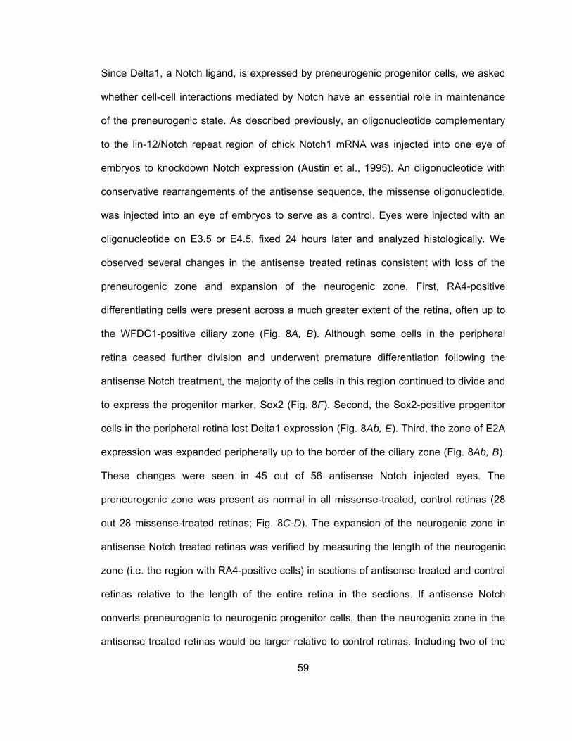
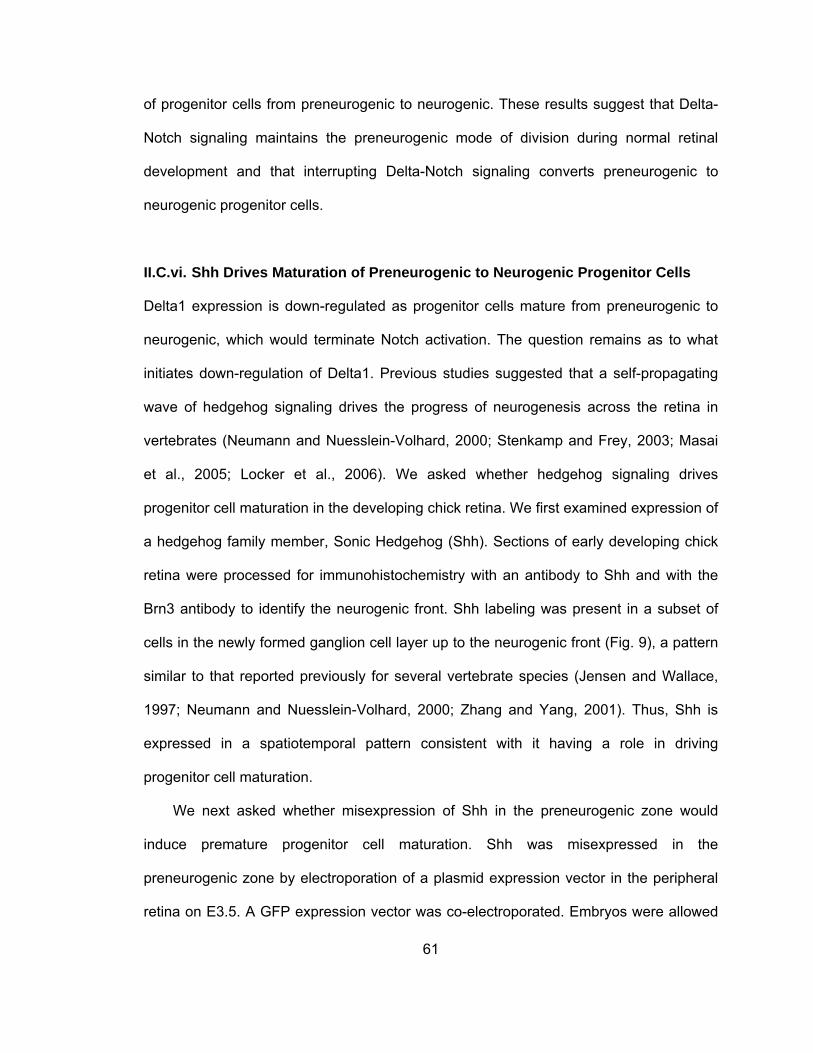
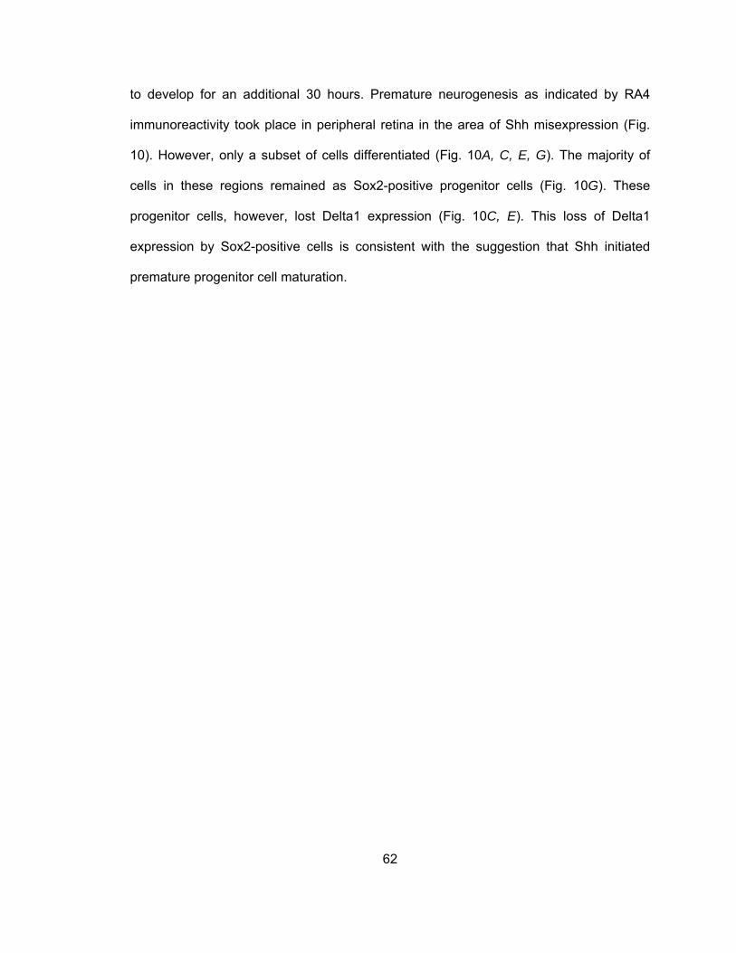

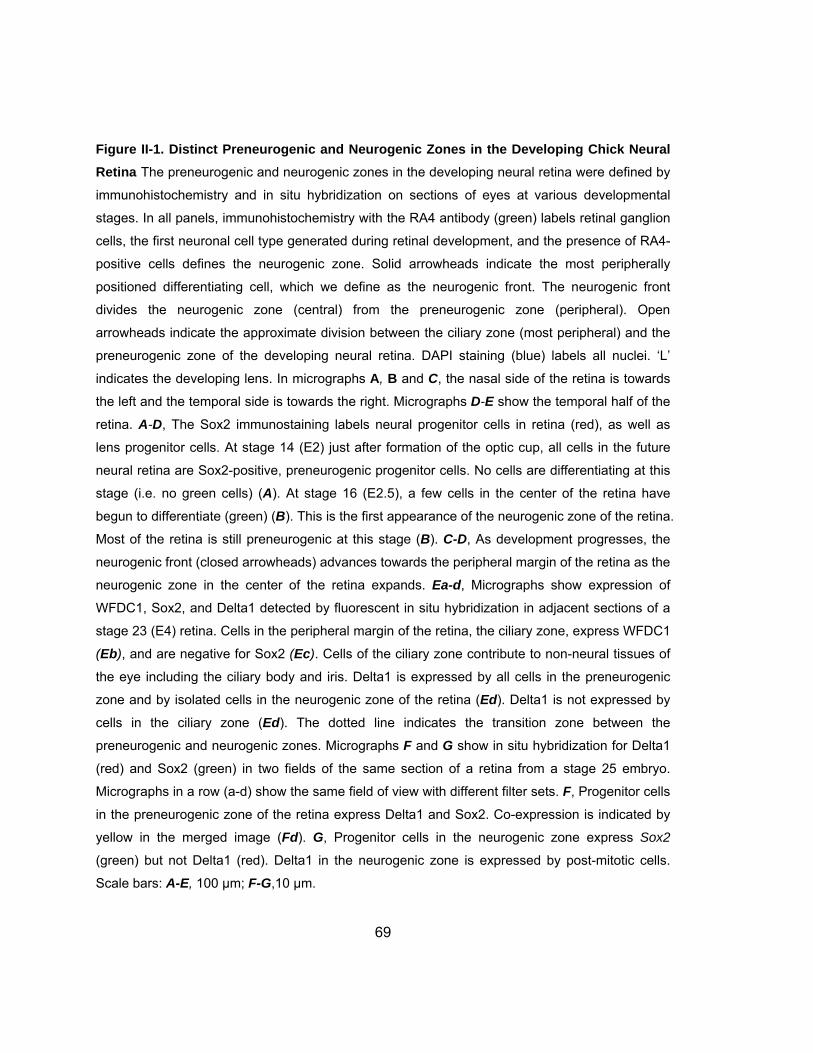
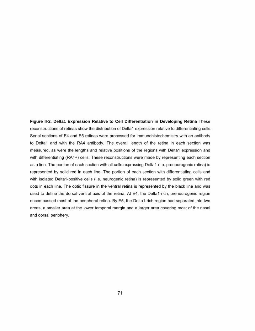
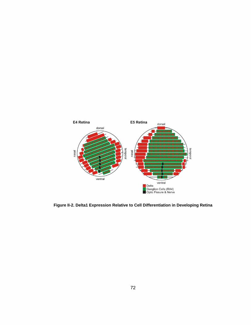
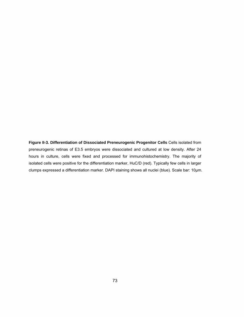
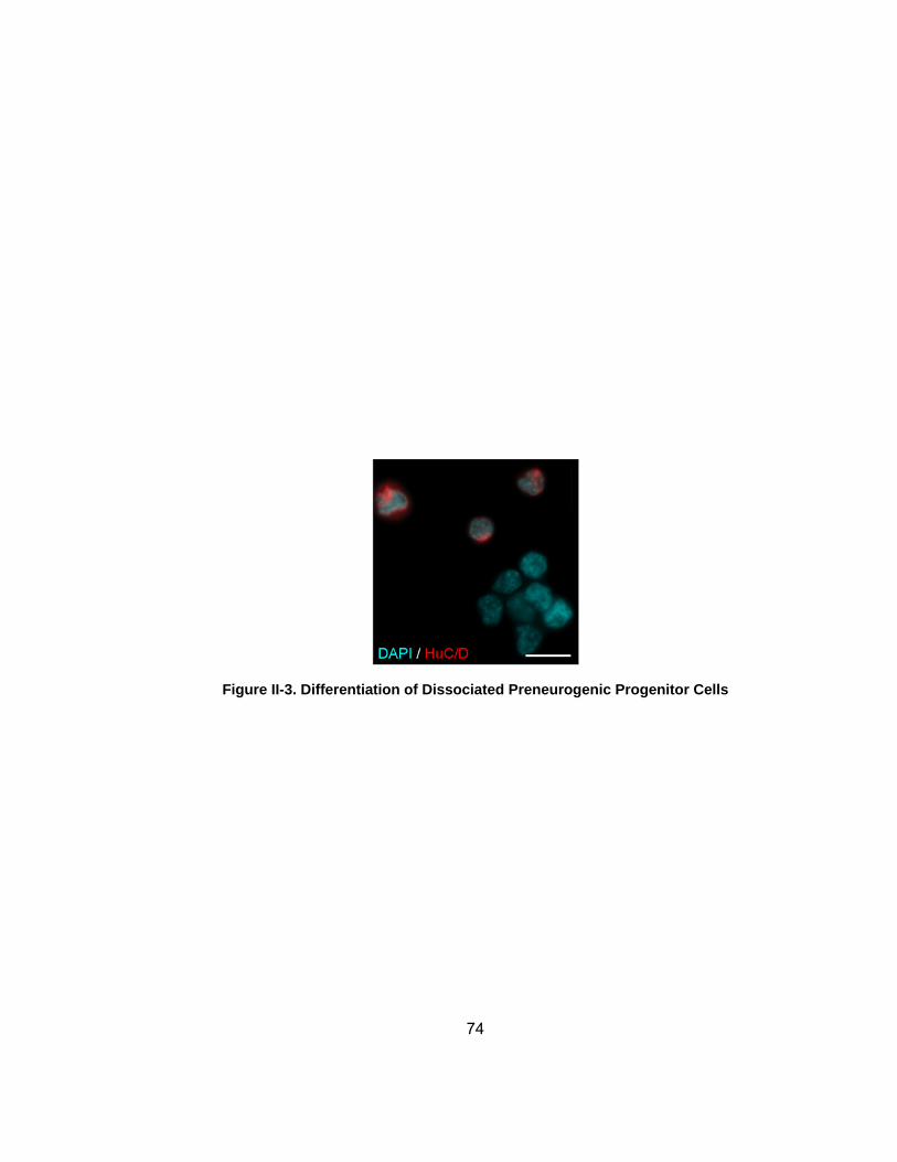
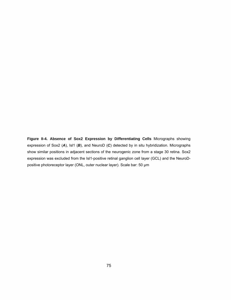
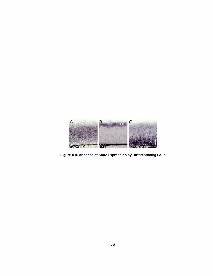
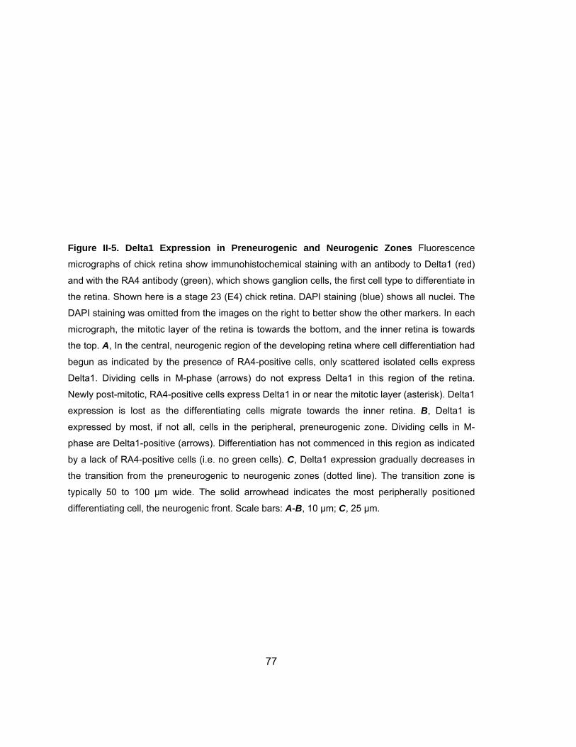
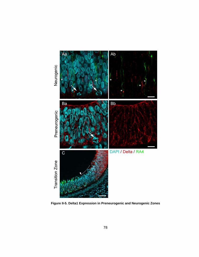
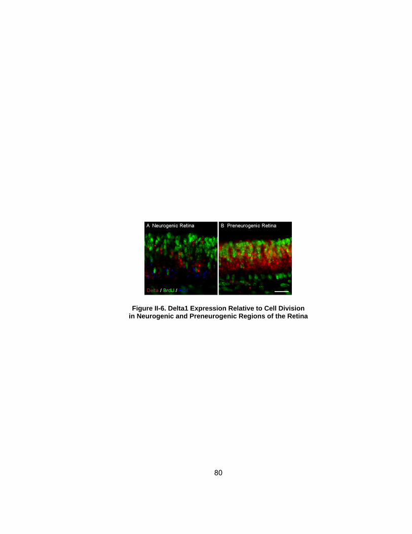
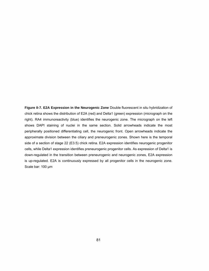

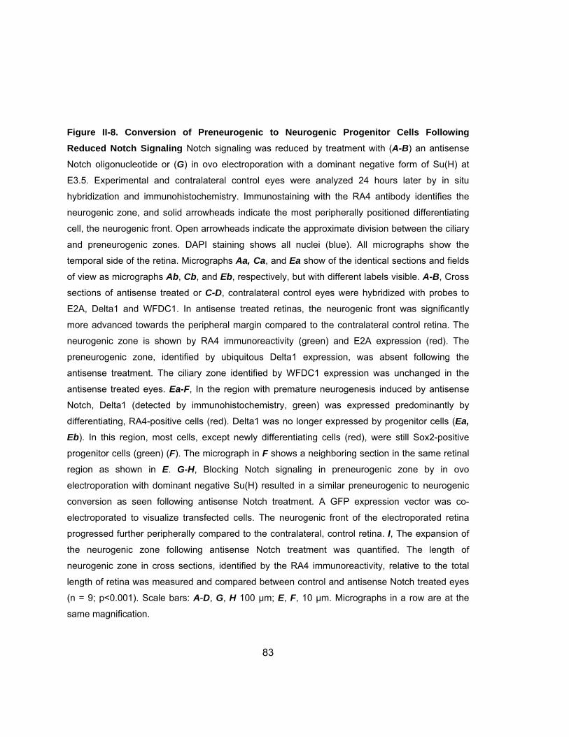
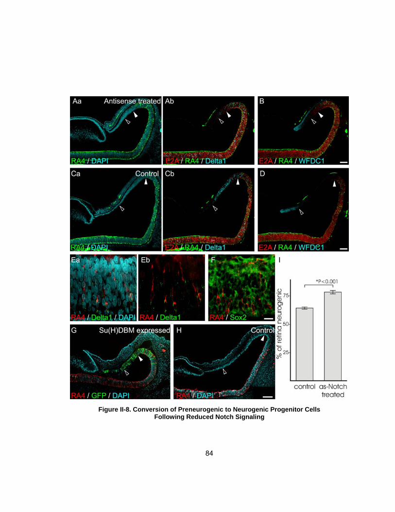
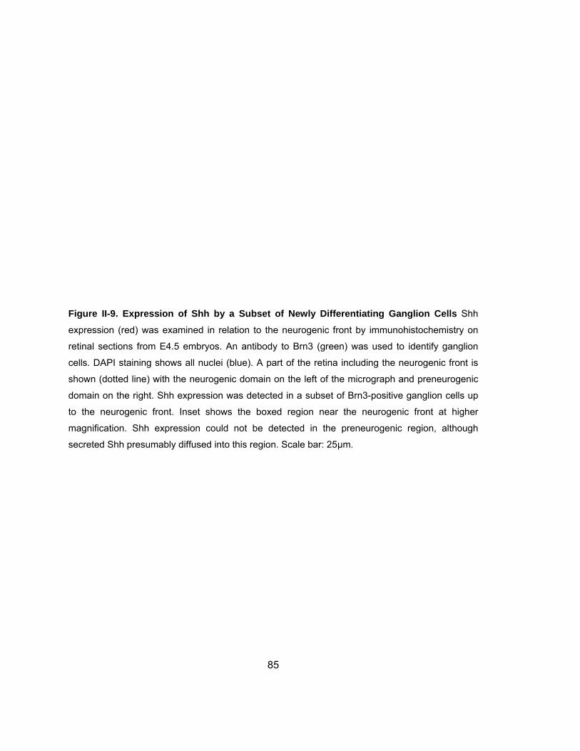
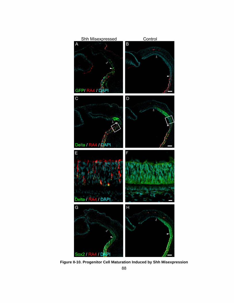
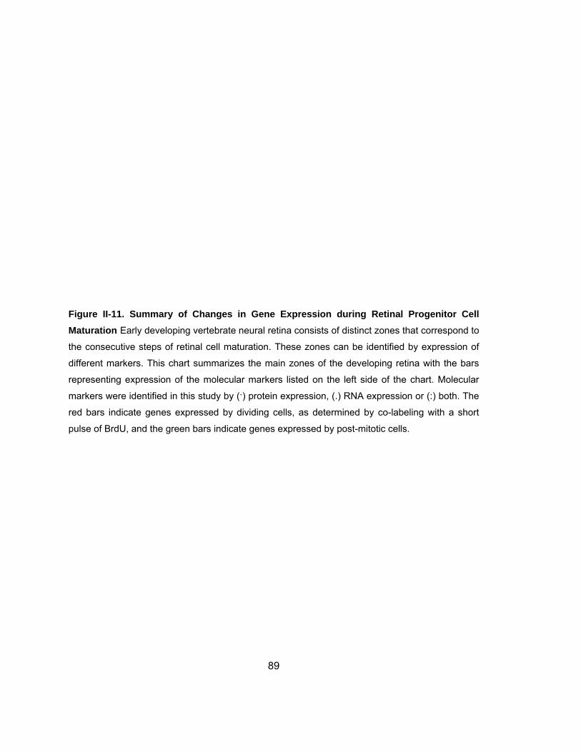
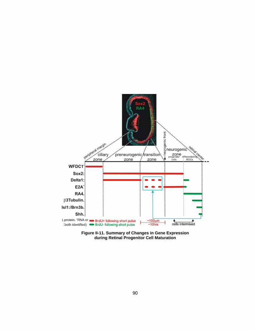

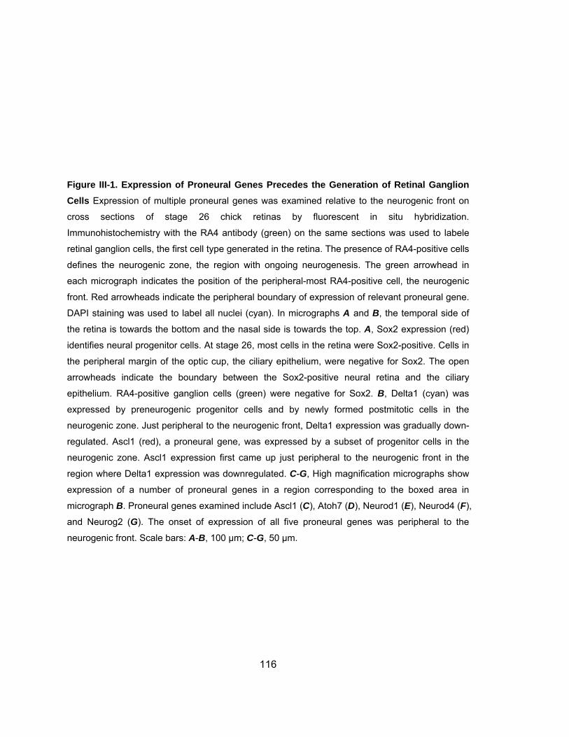
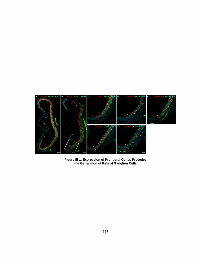
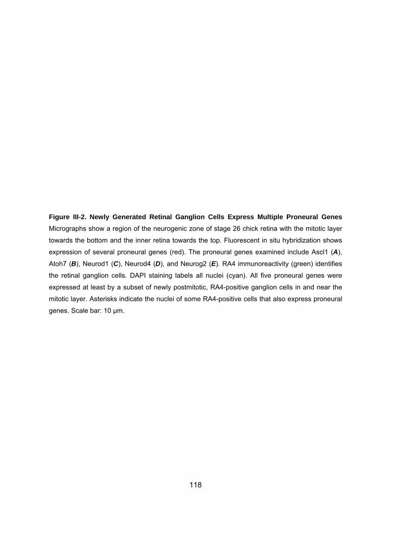
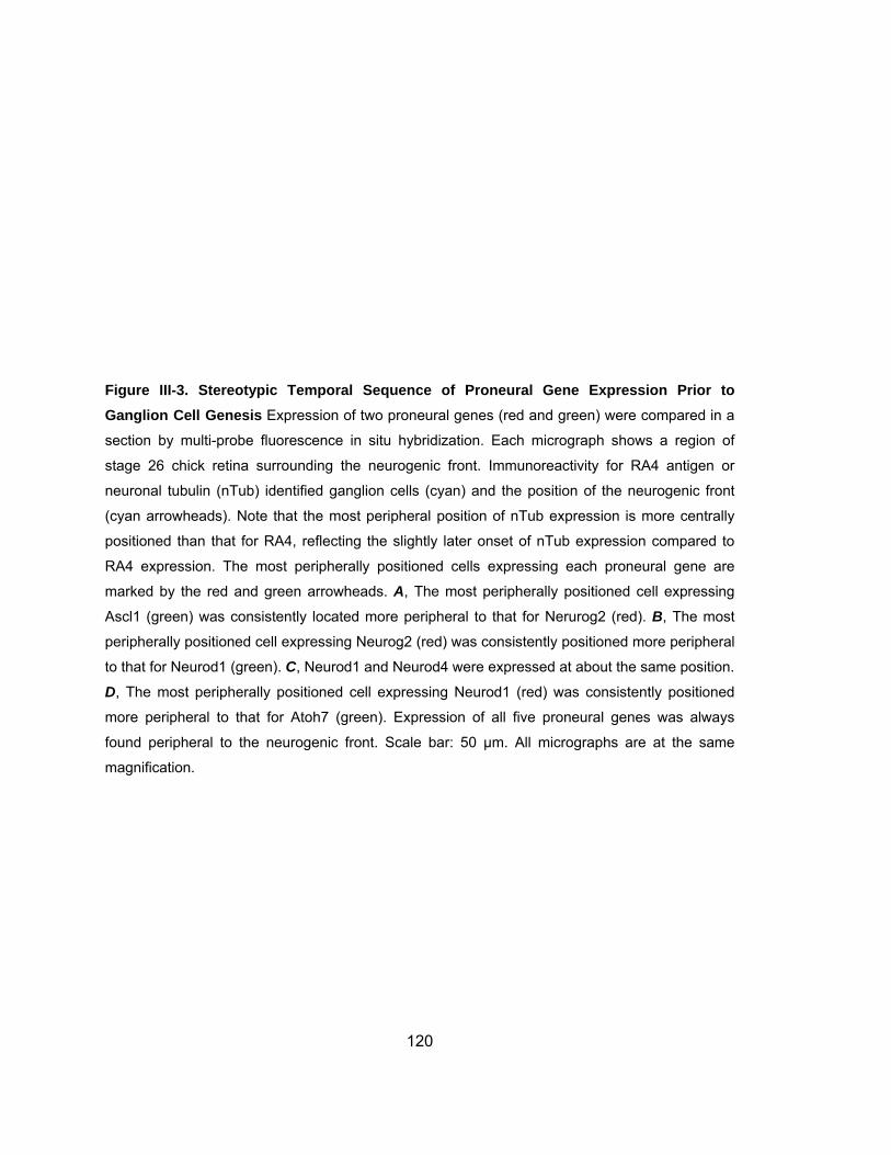
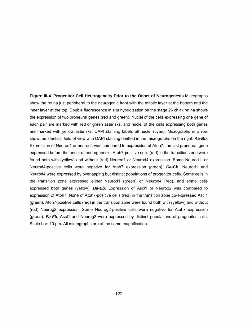
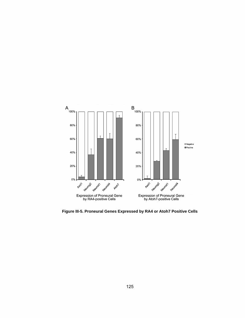
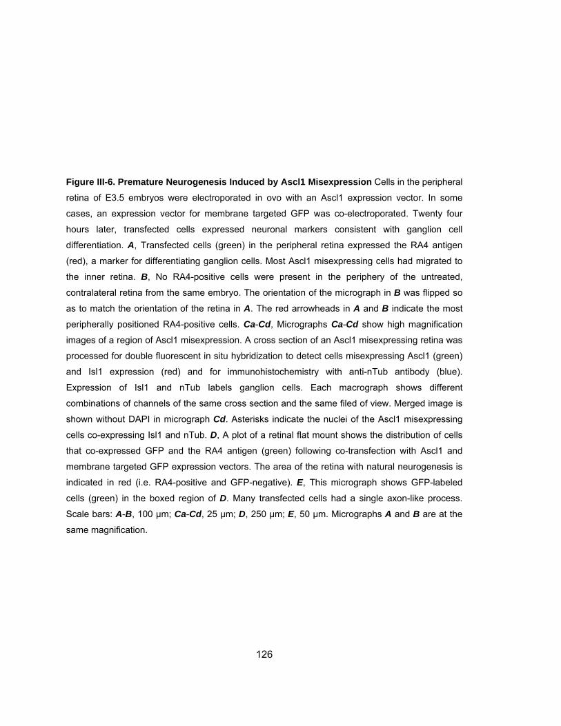
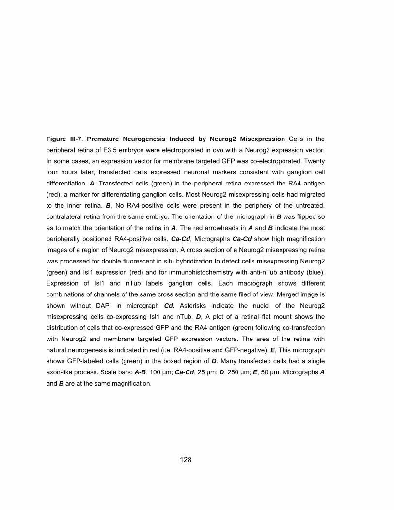
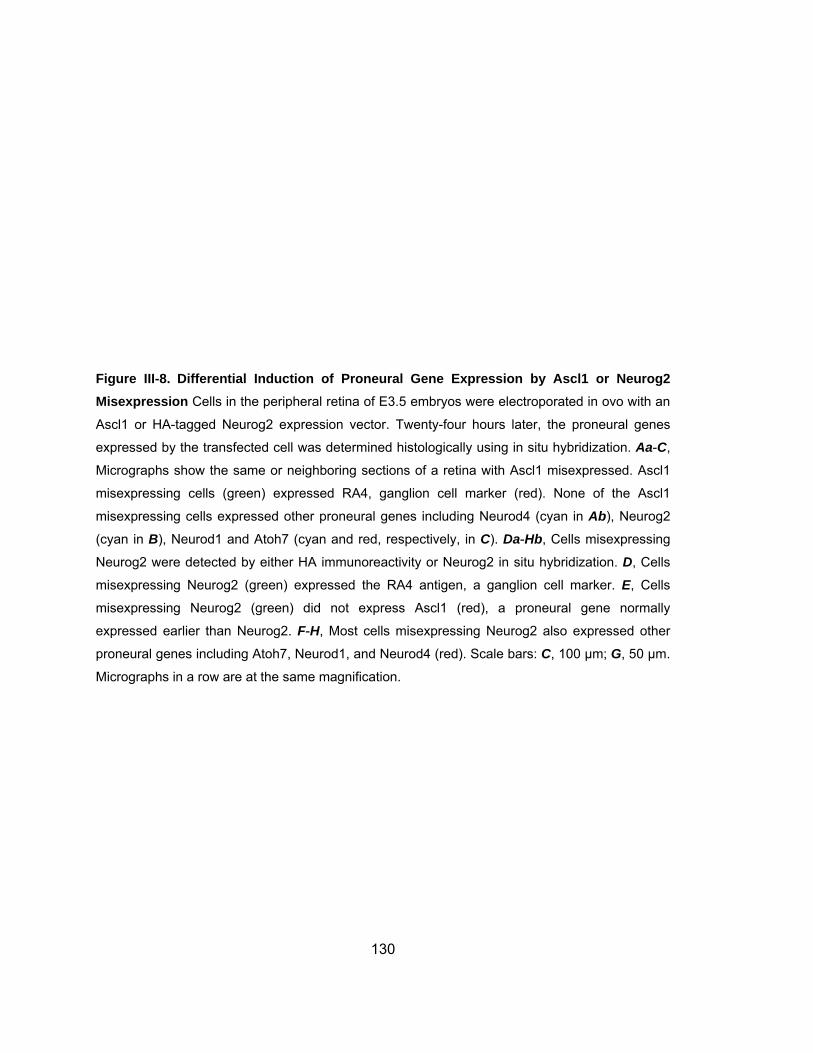
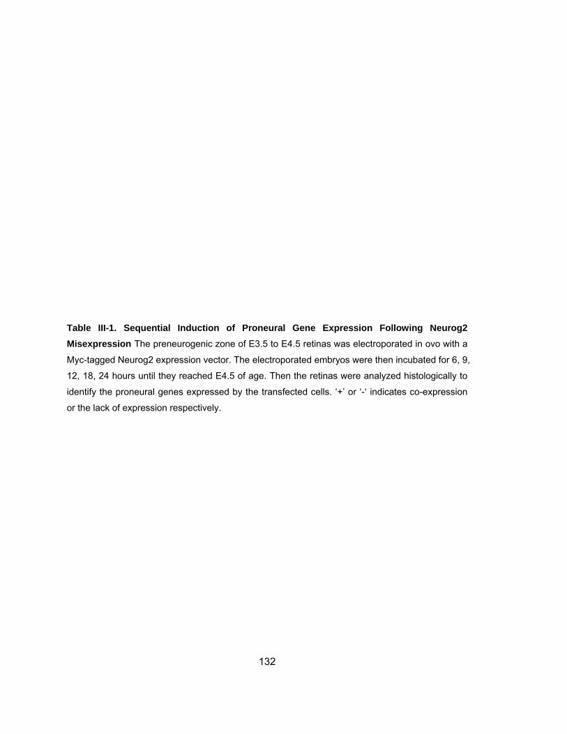


![Stem/progenitor cells in the cerebral cortex of the human ... · disease and Parkinson’s disease [14]. Stem/progenitor cells and neurogenesis in the . cerebral cortex during pre-](https://img.pdfslide.net/doc/110x75/5f17ee643585122f2e3c70e7/stemprogenitor-cells-in-the-cerebral-cortex-of-the-human-disease-and-parkinsonas.jpg)
![Neurogenesis and vascularization of the damaged brain using ... GJClub...progenitor metabolism [22]. Lactate is also a component of widely used biobased plastics made from poly-lactic-acid](https://img.pdfslide.net/doc/110x75/6018dadcdd210f39bf58f60b/neurogenesis-and-vascularization-of-the-damaged-brain-using-gjclub-progenitor.jpg)