Embed Size (px)
Citation preview
2Chapter
Handbook of Atypical Parkinsonism, eds. Carlo Colosimo, David E. Riley, and Gregor K. Wenning. Published by Cambridge University Press. © Cambridge University Press 2011.
Historical review Although the earlier literature contained several accounts of progressive supranuclear palsy (PSP) [ 1 ], including the famous case reported by Charcot in the nineteenth century [ 2 ], the fi rst detailed description of this condition came from John Cliff ord Richardson, John Steele, and Jerzy Olszewski in the early 1960s [ 3 ]. Th e term PSP was introduced by the same Canadian investigators in 1964 [ 4 ]. Th ey described a pre-senile sporadic neurodegenerative disease characterized clinically by a combination of supranuclear vertical ophthalmoplegia, pseudobulbar palsy, nuchal dystonia, and dementia. In their report they described the clin-ical features of nine patients and reported the neuropathological changes in seven of them. Th e predominant pathological changes were cell loss and gliosis in the brainstem, basal gan-glia, and cerebellum.
In 1986, Pollock and colleagues fi rst reported that the fi lamentous aggregates found in the brains of patients with PSP shared antigenic determinants with microtubule-associated pro-tein tau [ 5 ]. Histopathological studies then confi rmed that PSP is a tauopathy characterized by hyperphosphorylated tau protein deposition forming fi brillary aggregates (globose neuro-fi brillary tangles) in neurons and glia of numerous cerebral areas including the cerebral neo-cortex, pallidum, subthalamic nucleus, substantia nigra, periacqueductal gray matter, superior collicula, and dentate nucleus [ 6 ]. Th e abundant presence of these aggregates in all clinical PSP subtypes suggested that PSP should be considered a tauopathy along with corticobasal degen-eration (CBD), frontotemporal dementia (FTD), and Alzheimer’s disease (AD) [ 7 ].
Th e introduction of clinical diagnostic criteria, fi rst proposed by Lees in 1987, made it eas-ier to identify these patients clinically [ 8 ]. Others subsequently proposed diff erent sets of diag-nostic criteria, mainly based on personal experience [ 9 – 11 ]. New clinical diagnostic criteria were developed in 1996 during a workshop held under the auspices of the National Institute for Neurological Disorders and Stroke and the Society for PSP (NINDS-SPSP) [ 12 ]. Th is time the criteria were also validated on a clinical data set from autopsy-confi rmed cases of PSP. Although the NINDS-SPSP criteria ( Table 4.1 ) have gained wide acceptance in the research community, in movement disorder clinics, they remain to be validated prospectively .
Morphological features Macroscopic examination of the brain in PSP usually shows midbrain atrophy and substantia nigra depigmentation, sometimes with mild frontal lobe atrophy. Microscopic examination
Progressive supranuclear palsy
Carlo Colosimo , Giovanni Fabbrini, Alfredo Berardelli 4
9780521111973c04_p58-74.indd 589780521111973c04_p58-74.indd 58 12/8/2010 2:02:28 PM12/8/2010 2:02:28 PM
Chapter 4: Progressive supranuclear palsy 59
invariably discloses neuronal loss, gliosis, neurofi brillary tangles (NFTs), and neuropil threads (NTs) in the basal ganglia and brainstem. PSP is now considered a tauopathy, char-acterized by hyperphosphorylated tau protein aggregates. Tau protein binds to microtu-bules, and plays an important role in neuronal cytoskeletal stability. Alternative splicing of exons 2, 3, and 10 of the tau gene generates six tau isoforms. Th e inclusion or exclusion of exon 10 produces isoforms with four (4R) or three (3R) microtubule binding sites. Normal brains have similar levels of 4R and 3R, but in some neurodegenerative disorders this ratio is changed. Th e revised neuropathological criteria for PSP require high NFT and NT dens-ities in at least three of the following brain areas: striatum, oculomotor complex, medulla, or dentate nucleus [ 13 ]. Th e pathological changes oft en involve the neocortex, especially the
Table 4.1 NINDS/SPSP criteria
Basic features
• gradually progressive disorder
• onset age >40 yr
• no evidence of other diseases that could explain the clinical features as indicated by exclusion criteria*
Diagnosis of clinically possible PSP
• vertical supranuclear palsy#
OR
• slowing of vertical saccades AND • postural instability with falls within a year of disease onset
Diagnosis of clinically probable PSP
• vertical supranuclear palsy
AND
• prominent postural instability with falls within a year of disease onset
#Upward gaze is considered abnormal when pursuit or voluntary gaze, or both, have a restriction of ≥50% of the normal range Supportive features - symmetrical akinesia or rigidity, proximal more than distal - abnormal neck posture, especially retrocollis - poor or absent response of parkinsonism to levodopa - early dysphagia and dysarthria - early onset of cognitive impairment including two or more of the following: apathy, impairment in abstract
thought, decreased verbal fl uency, utilization or imitation behavior, or frontal release signs Exclusion criteria - recent history of encephalitis - alien limb syndrome - cortical sensory defi cits - focal frontal or temporoparietal atrophy - hallucinations or delusions unrelated to dopaminergic therapy - cortical dementia of Alzheimer type - prominent, early cerebellar symptoms - unexplained dysautonomia - neuroradiological evidence of relevant structural abnormality - Whipple’s disease, confi rmed by polymerase chain reaction
Reproduced with kind permission from Litvan et al . [12]
9780521111973c04_p58-74.indd 599780521111973c04_p58-74.indd 59 12/8/2010 2:02:31 PM12/8/2010 2:02:31 PM
Chapter 4: Progressive supranuclear palsy60
primary motor area of the frontal lobe, and severe cell loss in this area has been indicated as a possible contributor of the early falls [ 14 ].
In addition to their clinical similarities, PSP and CBD share certain pathological fea-tures. Th is clinicopathological overlap led some to consider the two diseases as diff erent ends of the same disease spectrum. A neuropathological fi nding of tau-positive tuft ed astrocytes is considered highly characteristic of PSP and diff erentiates it from CBD. Th ese astrocytic alterations probably refl ect the main degenerative process rather than a change secondary to gliosis. Th e ballooned neurons typically found in CBD are rarely observed in PSP. Th e tau aggregates in PSP and CBD contain mainly 4R isoforms and accumulate in neuronal and glial cells. How these aggregates form and how they aff ect diff erent regions of the brain remain unclear. Ultrastructural examination shows that PSP NFTs consist of mainly straight fi laments, whereas AD NFTs contain paired helical fi laments [ 15 ].
Genetics Th e importance of tau aggregates in causing PSP receives further support from genetic fi nd-ings. PSP is strongly associated with a specifi c haplotype in the tau gene (denoted as H1). Several reports from diff erent populations show that patients with PSP (like those with CBD) show the H1 haplotype and the H1/H1 genotype signifi cantly more frequently than controls [ 16 ]. Fine-mapping of this region detected a variation in a single intron regulating tau expression on the H1 background [ 17 ]. A genome-wide association study subsequently discovered a second major susceptibility locus on chromosome 11 that contains several interesting candidate genes [ 18 ].
Although PSP is usually a sporadic disease, some studies describe familial clustering. One pedigree with autosomal dominant PSP has been described and linked to a locus on chromosome 1q31, but the gene responsible for the disease in this family still has to be iden-tifi ed [ 19 ]. Although mutations in the tau gene may occasionally give rise to a clinical and pathological picture similar to PSP [ 20 , 21 ], screening of large cohorts of sporadic and famil-ial PSP cases has failed to identify tau gene mutations to date .
Clinical picture Presenting features Th e most frequently reported symptom at disease onset is balance problems, followed by personality changes, bulbar symptoms, and visual problems. PSP patients present with a peculiar akinetic-rigid motor disorder that in most cases diff ers from that observed in Parkinson’s disease (PD). Symptoms are more prominent in the axial segments whereas the limbs are relatively preserved [ 8 ]. Unlike the onset symptoms in PD, postural stability is compromised early on. In the classic PSP phenotype (recently re-named Richardson’s Syndrome, RS), the motor disorder usually responds poorly to levodopa. Th e poor motor response to dopaminergic drugs is helpful in diff erentiating between the classic PSP phenotype and PD [ 22 ]. A subgroup of PSP patients may nevertheless show a good, but usually short-lived, response to levodopa ( PSP-Parkinsonism [PSP-P]) [ 22 ]. In some patients, behavioral (apathy, disinhibition) and cognitive (progressive aphasia, apraxia of speech, memory loss) disorders may also be the presenting feature or accompany the motor disorder at disease onset.
9780521111973c04_p58-74.indd 609780521111973c04_p58-74.indd 60 12/8/2010 2:02:31 PM12/8/2010 2:02:31 PM
Chapter 4: Progressive supranuclear palsy 61
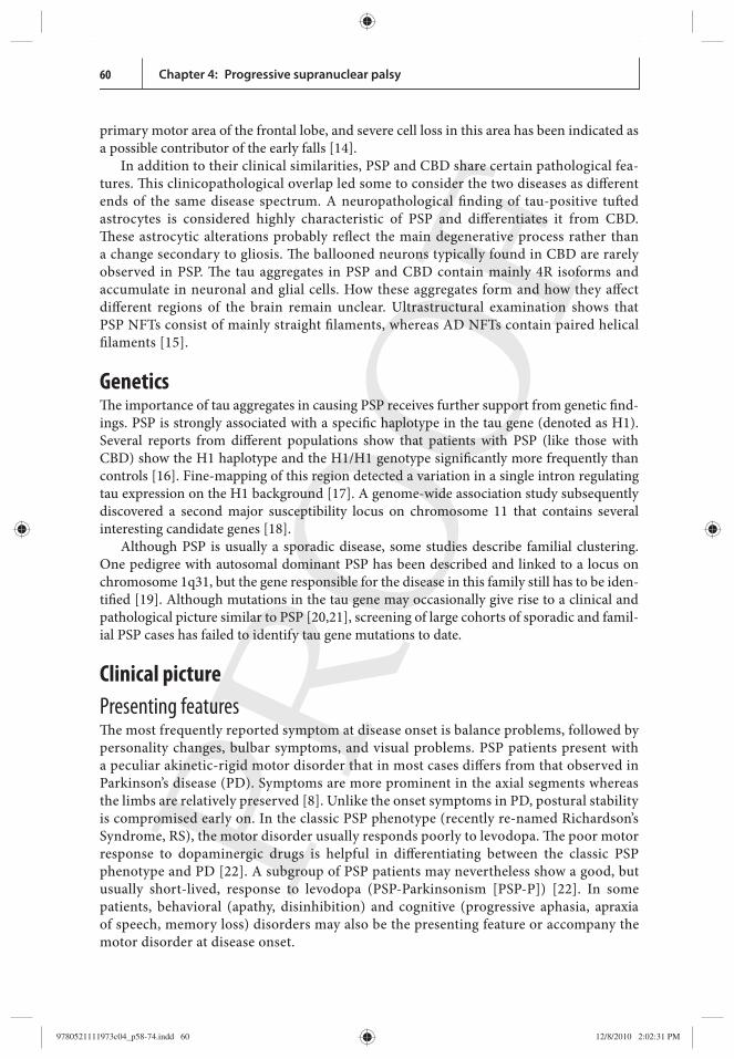
Core characteristics of the disease Patients with PSP generally manifest parkinsonian signs characterized by bradykinesia, rigidity, and disequilibrium with severe gait unsteadiness. Th e marked axial rigid-ity infl uences the posture, which may be characteristically erect like the original cases described by Richardson and colleagues (who described it as “ nuchal dystonia,” Figure 4.1 ) or more closely resembles the fl exed posture typical of PD. Progressive imbalance leads to repeated and frequent falls (usually backward). Postural tremor and less com-monly tremor at rest may be superimposed occasionally. Even so, a classical pill-rolling resting tremor has been reported in less than 20% of the subjects [ 23 ]. Patients with PSP frequently have dysphagia and a characteristic growling high-pitched severe dys-arthria, with mixed spastic and parkinsonian features [ 4 ]. PSP patients also manifest eyelid movement disorders, including blepharospasm and eyelid opening-and-closing apraxia. Although neurological examination in patients with PSP sometimes shows pyr-amidal signs, obvious spastic paraparetic gait or signifi cant pyramidal weakness should cast doubt on the clinical diagnosis of PSP.
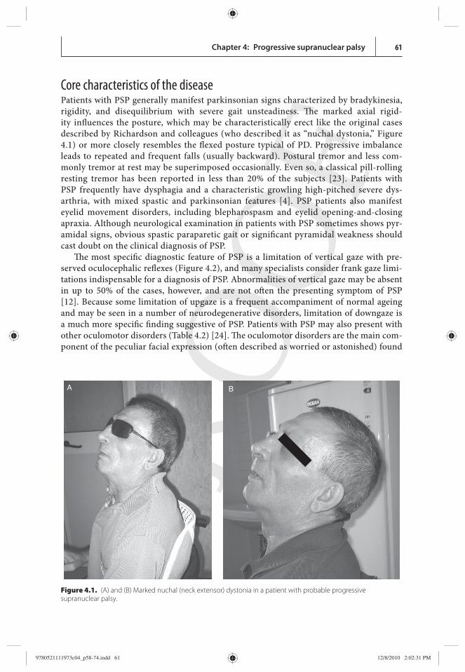
Th e most specifi c diagnostic feature of PSP is a limitation of vertical gaze with pre-served oculocephalic refl exes ( Figure 4.2 ), and many specialists consider frank gaze limi-tations indispensable for a diagnosis of PSP. Abnormalities of vertical gaze may be absent in up to 50% of the cases, however, and are not oft en the presenting symptom of PSP [ 12 ]. Because some limitation of upgaze is a frequent accompaniment of normal ageing and may be seen in a number of neurodegenerative disorders, limitation of downgaze is a much more specifi c fi nding suggestive of PSP. Patients with PSP may also present with other oculomotor disorders ( Table 4.2 ) [ 24 ]. Th e oculomotor disorders are the main com-ponent of the peculiar facial expression (oft en described as worried or astonished) found
A B
Figure 4.1. (A) and (B) Marked nuchal (neck extensor) dystonia in a patient with probable progressive supranuclear palsy.
9780521111973c04_p58-74.indd 619780521111973c04_p58-74.indd 61 12/8/2010 2:02:31 PM12/8/2010 2:02:31 PM
Chapter 4: Progressive supranuclear palsy62
in patients with PSP, together with the characteristic dystonic features of the frontalis, procerus, and corrugator muscles [ 25 ]. Th e physician needs to diff erentiate PSP from the numerous other neurological conditions that can initially manifest with oculomotor dys-function [ 26 – 29 ] ( Table 4.3 ).
Clinical variants In addition to RS, other clinical syndromes known to accompany PSP-tau pathology include PSP-P (the second most frequent aft er RS), corticobasal syndrome (CBS) [ 30 ], and pure akinesia with gait freezing (PAGF) [ 31 ]. Th ese syndromes diff er in their clinical features at disease onset and with disease duration. Patients with RD also have a shorter disease course than those with PSP-P and PAGF.
A B
C
D E
Figure 4.2 Vertical supranuclear ophthalmoplegia with preserved horizontal eye movements in a patient with classical progressive supranuclear palsy.
9780521111973c04_p58-74.indd 629780521111973c04_p58-74.indd 62 12/8/2010 2:02:35 PM12/8/2010 2:02:35 PM
Chapter 4: Progressive supranuclear palsy 63
Th e concept of PSP-P has evolved notably over the past 40 years. Th e literature repeat-edly refers to occasional cases of pathologically defi nite PSP whose neurological signs and disease course resemble that of PD. Only in recent years, however, did the large clinicopatho-logical series reported by Williams et al . [ 22 ] underline that this clinical presentation of PSP
Table 4.3 Other causes of supranuclear ophthalomoplegia associated with parkinsonism
PD
MSA-P
CBD
DLB
FTD and parkinsonism linked to chromosome 17
Huntington’s disease
Motor neuron disease
Genetic cerebellar ataxias (SCA 2, SCA 3, SCA 7, SCA 17)
Postencephalitic parkinsonism
Prion diseases
Progressive external ophthalmoplegia
Tumors compressing the brainstem (pinealoma, glioma)
Multi-infarct state
Myasthenia gravis
CNS lymphoma
Niemann–Pick type C disease
Drug-induced
Whipple’s disease
Calcifi cation of the basal ganglia
Table 4.2 Oculomotor abnormalities in progressive supranuclear palsy
Early stages
• Slowness of vertical saccadic movements • Hypometric saccades • Reduced blinking • Square wave jerks
Middle stages
• Supranuclear vertical gaze palsy • Lid retraction with very rare blinking (<3) • Impaired convergence • Apraxia of eyelid opening or closing
Late stages
• Supranuclear horizontal gaze palsy • Loss of oculocephalic refl exes • Blepharospasm • Disconjugate gaze
Modifi ed from Golbe [24]
9780521111973c04_p58-74.indd 639780521111973c04_p58-74.indd 63 12/8/2010 2:02:38 PM12/8/2010 2:02:38 PM
Chapter 4: Progressive supranuclear palsy64
is far more common than previously recognized. Th ese authors fi rst proposed designating this clinical disease phenotype PSP-P; this form may account for up to one-third of all cases of PSP. Patients with PSP-P typically present with tremor at rest, unilateral or asymmetrical bradykinesia, and a discrete or good response to levodopa. Th e initial clinical features are diffi cult to distinguish from those of PD. Multiple system atrophy of the parkinsonian type (MSA-P), particularly in patients without signs of full-blown dysautonomia, may be quite diffi cult to diff erentiate clinically from PSP-P [ 32 ].
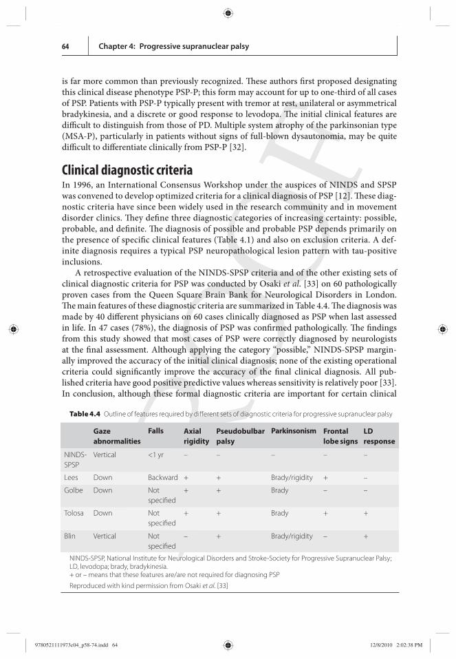
Clinical diagnostic criteria In 1996, an International Consensus Workshop under the auspices of NINDS and SPSP was convened to develop optimized criteria for a clinical diagnosis of PSP [ 12 ]. Th ese diag-nostic criteria have since been widely used in the research community and in movement disorder clinics. Th ey defi ne three diagnostic categories of increasing certainty: possible, probable, and defi nite. Th e diagnosis of possible and probable PSP depends primarily on the presence of specifi c clinical features ( Table 4.1 ) and also on exclusion criteria. A def-inite diagnosis requires a typical PSP neuropathological lesion pattern with tau-positive inclusions.
A retrospective evaluation of the NINDS-SPSP criteria and of the other existing sets of clinical diagnostic criteria for PSP was conducted by Osaki et al . [ 33 ] on 60 pathologically proven cases from the Queen Square Brain Bank for Neurological Disorders in London. Th e main features of these diagnostic criteria are summarized in Table 4.4 . Th e diagnosis was made by 40 diff erent physicians on 60 cases clinically diagnosed as PSP when last assessed in life. In 47 cases (78%), the diagnosis of PSP was confi rmed pathologically. Th e fi ndings from this study showed that most cases of PSP were correctly diagnosed by neurologists at the fi nal assessment. Although applying the category “possible,” NINDS-SPSP margin-ally improved the accuracy of the initial clinical diagnosis; none of the existing operational criteria could signifi cantly improve the accuracy of the fi nal clinical diagnosis. All pub-lished criteria have good positive predictive values whereas sensitivity is relatively poor [ 33 ]. In conclusion, although these formal diagnostic criteria are important for certain clinical
Table 4.4 Outline of features required by diff erent sets of diagnostic criteria for progressive supranuclear palsy
Gaze abnor malities
Falls Axial rigidity
Pseudo bulbar palsy
Parkin sonism Frontal lobe signs
LD response
NINDS-SPSP
Vertical <1 yr – – – – –
Lees Down Backward + + Brady/rigidity + –
Golbe Down Not specifi ed
+ + Brady – –
Tolosa Down Not specifi ed
+ + Brady + +
Blin Vertical Not specifi ed
– + Brady/rigidity – +
NINDS-SPSP, National Institute for Neurological Disorders and Stroke-Society for Progressive Supranuclear Palsy; LD, levodopa; brady, bradykinesia. + or – means that these features are/are not required for diagnosing PSP
Reproduced with kind permission from Osaki et al . [33]
9780521111973c04_p58-74.indd 649780521111973c04_p58-74.indd 64 12/8/2010 2:02:38 PM12/8/2010 2:02:38 PM
Chapter 4: Progressive supranuclear palsy 65
research fi elds, they add little to the problem of detecting early cases and screening tools need improving .
Time course of the disease PSP aff ects both genders, despite a slight male predominance. Th e disease progresses relent-lessly and has a signifi cantly shorter mean survival than PD [ 24 ]. Th e clinical symptoms commonly begin in the seventh decade, although occasionally as early as the fi ft h decade. Pooling several PSP case series yielded a median onset age of 63 years, with rare cases begin-ning as early as 40 years of age.
PSP is a chronically progressive disease characterized by the gradual onset of neurologic al symptoms with increasing disability. In a comparative clinicopathological study, latencies to onset of falls were short in patients with PSP, intermediate in MSA, dementia with Lewy bod-ies (DLB), and CBD, and long in PD [ 34 ]. Recurrent falls within the fi rst year aft er disease onset predicted PSP in 68% of the patients. Conversely, latency to onset, but not duration, of recurrent falls diff erentiates PD from other examples of parkinsonism. In another study, the progression to diff erent Hoehn and Yahr (HY) stages was evaluated in 81 pathologically con-fi rmed patients with parkinsonism. Latencies to each HY stage were longer in patients with PD than in those with atypical parkinsonism (AP). Development of a HY-III within one year of motor onset accurately predicted AP. Th e progression to each HY stage was unhelpful in distinguishing the various disorders with AP from each other. Once patients with PD and AP became wheelchair-bound, both had equally short survival times [ 35 ].
Th e prognosis of PSP remains poor. Th is is a progressive disorder associated with a short-ened life span, oft en leading to death within ten years aft er symptom onset. Mean survival ranges from 5.9 to 9.7 years according to the diff erent series .
Epidemiology Th e dearth of published epidemiological studies makes it diffi cult to determine incidence and prevalence rates for PSP. Th e estimated prevalence of PSP (per 100 000 in the popula-tion) in the various studies ranged from 1.3 to 4.9 [ 36 , 37 ]. Th e estimated annual incidence of PSP was about 0.3–1.1 cases per 100 000 persons or 5.3/100 000 people over the age of 50 years [ 38 ]. Th ese fi gures match those for other well-known neurodegenerative disor-ders such as Huntington’s disease or motor neuron disease. Th e analytical epidemiology of PSP is even poorer and more controversial. Although a retrospective study conducted in Switzerland showed an increased risk of PSP associated with arterial hypertension [ 39 ] other independent series failed to confi rm the fi nding [ 40 ]. Smoking habits seem to be similar to those in healthy controls. Th e fact that the inverse association with smoking found previously in PD is shared by MSA but not by PSP lends epidemiological support to the notion that diff erent smoking habits are associated with diff erent groups of neuro-degenerative disease [ 41 ].
Investigations Introduction Th e clinical diagnosis of PSP depends primarily on history and physical examination. Recent evidence, however, shows that additional investigations may be useful in diff erentiating PSP from other parkinsonian syndromes.
9780521111973c04_p58-74.indd 659780521111973c04_p58-74.indd 65 12/8/2010 2:02:38 PM12/8/2010 2:02:38 PM
Chapter 4: Progressive supranuclear palsy66
Computerized tomography Although CT scans sometimes disclose pathological changes including generalized or brain-stem atrophy in patients with PSP, current data suggest that this diagnostic tool is of limited use in routine clinical practice [ 8 ].
Routine magnetic resonance imaging Several studies have sought to improve the diagnostic accuracy of PSP by using various MRI techniques [ 42 ]. Routine and volumetric MRI imaging may show midbrain atrophy, a fi nding that helps in diff erentiating patients with PSP from healthy controls and those with PD and other disorders with AP. A study designed to provide a quantitative assessment of atrophy by measuring midbrain diameter or area found that a diameter of <17 mm on axial MRIs diff erentiates patients with PSP from healthy controls [ 43 ]. Other studies suggest that atrophy is better evaluated on mid-sagittal slices because they are not subject to variation in the scanning angle. Th e midbrain atrophy seen on these slices typically resembles a pen-guin or hummingbird silhouette [ 44 ]. In a study designed to measure the midbrain area in patients with PSP, Japanese investigators found a signifi cant area reduction in PSP versus PD and controls [ 45 ]. Th e relationship between the midbrain/pons areas was also signifi -cantly lower in patients with PSP than that observed in PD patients and controls. Although these measurements were helpful in diff erentiating PSP, MSA, and PD as groups, the data obtained overlapped among the groups of patients investigated, and therefore were not help-ful for an individual diagnosis.
In a study investigating morphometric MRI in PSP, MSA, PD, and controls using volu-metric T1-weighted sequences [ 46 ], Quattrone and colleagues measured the midbrain and pons areas, together with the medial cerebellar peduncle (MCP) and superior cerebellar peduncle (SCP) width. Th ey found that atrophy in PSP involves the midbrain and SCP whereas atrophy in MSA involves the the pons and the MCP. In patients with PD all brain areas measured had dimensions similar to those in control subjects. Single structure meas-urements did not discriminate among the diff erent diseases on an individual basis, because of substantial overlap. Th e investigators therefore proposed a new index calculated with a specifi c formula (area of pons/area of midbrain x MCP diameter/SCP diameter) obtained by combining the single measurements obtained in the various cerebral structures. Th e ‘magnetic resonance parkinsonism index’ (MRPI) was signifi cantly higher in PSP than in the other conditions, and could diff erentiate individual patients with PSP from those with MSA and PD. Th e promising results obtained with morphometric MRI should be con-fi rmed in studies conducted by other groups before proposing its use in routine clinical practice .
Diff usion-weighted imaging Diff usion-weighted imaging (DWI) is a useful diagnostic tool that can provide additional support for a diagnosis of AP, and especially for PSP. In their study, Seppi and coworkers found signifi cantly higher rADC (regional apparent diff usion coeffi cient) values in both the putamen and globus pallidus in patients with PSP than in those with PD [ 47 ]. Th e increased putaminal rADC values in PSP probably refl ect ongoing striatal degeneration, whereas most neuropathological studies reveal an intact striatum in PD. Despite these diff erences, increased putaminal rADC values are not able to discriminate PSP from MSA-P.
9780521111973c04_p58-74.indd 669780521111973c04_p58-74.indd 66 12/8/2010 2:02:38 PM12/8/2010 2:02:38 PM
Chapter 4: Progressive supranuclear palsy 67
Magnetic resonance volumetry Whether magnetic resonance volumetry will help in diff erentiating PSP from other dis-orders with AP remains to be confi rmed. Patients with PSP, MSA-P, and MSA-C had signifi -cantly lower mean striatal and brainstem volumes than patients with PD, and patients with MSA-P and MSA-C also showed a reduction in cerebellar volume [ 48 ]. Total intracranial volume-normalized MRI-based volumetric measurements provide a sensitive marker to dis-criminate PD from AP.
Voxel-based morphometry is an observer-unbiased volumetric procedure that can be used to investigate the entire brain. A study comparing patients with probable PSP and healthy controls showed that in patients with PSP several cortical areas in the frontal, tem-poral, and insular lobes were decreased in volume [ 49 ]. White matter comparisons also dis-closed a volume reduction in the frontotemporal regions and the mesencephalon. Th is brain atrophy pattern probably accounts for the cardinal PSP-associated behavioral defi cits .
Functional imaging Functional imaging methods for diff erential diagnosis in AP are techniques designed to investigate receptor binding and glucose metabolism. Studies of brain receptor binding in parkinsonism, by evaluating dopa-decarboxylase activity and the dopamine transporter (DAT) examine the presynaptic nigrostriatal neurons, and by evaluating the dopamine D 2 -receptors examine postsynaptic dopaminergic function. More recently, SPECT and PET lig-ands have become available to study cardiac sympathetic innervation.
Using PET, the Hammersmith group found that putaminal uptake of the presynaptic dopaminergic marker 18 F-fl uorodopa was reduced to a similar extent in PD and PSP [ 50 ]; in some patients with PSP, caudate uptake was also markedly reduced, as opposed to only a moderate reduction in PD [ 51 ]. Measurements of striatal dopamine D 2 -receptor densities using raclopride and PET also failed to diff erentiate between PD and AP, demonstrating a similar loss of densities in patients with advanced PD, PSP, and MSA [ 52 ].
SPECT evaluation of DAT using 123 I-β-CIT may be useful in diff erentiating true par-kinsonism from patients with essential tremor and patients with parkinsonism owing to a subcortical vascular encephalopathy. Although PSP cannot be distinguished from PD with this method alone [ 53 ], patients with PSP may show a more symmetrical DAT loss, con-sistent with the more symmetrical clinical motor dysfunction observed in this condition. SPECT imaging studies of patients with dopa-naїve parkinsonism have used 123 I-IBZM as a D 2 -receptor ligand [ 54 ]. Subjects with normal IBZM binding responded well to apomorph-ine and benefi tted from subsequent chronic dopaminergic therapy, whereas subjects with reduced binding failed to respond. In some of these patients, other clinical features atypical for PD developed during follow-up [ 55 ]. Despite these interesting fi ndings, because striatal IBZM binding is also reduced in other disorders with AP such as MSA, IBZM binding has limited predictive value for an early diagnosis of PSP [ 56 ].
Scintigraphic visualization of postganglionic sympathetic cardiac neurons was found to diff erentiate patients with PD from patients with AP [ 57 ]. Considering all reports published so far, standard scintigraphy with 123 I-metaiodobenzylguanidine (MIBG), a technique used for years to detect pheochromocytoma cells, correctly distinguished most patients with PD, all of whom had severely reduced cardiac MIBG uptake. Th is radioactive ligand method appears to be a highly sensitive and specifi c tool to discriminate between PD and AP within
9780521111973c04_p58-74.indd 679780521111973c04_p58-74.indd 67 12/8/2010 2:02:38 PM12/8/2010 2:02:38 PM

Chapter 4: Progressive supranuclear palsy68
two years aft er the onset of symptoms but it cannot distinguish PSP from other forms of AP such as MSA [ 58 ].
Neurophysiology Among the standard neurophysiological tests used in patients with abnormal eye move-ments, electro-oculographic recording may help in distinguishing patients with PSP from those with CBD at an early stage [ 59 ]. Patients with PSP have decreased horizontal saccadic amplitude and velocity but normal latency, whereas those with CBD show normal saccadic velocity and increased latency. Th e antisaccadic task (looking in the direction opposite to a visual stimulus), which correlates well with frontal lobe dysfunction, is markedly impaired in patients with PSP, although it may also be impaired in AD. Conversely, patients with PD or MSA-P have no or only slight saccadic impairment [ 60 ].
In a recent study by our group, when we recorded blink movements in patients with PSP we found that voluntary, spontaneous, and refl ex blinking all show abnormal kinematic features, and there was a correlation between abnormal kinematic variables and patients’ clinical features [ 61 ]. Our fi ndings suggest that abnormal blinking in patients with PSP refl ects the widespread cortical, subcortical, and brainstem degenerative changes related to this disease.
Other neurophysiological measures of brainstem function are abnormal, refl ecting the pathological alterations in the midbrain and pons typical of PSP. An absent or a severely reduced startle reaction has been described in patients with PSP, whereas it was only mildly aff ected in PD patients [ 62 ]. Th e orbicularis oculi response to an electrical stimulus is abnor-mal in patients with PSP in whom electrical median-nerve stimulation elicits a normal men-talis response [ 59 ]. Th ese fi ndings diff erentiate patients with PSP from those with PD, MSA, and CBD, in whom peripheral nerve stimulation invariably elicits simultaneous responses in the orbicularis oculi and mentalis muscles.
In patients with parkinsonism, diagnostic neurophysiological studies now commonly include external anal or urethral sphincter EMG. Owing to degeneration of Onuf ’s nucleus, the anal and urethral external sphincter muscles both undergo denervation and re-inner-vation [ 63 ]. Neurogenic changes in the sphincter muscles can be present in patients with PSP [ 64 ]. Sphincter EMG recordings nevertheless have limited diagnostic value in PSP because they almost invariably disclose similar abnormalities in patients with MSA as well [ 65 ]. Another disadvantage of sphincter-EMG recordings is confounding from non-specifi c abnormalities such as chronic constipation, previous pelvic surgery, or vaginal deliveries [ 66 ].
Finally, although somatosensory, visual, and brainstem evoked potentials are usually normal in PSP, the presence of abnormal motor potentials evoked by transcranial magnetic stimulation and a prolonged central motor conduction time suggests the involvement of pyramidal tracts. In addition, PSP patients show an increased cortical excitability as demon-strated by an abnormal input–output curve [ 67 ].
Other investigations Although several studies over the past 15 years have sought PSP biomarkers in cerebrospinal fl uid (CSF), none of them has provided fi ndings that can be applied in clinical practice. For example, Holmberg and colleagues [ 68 ] fi rst showed that the CSF neurofi lament (NFL)
9780521111973c04_p58-74.indd 689780521111973c04_p58-74.indd 68 12/8/2010 2:02:39 PM12/8/2010 2:02:39 PM
Chapter 4: Progressive supranuclear palsy 69
content was signifi cantly higher in patients with PSP and MSA than in those with PD, refl ect-ing the degree of ongoing neuronal degeneration aff ecting mainly the axonal compartment. Th ey also proposed that combining CSF-NFL dosing and a levodopa test may improve the diff erential diagnosis of parkinsonian syndromes [ 69 ]. Whereas the CSF-NFL test predicted 79% and levodopa tests predicted 85% correct diagnoses (PD vs. non-PD [MSA and PSP]), the combined test predicted 90% correct diagnoses.
CSF levels of total tau protein in patients with PSP were found to be similar to those in controls and patients with AD [ 70 ], but signifi cantly increased in patients with CBD. In con-trast, a recent study found that patterns of proteolytic tau fragments in CSF from patients with PSP diff ered from those in patients with other neurodegenerative conditions such as AD, FTD, CBD, and PD [ 71 ]. Th ese results using qualitative tau-measures are promising, and if confi rmed by other groups may indicate a possible biomarker for diagnosing patients with PSP early in the disease course .
Treatment Given the few randomized controlled studies so far conducted, the symptomatic manage-ment of PSP is based largely on empirical evidence.
General approach A number of therapeutical approaches, other than pharmacological, are important in PSP: for example, physical therapy, speech therapy, occupational therapy, and psychological support for patients and carers; macrogol-water solution [ 72 ] for constipation; and food thickeners, feeding via a nasogastric tube, or percutaneous endoscopic gastrostomy for dysphagia. (See also Chapter 8). Th ese management decisions should be based on careful clinical judgment, taking into account the patient’s and caregivers’ expectations.
Treatment of parkinsonism Most patients with PSP have parkinsonian features and these should be a major tar-get for therapeutic intervention. Unfortunately, dopaminergic treatment provides only modest results. Open-label or retrospective studies suggest that up to 30% of patients with PSP may benefi t from levodopa at least transiently [ 22 ]. Occasionally, a benefi -cial eff ect is evident only when seemingly unresponsive patients deteriorate aft er levo-dopa withdrawal. Results with dopamine agonists have been even more disappointing [ 24 ]. Anti-parkinsonian eff ects have been reported in a few patients with PSP treated with amantadine but an open study including subjects with PSP reported no signifi cant improvement [ 73 ].
Treatment of other clinical features PSP involves the dopaminergic and also the cholinergic systems [ 74 ]. Unfortunately, stud-ies with the cholinesterase inhibitor donepezil found no improvement in the cognitive dysfunction associated with this disease [ 75 ]. Blepharospasm, apraxia of eyelid opening, scialorrhea, as well as limb and nuchal dystonia may respond well to local injections of botulinum toxin A [ 76 ].
9780521111973c04_p58-74.indd 699780521111973c04_p58-74.indd 69 12/8/2010 2:02:39 PM12/8/2010 2:02:39 PM
Chapter 4: Progressive supranuclear palsy70
Neuroprotection trials Despite the disappointing results from the fi rst trials with coenzyme Q10 [ 77 ] and riluzole [ 78 ], several international groups are conducting multicenter intervention trials with pos-sible disease-modifying agents in PSP. Th ese trials should change our approach to PSP. For example, they will provide previously unavailable prospective data concerning disease pro-gression that can be used to identify reliable predictors of survival.
In addition, a specifi c rating instrument has recently been developed to standardize severity assessments in specialized clinics and research programs worldwide [ 79 ]. Th e PSP rating scale (PSPRS) is a prospectively validated clinical tool that represents a convenient global measure of disability and disease progression in PSP. Th e score on this disease-spe-cifi c scale increases at a rate of around 10 points per year in patients with clinically probable PSP, up to a maximum of 100 points. Th is new tool will be helpful for planning future phase III intervention trials more eff ectively during the next decade .
Conclusions Th e recently obtained molecular information along with fi ndings from clinical trials of disease-modifying agents hopefully should bring about a major change in our therapeutic approach to this devastating illness .
References 1. Brusa A , Stoehr R , Pramstaller PP.
Progressive supranuclear palsy: new disease or variant of postencephalitic parkinsonism? Mov Disord 2004 ; 19 : 247 –52.
2. Goetz CG. An early photographic case of probable progressive supranuclear palsy . Mov Disord 1996 ; 11 : 617 –8.
3. Richardson JC , Steele JC , Olszewski J. Supranuclear ophthalmoplegia, pseudobulbar palsy, nuchal dystonia and dementia . Trans Am Neurol Assoc 1963 ; 8 : 25 –9.
4. Steele JC , Richardson JC , Olszewski J. Progressive supranuclear palsy. A heterogeneous degeneration involving the brain stem, basal ganglia and cerebellum with vertical gaze and pseudobulbar palsy, nuchal dystonia and dementia . Arch Neurol 1964 ; 10 : 333 –59.
5. Pollock NJ , Mirra SS , Binder LI , et al . Filamentous aggregates in Pick ’ s disease, progressive supranuclear palsy, and Alzheimer ’ s disease share antigenic determinants with microtubule-associated protein, tau . Lancet ; 2(8517): 1211.
6. Daniel SE , de Bruin V , Lees AJ. Th e clinical and pathological spectrum of Steele-Richardson-Olszewski syndrome (progressive supranuclear palsy): a reappraisal . Brain 1995 ; 118 : 759 –70.
7. Williams DR. Tauopathies: classifi cation and clinical update on neurodegenerative diseases associated with microtubule-associated protein tau . Intern Med J 2006 ; 36 : 652 –60.
8. Lees AJ. Th e Steele–Richardson–Olszewski syndrome (progressive supranuclear palsy). In: Marsden CD , Fahn S , eds. Movement disorders 2 . London: Butterworths, 1987 ; 272–87.
9. Blin J , Baron JC , Dubois B , et al. Positron emission tomography study in progressive supranuclear palsy. Brain hypometabolic pattern and clinicometabolic correlations . Arch Neurol 1990 ; 47 : 747 –52.
10. Golbe LI , Davis PH. Progressive supranuclear palsy. In: Jankovic J , Tolosa E , eds. Parkinson’s disease and movement disorders . Baltimore: Williams and Wilkins, 1993 ; 145–61.
11. Tolosa E , Valldeoriola F , Marti MJ . Clinical diagnosis and diagnostic criteria of progressive supranuclear palsy
9780521111973c04_p58-74.indd 709780521111973c04_p58-74.indd 70 12/8/2010 2:02:39 PM12/8/2010 2:02:39 PM
Chapter 4: Progressive supranuclear palsy 71
(Steele-Richardson-Olszewski syndrome) . J Neural Transm Suppl 1994 ; 42 : 15 –31.
12. Litvan I , Agid Y , Calne D , et al. Clinical research criteria for the diagnosis of progressive supranuclear palsy (Steele-Richardson-Olszewski syndrome): report of the NINDS-SPSP International Workshop . Neurology 1996 ; 47 : 1 –9.
13. Hauw JJ , Daniel SE , Dickson D , et al. Preliminary NINDS neuropathologic criteria for Steele-Richardson-Olszewski syndrome (progressive supranuclear palsy) . Neurology 1994 ; 44 : 2015 –9.
14. Halliday GM , Macdonald V , Henderson JM. A comparison of degeneration in motor thalamus and cortex between progressive supranuclear palsy and Parkinson ’ s disease . Brain 2005 ; 128 : 2272 –80.
15. Montpetit V , Clapin DF , Guberman A. Substructure of 20 nm fi laments of progressive supranuclear palsy . Acta Neuropathol 1985 ; 68 : 311 –18.
16. Bennett P , Bonifati V , Bonuccelli U , et al. Direct genetic evidence for involvement of tau in progressive supranuclear palsy. European Study Group on Atypical Parkinsonism Consortium . Neurology 1998 : 51 : 982 –5.
17. Rademakers R , Melquist S , Cruts M , et al. High-density SNP haplotyping suggests altered regulation of tau gene expression in progressive supranuclear palsy . Hum Mol Genet 2005 ; 14 : 3281 –92.
18. Melquist S , Craig DW , Huentelman MJ , et al. Identifi cation of a novel risk locus for progressive supranuclear palsy by a pooled genomewide scan of 500,288 single-nucleotide polymorphisms . Am J Hum Genet 2007 ; 80 : 769 –78.
19. Ros R , Gomez GP , Hirano M , et al. Genetic linkage of autosomal dominant progressive supranuclear palsy to 1q31.1 . Ann Neurol 2005 ; 57 : 634 –41.
20. Pastor P , Pastor E , Carnero C , et al. Familial atypical progressive supranuclear palsy associated with homozigosity for the delN296 mutation in the tau gene . Ann Neurol 2001 ; 49 : 263 –7.
21. Morris HR , Osaki Y , Holton J , et al. Tau exon 10+16 mutation FTDP-17 presenting clinically as sporadic young onset PSP . Neurology 2003 ; 61 : 102 –4.
22. Williams DR , de Silva R , Paviour DC , et al. Characteristics of two distinct clinical phenotypes in pathologically proven progressive supranuclear palsy: Richardson ’ s syndrome and PSP-parkinsonism . Brain 2005 ; 128 : 1247 –58.
23. Collins SJ , Ahlskog JE , Parisi JE , et al . Progressive supranuclear palsy: neuropathologically based diagnostic clinical criteria . J Neurol Neurosurg Psychiatry 1995 ; 58 : 167 –73.
24. Golbe LI. Progressive supranuclear palsy. In: Beal M Flint , Lang AE , Ludolph A, eds . Neurodegenerative diseases . Cambridge: Cambridge University Press, 2005 ; 663–81.
25. Romano S , Colosimo C. Procerus sign in progressive supranuclear palsy . Neurology 2001 ; 57 : 1928 .
26. van Zagten M , Lodder J , Kessels F. Gait disorder and parkinsonian signs in patients with stroke related to small deep infarcts and white matter lesions . Mov Disord 1998 ; 13 : 89 –95.
27. Siderowf AD , Galetta SL , Hurtig HI , et al . Posey and Spiller and progressive supranuclear palsy: an incorrect attribution . Mov Disord 1998 ; 13 : 170 –4.
28. Averbuch-Heller L , Paulson GW , Daroff RB , et al . Whipple ’ s disease mimicking progressive supranuclear palsy: the diagnostic value of eye movement recording . J Neurol Neurosurg Psychiatry 1999 ; 66 : 532 –5.
29. Campdelacreu J , Kumru H , Tolosa E , et al . Progressive supranuclear palsy syndrome induced by clebopride . Mov Disord 2004 ; 19 : 482 –4.
30. Boeve BF , Maraganore DM , Parisi JE , et al. Pathologic heterogeneity in clinically diagnosed corticobasal degeneration . Neurology 1999 ; 53 : 795 –800.
31. Riley DE , Fogt N , Leigh RJ. Th e syndrome of ‘ pure akinesia ’ and its relationship to progressive supranuclear palsy . Neurology 1994 ; 44 : 1025 –9.
32. Colosimo C , Albanese A , Hughes AJ , et al . Some specifi c clinical features diff erentiate multiple system atrophy (striatonigral variety) from Parkinson ’ s disease . Arch Neurol 1995 ; 52 : 294 –8.
33. Osaki Y , Ben-Shlomo Y , Lees AJ , et al . Accuracy of clinical diagnosis of
9780521111973c04_p58-74.indd 719780521111973c04_p58-74.indd 71 12/8/2010 2:02:39 PM12/8/2010 2:02:39 PM
Chapter 4: Progressive supranuclear palsy72
progressive supranuclear palsy . Mov Disord 2004 ; 19 : 181 –9.
34. Wenning GK , Ebersbach G , Verny M , et al. Progression of falls in postmortem-confi rmed parkinsonian disorders . Mov Disord 1999 ; 14 : 947 –50.
35. Müller J , Wenning GK , Jellinger K , et al . Progression of Hoehn and Yahr stages in parkinsonian disorders: a clinicopathologic study . Neurology 2000 ; 55 : 888 –91.
36. Chio A , Magnani C , Schiff er D. Prevalence of Parkinson ’ s disease in northwestern Italy: comparison of tracer methodology and clinical ascertainment of cases . Mov Disord 1998 ; 13 : 400 –5.
37. Schrag A , Ben-Shlomo Y , Quinn NP. Prevalence of progressive supranuclear palsy and multiple system atrophy: a cross-sectional study . Lancet 1999 ; 354 : 1771 –5.
38. Bower JH , Maraganore DM , McDonnell SK , et al . Incidence of progressive supranuclear palsy and multiple system atrophy in Olmsted County, Minnesota, 1976 to 1990 . Neurology 1997 ; 49 : 1284 –8.
39. Ghika J , Bogousslavsky J. Presymptomatic hypertension is a major feature in the diagnosis of progressive supranuclear palsy . Arch Neurol 1997 ; 54 : 1104 –8.
40. Colosimo C , Osaki Y , Vanacore N , et al . Lack of association between progressive supranuclear palsy and arterial hypertension: a clinicopathological study . Mov Disord 2003 ; 18 : 694 –7.
41. Vanacore N , Bonifati V , Fabbrini G , et al. Smoking habits in multiple system atrophy and progressive supranuclear palsy . Neurology 2000 ; 54 : 114 –19.
42. Olanow CW. Magnetic resonance imaging in parkinsonism . Neurol Clin 1992 ; 10 : 405 –20.
43. Schrag A , Good CD , Miszkiel K , et al. Diff erentiation of atypical parkinsonian syndromes with routine MRI . Neurology 2000 ; 54 : 697 –702.
44. Kato N , Arai K , Hattori T. Study of the rostral midbrain atrophy in progressive supranuclear palsy . J Neurol Sci 2003 ; 210 : 57 –60.
45. Oba H , Yagishita A , Terada H , et al. New and reliable MRI diagnosis for progressive supranuclear palsy . Neurology 2005 ; 64 : 2050 –5.
46. Quattrone A , Nicoletti M , Messina D , et al. MR imaging index for diff erentiation of progressive supranuclear palsy from Parkinson disease and the Parkinson variant of multiple system atrophy . Radiology 2008 ; 246 : 214 –21.
47. Seppi K , Schocke MFH , Esterhammer R , et al. DWI discriminates PSP from PD, but not from the Parkinson variant of multiple system atrophy , Neurology 2003 ; 60 : 922 –7.
48. Schulz JB , Skalej M , Wedekind D , et al. Magnetic resonance imaging-based volumetry diff erentiates idiopathic Parkinson ’ s syndrome from multiple system atrophy and progressive supranuclear palsy . Ann Neurol 1999 ; 45 : 65 –74.
49. Brenneis C , Seppi K , Schocke M , et al . Voxel based morphometry reveals a distinct pattern of frontal atrophy in progressive supranuclear palsy . J Neurol Neurosurg Psychiatry 2004 ; 75 : 246 –9.
50. Brooks DJ , Ibanez V , Sawle GV , et al. Diff ering patterns of striatal 18-F-Dopa uptake in Parkinson`s disease, multiple system atrophy and progressive supranuclear palsy . Ann Neurol 1990 ; 28 : 547 –55.
51. Burn DJ , Sawle GV , Brooks DJ. Diff erential diagnosis of Parkinson ’ s disease, multiple system atrophy, and Steele-Richardson-Olszewski syndrome: discriminant analysis of striatal 18F-dopa PET data . J Neurol Neurosurg Psychiatry 1994 ; 57 : 278 –84.
52. Brooks DJ , Ibanez V , Sawle GV , et al. Striatal D2 receptor status in patients with Parkinson ’ s disease, striatonigral degeneration, and progressive supranuclear palsy, measured with 11C-raclopride and positron emission tomography . Ann Neurol 1992 ; 31 : 184 –92.
53. Brücke T , Asenbaum S , Pirker W , et al. Measurement of the dopaminergic degeneration in Parkinson ’ s disease with [ 123I ] beta-CIT and SPECT. Correlation with clinical fi ndings and comparison with multiple system atrophy and progressive supranuclear palsy . J Neural Transm Suppl 1997 ; 50 : 9 –24.
54. Schwarz J , Tatsch K , Arnold G , et al. 123I-iodobenzamide-SPECT in 83 patients with
9780521111973c04_p58-74.indd 729780521111973c04_p58-74.indd 72 12/8/2010 2:02:39 PM12/8/2010 2:02:39 PM
Chapter 4: Progressive supranuclear palsy 73
de novo parkinsonism . Neurology 1993 ; 43 : 17 –20.
55. Schwarz J , Tatsch K , Gasser T , et al. 123I-IBZM binding compared with long-term clinical follow-up in patients with de novo parkinsonism . Mov Disord 1998 ; 13 : 16 –19.
56. van Royen E , Verhoeff NF , Speelman JD , et al . Multiple system atrophy and progressive supranuclear palsy. Diminished striatal D2 dopamine receptor activity demonstrated by 123I-IBZM single photon emission computed tomography . Arch Neurol 1993 ; 50 : 513 –16.
57. Orimo S , Ozawa E , Nakade S , et al . (123)I-metaiodobenzylguanidine myocardial scintigraphy in Parkinson ’ s disease . J Neurol Neurosurg Psychiatry 1999 ; 67 : 189 –94.
58. Yoshita M. Diff erentation of idiopathic Parkinson ’ s disease from striatonigral degeneration and progressive supranuclear palsy using iodine-123 meta-iodobenzylguanidine myocardial scintigraphy . J Neurol Sci 1998 ; 155 : 60 –7.
59. Valls-Solé J. Neurophysiological aids to the diagnosis of progressive supranuclear palsy (PSP) . Suppl Clin Neurophysiol 2006 ; 58 : 249 –56.
60. Vidailhet M , Rivaud S , Gouider-Khouja N , et al. Eye movements in parkinsonian syndromes . Ann. Neurol 1994 ; 35 : 420 –6.
61. Bologna M , Agostino R , Gregori B , et al. Voluntary, spontaneous and refl ex blinking in patients with clinically probable progressive supranuclear palsy . Brain 2009 ; 132 : 502 –10.
62. Vidailhet M , Rothwell JC , Th ompson PD , et al . Th e auditory startle response in the Steele-Richardson-Olszewski syndrome and Parkinson ’ s disease . Brain 1992 ; 115 : 1181 –92.
63. Palace J , Chandiramani VA , Fowler CJ. Value of sphincter electromyography in the diagnosis of multiple system atrophy . Muscle Nerve 1997 ; 20 : 1396 –403.
64. Valldeoriola F , Valls-Sole J , Tolosa ES , et al. Striated anal sphincter denervation in patients with progressive supranuclear palsy . Mov Disord 1995 ; 10 : 550 –5.
65. Vodusek D. Sphincter EMG and diff erential diagnosis of multiple system atrophy . Mov Disord 2001 ; 16 : 600 –7.
66. Colosimo C , Inghilleri M , Ray Chaudhuri K. Parkinson ’ s disease misdiagnosed as multiple system atrophy by sphincter electromyography . J Neurol 2000 ; 247 : 559 –61.
67. Kühn AA , Grosse P , Holtz K , et al . Patterns of abnormal motor cortex excitability in atypical parkinsonian syndromes . Clin Neurophysiol 2004 ; 115 : 1786 –95.
68. Holmberg B , Rosengren L , Karlsson JE , et al . Increased cerebrospinal fl uid levels of neurofi lament protein in progressive supranuclear palsy and multiple-system atrophy compared with Parkinson ’ s disease . Mov Disord 1998 ; 13 : 70 –7.
69. Holmberg B , Johnels B , Ingvarsson P , et al . CSF-neurofi lament and levodopa tests combined with discriminant analysis may contribute to the diff erential diagnosis of Parkinsonian syndromes . Parkinsonism Relat Disord 2001 ; 8 : 23 –31.
70. Urakami K , Mori M , Wada K , et al. A comparison of tau protein in cerebrospinal fl uid between corticobasal degeneration and progressive supranuclear palsy . Neurosci Lett 1999 ; 259 : 127 –9.
71. Borroni B , Gardoni F , Parnetti L , et al. Pattern of tau forms in CSF is altered in progressive supranuclear palsy . Neurobiol Aging 2009 ; 30 : 34 –40.
72. Eichhorn TE , Oertel WH. Macrogol 3350/electrolyte improves constipation in Parkinson ’ s disease and multiple system atrophy . Mov Disord 2001 ; 16 : 1176 –7.
73. Colosimo C , Merello M , Pontieri FE. Amantadine in parkinsonian patients unresponsive to levodopa: a pilot study . J Neurol 1996 ; 243 : 422 –5.
74. Juncos JL , Hirsch EC , Malessa S , et al . Mesencephalic cholinergic nuclei in progressive supranuclear palsy . Neurology 1991 ; 41 : 25 –30.
75. Fabbrini G , Barbanti P , Bonifati V , et al. Donepezil in the treatment of progressive supranuclear palsy . Acta Neurol Scand 2001 ; 103 : 123 –5.
76. Ward AB , Molenaers G , Colosimo C , et al . Clinical value of botulinum toxin in neurological indications . Eur J Neurol 2006 ; 13( Suppl 4): 20 –6.
77. Stamelou M , Reuss A , Pilatus U , et al . Short-term eff ects of coenzyme Q10 in progressive supranuclear palsy: a
9780521111973c04_p58-74.indd 739780521111973c04_p58-74.indd 73 12/8/2010 2:02:40 PM12/8/2010 2:02:40 PM

Chapter 4: Progressive supranuclear palsy74
randomized, placebo-controlled trial . Mov Disord 2008 ; 23 : 942 –9.
78. Bensimon G , Ludolph A , Agid Y , et al. NNIPPS Study Group. Riluzole treatment, survival and diagnostic criteria in
Parkinson plus disorders: the NNIPPS study . Brain 2009 ; 132 : 156 –71.
79. Golbe LI , Ohman-Strickland PA. A clinical rating scale for progressive supranuclear palsy . Brain 2007 ; 130 : 1552 –65.
9780521111973c04_p58-74.indd 749780521111973c04_p58-74.indd 74 12/8/2010 2:02:40 PM12/8/2010 2:02:40 PM
![EfficacyofManipulativeAcupunctureTherapyMonitoredbyLSCI ...Bell’s palsy is an acute peripheral facial nerve palsy of un-knowncauseandaccountsfor50%ofallcasesoffacialnerve palsy [1]](https://img.pdfslide.net/doc/110x75/60a4deb9e0003e748e568e41/efficacyofmanipulativeacupuncturetherapymonitoredbylsci-bellas-palsy-is-an.jpg)