Embed Size (px)
Citation preview
1
Proteomics
Proteomics is the study of all proteins within organism. Challenges
1. The proteome is larger than the genome due to alternative splicing and protein modification.
As we have said before we need to know 2. All protein-protein interactions. 3. One protein or peptide may have multiple
functions depending on context. 4. Regulation of protein function. 5. Modification 6. Location
Location will help us to understand the proteins role in the cell, what its function is, and what controls its function.
7. Detection and quantitation
The concentration of all proteins changes by 10 orders of magnitude within the cell. Currently there are no easy methods for determining the concentrations over this large of a concentration range.
2
What areas are being studied in Proteomics? The areas being studied under the name of proteomics is changing and often growing as the field develops. I will present a few of the areas in this class. Some areas in proteomics are:
1. Mass spectrometer based proteomics This area is most commonly associated with proteomics and is a method to determine which proteins are expressed and the amounts of those proteins. 2. Array based proteomics This method uses various arrays to try and define the function of the proteins, regulation levels and interacting partners within the cell. 3. Informatics This is trying to define what information will be needed, stored, accessed and how it can be used to study the proteome of the cell. 4. Clinical 5. Orthagonalomics
3
MASS SPECTROMETER BASED PROTEOMICS
Principles Mass spectrometry is based on the fact that ions of differing charge and mass will move differently in a magnetic field. In proteomics the proteins are first separated by some means and then analyzed with a mass spectrometer. Let’s begin by discussing various means for separating proteins to prepare them for mass spectrometry and then discuss mass spectrometry and how it is used. Separating the genome The protein genome is separated by several different methods. Many researchers are first separating portions of the genome, such as isolating organelles, and then analyzing that portion. This is because often proteins of interest, regulatory proteins are in low abundance. The most commonly used method is 2-dimensional gel electrophoresis. First I will explain the individual methods that are coupled to create the two dimensions.
4
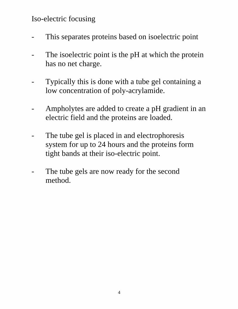
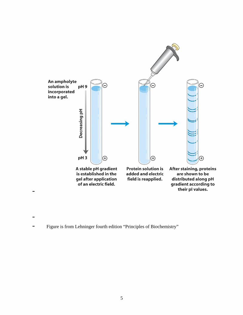
Iso-electric focusing - This separates proteins based on isoelectric point - The isoelectric point is the pH at which the protein
has no net charge. - Typically this is done with a tube gel containing a
low concentration of poly-acrylamide. - Ampholytes are added to create a pH gradient in an
electric field and the proteins are loaded. - The tube gel is placed in and electrophoresis
system for up to 24 hours and the proteins form tight bands at their iso-electric point.
- The tube gels are now ready for the second
method.
5
- - - Figure is from Lehninger fourth edition “Principles of Biochemistry”
6
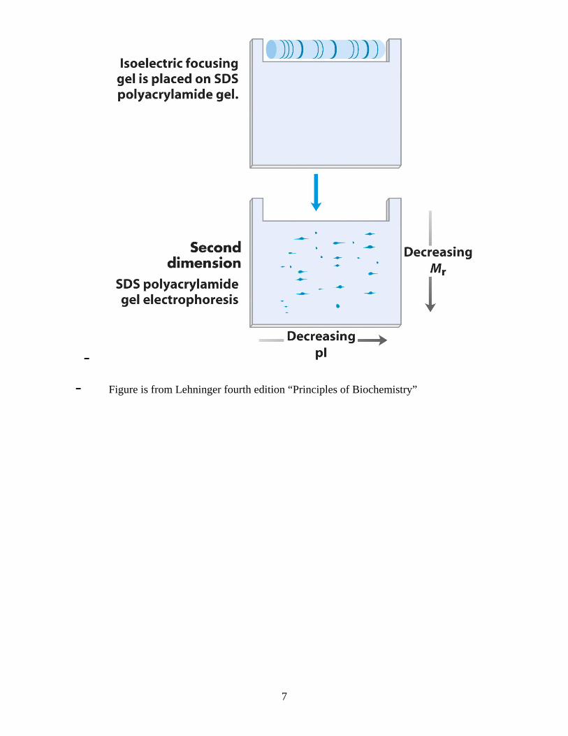
- SDS Poly-Acrylamide Gel Electrophoresis - The second dimension separates the proteins based
on size. - There are two parts, the stacking gel which
concentrates the sample and the running gel that is used to separate the proteins.
- The tube gel is soaked in a solution containing
chemical to denature the proteins including sodium dodecyl sulfate a detergent which gives the proteins a net negative charge. This means that all proteins will move in one direction.
- The tube gel is then put in the one long well in the
stacking gel, sealed in place with agarose, and the proteins subjected to an electric field to separate.
- The larger proteins are found at the top and the
smaller ones are found at the bottom of the gel. - In a 2D gel the proteins appear as spots on the gel
rather than bands. These spots can then be further processed or used for mass spectrometry directly.
- Further processing usually include trypsin
digestion
7
-
- Figure is from Lehninger fourth edition “Principles of Biochemistry”
8
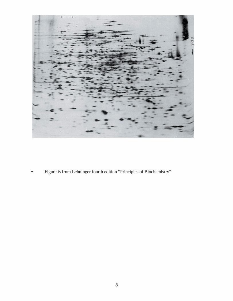
- Figure is from Lehninger fourth edition “Principles of Biochemistry”
9
10
Alternate methods of separation. Recently there has been a change in the way people are approaching separating the proteins. The first dimension is run in larger agarose tube gels with ampholytes. This has less resolution than polyacrylamide gels. The tubes are sliced and the proteins are allowed to diffuse out. The diffused proteins are then separated by capillary electrophoresis. Capillary electrophoresis has a much greater resolution for the proteins mass. Proteins are eluted from the capillary in the process and can be collected. They are readily available for mass spectrometry.
11
Mass Spectrometery
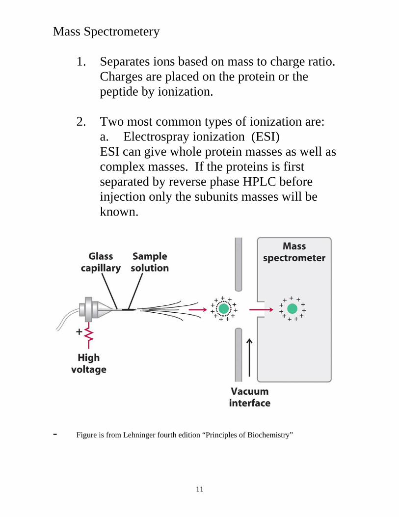
1. Separates ions based on mass to charge ratio. Charges are placed on the protein or the peptide by ionization.
2. Two most common types of ionization are:
a. Electrospray ionization (ESI) ESI can give whole protein masses as well as complex masses. If the proteins is first separated by reverse phase HPLC before injection only the subunits masses will be known.
- Figure is from Lehninger fourth edition “Principles of Biochemistry”
12
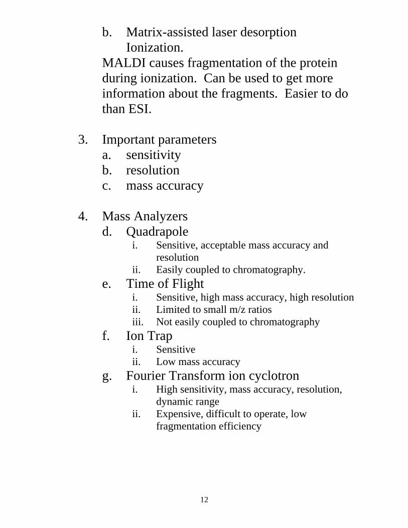
b. Matrix-assisted laser desorption Ionization.
MALDI causes fragmentation of the protein during ionization. Can be used to get more information about the fragments. Easier to do than ESI.
3. Important parameters a. sensitivity b. resolution c. mass accuracy
4. Mass Analyzers d. Quadrapole
i. Sensitive, acceptable mass accuracy and resolution
ii. Easily coupled to chromatography. e. Time of Flight
i. Sensitive, high mass accuracy, high resolution ii. Limited to small m/z ratios iii. Not easily coupled to chromatography
f. Ion Trap i. Sensitive ii. Low mass accuracy
g. Fourier Transform ion cyclotron i. High sensitivity, mass accuracy, resolution,
dynamic range ii. Expensive, difficult to operate, low
fragmentation efficiency
13
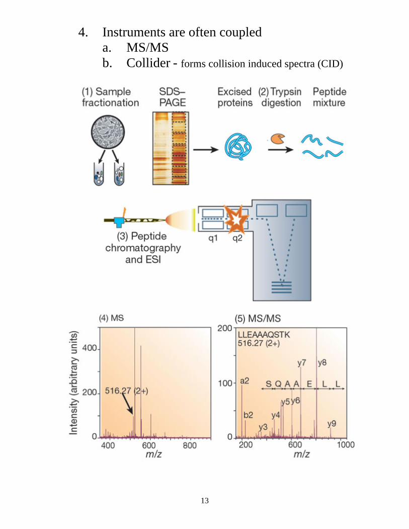
4. Instruments are often coupled a. MS/MS b. Collider - forms collision induced spectra (CID)
14
Protein identification and Quatitation
1. Protein separation by 2 dimensional electrophoresis or similar methods
2. Limited protein purification and automated MS.
Limited purification and MS
1. multiple chromatography stages a. ion exchange, reverse phase, affinity b. electrophoresis
2. To quantitate a. add stable isotopes b. post separation modification i. SH, NH2, N-linked carbohydrates c. incorporation of isotopes in culture
Protein Identification
1. Use collision induced spectra
i. provides sequence information ii. provides unique m/z spectra for each
peptide
2. Problem is large number of CID information i. need methods for searching ii. Filtering has been tried (limited success) iii. Most successful with human intervention. iv. Improving