Embed Size (px)
Citation preview
DOI: 10.2478/s11686-012-0021-x© W. Stefanski Institute of Parasitology, PASActa Parasitologica, 2012, 57(2), 114–121; ISSN 1230-2821
Protozoan and myxozoan infections in wild gilthead seabream (Sparus aurata L.)
from North Lake of Tunis, Tunisia
Sihem BahriDépartement de Biologie, Faculté des Sciences de Tunis, Université Tunis El Manar, 2092 Tunisie
AbstractA total of 150 gilthead seabream Sparus aurata L., from North Lake of Tunis, Tunisia, were studied for protozoan and myxo-zoan parasites. The parasitological survey revealed the presence of ectoparasites (Amyloodinium ocellatum Brown, 1931, Tri-chodina lepsii Lom, 1962 on the gills) and endoparasites (Ceratomyxa sparusaurati Sitjà-Bobadilla, Palenzuela etAlvarez-Pellitero, 1995 infecting the gallbladder, and Eimeria sparis Sitjà-Bobadilla, Palenzuela et Alvarez-Pellitero, 1996parasitizing the intestine). This is the first record of Amyloodinium ocellatum, Trichodina lepsii, Ceratomyxa sparusaurati, andEimeria sparis in S. aurata from Tunisian waters. Data on prevalence and intensity of infection are provided. A comparison ofthe present species with previously described species in cultured gilthead seabream from other Mediterranean countries is alsopresented. In this study Trichodina lepsii is identified for the first time in Sparus aurata. A taxonomic description of this speciesbased on silver nitrate method is provided.
KeywordsAmyloodinium ocellatum, Trichodina lepsii, Eimeria sparis, Ceratomyxa sparusaurati, Sparus aurata, North Lake of Tunis,Tunisia, prevalence, intensity of infection
Introduction
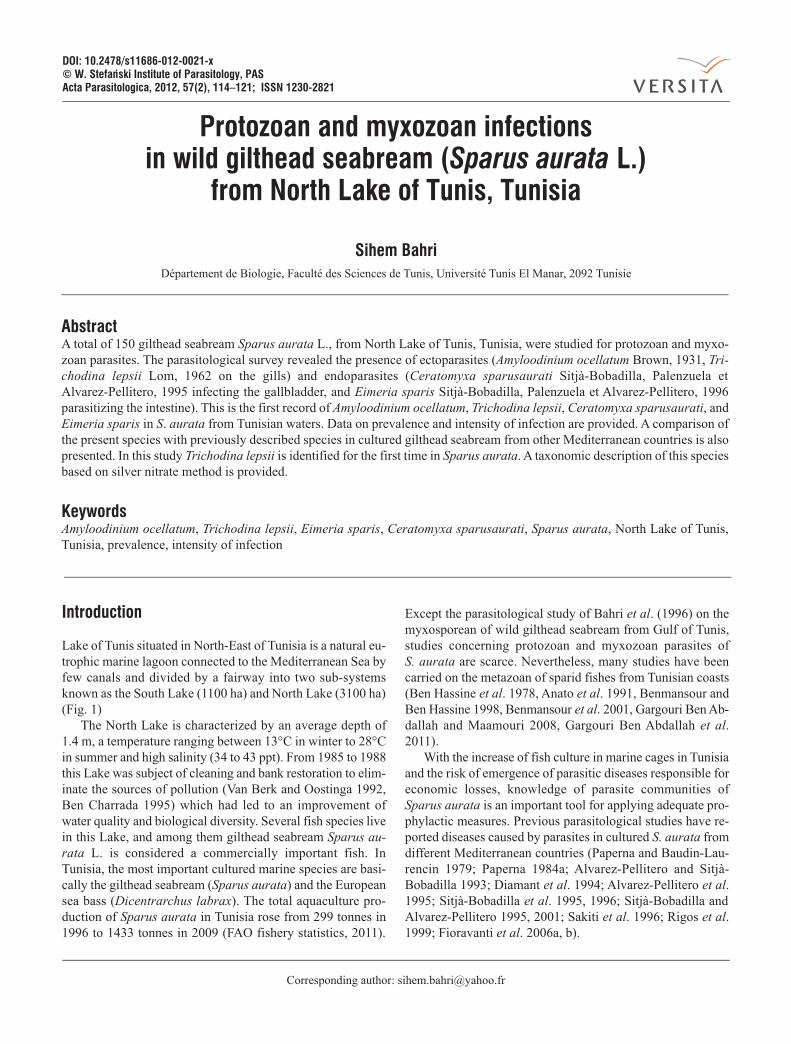
Lake of Tunis situated in North-East of Tunisia is a natural eu-trophic marine lagoon connected to the Mediterranean Sea byfew canals and divided by a fairway into two sub-systemsknown as the South Lake (1100 ha) and North Lake (3100 ha)(Fig. 1)
The North Lake is characterized by an average depth of1.4 m, a temperature ranging between 13°C in winter to 28°Cin summer and high salinity (34 to 43 ppt). From 1985 to 1988this Lake was subject of cleaning and bank restoration to elim-inate the sources of pollution (Van Berk and Oostinga 1992,Ben Charrada 1995) which had led to an improvement ofwater quality and biological diversity. Several fish species livein this Lake, and among them gilthead seabream Sparus au-rata L. is considered a commercially important fish. InTunisia, the most important cultured marine species are basi-cally the gilthead seabream (Sparus aurata) and the Europeansea bass (Dicentrarchus labrax). The total aquaculture pro-duction of Sparus aurata in Tunisia rose from 299 tonnes in1996 to 1433 tonnes in 2009 (FAO fishery statistics, 2011).
Except the parasitological study of Bahri et al. (1996) on themyxosporean of wild gilthead seabream from Gulf of Tunis,studies concerning protozoan and myxozoan parasites ofS. aurata are scarce. Nevertheless, many studies have beencarried on the metazoan of sparid fishes from Tunisian coasts(Ben Hassine et al. 1978, Anato et al. 1991, Benmansour andBen Hassine 1998, Benmansour et al. 2001, Gargouri Ben Ab-dallah and Maamouri 2008, Gargouri Ben Abdallah et al.2011).
With the increase of fish culture in marine cages in Tunisiaand the risk of emergence of parasitic diseases responsible foreconomic losses, knowledge of parasite communities ofSparus aurata is an important tool for applying adequate pro-phylactic measures. Previous parasitological studies have re-ported diseases caused by parasites in cultured S. aurata fromdifferent Mediterranean countries (Paperna and Baudin-Lau-rencin 1979; Paperna 1984a; Alvarez-Pellitero and Sitjà-Bobadilla 1993; Diamant et al. 1994; Alvarez-Pellitero et al.1995; Sitjà-Bobadilla et al. 1995, 1996; Sitjà-Bobadilla andAlvarez-Pellitero 1995, 2001; Sakiti et al. 1996; Rigos et al.1999; Fioravanti et al. 2006a, b).
Corresponding author: [email protected]
Protozoan and myxozoan infections in Sparus aurata 115
Therefore, the aim of the present work was to evaluate therisk of protozoan and myxozoan infections in wild giltheadseabream from North Lake of Tunis. Histological observationswere also carried out in order to clarify the possible patho-genic effects of parasites on infected organs.
Materials and methods
From 2009 to 2010, 150 specimens of Sparus aurata rangingfrom 80–250 g in weight were collected from North Lake ofTunis. Fish were euthanized by lethal dose of MS 222 andtransported to the laboratory for parasitological examination.Environmental variables were measured using a WTW multi-parameter (Multi 340i) for water temperature (°C) and salin-ity (ppt) in each season (Table I).
Fresh smears of skin and portions of gills from the eightgill arches were examined for the presence of parasites. Forthe observation of the adhesive disc of trichodinids, smearsfrom infected fish were air dried and impregnated for 10 minin 2% aqueous AgNo
3solution (Klein 1958). Measurements
(in µm) of trichodinids were made according to the recom-mendations proposed by Lom (1958). Details of denticleswere presented according to Van As and Basson (1989).
Fresh spores of the myxosporean isolated from the gall-bladder were measured according to the criteria described byLom and Arthur (1989). The oocysts of the coccidian were
measured from fresh smear under light microscope. Photo-graphs of parasites were taken with a Nikon E600 microscopeusing differential interference contrast optics.
Prevalence of infection was defined as percentage of fishinfected. Intensity of infection was semiquantitatively evalu-ated according to Alvarez-Pellitero et al. (1995) with therange: (+) 1–5; (++) 6–10; (+++) 11–25; (++++) 26–50;(+++++) 51–100; (++++++) > 100 parasites per fish.
For histological examination, fragments of infected gills,gallbladders and intestine were fixed in 10% neutral bufferedformalin for 24 h. After fixation, the tissues were dehydratedthrough ethanol series, equilibrated in xylene and embedded inparaffin according to standard histological techniques. Sec-tions of 5 µm were cut and stained with haematoxylin-eosinbefore examination under light microscope.
Results
Specimens of Sparus aurata caught from North Lake of Tuniswere found to be parasitised by two ectoparasites, Amyloodi-nium ocellatum and Trichodina lepsii and two endoparasites,the cœlozoic myxozoan Ceratomyxa sparusaurati and the api-complexan Eimeria sparis. All species were previously re-ported to infect S. aurata from several Mediterranean aqua-culture systems, except Trichodina lepsii which is identified forthe first time in gilthead seabream.
Fig. 1. Map showing location of study site (*) in North Tunisia
Table I. Water parameters measured at the sampling period in North Lake of Tunis
Parameters Autumn Winter Spring Summer
Temperature (°C) 20.8 12 18.4 28
Salinity (ppt) 38.6 34.4 37.6 42.6
Sihem Bahri116
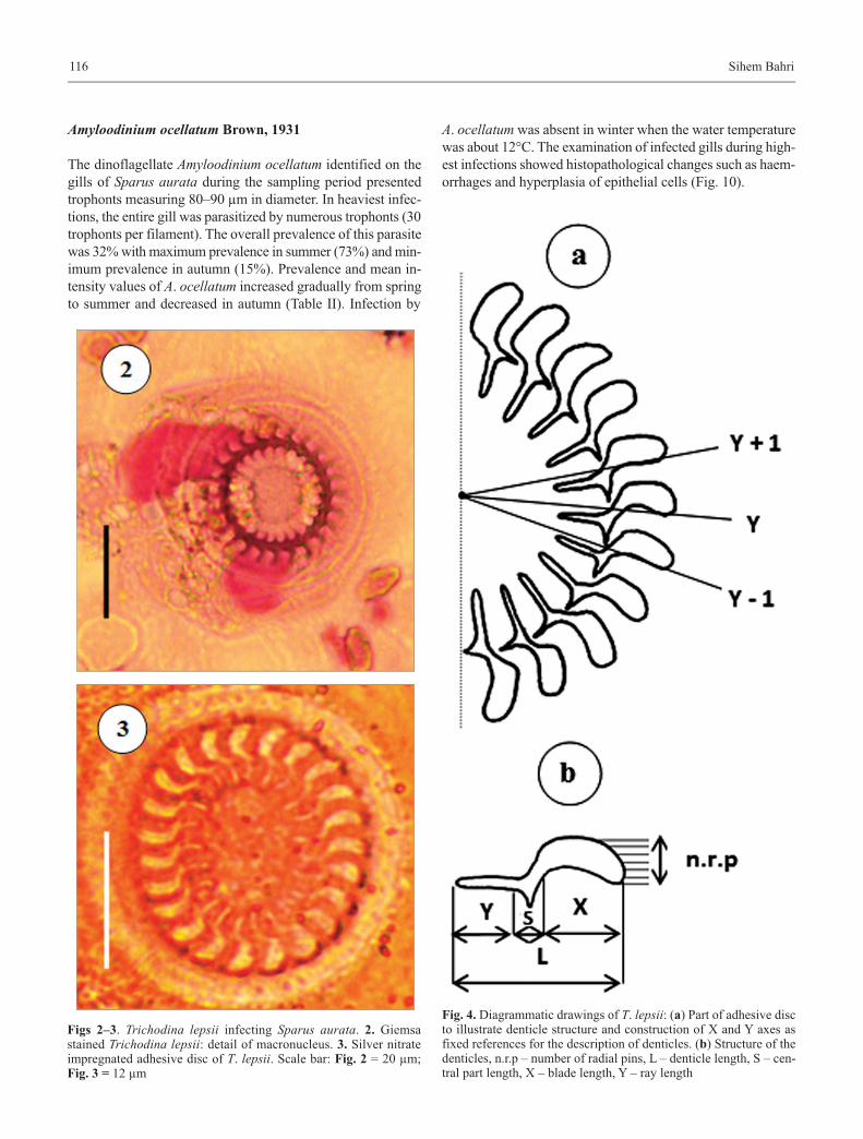
Amyloodinium ocellatum Brown, 1931
The dinoflagellate Amyloodinium ocellatum identified on thegills of Sparus aurata during the sampling period presentedtrophonts measuring 80–90 µm in diameter. In heaviest infec-tions, the entire gill was parasitized by numerous trophonts (30trophonts per filament). The overall prevalence of this parasitewas 32% with maximum prevalence in summer (73%) and min-imum prevalence in autumn (15%). Prevalence and mean in-tensity values of A. ocellatum increased gradually from springto summer and decreased in autumn (Table II). Infection by
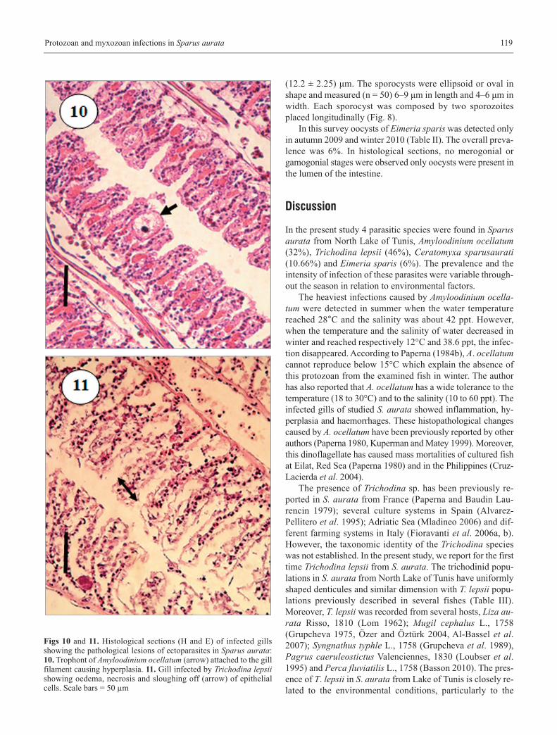
A. ocellatum was absent in winter when the water temperaturewas about 12°C. The examination of infected gills during high-est infections showed histopathological changes such as haem-orrhages and hyperplasia of epithelial cells (Fig. 10).
Fig. 4. Diagrammatic drawings of T. lepsii: (a) Part of adhesive discto illustrate denticle structure and construction of X and Y axes asfixed references for the description of denticles. (b) Structure of thedenticles, n.r.p – number of radial pins, L – denticle length, S – cen-tral part length, X – blade length, Y – ray length
Figs 2–3. Trichodina lepsii infecting Sparus aurata. 2. Giemsastained Trichodina lepsii: detail of macronucleus. 3. Silver nitrateimpregnated adhesive disc of T. lepsii. Scale bar: Fig. 2 = 20 µm;Fig. 3 = 12 µm
Protozoan and myxozoan infections in Sparus aurata 117
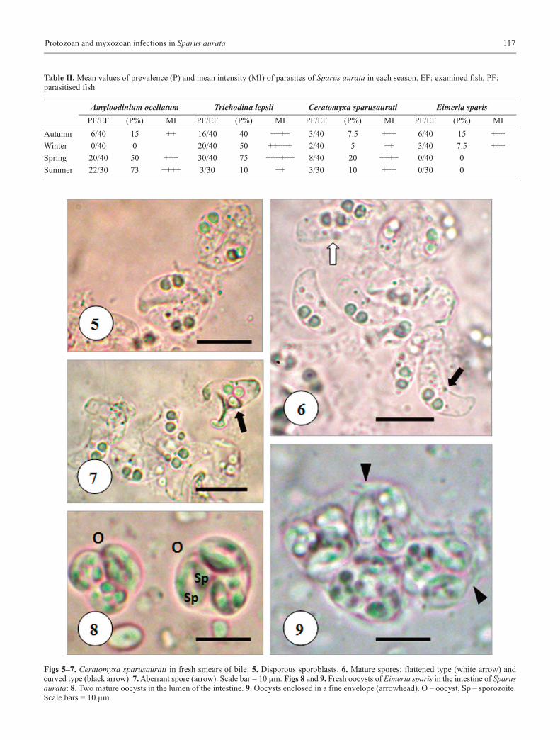
Table II. Mean values of prevalence (P) and mean intensity (MI) of parasites of Sparus aurata in each season. EF: examined fish, PF: parasitised fish
Amyloodinium ocellatum Trichodina lepsii Ceratomyxa sparusaurati Eimeria sparisPF/EF (P%) MI PF/EF (P%) MI PF/EF (P%) MI PF/EF (P%) MI
Autumn 6/40 15 ++ 16/40 40 ++++ 3/40 7.5 +++ 6/40 15 +++
Winter 0/40 0 20/40 50 +++++ 2/40 5 ++ 3/40 7.5 +++
Spring 20/40 50 +++ 30/40 75 ++++++ 8/40 20 ++++ 0/40 0
Summer 22/30 73 ++++ 3/30 10 ++ 3/30 10 +++ 0/30 0
Figs 5–7. Ceratomyxa sparusaurati in fresh smears of bile: 5. Disporous sporoblasts. 6. Mature spores: flattened type (white arrow) andcurved type (black arrow). 7. Aberrant spore (arrow). Scale bar = 10 µm. Figs 8 and 9. Fresh oocysts of Eimeria sparis in the intestine of Sparusaurata: 8. Two mature oocysts in the lumen of the intestine. 9. Oocysts enclosed in a fine envelope (arrowhead). O – oocyst, Sp – sporozoite.Scale bars = 10 µm
Sihem Bahri118
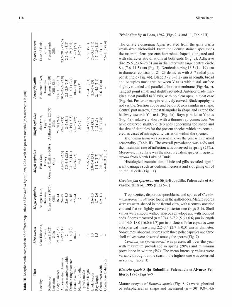
Trichodina lepsii Lom, 1962 (Figs 2–4 and 11, Table III)
The ciliate Trichodina lepsii isolated from the gills was asmall-sized trichodinid. From the Giemsa stained specimensthe macronucleus presents horseshoe-shaped, elongated andwith characteristic dilations at both ends (Fig. 2). Adhesivedisc 25.5 (23.6–28.8) µm in diameter with large central circle8.4 (7.6–11.5) µm (Fig. 3). Denticulate ring 16.5 (14–19) µmin diameter consists of 21–23 denticles with 5–7 radial pinsper denticle (Fig. 4b). Blade 3 (2.8–3.2) µm in length, broadand occupies most area between Y axes with distal surfaceslightly rounded and parallel to border membrane (Figs 4a, b).Tangent point small and slightly rounded. Anterior blade mar-gin almost parallel to Y axis, with no clear apex in most case(Fig. 4a). Posterior margin relatively curved. Blade apophysisnot visible. Section above and below X axis similar in shape.Central part narrow, almost triangular in shape and extend thehalfway towards Y-1 axis (Fig. 4a). Rays parallel to Y axes(Fig. 4a), relatively short with a thinner ray connection. Wehave observed slightly differences concerning the shape andthe size of denticles for the present species which are consid-ered as cases of intraspecific variation within the species.
Trichodina lepsii was present all over the year with markedseasonality (Table II). The overall prevalence was 46% andthe maximum rate of infection was observed in spring (75%).Moreover, this ciliate was the most prevalent species in Sparusaurata from North Lake of Tunis.
Histological examination of infested gills revealed signif-icant damages such as oedema, necrosis and sloughing off ofepithelial cells (Fig. 11).
Ceratomyxa sparusaurati Sitjà-Bobadilla, Palenzuela et Al-
varez-Pellitero, 1995 (Figs 5–7)
Trophozoites, disporous sporoblasts, and spores of Cerato-myxa sparusaurati were found in the gallbladder. Mature sporeswere crescent-shaped in the frontal view, with a convex anteriorend and flat or slightly curved posterior one (Figs 5–6). Shellvalves were smooth without mucous envelope and with roundedends. Spores measured (n = 30) 4.2–7.2 (5.6 ± 0.6) μm in lengthand 14.0–18.0 (16.0 ± 1.7) μm in thickness. Polar capsules weresubspherical measuring 2.2–3.4 (2.7 ± 0.3) μm in diameter.Sometimes, abnormal spores with three polar capsules and threeshell valves were observed among the spores (Fig. 7).
Ceratomyxa sparusaurati was present all over the yearwith maximum prevalence in spring (20%) and minimumprevalence in winter (5%). The mean intensity values werevariable throughout the season, the highest one was observedin spring (Table II).
Eimeria sparis Sitjà-Bobadilla, Palenzuela et Alvarez-Pel-
litero, 1996 (Figs 8–9)
Mature oocysts of Eimeria sparis (Figs 8–9) were sphericalor subspherical in shape and measured (n = 30) 9.8–14.6 T
ab
le I
II.
Mor
phom
etri
c co
mpa
riso
n of
dif
fere
nt p
opul
atio
ns o
f Tr
icho
dina
leps
iiL
om, 1
962
wit
h th
e pr
esen
t m
ater
ial
(mea
sure
men
ts i
n μ
m)
Host
Liza
aur
ata
Mug
il ce
phal
usM
ugil
ceph
alus
Mug
il ce
phal
usPe
rca
fluva
tilis
Spar
us a
urat
a
Loc
alit
y
Ref
eren
ceL
ocat
ion
Bod
y di
amet
erA
dhes
ive
disc
dia
met
erB
orde
r m
embr
ane
wid
thD
enti
cle
ring
dia
met
erD
enti
cle
num
ber
Num
ber
of r
adia
l pi
ns/d
enti
cle
Den
ticl
e le
ngth
B
lade
len
gth
Ray
len
gth
Cen
tral
par
t w
idth
Cen
tral
cir
cle
diam
eter
Lak
e T
abac
aria
, R
uman
ia
Lom
(19
62)
G
ills
28–4
2 (3
5)19
–27
(21)
211
–14
(12)
18–2
5– 3 2.5
1.5 1 –
Bou
rgas
Lak
e,
Bul
gari
aG
rupc
heva
(19
75)
Gil
ls36
–44
24–2
72.
6–3.
514
–16
22–2
45 –
2.6–
3.5
1.7–
2.6
0.9–
1.3
–
Bla
ck S
ea,
Tur
key
Öze
r an
d Ö
ztür
k (2
004)
Gil
ls–
19.2
–27
(22.
5)2.
3–3.
4 (2
.8)
13.4
–15
(14.
1)19
–24
(22)
4–5
5.2–
6 (5
.6)
2.8–
3.4
(3.1
)1.
4–2
(1.7
)0.
6–1
(0.8
)9.
4–10
.9 (
10.1
)
Lak
e Q
arun
, E
gypt
Al-
Bas
sel
etal
. (20
07)
Gil
ls25
–30
(28.
4)22
–27
(23)
2–5
(3.1
)13
–19
(15.
5)21
–26
(22)
5–7
(6)
3–4.
5 (3
.5)
3–4
(3.2
)1–
2 (1
.3)
2.5–
3 (2
.8)
–
Bla
ckm
an R
iver
, T
asm
ania
Bas
son
(201
0)G
ills
, Ski
n 25
.4–3
1.3
(27.
7)20
.5–2
5.6
(22.
8)2.
1–2.
9 (2
.5)
11–1
6.1
(13.
4)18
–22
(20)
6–8
(7)
2.7–
4.1
(3.3
)2.
5–3.
6 (3
)2.
4–4.
3 (3
.1)
0.6–
1 (0
.8)
–
Lak
e of
Tun
is,
Tun
isia
Pre
sent
stu
dyG
ills
–23
.6–2
8.8
(25.
5)2.
2–4.
4 (3
.8)
14–1
9 (1
6.5)
21
–23
(22)
5–7
(6)
5.7–
6 (5
.7)
2.8–
3.2
(3.1
)
1.7–
1.9
(1.8
) 1–
1.2
(1.1
)7.
6–11
.5 (
8.4)
Protozoan and myxozoan infections in Sparus aurata 119
(12.2 ± 2.25) μm. The sporocysts were ellipsoid or oval inshape and measured (n = 50) 6–9 μm in length and 4–6 μm inwidth. Each sporocyst was composed by two sporozoitesplaced longitudinally (Fig. 8).
In this survey oocysts of Eimeria sparis was detected onlyin autumn 2009 and winter 2010 (Table II). The overall preva-lence was 6%. In histological sections, no merogonial orgamogonial stages were observed only oocysts were present inthe lumen of the intestine.
Discussion
In the present study 4 parasitic species were found in Sparusaurata from North Lake of Tunis, Amyloodinium ocellatum(32%), Trichodina lepsii (46%), Ceratomyxa sparusaurati(10.66%) and Eimeria sparis (6%). The prevalence and theintensity of infection of these parasites were variable through-out the season in relation to environmental factors.
The heaviest infections caused by Amyloodinium ocella-tum were detected in summer when the water temperaturereached 28°C and the salinity was about 42 ppt. However,when the temperature and the salinity of water decreased inwinter and reached respectively 12°C and 38.6 ppt, the infec-tion disappeared. According to Paperna (1984b), A. ocellatumcannot reproduce below 15°C which explain the absence ofthis protozoan from the examined fish in winter. The authorhas also reported that A. ocellatum has a wide tolerance to thetemperature (18 to 30°C) and to the salinity (10 to 60 ppt). Theinfected gills of studied S. aurata showed inflammation, hy-perplasia and haemorrhages. These histopathological changescaused by A. ocellatum have been previously reported by otherauthors (Paperna 1980, Kuperman and Matey 1999). Moreover,this dinoflagellate has caused mass mortalities of cultured fishat Eilat, Red Sea (Paperna 1980) and in the Philippines (Cruz-Lacierda et al. 2004).
The presence of Trichodina sp. has been previously re-ported in S. aurata from France (Paperna and Baudin Lau-rencin 1979); several culture systems in Spain (Alvarez-Pellitero et al. 1995); Adriatic Sea (Mladineo 2006) and dif-ferent farming systems in Italy (Fioravanti et al. 2006a, b).However, the taxonomic identity of the Trichodina specieswas not established. In the present study, we report for the firsttime Trichodina lepsii from S. aurata. The trichodinid popu-lations in S. aurata from North Lake of Tunis have uniformlyshaped denticules and similar dimension with T. lepsii popu-lations previously described in several fishes (Table III).Moreover, T. lepsii was recorded from several hosts, Liza au-rata Risso, 1810 (Lom 1962); Mugil cephalus L., 1758(Grupcheva 1975, Özer and Öztürk 2004, Al-Bassel et al.2007); Syngnathus typhle L., 1758 (Grupcheva et al. 1989),Pagrus caeruleostictus Valenciennes, 1830 (Loubser et al.1995) and Perca fluviatilis L., 1758 (Basson 2010). The pres-ence of T. lepsii in S. aurata from Lake of Tunis is closely re-lated to the environmental conditions, particularly to the
Figs 10 and 11. Histological sections (H and E) of infected gillsshowing the pathological lesions of ectoparasites in Sparus aurata:10. Trophont of Amyloodinium ocellatum (arrow) attached to the gillfilament causing hyperplasia. 11. Gill infected by Trichodina lepsiishowing oedema, necrosis and sloughing off (arrow) of epithelialcells. Scale bars = 50 µm
Sihem Bahri120
temperature and the salinity. In this study we noted that thehighest prevalence and mean intensities of T. lepsii wererecorded during winter and spring, while the lowest infectionrate occurred in summer when the temperature and the salin-ity of water were high, respectively 28°C and 42.6 ppt. Manyauthors have determined the relationship between seasonal dy-namics of trichodinids and water quality and reported that themaximum rate of infection was in winter and/or in spring (AbuEl-Wafa 1988; Hassan 1999; Özer and Erdem 1999; Özer2000, 2003; Jerônimo et al. 2011). Previous studies have re-ported that the seasonal prevalence and intensity of tricho-dinids can be affected by organic pollution such as the level ofnitrite, nitrate and phosphate (Ogut and Palm 2005) and thedeposition of pig manure (Jerônimo et al. 2011). The fish ex-amined in this study presented at higher infection, necrosis,oedema and hyperplasia of the gills. These pathological le-sions have been previously observed associated with Tri-chodina spp. infection in several fish species by other authors(Hassan 1999, Afifi et al. 2000, Yemmen et al. 2011).
In the current study, the seasonal variations of Ceratomyxasparusaurati prevalence are similar to those reported by Palen-zuela et al. (1997) in intensive open system (i.e. higher inspring and summer and lower in autumn and winter). In gen-eral, many factors are involved in seasonal fluctuations ofmyxosporeans such as temperature (Alvarez-Pellitero andSitjà-Bobadilla 1993, Alvarez-Pellitero et al. 1995), farmingsystems and size categories (Palenzuela et al. 1997, Fioravantiet al. 2006a, b). Fish heavily infected with C. sparusauratipresent enlarged gallbladders and abdominal distension(Palenzuela et al. 1997). Nevertheless, we have not observedexternal sign of disease in the infected fish during the survey.Further myxosporeans have been reported in gilthead sea-bream, Enteromyxum leei Diamant, Lom et Dyková, 1994;Polysporoplasma sparis Sitjà-Bobadilla et Alvarez-Pellitero,1995; Henneguya sp. Bahri, Ben Hassine et Marques, 1996and Leptotheca sparidarum Sitjà-Bobadilla et Alvarez-Pel-litero, 2001 which have not been observed in the sampled fishduring our study.
Our observations on Eimeria sparis are limited to theoocysts and sporozoites. The different stages of merogony andgamogony were not observed. Comparatively, fish in the pres-ent study presented lower prevalence and infection intensitiesthan those observed by Alvarez-Pellitero et al. (1995) in S. au-rata from different cultured systems in Spain. On the contrary,Fioravanti et al. 2006b observed a very low prevalence (0.2%)of this parasite in cultured S. aurata from Italy.
In conclusion, prevalence and infection intensity of proto-zoan ectoparasites in wild S. aurata from North Lake of Tunisare influenced by environmental factors basically temperatureand salinity. However, their pathogenicity was related to theintensity of parasites and to the host-inherent parameters. Inthe other hand, seasonal variations of the myxosporean Cera-tomyxa sparusaurati and the apicomplexan Eimeria sparisseems to be mostly related to the presence of their intermedi-ate hosts in the lake.
ReferencesAbu El-Wafa S.A.D. 1988. Protozoa parasites of some freshwater
fishes in Behera Governorate. M.V.Sc. Thesis, AlexandriaUniversity.
Afifi S.H., Thobiati AL., Hazaa M.S. 2000. Parasitic lesions in Niletilapia Oreochromis niloticus from fish farms in Saudi Ara-bia. Assiut Veterinary Medical Journal, 42, 183–194.
Al-Bassel D.A., Abdel-Baki A.S., Atwa M.S. 2007. Trichodinid ec-toparasites (Ciliophora: Peritrichia) of Mugil cephalus Lin-naeus, 1758 from Lake Qarun. Egyptian Journal of AquaticBiology and Fisheries, 11, 13–26.
Alvarez-Pellitero P., Sitjà-Bobadilla A. 1993. Pathology of Myxo-sporea in marine fish culture. Diseases of Aquatic Organisms,17, 229–238. DOI: 10.3354/dao017229.
Alvarez-Pellitero P., Sitjà-Bobadilla A., Franco-Sierra A., PalenzuelaO. 1995. Protozoan parasites of gilthead seabream, Sparus au-rata L., from different culture systems in Spain. Journal ofFish Diseases, 18, 105–115. DOI:10.1111/j.1365-2761.1995.tb00268.x.
Anato C.B., Ktari M.H., Dossou C.H. 1991. La parasitofaune méta-zoaire de Boops boops (Linné, 1758), poisson téléostéen Spa-ridae des côtes tunisiennes. Oebalia, 17, 259–266.
Bahri S., Ben Hassine O.K., Marques A. 1996. Henneguya sp. (Myxo-sporea, Bivalvulida) infecting the gills of wild giltheadseabream Sparus aurata L., from the coast of Tunisia. Bulletinof the European Association of Fish Pathologists, 16, 51–53.
Basson L. 2010. First records of trichodinid ectoparasites (Cilio-phora: Peritrichia) from introduced freshwater fishes in Tas-mania, Australia, with comments on pathogenicity. ActaProtozoologica, 49, 253–265.
Ben Charrada R. 1995. Impact des aménagements de restauration surla qualité des eaux et des peuplements benthiques du lac deTunis. Marine Life, 5, 51–64.
Ben Hassine O.K., Essafi K., Raibaut A. 1978. Les Lernaeopodidés,Copépodes parasites de Sparidés de Tunisie. Archives de l’Ins-titut Pasteur de Tunis, 554, 431–454.
Benmansour B., Ben Hassine O.K. 1998. Preliminary analysis of par-asitic copepod species richness among coastal fishes ofTunisia. Italian Journal of Zoology, 65:S1, 341–344. DOI:10.1080/11250009809386844.
Benmansour B., Ben Hassine O.K., Diebakate C., Raibaut A. 2001.Sur deux espèces de Copépodes Lernaeopodidae (Siphono-stomatoida) parasites du marbré Lithognathus mormyrus (Lin-naeus, 1758) (Pisces, Sparidae). Zoosystema, 23, 695–703.
Cruz-Lacierda E.R., Maeno Y., Pineda A.J.T., Matey V.E. 2004. Massmortality of hatcheryreared milkfish (Chanos chanos) andmangrove red snapper (Lutjanus argentimaculatus) caused byAmyloodium ocellatum (Dinoflagellida). Aquaculture, 236,85–94.
Diamant A., Lom J., Dykova I. 1994. Myxidium leei n. sp., a patho-genic myxosporean of cultured seabream Sparus aurata. Dis-eases of Aquatic Organisms, 20, 137–141. DOI: 10.3354/dao020137.
FAO. ©2011. Statistiques sur les pêches et l’aquaculture. Produc-tion mondiale de l’aquaculture 1950–2009 (Fishstat). In :FAO Département des pêches et de l’aquaculture (en ligne),Rome.
Fioravanti M.L., Caffara M., Florio D., Gustinelli A., Marcer F.2006a. A parasitological survey of European sea bass (Dicen-trarchus labrax) and gilthead seabream (Sparus aurata), cul-tured in Italy. Veterinary Research Communications, 30(Suppl. 1), 249–252. DOI: 10.1007/s11259-006-0053-5.
Fioravanti M.L., Caffara M., Florio D., Gustinelli A., Marcer F., Qua-glio F. 2006b. Parasitic diseases of marine fish: epidemiolog-ical and sanitary considerations. Parasitologia, 48, 15–18.
Protozoan and myxozoan infections in Sparus aurata 121
Gargouri Ben Abdallah L., Maamouri F. 2008. Digenean fauna di-versity in sparid fish from Tunisian coasts. Bulletin of the Eu-ropean Association of Fish Pathologists, 28, 129–137.
Gargouri Ben Abdallah L., Antar R., Maamouri F. 2011. Diversity ofthe digenean fauna in sparid fishes from the Lagoon of Bi-zerte in Tunisia. Acta Parasitologica, 56, 34–39. DOI: 10.2478/s11686-011-0007-0.
Grupcheva G.I. 1975. Parasitic Infusoria (Peritricha, Urceolariidae)on some fishes from the Bourgas Lake. Acta Zoologica Bul-garica, 1, 77–83.
Grupcheva G.I., Lom J., Dykova I. 1989. Trichodinids (Ciliata: Urce-olariidae) from gills of some marine fishes with the descrip-tion of Trichodina zaikai sp. n. Folia Parasitologica, 36,193–207.
Hassan M.A.H. 1999. Trichodiniasis in farmed freshwater Tilapia inEastern Saudi Arabia. Journal of King Abdulaziz University:Marine Sciences, 10, 157–168.
Jerônimo G.T., Speck G.M., Cechinel M.M., Gonçalves E.L.T., Mar-tins M.L. 2011. Seasonal variation on the ectoparasitic com-munities of Nile tilapia cultured in three regions in southernBrazilian. Brazilian Journal of Biology, 71, 365–373.
Klein B.M. 1958. The dry silver method and its proper use. Journalof Protozoology, 5, 99–103. DOI: 10.1111/j.1550-7408.1958.tb02535.x.
Kuperman B.I., Matey V.E. 1999. Massive infestation by Amyloo-dinium ocellatum (Dinoflagellida) of fish in a highly salinelake, Salton Sea, California, USA. Diseases of Aquatic Or-ganisms, 39, 65–73. DOI: 10.3354/dao039065.
Lom J. 1958. A contribution to the systematics and morphology ofendoparasitic trichodinids from amphibians of uniform spe-cific characteristics. Journal of Protozoology, 5, 251–263.
Lom J. 1962. Trichodinid ciliates from fishes of the Rumanian BlackSea Coast. Parasitology, 52, 49–61. DOI: 10.1017/S0031182000023982.
Lom J., Arthur J.R. 1989. A guideline for the preparation of speciesdescription in Myxosporea. Journal of Fish Diseases, 12,151–156. DOI: 10.1111/j.13652761.1989.tb00287.x.
Loubser G.J., Van As J.G., Basson L. 1995. Trichodinid ectopara-sites (Ciliophora: Peritrichida) of some fishes from the Bayof Dakar, Senegal (West Africa). Acta Protozoologica, 34,211–216.
Mladineo I. 2006. Parasites of Adriatic cage reared fish. Acta Adria-tica, 47, 23–28.
Ogut H., Palm H.W. 2005. Seasonal dynamics of Trichodina spp. onwhiting (Merlangius merlangus) in relation to organic pollu-tion on the eastern Black Sea coast of Turkey. ParasitologyReaserch, 96, 149–153. DOI: 10.1007/s00436-005-1346-2.
Özer A. 2000. The occurrence of three species of Trichodina (Cilio-phora: Peritrichia) on Cyprinus carpio in relation to cultureconditions, seasonality and host characteristics. Acta Proto-zoologica, 39, 61–66. DOI: 10.1080/002229399300209.
Özer A. 2003. Trichodina domerguei Wallengren, 1987 (Ciliophora: Peri-trichia) infestations on the Round Goby, Neogobius melanosto-mus Pallas, 1811 in relation to seasonality and host factors. Com-parative Parasitology, 70, 132–135. DOI: 10.1654/4073.
Özer A., Erdem O. 1999. The relationship between occurrence of ec-toparasites, temperature and culture conditions; a comparisonof farmed and wild common carp (Cyprinus carpio L., 1758)in the Sinop region of northern Turkey. Journal of NaturalHistory, 33, 483–491.
Özer A., Öztürk T. 2004. Trichodina puytoraci Lom, 1962 and Tri-chodina lepsii Lom, 1962 (Peritrichida: Ciliophora) infesta-tions on mugilids caught at the Black Sea Coast of Sinop,Turkey. Turkish Journal of Zoology, 28, 179–182.
Palenzuela O., Sitjà-Bobadilla A., Alvarez-Pellitero P. 1997. Cerato-myxa sparusaurati (Protozoa: Myxosporea) infections in cul-tured gilthead seabream Sparus aurata (Pisces: Teleostei)from Spain: Aspects of the host-parasite relationship. Para-sitology Research, 83, 539–548.
Paperna I. 1980. Amyloodinium ocellatum (Brown, 1931) (Dinofla-gellida) infestations in cultured marine fish at Eilat, Red Sea:epizootiology and pathology. Journal of Fish Diseases, 3,363–372.
Paperna I. 1984a. Review of diseases affecting cultured Sparus au-rata and Dicentrarchus labrax. In: (Eds. G. Barnabé and R.Billard) L’Aquaculture du Bar et des Sparidés. INRA Publ.Paris, pp. 465–482.
Paperna I. 1984b. Reproduction cycle and tolerance to temperatureand salinity of Amyloodinium ocellatum (Brown, 1931) (Di-noflagellida). Annales de Parasitologie Humaine et Compa-rée, 59, 7–30.
Paperna I., Baudin-Laurencin F. 1979. Parasitic infections of sea bass,Dicentrarchus labrax, and gilthead seabream, Sparus aurata,in mariculture facilities in France. Aquaculture, 16, 173–175.
Rigos G., Christophilogiannis P., Yiagnisi M., Andriopoulou A.,Koutsodimou M., Nengas I., Alexis M. 1999. Myxosporeaninfections in Greek mariculture. Aquaculture International, 7,361–364.
Sakiti N., Tarer V., Jacquemin D., Marques A. 1996. Présence en Mé-diterranée occidentale d’une Myxosporidie histozoïque pa-thogène dans les élevages de la daurade, Sparus aurata.Annales des Sciences Naturelles, Zoologie, Paris, 17, 123–127.
Sitjà-Bobadilla A., Alvarez-Pellitero P. 1995. Light and electron mi-croscopic description of Polysporoplasma n. g. from Sparusaurata (L.), and Polysporoplasma mugilis n. sp. from Liza au-rata. European Journal of Protistology, 31, 77–89.
Sitjà-Bobadilla A., Alvarez-Pellitero P. 2001. Leptotheca sparidiumn. sp. (Myxosporea: Bivalvulida), a parasite from culturedcommon dentex (Dentex dentex L.) and gilthead seabream(Sparus aurata L.) (Teleostei: Sparidae). Journal of Eukaryo-tic Microbiology, 48, 627–639. DOI: 10.1111/j.1550-7408.2001.tb00202.x.
Sitjà-Bobadilla A., Palenzuela O., Alvarez-Pellitero P. 1995. Cerato-myxa sparusaurati n. sp. (Myxosporea: Bivalvulida), a newparasite from cultured gilthead seabream (Sparus aurata L.):Light and electron microscopic description. Journal of Eu-karyotic Microbiology, 42, 529–539. DOI: 10.1111/j.1550-7408.1995.tb05901.x.
Sitjà-Bobadilla A., Palenzuela O., Alvarez-Pellitero P. 1996. Lightmicroscopic description of Eimeria sparis sp. nov. and Gous-sia sparis sp. nov. (Protozoa: Apicomplexa) from Sparus au-rata L. (Pisces: Teleostei). Parasitology Research, 82, 323–332.
Yemmen C., Quilichini Y., Ktari M.H., Marchand B., Bahri S. 2011.Morphological, ecological and histopathological studies ofTrichodina gobii Raabe, 1959 (Ciliophora: Peritrichida) in-fecting the gills of Solea aegyptiaca. Protistology, 6, 258–263.
Van As J.G., Basson L. 1989. A further contribution to the taxonomyof the Trichodinidae (Ciliophora: Peritricha) and a review ofthe taxonomic status of some fish ectoparasitic trichodinids.Systematic Parasitology, 14, 157–179.
Van Berk A.H., Oostinga H. 1992. North lake of Tunis and its shores:Restauration and development. Terra Aqua, 49, 23–32.
(Accepted February 29, 2012)