Embed Size (px)
Citation preview
Protozoans/Parasites
What We Will Cover: Parasitemias
• Flagellated protozoans (P. Mastigophora)• Flagellated algae (C. Dinoflagellida)• Ciliates (protozoans)• Myxozoans (protozoans)• Digenetic Trematodes (flukes)• Cestodes (tapeworms)
(1) Flagellated Protozoans
• Flagellates are protozoans: simple, single-celled animals (over 50,000 recognized species)
• very small (15-30 M), body elongate, leaf-like appearance, up to 975,000/mL of blood
• flagellum arises posteriorly and can be connected to other parts of body, pulls animal through the blood
• most famous are Trypanasoma, Trypanoplasma, Ichthyobodo necatur
Ichthyobodo necatur
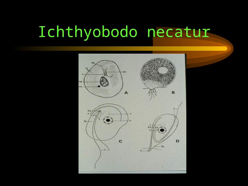
• A Mastigophoran, but a member of Class Diplomonadea
• also small, but flat and ovoid when swimming• has 2-4 flagella arising from a basal body
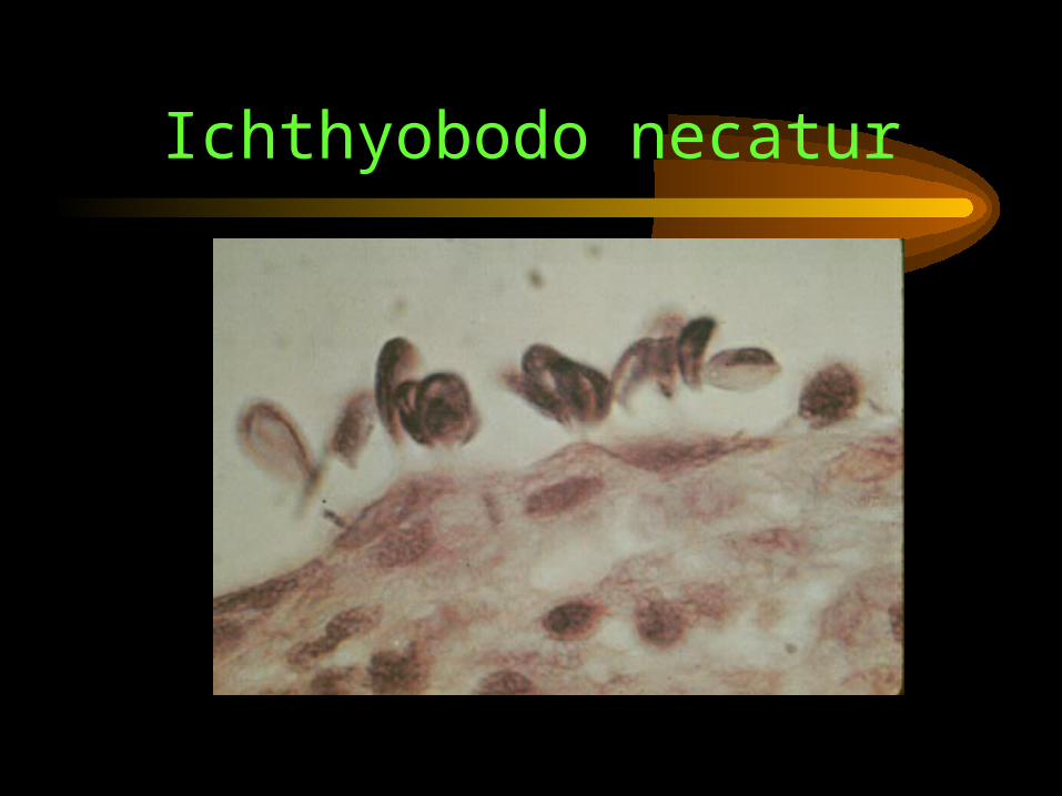
(kinetosome) at anterior end• obligate parasite, poor swimmer, attaches to gills,
but not good at attaching• uses a sucking organelle to penetrate host• tissue penetrated becomes necrotic
Icthyobodo necatur• Largely affects young, undernourished carp and
trout
• can also parasitize frogs/tadpoles
• wild fish/frogs serve as reservoirs, found everywhere
• seasonality affect resulting from salmonid hatchery stocking seasons (April - May)
• affects smolts by attaching to gills and not allowing them to adapt to seawater
Ichthyobodo necatur
• Pathogenicity: dull spots on body (blue slime), pale gills, hemorrhaging, fin necrosis, loss of appetite, flashing, moribund fish
• Control: salmonids need prophylaxis with formalin (1:4000 for 1 hr); carp need 1% salt bath 30 minutes repeated 3-4 times
Ichthyobodo necatur
Ichthyobodo necatur

(2) Flagellated algae:Oodinium/Amylodinium
• Members of Subkingdom Protozoa, Phylum Mastigophora (flagellates), Class Dinoflagellida (dinoflagellates)
• Two major genera: Oodinium (freshwater) and Amylodinium (saltwater)
• Both attach to skin and gills causing condition known as velvet or rust disease (from chlorophyll in parasite)
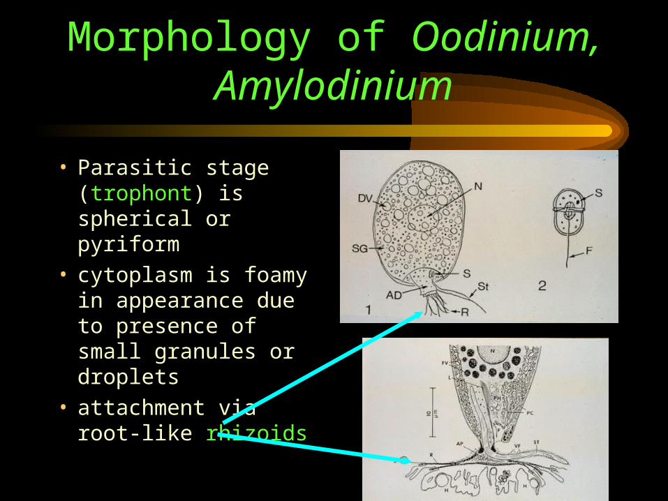
Morphology of Oodinium, Amylodinium
• Parasitic stage (trophont) is spherical or pyriform
• cytoplasm is foamy in appearance due to presence of small granules or droplets
• attachment via root-like rhizoids
Life Cycle of Oodinium/Amylodinium
• Parasitic trophont found on fish for about one week feeding on cytoplasm
• eventually retracts rhizoids, drops off and encysts• encysted form known as “tomont”, hard to kill with
chemicals• tomont undergoes mitotic division (8), ultimately
producing up to 256 dinospores• one dinospore can equal 1 billion new parasites• dinospores break out of cyst and seek new hosts
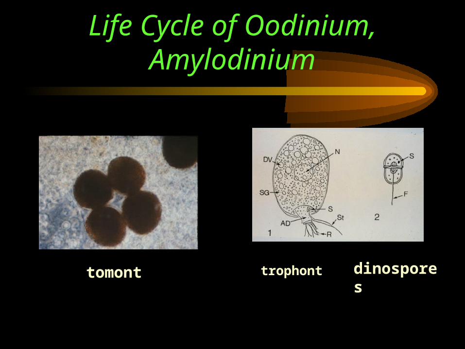
Life Cycle of Oodinium, Amylodinium
tomont dinosporestrophont
Host/parasite Relationship
• Broad specificity: sea bream, sea bass, mullet, tilapia, striped bass
• wide geographic distribution (here in Gulf of Mexico)
• cause little problem in nature, usually result of crowding
• outbreaks can be very explosive, Gulf Coast Research Lab lost almost all striped bass to this in 1976
Pathology/Control• Damage due to penetration of rhizoids
• Affects epithelium of skin, gills, nasal cavities, eyes and mouth
• Parasites produce lytic excretion causing inflammation, sometimes necrosis, secondary infections with bacteria/fungi
• Control: difficult due to rapid reproduction, no apparent acquired immunity, can encyst
• Treatment: copper (fair), metronidazole (14 mg/L)
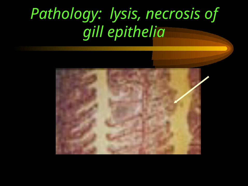
Pathology: lysis, necrosis of gill epithelia
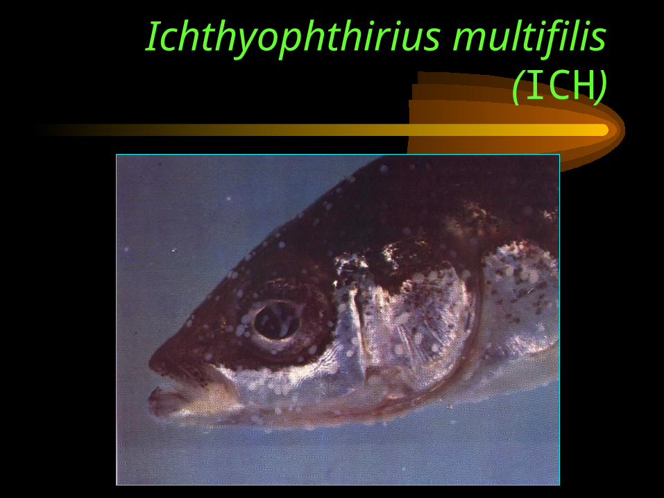
(3)Ciliates: Ichthyophthirius multifilis (ICH)
• Another single-celled protozoan type
• adult is round in shape, up to 1 mm in diameter, known as “trophont” (rem? Same as Amylodinium)
• short cilia in rows over entire cell, obvious as free-living stages “tumble” through the water
• Life Cycle: the trophont attaches to gills or skin, after 7-10 days, the trophont drops off and is called a “tomont” (same, also), tomont attaches to substrate and encysts, cyst ruptures releasing swarmers known as “theronts”
• theronts are the parasites (have perferatorium), also use hyaluronidase, only for less than 20 hrs, displace normal tissue as they grow
Ichthyophthirius multifilis (ICH)
• Signs: white pustules in advanced cases, sometimes called white spot disease; if found on gills, not found on body
• Behavioral changes: fish scratch against bottom (flash), hide in corners, twitching fins
• death in 20-26 days, thought to be due to osmoregulatory failure in most cases
• Host/parasite range: broad, mainly in catfish/salmonids
Ichthyophthirius multifilis (ICH)
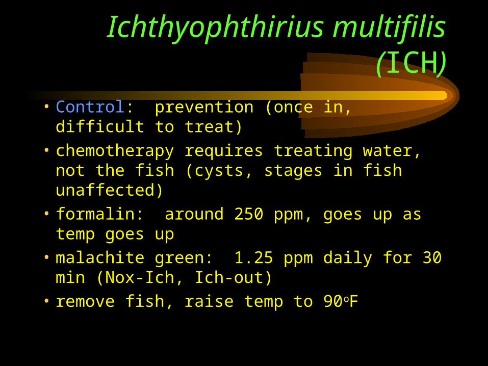
• Control: prevention (once in, difficult to treat)
• chemotherapy requires treating water, not the fish (cysts, stages in fish unaffected)
• formalin: around 250 ppm, goes up as temp goes up
• malachite green: 1.25 ppm daily for 30 min (Nox-Ich, Ich-out)
• remove fish, raise temp to 90oF
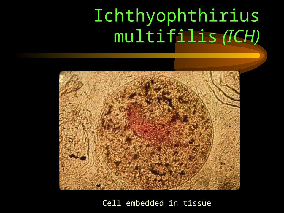
Ichthyophthirius multifilis (ICH)
Cell embedded in tissue
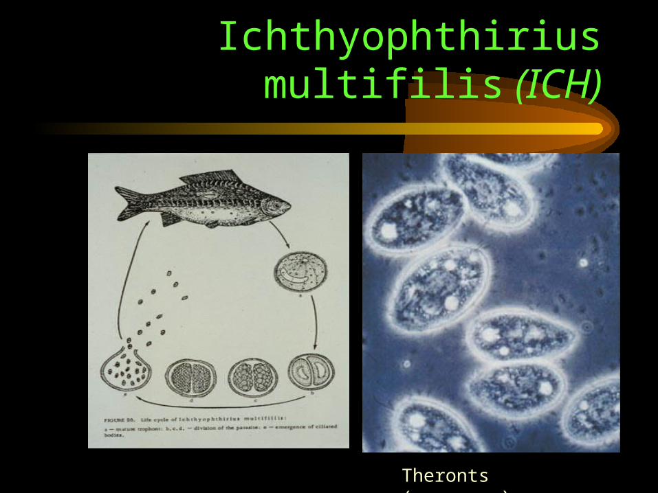
Ichthyophthirius multifilis (ICH)
Theronts (swarmers)
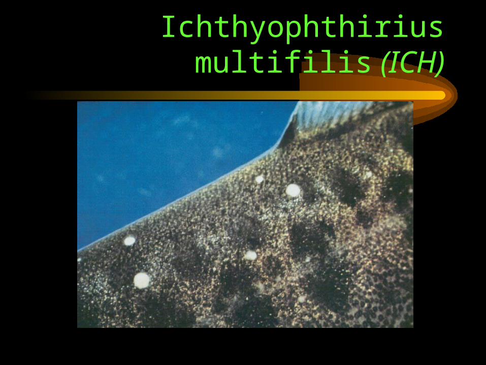
Ichthyophthirius multifilis (ICH)
Ichthyophthirius multifilis (ICH)
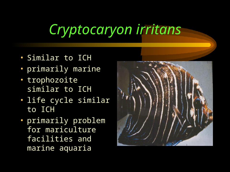
Cryptocaryon irritans
• Similar to ICH• primarily marine• trophozoite similar to
ICH• life cycle similar to
ICH• primarily problem for
mariculture facilities and marine aquaria
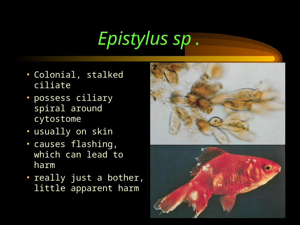
Epistylus sp.
• Colonial, stalked ciliate
• possess ciliary spiral around cytostome
• usually on skin• causes flashing, which
can lead to harm• really just a bother,
little apparent harm
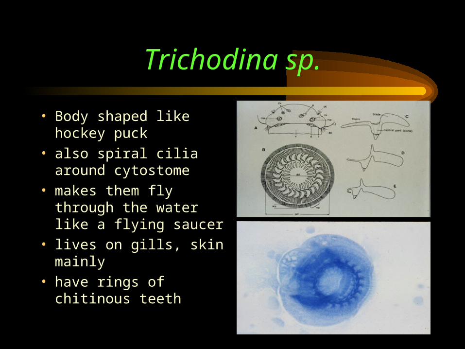
Trichodina sp.
• Body shaped like hockey puck
• also spiral cilia around cytostome
• makes them fly through the water like a flying saucer
• lives on gills, skin mainly• have rings of chitinous
teeth

Stop here: next time, myxozoans and P. Platyhelminthes!
(4) Myxozoans: Myxobolus cerebralis
• Rather odd, exclusively endoparasites
• could be Cnidarians (Phylum Cnidaria)
• multicellular during adult life, with various cell types
• now don’t know what to call them
• anyhow, we will discuss whatever they are in the context of what they cause: whirling disease (Salmonid Whirling Disease)
Salmonid Whirling Disease
• Important characteristic: can produce spore that is highly resistant (15 yrs dessication), associated with dispersal
• Life Cycle: infective stage gets into fish upon contact with skin, produces amoebula known as “trophozoite”, site attacked is species specific, most visible stage is the spore, spore released to environment, consumed by oligochaete, grow and released to environment
• fish eats oligochaete or encounters free spores
Salmonid Whirling Disease
• Usually found in salmonids, but not a contagious parasite
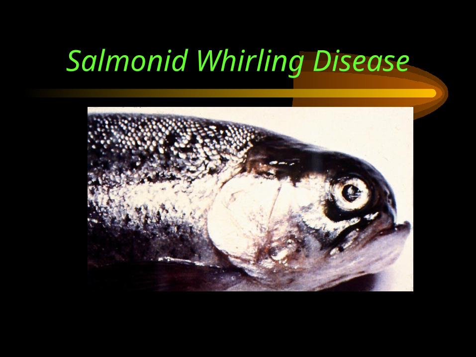
• Pathology: development in cartilage, usually young fish, can have carriers showing no signs, fish exhibits whirling (tail chasing) when feeding or alarmed, whirling caused by destruction of inner ear by spores
• can cause “blacktail” by controlling production of chromatophores in spinal column, also pugnose, skeletal deformities
Salmonid Whirling Disease
• Diagnosis: remove gill arch, grind and allow to settle, check supernatant for spores
• other methods: cook head/plankton centri- fuge, pepsin-trypsin digestion/centrifuge
• Fluorescent Antibody Test (FAT) w/rabbit
• Transmission: direct during first year, indirect via annelid, contamination (cyst)
• Hosts: trout, salmon, char, grayling
Salmonid Whirling Disease
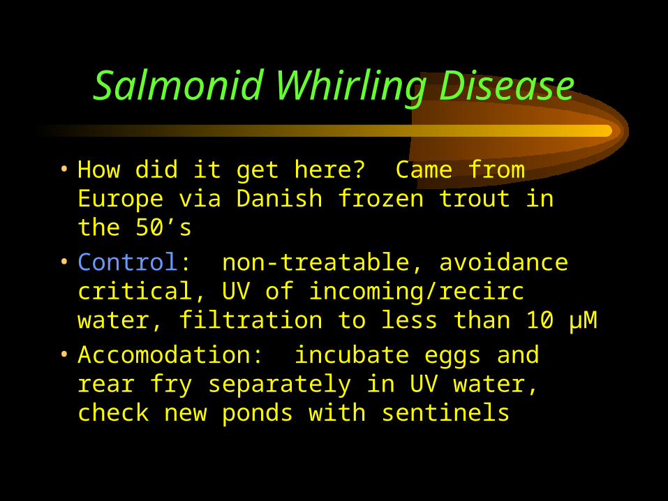
• How did it get here? Came from Europe via Danish frozen trout in the 50’s
• Control: non-treatable, avoidance critical, UV of incoming/recirc water, filtration to less than 10 µM
• Accomodation: incubate eggs and rear fry separately in UV water, check new ponds with sentinels
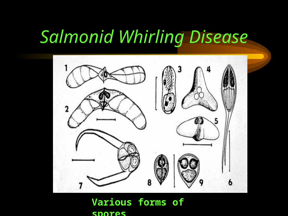
Salmonid Whirling Disease
Various forms of spores
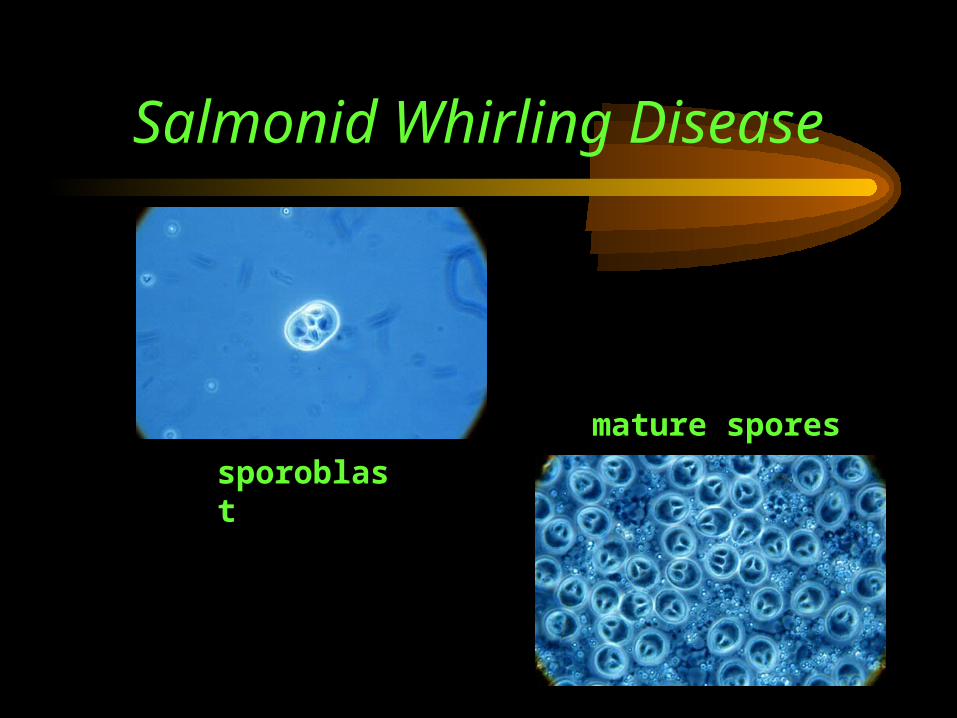
Salmonid Whirling Disease
sporoblast
mature spores
Salmonid Whirling Disease
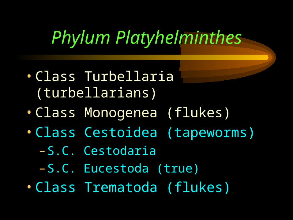
Phylum Platyhelminthes
• Class Turbellaria (turbellarians)
• Class Monogenea (flukes)
• Class Cestoidea (tapeworms)– S.C. Cestodaria
– S.C. Eucestoda (true)
• Class Trematoda (flukes)
(5) Cestodes: tapeworms• Tapeworms are members of the Phylum
Platyhelminthes which includes the classes Turbellaria, Monogenea, Trematoda and Cestoidea (tapeworms)
• the tapeworms are unique in that they have no digestive tract and, thus, are parasitic of many invertebrates
• we are concerned with the Subclass Eucestoda, the true tapeworms
Cestodes• Occur in all classes of vertebrates and some
invert’s, common in wild fish, mainly unsightly• all have three different body regions: scolex
(attachment), neck (buds off proglotids), strobila (remainder)
• proglottids formed by the asexual budding of neck
• all proglottids develop both male and female sexual organs
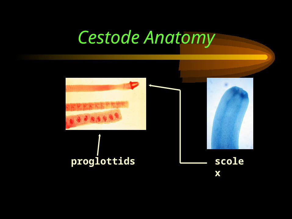
Cestode Anatomy
scolexproglottids
Cestodes
• Gravid proglottids are usually full of eggs, gonads degenerated
• Egg release: apolysis (release of whole proglottids) or anapolysis (eggs extruded through a common pore)
• Morphology: no gut; have nervous, osmoregulatory, reproductive systems for both males and females (male develops first)
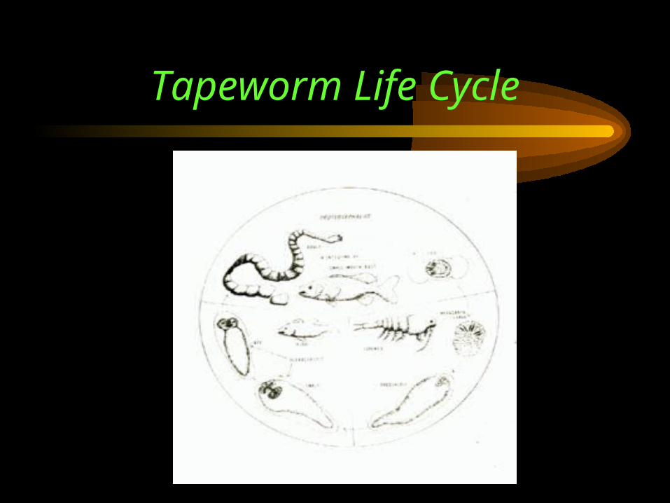
Tapeworm Life Cycle
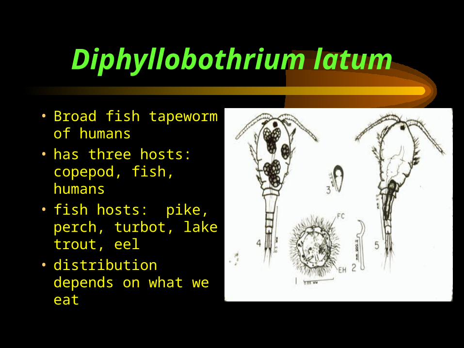
Diphyllobothrium latum
• Broad fish tapeworm of humans
• has three hosts: copepod, fish, humans
• fish hosts: pike, perch, turbot, lake trout, eel
• distribution depends on what we eat
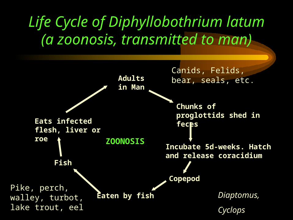
Life Cycle of Diphyllobothrium latum (a zoonosis, transmitted to man)
Adultsin Man
Chunks of proglottids shed in feces
Incubate 5d-weeks. Hatchand release coracidium
Copepod
Diaptomus,
Cyclops
Eaten by fish
Fish
Eats infected flesh, liver or roe
Pike, perch, walley, turbot, lake trout, eel
Canids, Felids, bear, seals, etc.
ZOONOSIS

Diphyllobothrium latum
• Effects on humans: abdominal pain, blockage of gut, vit B12 deficiency
• Effects on fish: visceral adhesion, sterility, decreased market value
• Control: cook fish, proper freezing
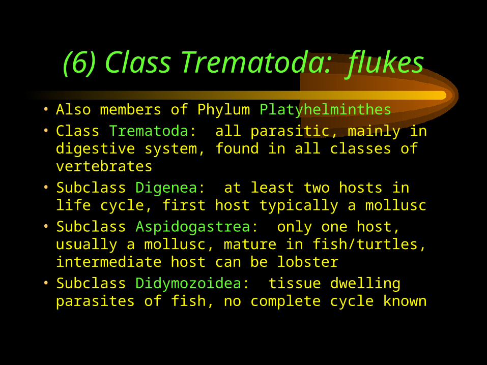
(6) Class Trematoda: flukes
• Also members of Phylum Platyhelminthes• Class Trematoda: all parasitic, mainly in digestive
system, found in all classes of vertebrates• Subclass Digenea: at least two hosts in life cycle, first
host typically a mollusc• Subclass Aspidogastrea: only one host, usually a
mollusc, mature in fish/turtles, intermediate host can be lobster
• Subclass Didymozoidea: tissue dwelling parasites of fish, no complete cycle known
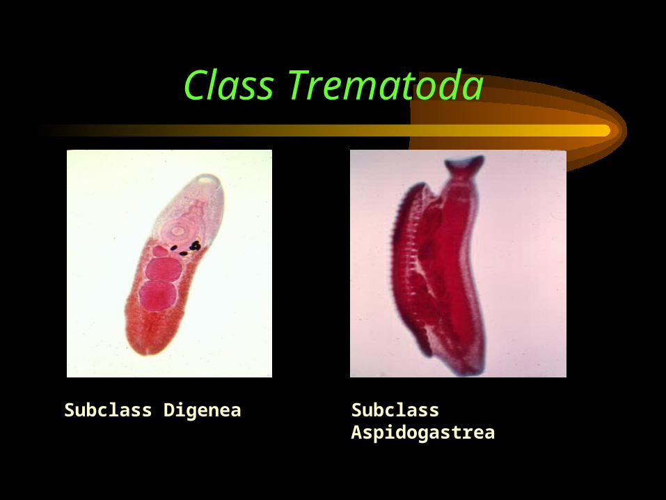
Class Trematoda
Subclass AspidogastreaSubclass Digenea
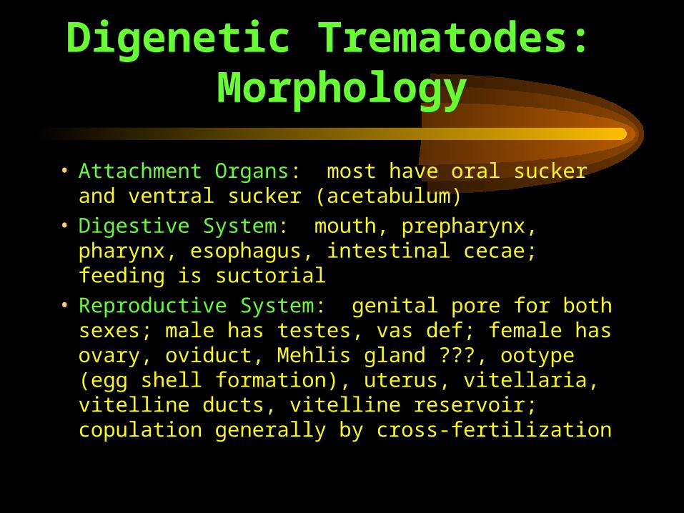
Digenetic Trematodes: Morphology
• Attachment Organs: most have oral sucker and ventral sucker (acetabulum)
• Digestive System: mouth, prepharynx, pharynx, esophagus, intestinal cecae; feeding is suctorial
• Reproductive System: genital pore for both sexes; male has testes, vas def; female has ovary, oviduct, Mehlis gland ???, ootype (egg shell formation), uterus, vitellaria, vitelline ducts, vitelline reservoir; copulation generally by cross-fertilization
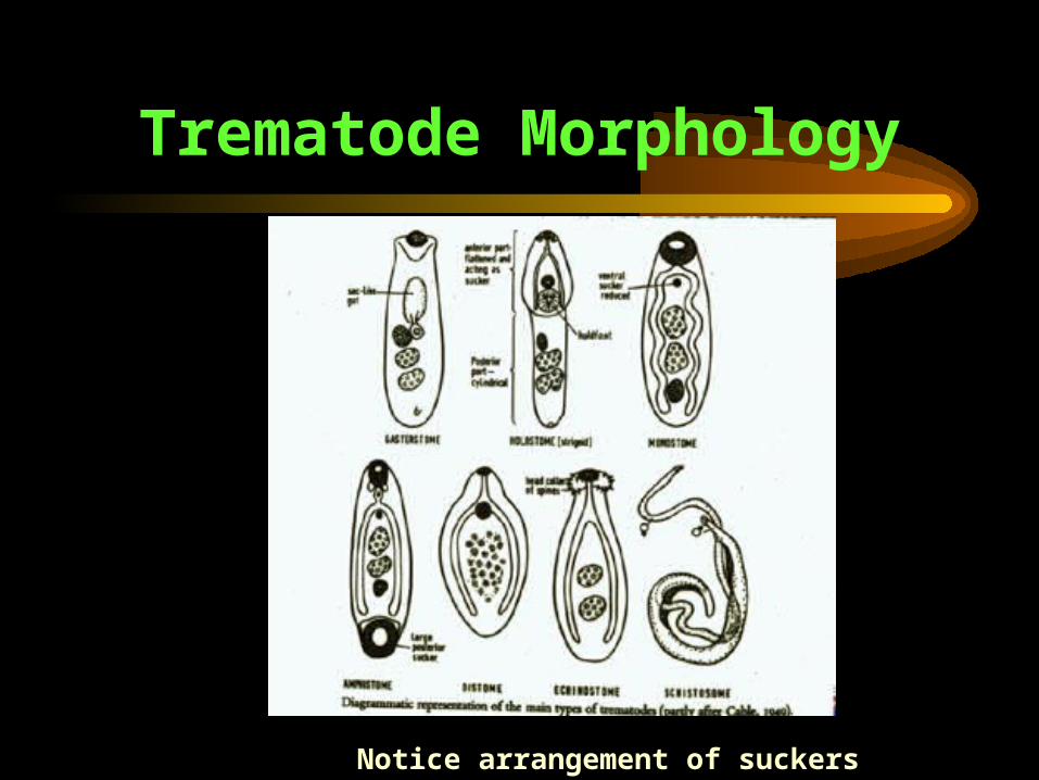
Trematode Morphology
Notice arrangement of suckers
Life Cycle of Trematodes
• Adult parasite: sexually reproducing stage of the parasite
• Definitive host: host of adult parasite
• Intermediate hosts: all hosts in life cycle other than the definitive host (usually numbered, starting with first after definitive host)
• adults live mainly in digestive tract, but also in blood, gall bladder, muscle, other organs
• eggs leave adult via feces, some ready to hatch after entering water, most need period of development
• in most cases eggs must be laid in water
Life Cycle of Trematodes• Larval stage hatching from egg is known as miracidium
(ciliated, free-swimming), only goal is to find/penetrate intermediate host
• first host is usually a snail; miracidia find intermediate host via photoreception, chemoreception, tangoreception, statoreception; snail mucus is attractive
• Asexual Reproduction: occurs in first intermediate host as either sporocysts (thin-walled germinal sac) or rediae (same, but with pharynx and gut); browse through tissues of snail
Life Cycle of Trematodes
• Goal: produce large numbers of cercaria, making up for losses in complex life cycle
• This is the difference between monogenetic and digenetic trematodes: monogenetic = one offspring; digenetic = produce cercaria = many offspring
• Cercariae: the second free-living larval stage, their fate depends on species– penetrates or is ingested by definitive host and develops into adult
(Sanouinicola)
– penetrates or is eaten by second intermediate host and encysts as metacercariae
– encysts on substrate and waits to be eaten by definitive host
Life Cycle of Trematodes
• Metacercariae: quiescent or resting stage, arrested development until definitive host eats secondary host, morphology varies with species
• after consumption, metacercariae excyst and develop into adults in relatively short period of time
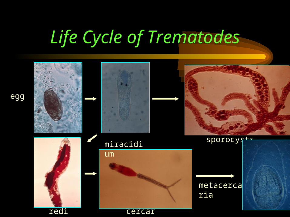
Life Cycle of Trematodes
egg
miracidiumsporocysts
rediae cercaria
metacercaria
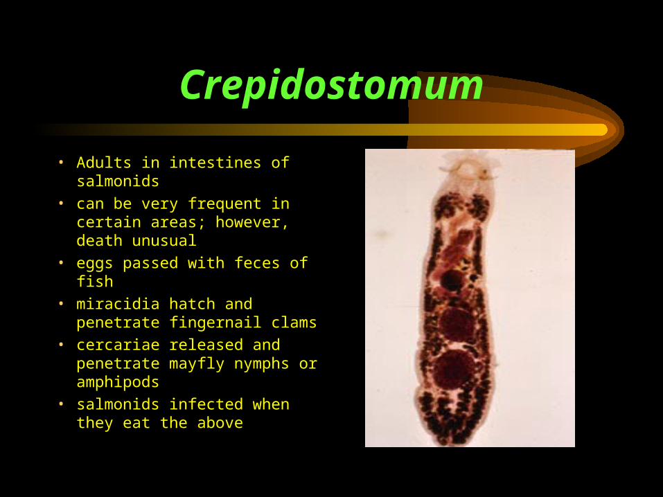
Crepidostomum
• Adults in intestines of salmonids
• can be very frequent in certain areas; however, death unusual
• eggs passed with feces of fish
• miracidia hatch and penetrate fingernail clams
• cercariae released and penetrate mayfly nymphs or amphipods
• salmonids infected when they eat the above
Sanguinicola
• Lacking suckers, ceca are X or H-shaped, numerous testes
• adults live in cyprinids, salmonids, etc.• found in bulbous arteriosus, ventral aorta, gill
vessels, kidneys• eggs released in bloodstream, hatch in gill
capillaries, release miracidiae• miracidiae penetrate Oxytrema snail, produce
sporocysts
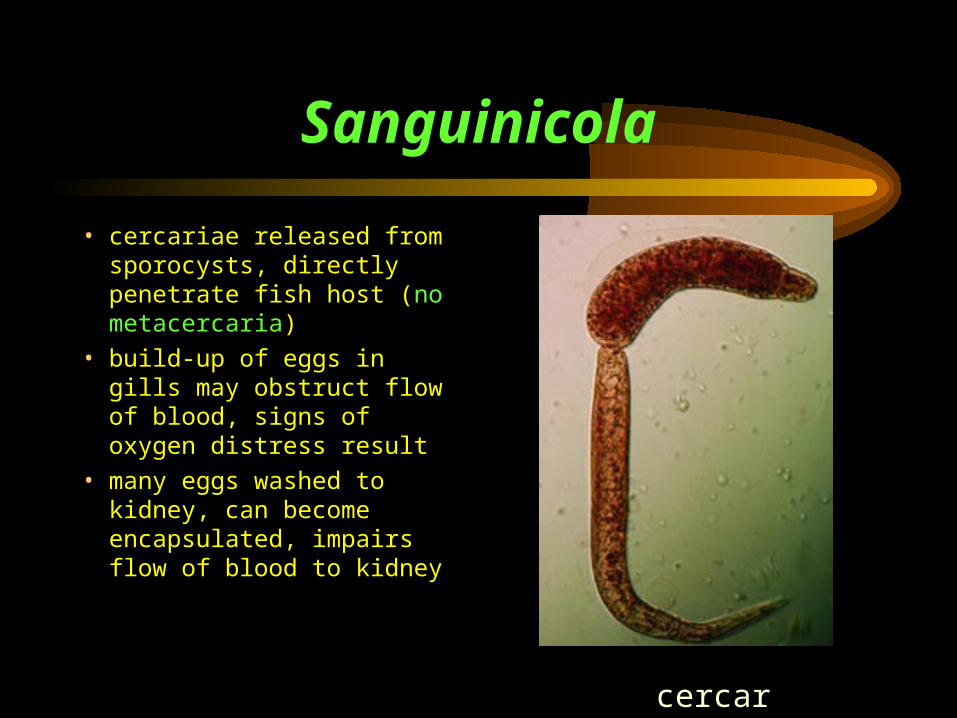
Sanguinicola
• cercariae released from sporocysts, directly penetrate fish host (no metacercaria)
• build-up of eggs in gills may obstruct flow of blood, signs of oxygen distress result
• many eggs washed to kidney, can become encapsulated, impairs flow of blood to kidney
cercaria
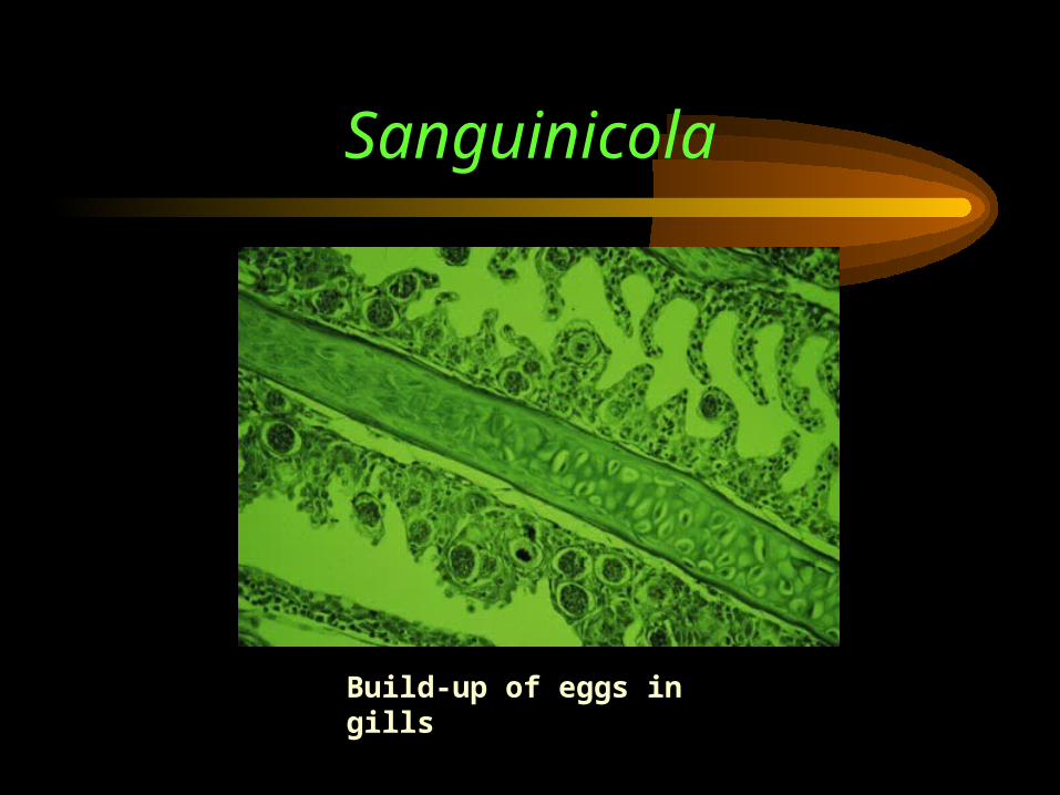
Sanguinicola
Build-up of eggs in gills