Embed Size (px)
Citation preview
Citation: Egypt. Acad. J. Biolog. Sci. (G. Microbiology) Vol.8 (2)pp. 11- 21 (2016)
Egyptian Academic Journal of Biological Sciences is the official English language journal of
the Egyptian Society for Biological Sciences, Department of Entomology, and Faculty of
Science Ain Shams University.
Microbiology journal is one of the series issued twice by the Egyptian Academic Journal of
Biological Sciences, and is devoted to publication of original papers related to the research
across the whole spectrum of the subject. These including bacteriology, virology, mycology
and parasitology. In addition, the journal promotes research on the impact of living organisms
on their environment with emphasis on subjects such a resource, depletion, pollution,
biodiversity, ecosystem…..etc www.eajbs.eg.net
Provided for non-commercial research and education use.
Not for reproduction, distribution or commercial use.
Vol. 8 No. 2 (2016)
Citation: Egypt. Acad. J. Biolog. Sci. (G. Microbiology) Vol.8 (2)pp. 11- 21 (2016)
Egypt. Acad. J. Biolog. Sci., 8(2): 11-21 (2016)
Egyptian Academic Journal of Biological Sciences
G. Microbiology
ISSN: 2090-0872 www.eajbs.eg.net
Incidence of Human Cytomegalovirus Viremia among Egyptian Hepatitis
C - Patients with Hepatocellular Carcinoma
Ahmed Khedr 1, Marwa K. Ibrahim
1, Ahmed B. Barakat
2, Mohamed S. Salama
3,
Kouka S. Abdel-wahab 4, and Mostafa K. El-Awady
1.
1-Department of Microbial biotechnology, Genetic Engineering Division, National Research
Centre, Cairo Egypt.
2- Department of Microbiology, Faculty of Science, Ain Shams University, Cairo, Egypt.
3- Molecular biology Laboratory, Faculty of Science, Ain Shams University.
4- Department of Microbiology, Faculty of Medicine, Al-Azhar University,Cairo, Egypt. E. mail: [email protected]
ARTICLE INFO ABSTRACT Article History
Received: 20/7/2016
Accepted: 30/8/2016
_________________
Keywords:
Human Cytomegalovirus
Hepatitis C virus
Hepatocellular Carcinoma
Background: Hepatocellular carcinoma (HCC) is the major hepatic complication that
may arise after many years of hepatitis C virus (HCV) infection. In Egypt, HCV
represents the major health problem, also human cytomegalovirus (HCMV) is known
as one of the highest un-resolved latent infections among general population. HCMV
viremia in the co-infection with HCV may cause life threatening in HCC patients. Our
study aimed to detect HCMV viremia in HCV–patients experienced HCC, and to
investigate its role in disease worsening. Methods: In HCC patients, HCV-RNA viral
load was determined by real- time polymerase chain reaction. In HCV-HCC patients,
HCMV-DNA was detected by amplification of gB gene region using nested-
polymerase chain reaction. Results: HCMV-DNA was detected in 4/73 in control
subjects with prevalence rate of 5.4%, whereas HCMV–DNA was recorded in 24/75
HCV-HCC patients with prevalence rate of 32 %. Data on the level of alpha feto
protein (AFP) was available for 59 out of 75 HCV-HCC patients, this enabled us to
differentiate between low risk HCC group of 40/59 patients with AFP < 500 ng/ml,
from them HCMV- DNA was detected in 14/40 patients with prevalence rate 35%,
and high risk HCC group of 19/59 patients with AFP > 500 ng /ml, from them
HCMV-DNA was reported in 9/19 patients with prevalence rate 47.37%. High
significant prevalence rate of HCMV-DNA (P < 0.001) among control and HCC
subjects was reported. Significant change in HCMV – DNA prevalence between low
and high risk HCC groups could not be achieved but tendency of higher prevalence
rate in HCMV- DNA was observed towards HCC high risk group of patients.
Conclusion: we illustrated information about HCMV/ HCV co-infection in HCC
patients, which referred to the association of HCMV viremia with HCV-HCC
patients, as well as the tendency of elevation in HCMV viremia from low to high risk
HCC patients depending on AFP threshold of 500 ng/ml.
INTRODUCTION
Hepatocellular carcinoma (HCC) may develop and take its progressive nature after 20
to 40 years of chronic HCV infection. HCV is a major cause of chronic liver inflammation,
hepatic fibrosis and initiation of neoplastic clones with irreversible genetic and epigenetic
changes, then development and progression of the malignant clones into a carcinogenic tissue
(Hoshida et al., 2014; Goossens and Hoshida, 2015).
Khedr et al. 12
Unlike HBV which can incorporate
into the host human genome leading to direct
carcinogenic activity, HCV is RNA virus
with limited incorporation of its genetic
material into the host’s genome (Mailly et
al., 2013; Billerbeck et al., 2013). Therefore,
the carcinogenic potential of HCV may
involve indirect mechanisms, which may
refer to interaction of HCV with cellular
pathways causing field cancerization area
which provide cirrhotic micro-environment
that allows initiation and promotion of
neoplastic clones by facilitating genetic
alterations and cellular transformation
(Aihara et al., 1994). Additionally, the direct
carcinogenic effects of HCV may be
achieved by the mechanisms involving viral
proteins interference with host cell
metabolism (Koike, 2005). Human cyto-
megalovirus (HCMV) is a beta - herpes virus
that may affect 60-80% of the human
population worldwide (Offermanns and
Rosenthal, 2008; Bader El Din et al., 2010),
and approxi-mately 100 % seroprevalence in
Africa (EL- Bassuoni et al., 2014). HCMV
genomic DNA is a linear, double-stranded
molecule, surrounded by a protein lining,
called matrix, which contains highly
immunogenic phosphoproteins that can
interfere with cellular cycle of the host cell.
This lining is surrounded by glycoproteins
(gB, gN, gO, gH, gM, gL) essential for the
virus to carry out its infectivity and entrance
to the host cell. The chief cells that act as
reservoirs of HCMV are the endothelial
cells, fibroblasts and myeloid cells
(Rowshani et al., 2005). The HCMV infec-
tion is characterized by alternating periods of
latency and reactivation. The infection of
endothelial cells and macrophages may
experience an important role in virus latency,
and this seems to be as a critical point in the
maintenance of HCMV during host life
(Jarvis et al., 2002). Reactivation of the virus
is usually observed during periods of down-
regulation of the immunity, such as drug
therapy and disease-related stress, or during
activation of the immunity such as
inflammatory diseases, or co-infection with
other viruses (Prosch et al., 2000; Gandhi et
al., 2004). HCMV viremia may act as a
predictor of high mortality in patients with
malignancy (Wang et al., 2010). Despite the
few published data regarding this issue,
HCMV viremia may be referred to be as a
risk factor during the course of
chemotherapy (Han et al., 2007; Kuo et al.,
2007). Alpha-fetoprotein (AFP) is the most
famous and classical tumor marker used for
HCC evaluation. AFP acts as a transporter
molecule for many ligands, such as fatty
acids, bilirubin, heavy metals, and other
molecules in addition to different drugs
(Mizejewski, 2001). Actually, significantly
higher AFP serum levels may show an
association with liver tumors (Marrero et al.,
2009). It has been proven that AFP serum
concentration may peak in a parallel way
with HCC tumor size (Kokudo et al., 2009).
In such regard, The current united- network
for organ sharing (UNOS) criteria for
transplantation of the liver had considered
patients of elevated AFP level > 500 as high
risk patients taking priority in liver
translation (Kemmer et al., 2006).
MATERIALS AND METHODS
Subjects of study:
Seventy five patients with proven HCV
infection who diagnosed with HCC were
enrolled in the current study. The 75 cases
included 63/75 (84 %) males and 12/75 (16
%) females, age ranged from 38 to 69 years
(mean age 57.8) years. Also, negative control
of seventy three subjects (negative for HCV-
Ab, HBs-Ag and HIV Ab) were included in
this study. The 73 subjects included 48/73
(65.75 %) males, and 25/73 (34.24 %)
females, age ranged from 22 to 55 years
(mean age 37.3). They were all Egyptians.
Informed consent was taken from each
subject before collecting blood samples.
HCV-HCC patients were positive for anti-
HCV antibody and HCV-RNA. They were
subjected to routine serum investigations
Incidence of Human Cytomegalovirus viremia among Egyptian Hepatitis C - patients
13
included liver function tests, alanine amino-
transeferase (ALT), aspartate amino-
transfrase (AST), bilirubin total (Bil T),
bilirubin direct (BilD), Alkaline Phosphatase
(Alk Ph), albumin (Alb) and, alpha feto
protein (AFP) as a tumor marker.
Methods:
HCV-RNA determination: RNA extracted
from 140 µl of serum or plasma using
QIAamp viral -RNA min kit (Qiagen,
Valencia, California,USA). Initially, the
samples were lysed using the buffer and 100
% ethanol for inactivation of RNase and
yield intact viral-RNA. Subsequently, the
HCV-RNA was purified in 3-steps procedure
according to the Qiagen protocol including
centrifugation at 8000 rpm. The RNA was
eluted into 60 µl of RNase free buffer, and
then HCV-RNA was determined by real time
PCR using atrus HCV RG RT-PCR kit
(Qiagen) that utilizes reverse-transcription
polymerase chain reaction (RT-PCR) with
Rotor gene Q instrument (Qiagen). It
contains master A, and B that contain
enzymes and regents for reverse -
transcription and amplification of 240 bp
region of HCV genome. Fluorescent
oligonucleotide probes specifically bind and
detect the amplified product. Monitoring of
fluorescent intensities during real time PCR
run, allowing detection as well as quanti-
fication of the product without re-open the
reaction tube after PCR run. This was done
by generating standard curve using 4
supplied quantitation standard. In 50 µl PCR
reaction tube contained 12 µl master A and
18 µl master B, unknown RNA sample of 20
µl was added then applied to real time PCR
device according to the following thermal
profile: Reverse - transcription of RNA at
50°C for 30 sec, starting activation of hot-
start enzyme at 95°C for 15 min, ampli-
fication of cDNA (50 cycle) at 95°C for 30
sec, 50°C for 60 sec, and 72°C for 30 sec.
HCMV-DMA detection: Extraction of DNA: Genomic DNA was
extracted from EDITA blood samples by
QIAamp DNA purification Kits (Qiagen)
according to manufacturer protocol. After
digestion with proteinase K, QIAamp Mini
spin columns were used for subsequent
salting out of cellular proteins by centri-
fugation steps at 8000 rpm, that allowed
optimal binding of the DNA to the QIAamp
membrane followed by DNA elution and
final storage at -20°C until required.
HCMV-DMA detection by PCR: HCV
DNA was amplified using primers derived
from the gB region of HCMV genome. PCR
protocols were followed as described
previously by (Fox et al., 1995; Jones et al.,
2000; Tabll et al., 2011) in two round PCR.
First-round PCR: The master mix prepared
by addition of 10 pmole of each primer
HCMV1 and HCMV2 (Biolegio, Nether-
lands), 0.2 mmol/l from each dNTP
(Promega, Madison, USA) 0.1 U Taq
polymerase enzyme (Promega, Madison,
USA), 1 x Taq buffer, 5 µl of DNA template,
and completed with distilled H2O to a final
volume of 20 µl. One negative control (in
which water replaced the DNA sample as a
check for contamination) and one positive
control were incorporated into each run. In
the second – round PCR: One µl of first-
round PCR product was added to the
reaction mixture, which is the same as the
first-round reaction mixture except for using
HCMV3 and HCMV4 as nested primers
(Biolegio, Netherlands). The thermal cycling
protocol was as follows: 1 min at 94°C, 1
min at 55°C and 1 min at 72°C for 30 cycles
using Biometra T1 thermal cycler (Biometra,
Germany), then the nested PCR product was
analyzed on agarose-gel electrophoresis.
Results were positively documented with a
visible 100-bp product on the gel. Primers
used for the first- and second-round PCR
were as follows: CMV1:5'GAGGACAACG
AAATCCTGTTGGGCA3';CMV2:5'GTCG
ACGGTGGAGATACTGCTGAGG3';
CMV3:5'ACCACCGCACTGAGGAATGT
CAG3';CMV4:5TCAATCATGCGTTTGA
AGAGGTA3'.
Khedr et al. 14
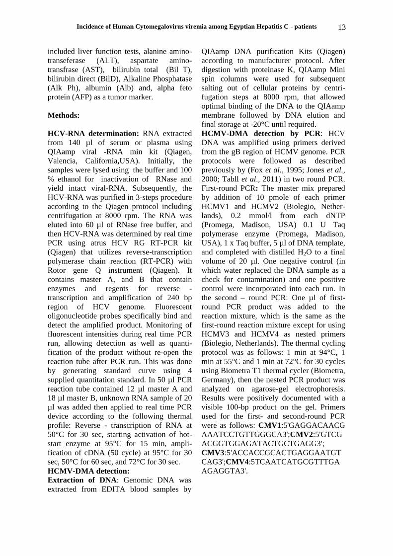
RESULTS
Incidence of HCMV-DNA among HCV-
HCC patients versus controls: The nested PCR product of HCMV gB
gene in were reported in Table 1. In control
subjects, HCMV-DNA positivity were
detected in 4/73 (5.48%), and HCMV-DNA
negativity were detected in 69/73 (94.52 %)
whereas, in HCV-HCC patients, HCMV-
DNA positivity were detected in 24/75
(32.00%), and HCMV-DNA negativity were
detected in 51/75 (68 %) patients. Chi-square
test applied to compare between two groups
regarding prevalence of HCMV –DNA
resulted in high significant correlation
towards HCC patients with p value of
<0.001*. Also, receiver operating character-
istic (ROC) Analysis for both groups had
referred to that HCMV might consider as a
predictive factor by which non HCV subjects
when get infected with HCV might develop
HCC by 32 % (referring to sensitivity)
depending on detection of HCMV-DNA with
PPV of 85.71%. HCMV-DNA incidence was
obviously illustrated in Figure 1.
Table 1: Statistical expressions and comparison between control and HCC groups of patients.
HCMV-
DNA
Groups Chi-Square
Sen
s.
Sp
ec.
PP
V
NP
V
Acc
ura
cy
Control HCC Total
N % N % N % X2 P-value
Pos. 4 5.48 24 32.00 28 18.92
15.277 <0.001*
32
.00
94
.52
85
.71
57
.50
62
.84
Neg 69 94.52 51 68.00 120 81.08
Total 73 100.00 75 100.00 148 100.00
Where; Pos. = positive, Neg = negative N= number, HCC= hepatocellular carcinoma, Sens= sensitivity, Spec=
specificity, PPV= positive - predictive value, NPV= negative - predictive value, and * = significant value.
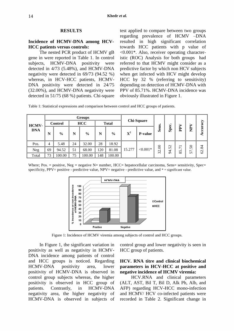
Figure 1: Incidence of HCMV viremia among subjects of control and HCC groups.
In Figure 1, the significant variation in
positivity as well as negativity in HCMV-
DNA incidence among patients of control
and HCC groups is noticed. Regarding
HCMV-DNA positivity area, lower
positivity of HCMV-DNA is observed in
control group subjects whereas, the higher
positivity is observed in HCC group of
patients. Contrastly, in HCMV-DNA
negativity area, the higher negativity of
HCMV-DNA is observed in subjects of
control group and lower negativity is seen in
HCC group of patients.
HCV. RNA titre and clinical biochemical
parameters in HCV-HCC at positive and
negative incidence of HCMV viremia:
HCV.RNA and clinical parameters
(ALT, AST, Bil T, Bil D, Alk Ph, Alb, and
AFP) regarding HCV-HCC mono-infection
and HCMV/ HCV co-infected patients were
recorded in Table 2. Significant change in
HCMV-DNA
Incidence of Human Cytomegalovirus viremia among Egyptian Hepatitis C - patients
15
such parameters is not seen except in Alk Ph
exhibits a significance change towards HCV-
HCC mono-infection (p value = 0.048*).
Table 2: HCV.RNA titre and clinical biochemical parameters regarding incidence of HCMV viremia in HCV-
HCC patients.
Parameters
HCMV-DNA T-Test
Positive Negative
Mean ± SD Mean ± SD T P-value
HCV-RNA
(IU/mL) 387111.111 ± 222876.897 448583.333 ± 354407.946 0.50 0.617
ALT (U/mL) 58.957 ± 50.220 66.486 ± 59.042 -0.610 0.545
AST(U/mL) 73.667 ± 73.375 87.000 ± 82.448 0.059 0.953
Bil T (mg/dL ) 3.317 ± 1.700 4.566 ± 5.287 -0.974 0.335
Bil D (mg/dL ) 1.625 ± 0.998 7.320 ± 6.972 -1.587 0.144
Alk Ph (U/mL) 101.000 ± 22.554 338.125 ± 204.579 -2.256 0.048*
Alb (g/dL) 2.794 ± 1.170 2.838 ± 0.572 -0.182 0.856
AFP (ng/mL) 793.458 ± 1589.200 1116.356 ± 2310.626 -0.956 0.346
Where; ALT= alanine amino- transferase, AST = aspartate amino- transferase, Bil T = Bilirubin total, Bil D =
bilirubin direct, Alp Ph = alkaline phosphatase, Alb = albumin, AFP = alpha feto protein, positive =
HCMV/HCV co-infection, Negative= HCV mono-infection, and * = significant value.
HCV. RNA titre and clinical biochemical
parameters regarding HCC Sub-grouping
according to AFP threshold of 500 ng/ml:
Level of AFP was determined in 59 out
of 75 HCV-HCC patients. AFP threshold of
500 ng/ml was served in classification of
HCC patients into 40/59 patients of low risk
(HCC1) as well as 19/59 patients of high risk
(HCC2). In Table 3, student-t test appli-
cation result in significantly bad outcome in
clinical parameters of AST, Alb, and AFP.
Regarding HCC2 (AFP > 500 ng/ml),
whereas non significant change is seen in
HCV-RNA, ALT, Bil T, Bil D, and Alk Ph
parameters.
Table 3: HCV.RNA titre and clinical biochemical parameters regarding low and high risk HCV-HCC patients.
Parameters
HCC patients T-test
HCC1(AFP <500) HCC2(AFP>500)
Mean ± SD Mean ± SD T P-value
HCV-RNA (IU/mL) 365800.000 ± 332127.815 563333.333 ± 131856.993 -1.396 0.179
ALT (U/mL) 55.410 ± 50.063 80.105 ± 63.142 -1.616 0.112
AST(U/mL) 65.568 ± 55.929 114.000 ± 105.165 -2.259 0.028*
Bil T (mg/dL ) 3.399 ± 3.826 5.532 ± 5.231 -1.715 0.092
Bil D (mg/dL ) 7.287 ± 9.290 4.800 ± 5.514 0.578 0.576
Alk Ph (U/mL) 376.333 ± 145.039 220.000 ± 208.378 1.188 0.262
Alb (g/dL) 3.053 ± 0.760 2.400 ± 0.742 3.041 0.004*
AFP (ng/mL) 161.984 ± 157.236 2734.684 ± 2959.608 -5.535 <0.001*
Where; ALT= alanine amino- transferase, AST = aspartate amino- transferase, Bil T = bilirubin total, Bil D =
bilirubin direct , and Alk Ph = alkaline phosphatase, Alb = albumin, AFP = alpha feto protein, HCC=
hepatocellular carcinoma , HCC1 = low risk HCC, HCC2= high risk HCC, and * = significant value.
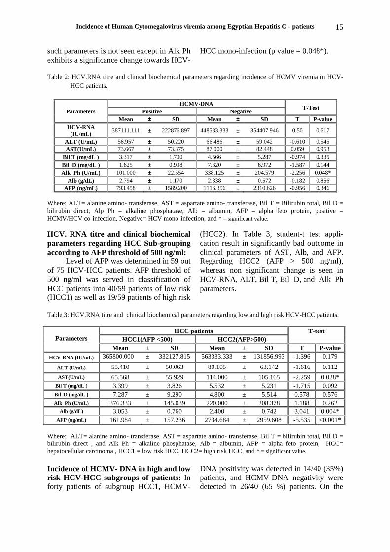
Incidence of HCMV- DNA in high and low
risk HCV-HCC subgroups of patients: In
forty patients of subgroup HCC1, HCMV-
DNA positivity was detected in 14/40 (35%)
patients, and HCMV-DNA negativity were
detected in 26/40 (65 %) patients. On the
Khedr et al. 16
other hand, HCMV-DNA positivity in
subgroup HCC2 was reported as 9/19 (47.37
%) and HCMV- DNA negativity was
reported as 10/19 (53 %). Correlations
among HCV-HCC patients in the two
subgroups were done depending on HCMV-
DNA prevalence by application of chi-
Square test. The results in Table (4) refer to
non-significance change in HCMV incidence
between the two subgroups. Also, ROC
analysis in HCC1 and HCC2 subgroups refer
to the consideration that HCMV may act as a
predictive factor by which HCV –HCC1 may
develop to HCV-HCC2 by 47.37%,
(referring to sensitivity) depending on
HCMV verimia, with PPV of 39.13 %.
Incidence of HCMV - DNA viremia is
obviously illustrated in Figure 2.
Table 4: Statistical expressions and comparison between HCC1 and HCC2 subgroups of patients.
HCMV-
DNA
HCV- HCC patients Chi-Square
Sen
s.
Sp
ec.
PP
V
NP
V
Acc
ura
cy
HCC1
HCC2
Total
N % N % N % X2 P-
value
Pos 14 35 9 47.37 23 32.00
0.3
90
0.5
32
47
.37
65
39
.13
72
.22
59
.32
Neg 26 65 10 52.63 36 68.00
Total 40 100.00 19 100.00 59 100.00
Where; Pos = positive, Neg = negative, HCC= hepatocellular carcinoma, HCC1 = low risk HCC, HCC2= high
risk HCC, Sens= sensitivity, Spec= specificity, PPV= positive -predictive value, NPV= negative -predictive
value.
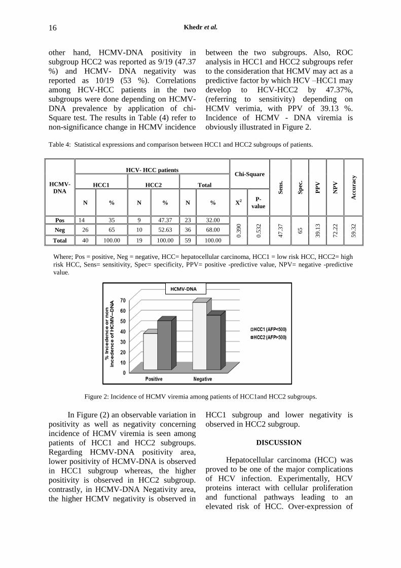
Figure 2: Incidence of HCMV viremia among patients of HCC1and HCC2 subgroups.
In Figure (2) an observable variation in
positivity as well as negativity concerning
incidence of HCMV viremia is seen among
patients of HCC1 and HCC2 subgroups.
Regarding HCMV-DNA positivity area,
lower positivity of HCMV-DNA is observed
in HCC1 subgroup whereas, the higher
positivity is observed in HCC2 subgroup.
contrastly, in HCMV-DNA Negativity area,
the higher HCMV negativity is observed in
HCC1 subgroup and lower negativity is
observed in HCC2 subgroup.
DISCUSSION
Hepatocellular carcinoma (HCC) was
proved to be one of the major complications
of HCV infection. Experimentally, HCV
proteins interact with cellular proliferation
and functional pathways leading to an
elevated risk of HCC. Over-expression of
HCMV-DNA
Incidence of Human Cytomegalovirus viremia among Egyptian Hepatitis C - patients
17
HCV proteins promotes cellular prolif-
eration, transformation, and tumor formation
in mice, referring to the direct effect of viral
proteins to activate oncogenic pathways
(Moriya et al., 1998; Fukutomi et al., 2005).
The viral core protein inhibits tumor
suppressor genes [TSG] (Kao et al., 2004;
Machida et al., 2009). HCV protein core
induces mitochondrial dysfunction, oxidative
bad outcome, and elevates cell growth
signals (Koike, 2009), this activates somatic
mutations regarding telomerase reverse-
transcriptase in early neoplastic process of
HCC with HCV and other etiologies (Nault
et al., 2013). Generally, hepatocyte
proliferation is declined at the cirrhosis stage
after several rounds of regeneration with
telomere shortening that promotes cellular
senescence to prevent carcinogenesis
(Herbig et al., 2004; Goossens et al., 2015).
HCMV infection was appeared to be a
dangerous issue in patients with
malegnancies (Wang et al., 2011). The
association of HCMV infection with HCC is
not completely defined till now (EL-
Bassuoni et al., 2014). The diagnosis of
HCMV active infection was depended on the
detection of HCMV replication in the blood
(HCMV-DNA) that referred to as HCMV
viremia and virus reactivation in absence of
effective immunity leading to serious disease
(Gandehi and Khanna, 2004). In the
current study, the incidence of HCMV
viremia was reported with significantly
higher percentage 32 % of entire HCV-HCC
patients, compared with 5.4 % in control
subjects with p-value of (<0.001). This result
may refer to the higher association of HCMV
viremia with HCV-HCC patients compared
with control subjects. In such regard, ROC
curve analysis referred to predictive value of
HCMV virema may be a predictor of HCC in
HCV patients by 32%. Incidence of HCMV
viremia in control subjects may be slightly
similar to that reported in control group in a
previous report (Taherkhani et al., 2015)
who reported prevalence rate of (0%) in
their work to study prevalence of HCMV in
patients with ulcerative colitis (Taherkhani et
al., 2015). On the other hand, the present
data on the incidence of HCMV viremia in
entire HCV-HCC patients agree with those
reported in a recent study regarding the
association of HCMV with HCC patients, El-
Bassiuoni et al., (2014), reported HCMV
prevalence rate with as 26.7 % in HCC
patients (El-Bassiuoni et al., 2014). Among
the 75 HCC patients, AFP results are
available for 59 which were divided into low
[ < 500 ng/ml ] and high risk [ > 500 ng/ml ]
according to Kemer et at., (2006). Burnett
et al., (2013) concluded that HCC staging
according to AFP level might be a predictor
of prognosis in non-cirrhotic HCC patients.
In the current study, the prevalence of
HCMV viremia was observed in 14/40
[35%] of the low risk HCC1 patients group
and in 9/19 [ 47.37% ] of the high risk HCC2
patients group. Despite the markedly higher
difference in HCMV prevalence in HCC1
than HCC2, the difference was not
statistically significant. El- Bassiuoni et al,
(2014) reported a 26.7% prevalence of
HCMV in HCC patients regardless of the
risk score of the patients. The present results
regarding HCMV incidence in HCC patients
agree with those of Lepiller et al., (2011),
who reported significantly higher prevalence
rate of HCMV in HCC patient than in non
HCC subjects. During the determination of
HCMV prevalence among patients from
different departments in a university hospital
in France. Lepiller et al., (2011) detected
HCMV-DNA in tissues of liver tumor areas,
whereas it could not be detected in adjacent
non tumor healthy tissues, indicating the
unique role of HCMV in hepatocellular
carcinoma. Lepiller et al., (2011) reported
that elimination of HCMV infection by
antiviral drugs or vaccination may reduce
HCC related mortality (Lepiller et al., 2011).
One of the evidences that support HCMV
contribution in HCC and tumor progression
from low to high risk disease was the
suggestion emerged by in vitro study which
proved that HCMV could transform
embryonic fibroblast cells in cell culture and
could promote chromosomal damage and
Khedr et al. 18
exerted mutations, however HCMV could
not be categorized as an oncogenic virus
(Michells et al., 2009; Lepiller et al., 2011).
Oncomodulation was the accepted
mechanism by which HCMV could
contribute in tumor progression (Clnat et al.,
1996; Lepiller et al., 2011), in such
mechanism, HCMV could infect tumor
tissues and acted as a cofactor in amplifying
processes of carcino-genesis without the
necessity of the tumor to be initiated firstly.
This was supported by experiments which
revealed that HCMV proteins could enhance
and influence tumor growth by interference
with cell cycle (Lepiller et al., 2011).
Elevation of interlukin 6 (IL-6) was found to
be accompanied with HCMV replication and
its reactivation. It is widely accepted that IL-
6 level would act as a critical factor
especially in liver inflammation as well as
liver cancer (Park et al., 2010). Moreover,
IL-6 mRNA expression could be up-
regulated by HCMV infection, hence IL-6
production might depend on transcriptional
factors (Dondorfer et al., 1994: Lepiller et
al., 2011), that might be stimulated by
HCMV and HCV, when IL-6 is secreted
from activated monocytes. It could
systematically act on hepatocyte and bind to
non-signaling receptor IL-6R on hepatocyte
surface, causing Janus kinase (JAK)
activation that could in turn activates signal
transducer and activator of transcription 3
(STAT3) as a major trans-criptional
oncogenic factor initiating trans-criptional
program of cell growth and differentiation
(Heinrich et al., 2003). The above findings
might explain the role of HCMV in HCC
development beside HCV, in addition to its
role in tumor worsening from low to high
risk HCC patients that reported 35% HCMV
viremia prevalence compared with 47.37 %
HCMV viremia prevalence reported in high
risk HCC patients. Conclusion: In the
current study we demonstrated information
regarding HCMV/ HCV co-infection in HCC
patients, which referred to the association of
HCMV viremia with HCV-HCC patients, as
well as tendency of elevated HCMV viremia
in high risk HCC patients, thus suggesting
that HCMV viremia contributes in disease
worsening. Further studies are still required
to assess the significant role of HCMV
viremia in HCC progression in HCV
patients.
REFERENCES
Aihara, T., Noguchi, S., Sasaki, Y., Nakano,
H., Imaoka, S. (1994): Clonal analysis
of regenerative nodules in hepatitis C.
virus-induced liver cirrhosis.
Gastroenterology, 107:1805-1811.
Bader El Din, N.G., Abd El Meguid, M.,
Tabll, A. A., Anany, M. A., Esmat, G.,
Zayed, N., Helmy, A., El Zayady, A.,
Barakat, A., and El Awady, M.K.
(2010): Human cytomegalovirus
infection inhibits response of chronic
hepatitis-C-virus-infected patients to
interferon-based therapy. Gastroenterol
and Hepatol, 26:55–62
Billerbeck, E., de Jong, Y., Dorner, M., de la
Fuente, C., Ploss, A. (2013): Animal
models for hepatitis C. Curr Top
Microbiol Immunol; 369:49-86.
Burnett, N.P., Dunki-Jacobs, E.M.,
Callender, G.G., Anderson, R.J.,
Scoggins, C.R., McMasters, K.M.,
Martin, R.C. (2013): Evaluation of
alpha-fetoprotein staging system for
hepatocellular carcinoma in noncirrhotic
patients. Am Surg., 79(7):716-22.
Cinatl, J., Cinatl, J., Vogel, J.U., Rabenau,
H., Kornhuber, B., Doerr, H.W.(1996):
Modulatory effects of human
cytomegalovirus infection on malignant
properties of cancer cells. Intervirology ,
39:259-269.
Dendorfer, U., Oettgen, P., Libermann, T.A
(1994): Multiple regulatory elements in
the interleukin-6 gene mediate induction
by prostaglandins, cyclic AMP, and
lipopolysaccharide. Mol Cell Biol.,
14:4443-4454.
EL- Bassuoni, M.A., Fathey, O., Talaat,
R.M., E-lRefaai, M. M. (2014):
Correlation Study of (CMV) in the
Presence of (HCV) and/or (HBV) in
Hepato-cellular Carcinoma (HCC)
Incidence of Human Cytomegalovirus viremia among Egyptian Hepatitis C - patients
19
Patients Egyptian Journal of Medical
Microbiology, 23: 89-93.
Fox J, Zuckermann M, Brink N, Neild PB,
Gazzard G, Tedder R, Miller R (1995):
Detection of herpesviral DNA by nested
polymerase chain reaction in CSF of
HIV-infected individuals with
neurological disease: a prospective
evaluation. J Infect Dis., 172:1087-
1090.
Fukutomi T, Zhou Y, Kawai S, Eguchi H,
Wands JR, Li J. (2005): Hepatitis C
virus core protein stimulates hepatocyte
growth: correlation with upregulation of
wnt-1 expression. Hepatology, 41:1096-
1105.
Gandhi, M.K., and Khanna, R. (2004):
Human cytomegalovirus: clinical
aspects, immune regulation, and
emerging treatments. Lancet. Infect.
Dis., 4: 725-38.
Goossens, N., and Hoshida, Y., (2015):
Hepatitis C virus- induced
hepatocellular carcinoma. Molecular
and Clinical Hepatology, 21: 105-114.
Han, X.Y. (2007): Edidemiologic analisis of
reactivated cytomegalovirus antigenimia
in patients with cancer J Clin Microbiol.,
45:1126-1132.
Heinrich PC, Behrmann I, Haan S,
Hermanns HM, Muller-Newen G,
Schaper F. (2003): Principles of
interleukin (IL)-6-type cytokine
signalling and its regulation. Biochem J ,
374:1-20.
Herbig, U., Jobling, W.A., Chen, B.P., Chen,
D.J., Sedivy, J.M. (2004): Telomere
shortening triggers senescence of human
cells through a pathway involving ATM,
p53, and p21 (CIP1), but not p16
(INK4a). Mol Cell, 14:501-513.
Hoshida, Y., Fuchs, B.C., Bardeesy, N.,
Baumert, T.F., Chung, R.T. (2014).
Patho genesis and prevention of
hepatitis C virus-induced hepatocellular
carcinoma. J Hepatology, 61 (Suppl 1):
S79-S90.
Jarvis, M., and Nelson, J. (2002): Human
CMV persistence and latency in
endothelial cells and macrophages.
Curr. Opin. Microbio., l(5): 403-7.
Jones, R., Lynne, N., Beattie, B.,
Westmoreland D., Fox, J. (2000):
Development and Application of a PCR-
Based Method Including an Internal
Control for Diagnosis of Congenital
Cytomegalovirus Infection. J Clinical
microbiology, 38(1):1-6.
Kao, C.F., Chen, S.Y., Chen, J.Y., Lee, Y.H.
(2004): Modulation of p53 transcription
regulatory activity and post-translational
modification by hepatitis C virus core
protein. Oncogene, 23:2472-2483.
Kemmer, N., Neff, G., Kaiser, T., Zacharias,
V., Thomas, M., Tevar, A . Satwah, S.,
Shukla, R., and Buell, J. (2006): An
Analysis of the UNOS Liver Transplant
Registry: High Serum Alpha-Fetoprotein
Does Not Justify an Increase in MELD
Points for Suspected Hepatocellular
Carcinoma. Liver transplantation, 12:
1519- 1522
Koike, K. (2005): Molecular basis of
hepatitis C virus-associated hepatocar-
cinogenesis: lessons from animal model
studies. Clin Gastroenterol Hepatol,
3(2):S132-135.
Koike, K. (2009): Steatosis, liver injury, and
hepatocarcinogenesis in hepatitis C viral
infection. J Gastroenterol, 44: 82-88.
Kokudo, N., and Makuuchi, M. (2009):
Evidence-based clinical practice
guidelines for hepatocellular carcinoma
in Japan: the J- HCC guidelines.
Gastroenterology, 44 (Suppl 19):119-
121
Kuo, C.P., Wu, C.L., Ho, H.T., Chen, C.G.,
Liu, S.I., and Lui, Y. T. (2007):
Detection of cytomegalovirus
reactivation in cancer patients receiving
chemotherapy. Clinical microbiology
and infection, 14: 221-227.
Lepiller, Q., Tripathy, M.K., Martino, V.D.,
Kantelip, B., and Herbein, G. (2011):
Increased HCMV seroprevalenee in patients
with hepatocellular carcinoma. Virology
Journal, 8: 485.
Machida, K., Liu, J.C., McNamara, G.,
Khedr et al. 20
Levine, A., Duan, L., Lai, M.M. (2009):
Hepatitis C virus causes uncoupling of
mitotic checkpoint and chromosomal
polyploidy through the Rb pathway. J
Virol, 83:12590-12600.
Mailly, L., Robinet, E., Meuleman, P.,
Baumert, T.F., Zeisel, M.B. (2013):
Hepatitis C virus infection and related
liver disease: the quest for the best
animal model. Front Microbiol, 4:213.
Marrero, J.A., Feng, Z., Wang, Y., Nguyen,
M.H., Befeler, A.S., Roberts, L.R.
(2009): Alpha-fetoprotein,des-gamma
carboxypr-othrombin, and lectin-bound
alpha-fetoprotein in early hepatocellular
carcinoma. Gastroenterology.,
137(1):110-118.
Michaelis, M., Doerr, H.W., and Cinatl, J.
(2009): The story of human
cytomegalovirus and cancer: increasing
evidence and open questions. Neoplasia,
11:1-9.
Mizejewski, G.J. (2001): Alpha feto protein
structure and function: revalence to
isoforms, epitopes, and conformational
variants. Exp Biol Med (Maywood),
226(5): 377-408.
Moriya, K., Fujie, H., Shintani, Y.,
Yotsuyanagi, H., Tsutsumi, T.,
Ishibashi, K., (1998): The core protein
of hepatitis C virus induces
hepatocellular carcinoma in transgenic
mice. Nat Med., 4:1065-1067.
Nault, J.C., Mallet, M., Pilati, C., Calderaro,
J., Bioulac-Sage, P., Laurent ,C., (2013):
High frequency of telomerase reverse-
transcriptase promoter somatic
mutations in hepatocellular carcinoma
and preneoplastic lesions. Nat Commun,
4: 2218.
Offermanns, S., Rosenthal, W. (2008):
Encyclopedia of Molecular
Pharmacology, 2nd edn. Berlin,
Heidelberg, New York: Springer-
Verlag, 437-438.
Park, E.J., Lee, J.H., Yu, G.Y., He, G., Ali,
S.R., Holzer, R.G., Osterreicher, C.H.,
Takahashi, H., Karin, M. (2010):
Dietary and genetic obesity promote
liver inflammation and tumorigenesis by
enhancing IL-6 and TNF expression.
Cell., 140:197-208.
Prosch, S., Wendt, C., Reinke, P. (2000): A
novel link between stress and human
cytomegalovirus (HCMV) infection:
sympathetic hyperactivity stimulates
HCMV activation. Virology, 272: 357-
65.
Rowshani, A., Bemelman, F., Van-
Leeuwen, E., Van- Lier R., Berge, I.J.
(2005): Clinical and immunologic
aspects of CMV infection in solid organ
transplant recipients. Transplantation,
79:381-386.
Tabll, A., Shoman, S., Ghanem, H., Nabil,
M., Bader El Din, N.G. and El- Awady,
M.K. (2011): Assessment of human
cytomegalovirus coinfection in Egyptian
chronic HCV patients Virology Journal,
8:343.
Taherkhani, R., Farashadpour, F., Makvandi,
M., Hamidifard, M., Esmailizadeh, M.,
Ahmadi, B., and, Heidari, H.. (2015):
Determination of cytomegalovirus
prevalence and glycoprotein B
Genotypes among ulcerative colitis
patients in Ahfaz, Iran. Jundishapur J
Micrbiol, (8)2:e17458.
Wang, Y.C., Wang, N.C., Lin, G.C., Perneg,
C.L,Yeh, K.M.,Yang, Y.S., Chiu, C.H.,
and Chang, F.Y. (2001): Risk factors
and outcomes of cytomegalovirus
viremia in cancer patients: Astudy from
a medical center in northern Taiwan J
Microbio Immunol and infection, 44:
442-448.
Wang, Y.C., Wang, N.C.Lin, G.C., Perneg,
C.L., Yeh, K.M., Yang, Y.S., Chiu,
C.H., and Chang, F.Y. (2010): Risk
factors and outcomes of
cytomegalovirus verimia in cancer
patients: a study from a medical center
in northern Taiwan. Journal of
microbiology and immunology and
infection, 44: 442-448.
Incidence of Human Cytomegalovirus viremia among Egyptian Hepatitis C - patients
21
ARABIC SUMMERY
المصريين "سى"معدل انتشار الفيروس المضخم للخاليا فى مرضى االلتهاب الكبدى الفيروسى
بسرطان الكبد المصابين
احمد خضر محمد1
ابراهيم خليل ى، مرو 1
، احمد بركات بركات 2مة، محمد سيد سال
3كوكا سعد الدين عبد الوهاب ،
4
، مصطفى كامل العوضى 1
-شعبة بحوث الهندسة الوراثية و البيوتكنولوجى –قسم التكنولوجيا الحيوية الميكروبية -1
.مصر –القاهرة -المركز القومى للبحوث
.مصر -القاهرة –جامعة عين شمس –كلية العلوم –قسم الميكروبيولوجى - 2
.مصر -القاهرة –جامعة عين شمس –كلية العلوم –معمل البيولوجيا الجزيئية - 3
.مصر –القاهرة -جامعة االزهر -كلية الطب -قسم الميكروبيولوجى - 4
[email protected]: بريد الكترونى
، "سى"بالفيروس الكبدىتنشأ بعد عدة سنوات من االصابة يعتبر مرض سرطان الكبد من المضاعفات الخطيرة التى: الخلفية العلمية
يعتبر واحد من اكثر حيث و تمثل االصابة بهذا الفيروس مشكلة طبية كبيرة فى مصر ، و ايضا االصابة بالفيروس المضخم للخاليا
مزدوجة مع الفيروس ودة كعدوى لفيروس المضخم للخاليا النشط فى وجاف, دية التى تظل كامنة فى المرضىاالمراض المع
، و قد هدفت دراستنا الى رصد معدل االصابة "سى"قد يهدد حياة مرضى سرطان الكبد الناشىء عن الفيروس الكبدى " سى"الكبدى
. فى مرضى سرطان الكبد" سى"النشطة بالفيروس المضخم للخاليا كعدوى مزدوجة مع الفيروس الكبدى
بى سى )من مرضى السرطان الكبدى بتقنية تفاعل البلمرة المتسلسل 57للخاليا فى تم فحص وجود الفيروس المضخم :المنهجية
ادة بعد الكشف المسبق لوجود االجسام المض( لاير تيم بى سى ار)لهم بتقنية " سى"و الذين قد تم تحديد وجود الفيروس الكبدى ( ار
مجموعة ) "سى"الشخاص الغير مصابيين بالفيروس من ا 53الفيروس المضخم للخاليا فى للفيروس، باألضافة لفحص وجود
. بنفس التقنية( الكنترول
% 32فى مجموعة الكنترول بينما وصلت النسبة الى % 7،4وجد ان معدل انتشار الفيروس المضخم للخاليا كان بنسبة : النتائج
بمعدل اقل من ( قيمة بى) مل االرتباط االحصائى اية لمعالت قيمة عو ظهر" سى"فى مرضى سرطان الكبد المصابيين بالفيروس
و تمريض منهم لقياسات دالئل االورام الفا في 75، و بعد تقسيم هوالء المرضى الى مجموعتين اعتمادا على ما توفر ل 1,,،,
% 45،35زاد الى للفيروس المضخم للخاليا و الذى % 37بروتين، فقد سجلت المجموعة االقل خطورة لسرطان الكبد معدل انتشار
فى المجموعة االكثر خطورة لسرطان الكبد و لكن بدون الوصول الى الرتباط احصائى لقيمة ال بى ، حيث ان التقسيم اعتمد على
. لأللفا فيتوبروتين يليترمل/جرامنانو ,,7المعدل
لمعدل االصابة النشطة بالفيروس المضخم للخاليا فى مرضى سرطان الكبد اظهرت الدراسة االرتباط االحصائى العالى: الخالصة
مقارنة باالشخاص الكنترول ، بينما مع االصابة النشطة للفيرس المضخم للخاليا " سى"المصابين بااللتهاب الكبدى الفيروسى
يليترمل /نانوجرام ,,7مادا على المعدل اعتظهرت القابلية فى زيادة معدل االصابة فى المجموعة االكثر خطورة لسرطان الكبد
.لدالئل االورام الفا فيتو بروتين ولكن بدون الحصول على معامل ارتباط احصائى، و الذى يحتاج دراسات مستقبلية فى هذا الصدد