Embed Size (px)
Citation preview
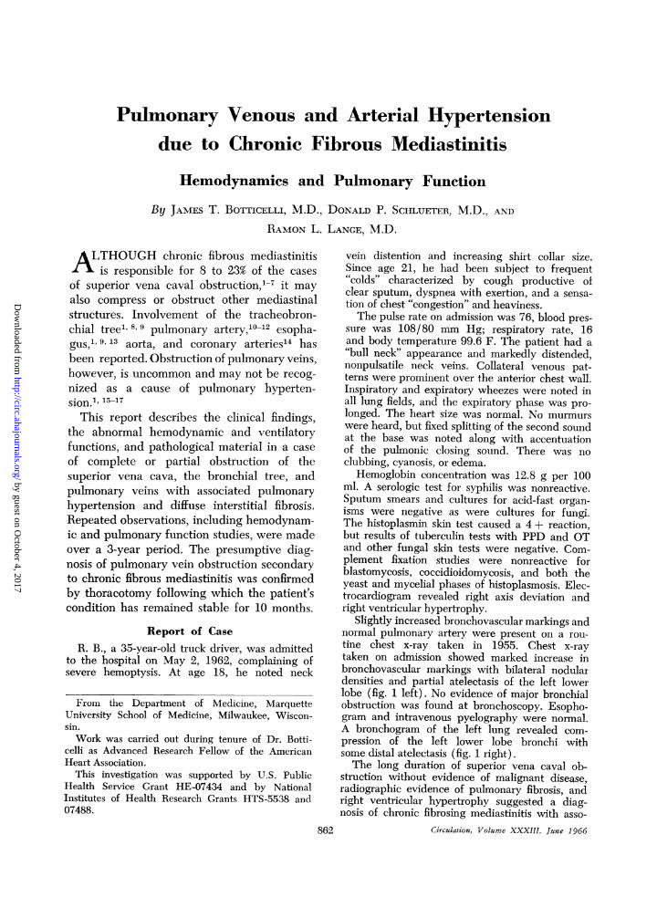
Pulmonary Venous and Arterial Hypertensiondue to Chronic Fibrous Mediastinitis
Hemodynamics and Pulmonary Function
By JAMES T. BOTTICELLI, M.D., DONALD P. SCHLUETER, M.D., AND
RAMON L. LANGE, M.D.
ALTHOUGH chronic fibrous mediastinitisis responsible for 8 to 23% of the cases
of superior vena caval obstruction,1-7 it mayalso compress or obstruct other mediastinalstructures. Involvement of the tracheobron-chial tree" 8, 9pulmonary artery,10-12 esopha-gus," 9, 13 aorta, and coronary arteries14 hasbeen reported. Obstruction of pulmonary veins,however, is uncommon and may not be recog-nized as a cause of pulmonary hyperten-sion.", 15-17
This report describes the clinical findings,the abnormal hemodynamic and ventilatoryfunctions, and pathological material in a caseof complete or partial obstruction of thesuperior vena cava, the bronchial tree, andpulmonary veins with associated pulmonaryhypertension and diffuse interstitial fibrosis.Repeated observations, including hemodynam-ic and pulmonary function studies, were madeover a 3-year period. The presumptive diag-nosis of pulmonary vein obstruction secondaryto chronic fibrous mediastinitis was confirmedby thoracotomy following which the patient'scondition has remained stable for 10 months.
Report of CaseR. B., a 35-year-old truck driver, was admitted
to the hospital on May 2, 1962, complaining ofsevere hemoptysis. At age 18, he noted neck
From the Department of Medicine, MarquetteUniversity School of Medicine, Milwaukee, Wiscon-sin.Work was carried out during tenure of Dr. Botti-
celli as Advanced Research Fellow of the AmericanHeart Association.
This investigation was supported by U.S. PublicHealth Service Grant HE-07434 and by NationalInstitutes of Health Research Grants HTS-5538 and07488.
862
vein distention and increasing shirt collar size.Since age 21, he had been subject to frequent"colds" characterized by cough productive ofclear sputum, dyspnea with exertion, and a sensa-tion of chest "congestion" and heaviness.The pulse rate on admission was 76, blood pres-
sure was 108/80 mm Hg; respiratory rate, 16and body temperature 99.6 F. The patient had a"bull neck" appearance and markedly distended,nonpulsatile neck veins. Collateral venous pat-terns were prominent over the anterior chest wall.Inspiratory and expiratory wheezes were noted inall lung fields, and the expiratory phase was pro-longed. The heart size was normal. No murmurswere heard, but fixed splitting of the second soundat the base was noted along with accentuationof the pulmonic closing sound. There was noclubbing, cyanosis, or edema.
Hemoglobin concentration was 12.8 g per 100ml. A serologic test for syphilis was nonreactive.Sputum smears and cultures for acid-fast organ-isms were negative as were cultures for fungi.The histoplasmin skin test caused a 4 + reaction,but results of tuberculin tests with PPD and OTand other fungal skin tests were negative. Com-plement fixation studies were nonreactive forblastomycosis, coccidioidomycosis, and both theyeast and mycelial phases of histoplasmosis. Elec-trocardiogram revealed right axis deviation andright ventricular hypertrophy.
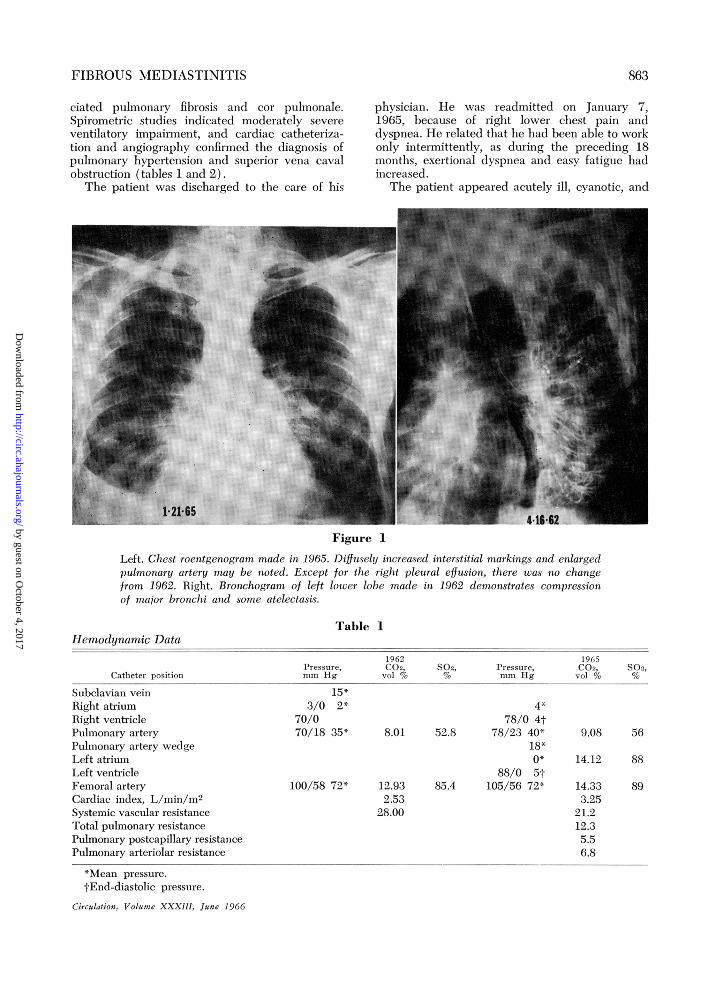
Slightly increased bronchovascular markings andnormal pulmonary artery were present on a rou-tine chest x-ray taken in 1955. Chest x-raytaken on admission showed marked increase inbronchovascular markings with bilateral nodulardensities and partial atelectasis of the left lowerlobe (fig. 1 left). No evidence of major bronchialobstruction was found at bronchoscopy. Esopho-gram and intravenous pyelography were normal.A bronchogram of the left lung revealed com-pression of the left lower lobe bronchi withsome distal atelectasis (fig. 1 right).The long duration of superior vena caval ob-
struction without evidence of malignant disease,radiographic evidence of pulmonary fibrosis, andright ventricular hypertrophy suggested a diag-nosis of chronic fibrosing mediastinitis with asso-
Circulation, Volume XXXIII, June 1966
by guest on October 4, 2017
http://circ.ahajournals.org/D
ownloaded from
FIBROUS MEDIASTINITIS
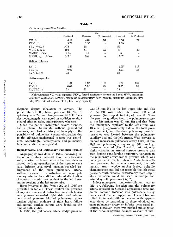
ciated pulmonary fibrosis and cor pulmonale.Spirometric studies indicated moderately severeventilatory impairment, and cardiac catheteriza-tion and angiography confirmed the diagnosis ofpulmonary hypertension and superior vena cavalobstruction (tables 1 and 2).
The patient was discharged to the care of his
physician. He was readmitted on January 7,1965, because of right lower chest pain anddyspnea. He related that he had been able to workonly intermittently, as during the preceding 18months, exertional dyspnea and easy fatigue hadincreased.
The patient appeared acutely ill, cyanotic, and
Figure 1
Left. Chest roentgenogram made in 1965. Diffusely increased interstitial markings and enlargedpulmonary artery may be noted. Except for the right pleural effusion, there was no changefrom 1962. Right. Bronchogram of left lower lobe made in 1962 demonstrates compressionof major bronchi and some atelectasis.
Table 1Hemodynamic Data
1962 1965Pressure, C02, S02, Pressure, C02, S02,
Catheter position mm Hg vol % % mm Hg vol % %
Subelavian veinRight atriumRight ventriclePulmonary arteryPulmonary artery wedgeLeft atriumLeft ventricleFemoral arteryCardiac index, L/min/m2Systemic vascular resistanceTotal pulmonary resistancePulmonary postcapillary resistancePulmonary arteriolar resistance
15*3/0 2*
70/070/18 35*
100/58 72* 1
4*78/0 4t
8.01 52.8 78/23 40*18*0*
88/0 5t12.93 85.4 105/56 72*2.53l8.00
*Mean pressure.tEnd-diastolic pressure.
Circulation, Volume XXXIII, June 1966
9.08 56
14.12 88
8914.333.25
21.212.35.56.8
863
by guest on October 4, 2017
http://circ.ahajournals.org/D
ownloaded from
BOTTICELLI ET AL.
Table 2Pulmnonary Function Studies
1962Predicted Observed % Predicted
VC, LFEV1, LFEV1/VC, %MVV, L/minMMEF, L/secMEFRO.2-1.2' L/sec
Helium dilution
RV, LTLC, LRV/TLC, %
Plethysmographic
RV, LTLC, LRV/TLC, %
4.613.72
>75160>2.2>7.0
4.092.3558811.13.4
1965Observed % Predicted
88 3.56- 1.89- 5357 69- 0.71- 2.67
1.416.02
23
1.416.0223
1.655.2132
1.875.96
31
13299
1.795.3533
Abbreviations: VC, vital capacity; FEV1, forced expiratory volume in 1 sec; MVV, maximumvoluntary ventilation; MMEF, maximum midexpiratory flow; MEFR, maximum expiratory flowrate; RV, residual volume; TLC, total lung capacity.
dyspneic despite inhalation of oxygen. Thepulse rate was 96, blood pressure 120/80, re-
spiratory rate 24, and temperature 98.6 F. Ten-der hepatomegaly was noted in addition to rightpleural effusion, rales, and expiratory wheezes.
Since the patient manifested severe dyspnea,had a pleural transudate without generalizedanasarca, and had a history of hemoptysis, thepossibility of pulmonary venous obstruction dueto the adhesive mediastinal process was consid-ered. Accordingly, hemodynamic and pulmonaryfunction studies were repeated.
Hemodynamic and Pulmonary Function Studies
Angiography was done in 1962. Following in-jection of contrast material into the subclavianvein, marked collateral circulation was demon-strated, with no opacification of the superior vena
cava. Inferior vena caval injection revealed en-
largement of the main pulmonary artery,without evidence of constriction of major pul-monary arteries. In addition, reduced distributionof contrast material was evident in the left lowerand in portions of the left upper lobe.Hemodynamic studies from 1962 and 1965 are
presented in table 1. These confirm the presenceof superior vena caval obstruction since subclavianvein pressure exceeded right atrial and inferiorvena caval pressure. Marked pulmonary hyper-tension without evidence of right heart failureand normal cardiac output were found at thetime of both studies.
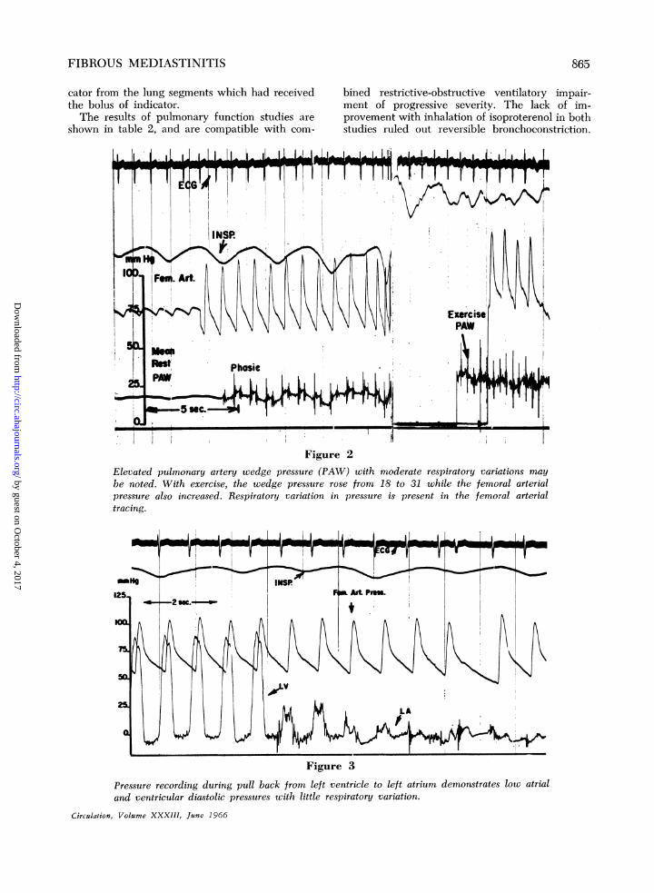
In 1965, the pulmonary artery wedge pressure
was 18 mm Hg in the left upper lobe and alsoin the left lower lobe. The mean left atrialpressure (transseptal technique) was 0. Sincethe pressure gradient from the pulmonary arteryto the left atrium was 40 mm Hg and that fromthe "pulmonary capillary" to the left atrium was
18 mm Hg, approximately half of the total pres-sure gradient, and therefore pulmonary vascularresistance was located between the pulmonarycapillary bed and the left atrium. With exercise, a
marked increase in pulmonary artery (105/38 mmHg) and pulmonary artery wedge (31 mm Hg)pressures occurred (figs. 2 and 3). At rest, onlyslight variation in arterial systolic pressure was
seen despite considerable respiratory variation inthe pulmonary artery wedge pressure which wasnot apparent in the left atrium. Aside from arti-facts produced by catheter movement, the pul-monary artery wedge tracing lacked pulsatilecharacteristics and did not resemble the left atrialpressure. With exercise, considerably more respir-atory variation could be seen in wedge andarterial systolic pressures (fig. 2).
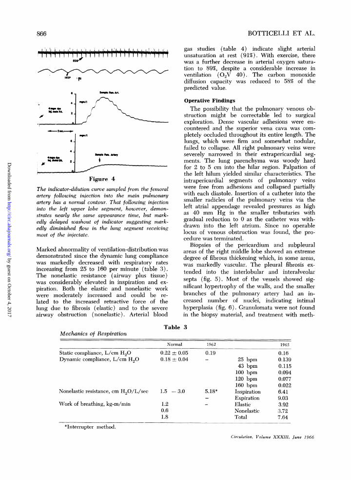
Indocyanine-green indicator-dilution curves
(fig. 4), following injection into the pulmonaryartery, revealed an 8-second appearance time andnormal contour. Injection into pulmonary arterybranches of the left upper and lower lobes re-
sulted in indicator-dilution curves with appear-
ance times corresponding to those obtained on
main pulmonary artery or inferior vena caval in-jection. However, there was marked prolongationof the curve suggesting delayed washout of indi-
Circulation, Volume XXXIII, June 1966
77
43
11787
12789
864
by guest on October 4, 2017
http://circ.ahajournals.org/D
ownloaded from
FIBROUS MEDIASTINITIS
cator from the lung segments which had receivedthe bolus of indicator.The results of pulmonary function studies are
shown in table 2, and are compatible with com-
bined restrictive-obstructive ventilatory impair-ment of progressive severity. The lack of im-provement with inhalation of isoproterenol in bothstudies ruled out reversible bronchoconstriction.
5T 11 1 I 1 1Trrv1 v7r
Figure 2Elevated pulmonary artery wedge pressure (PAW) with moderate respiratory variations maybe noted. With exercise, the wedge pressure rose from 18 to 31 while the femoral arterialpressure also increased. Respiratory variation in pressure is present in the femoral arterialtracing.
Figure 3
Pressure recording during pull back from left ventricle to left atrium demonstrates low atrialand ventricular diastolic pressures with little respiratory variation.
Circulation, Volume XXXIJI, June 1966
865
by guest on October 4, 2017
http://circ.ahajournals.org/D
ownloaded from
BOTTICELLI ET AL.
1I
INSP p
6 SampkFeF Ar.
Figr/I 44Thdi
Figure 4
The indicator-dilution curve sampled from the femoralartery following injection into the main pulmonaryartery has a normal cont.our. That following injectioninto the left upper lobe segment, however, demon-strates nearly the same appearance time, but mark-edly delayed washout of indicator suggesting mark-edly diminished flow in the lung segment receivingmost of the injectate.
Marked abnormality of ventilation-distribution wasdemonstrated since the dynamic lung compliancewas markedly decreased with respiratory ratesincreasing from 25 to 160 per minute (table 3).The nonelastic resistance (airway plus tissue)was considerably elevated in inspiration and ex-piration. Both the elastic and nonelastic workwere moderately increased and could be re-lated to the increased retractive force of thelung due to fibrosis (elastic) and to the severeairway obstruction (nonelastic). Arterial blood
TaMechanics of Respiration
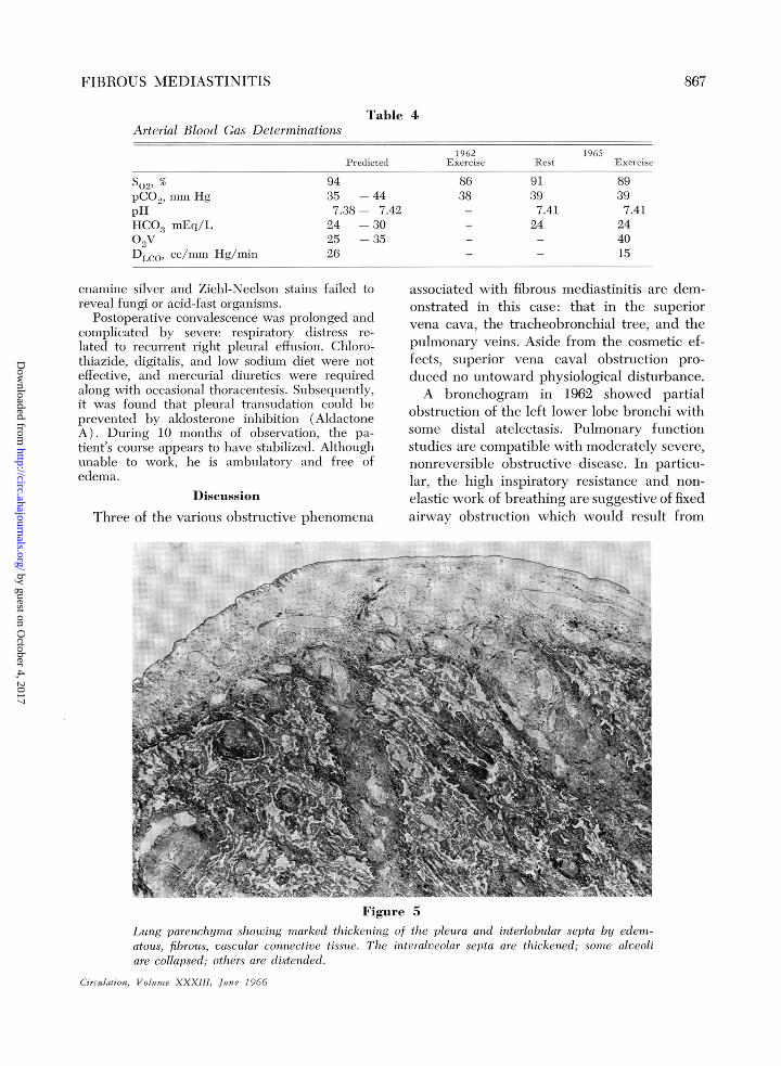
gas studies (table 4) indicate slight arterialunsaturation at rest (91%). With exercise, therewas a further decrease in arterial oxygen satura-tion to 89%, despite a considerable increase inventilation (02V 40). The carbon monoxidediffusion capacity was reduced to 58% of thepredicted value.
Operative FindingsThe possibility that the pulmonary venous ob-
struction might be correctable led to surgicalexploration. Dense vascular adhesions were en-countered and the superior vena cava was com-pletely occluded throughout its entire length. Thelungs, which were firm and somewhat nodular,failed to collapse. All right pulmonary veins wereseverely narrowed in their extrapericardial seg-ments. The lung parenchyma was woody hardfor 2 to 5 cm into the hilar region. Palpation ofthe left hilum yielded similar characteristics. Theintrapericardial segments of pulmonary veinswere free from adhesions and collapsed partiallywith each diastole. Insertion of a catheter into thesmaller radicles of the pulmonary veins via theleft atrial appendage revealed pressures as highas 40 mm Hg in the smaller tributaries withgradual reduction to 0 as the catheter was with-drawn into the left atrium. Since no operablelocus of venous obstruction was found, the pro-cedure was terminated.
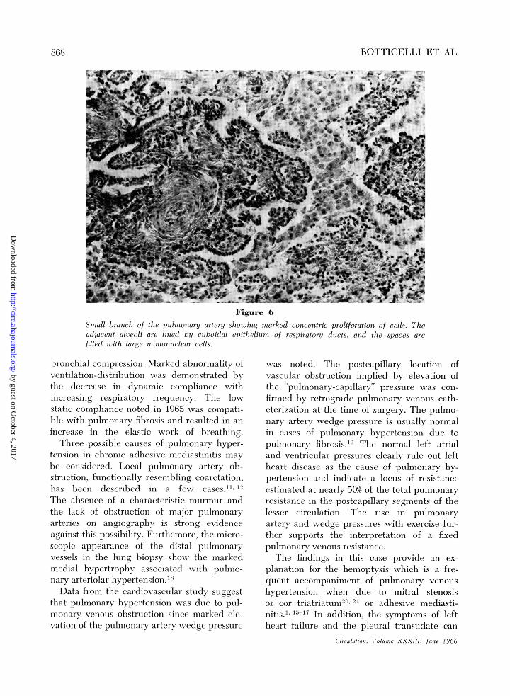
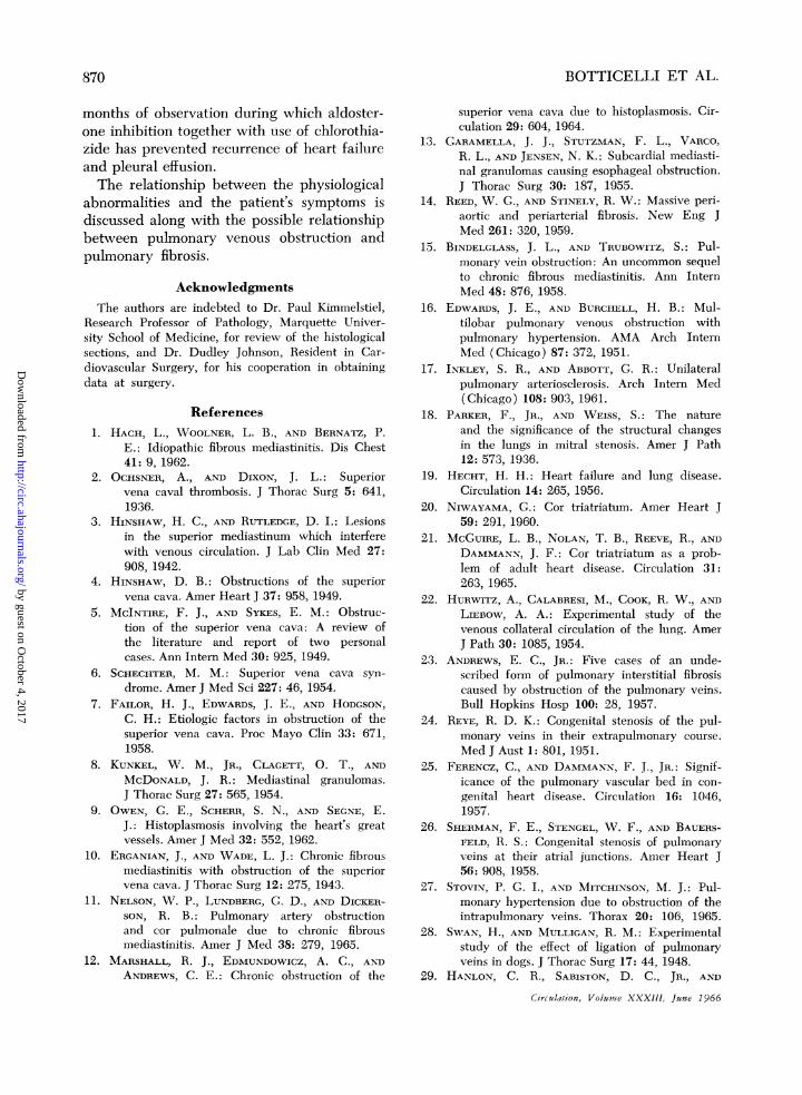
Biopsies of the pericardium and subpleuralareas of the right middle lobe showed an extremedegree of fibrous thickening which, in some areas,was markedly vascular. The pleural fibrosis ex-tended into the interlobular and interalveolarsepta (fig. 5). Most of the vessels showed sig-nificant hypertrophy of the walls, and the smallerbranches of the pulmonary artery had an in-creased number of nuclei, indicating intimalhyperplasia (fig. 6). Granulomata were not foundin the biopsy material, and treatment with meth-
ible 3
Normal
Static compliance, L/cm H20Dynamic compliance, L/cm H20
Nonelastic resistance, cm H20/L/sec
Work of breathing, kg-m/min
1962
0.22 0.05 0.190.18 + 0.04 -
1.5 -3.0 5.18*
1.2 -
0.61.8
*Interrupter method.
Circulation, Volume XXXIII, June 1966
25 bpm43 bpm100 bpm120 bpm160 bpmInspirationExpirationElasticNonelasticTotal
1965
0.160.1390.1150.0940.0770.0226.419.033.923.727.64
866
by guest on October 4, 2017
http://circ.ahajournals.org/D
ownloaded from
FIBROUS MEDIASTINITIS
Table 4Arterial Blood Gas Determinations
Predicted
so2, %pCO2, mm HgpHHCO3 mEq/L02VDLCO, cc/mm Hg/min
9435 -447.38- 7.4224 -3025 -3526
1962Exercise
8638
Rest
91397.4124
1965Exercise
89397.41
244015
enamine silver and Ziehl-Neelson stains failed toreveal fungi or acid-fast organisms.
Postoperative convalescence was prolonged andcomplicated by severe respiratory distress re-lated to recurrent right pleural effusion. Chloro-thiazide, digitalis, and low sodium diet were noteffective, and mercurial diuretics were requiredalong with occasional thoracentesis. Subsequently,it was found that pleural transudation could beprevented by aldosterone inhibition (AldactoneA). During 10 months of observation, the pa-tient's course appears to have stabilized. Althoughunable to work, he is ambulatory and free ofedema.
Discussion
Three of the various obstructive phenomena
associated with fibrous mediastinitis are dem-onstrated in this case: that in the superiorvena cava, the tracheobronchial tree, and thepulmonary veins. Aside from the cosmetic ef-fects, superior vena caval obstruction pro-duced no untoward physiological disturbance.A bronchogram in 1962 showed partial
obstruction of the left lower lobe bronchi withsome distal atelectasis. Pulmonary functionstudies are compatible with moderately severe,nonreversible obstructive disease. In particu-lar, the high inspiratory resistance and non-elastic work of breathing are suggestive of fixedairway obstruction which would result from
Figure 5Lung parenchyma showing marked thickening of the pleura and interlobular septa by edem-atous, fibrous, vascular connective tissue. The interalveolar septa are thickened; some alveoliare collapsed; others are distended.
Circulation, Volume XXXIII, June 1966
867
by guest on October 4, 2017
http://circ.ahajournals.org/D
ownloaded from
BOTTICELLI ET AL.
Figure 6Small branch of the pulmonary artery showing marked concentric proliferation of cells. Theadjacent alveoli are lined by culboidal epithelium of respiratory ducts, and the spaces arefilled with large mononuclear cells.
bronchial compression. Marked abnormality ofventilation-distribution was demonstrated bythe decrease in dynamic compliance withincreasing respiratory frequency. The lowstatic compliance noted in 1965 was compati-ble with pulmonary fibrosis and resulted in anincrease in the elastic work of breathing.
Three possible causes of pulmonary hyper-tension in chronic adhesive mediastinitis maybe considered. Local pulIoMary artery ob-struction, functionally resembling coarctation,has been described in a few cases."' 12The absence of a characteristic murmur andthe lack of obstruction of major pulmonaryarteries on angiography is strong evidenceagainst this possibility. Furthemore, the micro-scopic appearance of the distal pulmonaryvessels in the lung biopsy show the markedmedial hypertrophy associated with pulmo-nary arteriolar hypertension.18Data from the cardiovascular study suggest
that pulmonary hypertension was due to pul-monary venous obstruction since marked ele-vation of the pulmonary artery wedge pressure
was noted. The postcapillary location ofvascular obstruction implied by elevation ofthe "pulmonary-capillary" pressure was con-firmed by retrograde pulmonary venous cath-eterization at the time of surgery. The pulmo-nary artery wedge pressure is usually normalin cases of pulmonary hypertension due topulmonary fibrosis.'9 The normal left atrialand ventricular pressures clearly rule out leftheart disease as the cause of pulmonary hy-pertension and indicate a locus of resistanceestimated at nearly 50% of the total pulmonaryresistance in the postcapillary segments of thelesser circulation. The rise in pulmonaryartery and wedge pressures with exercise fur-ther supports the interpretation of a fixedpulmonary venous resistance.The findings in this case provide an ex-
planation for the hemoptysis which is a fre-quent accompaniment of pulmonary venoushypertension when due to mitral stenosisor cor triatriatum20, 21 or adhesive mediasti-nitis.' "'1 In addition, the symptoms of leftheart failure and the pleural transudate can
Circulation. Volume XXXIII, June 1966
868
by guest on October 4, 2017
http://circ.ahajournals.org/D
ownloaded from

FIBROUS MEDIASTINITIS
be related to the pulmonary venous and con-sequent pulmonary capillary hypertension.The markedly delayed washout of indicator
following segmental pulmonary arterial injec-tion probably reflects decreased flow in thesegments receiving most of the injectate. Thismay be due to partial atelectasis in the caseof the left lower lobe, but more likely reflectsvariations in the degree of pulmonary venousobstruction in various segments with conse-quent uneven flow distribution. Hurwitz andassociates22 have studied the venous collateralcirculation in dogs following pulmonary ve-nous ligation and have shown that collateralveins drain into the azygos system via pleuraladhesions and return blood to the right heart.This collateral system is capable of carryingup to 20% of the flow of an unobstructedlung. A significant shunt (left-to-right) is ruledout in this case by the normal contour of theindicator-dilution studies following inferiorcaval and proximal pulmonary arterial injec-tion.The vascular changes in the pulmonary arte-
rioles closely resemble those reported in mitralstenosis and other forms of secondary pul-monary hypertension. These so-called "pro-tective" lesions have been described in pul-monary lobes with pulmonary venousobstruction while those areas without venousobstruction are spared.3 1 23Andrews23 has suggested that interstitial
fibrosis is the result of complete, partial, orintermittent pulmonary venous obstruction.His evidence is not convincing, however, sincetwo of his five patients had left atrial enlarge-ment without definite pulmonary venous ob-struction, two had mediastinal fibrosis andthe fifth had several congenital defectsin addition to left pulmonary venous stenosis.Additional reports of congenital pulmonaryvenous stenosis24-26 have described pulmonaryfibrosis, and Stovin and Mitchinson27 havereported a case of pulmonary fibrosis withobstruction of intrapulmonary veins. The rarityof extensive pulmonary fibrosis in mitralstenosis, most cases of atrial thrombosis, atrialmyxoma, cor triatriatum, and chronic severeleft heart failure suggest that pulmonaryCirculation, Volume XXXIII, June 1966
fibrosis may not be causally related to pul-monary venous hypertension in man. Reportsof animal experiments28-30 in which pulmonaryfibrosis followed acute pulmonary venousligation have doubtful application since acutehemorrhage and infection invariably followacute ligation. A common etiology for pul-monary and mediastinal fibrosis is a moreattractive hypothesis.A common infectious cause for the pul-
monary fibrosis and mediastinitis may existin our case. Histoplasmosis has been frequentlyimplicated in the past decade9 31-33 and issuggested by the 4+reaction to skin-testingin this patient. Despite the negative comple-ment-fixation studies, negative histologicalexamination, and negative fungus culture, his-toplasmosis may be present.34 36 On the otherhand, a positive skin test alone is inconclusiveevidence. Other etiological agents which havebeen reported, such as tuberculosis, syphilis,and actinomycosis were eliminated from con-sideration by history and appropriate studies.Hawk and Hazard35 pointed out the gross
and histological similarity of fibrous medias-tinitis, fibrosing retroperitonitis, and Riedel'sstruma. These conditions may represent var-iable manifestations of a tissue response toone or more undetermined factors.
SummaryA patient with chronic fibrous mediastinitis
presented with bronchial, superior vena caval,and pulmonary venous obstruction, pulmonaryhypertension, and pulmonary fibrosis. Pulmo-nary venous obstruction was confirmed atsurgery and explains the hemodynamic find-ings of elevated pulmonary artery and pulmo-nary wedge pressures despite low left atrialpressure. Pulmonary function studies showedmarked alterations in ventilation-distribution,diffusion, work of breathing, and fixed airwayresistance.
It is concluded that the pulmonary arterialhypertension was due, in large part, to thepulmonary venous hypertension caused bypulmonary venous obstruction. Unsuccessfulsurgical intervention has been followed by 10
869
by guest on October 4, 2017
http://circ.ahajournals.org/D
ownloaded from
BOTTICELLI ET AL.
months of observation during which aldoster-one inhibition together with use of chlorothia-zide has prevented recurrence of heart failureand pleural effusion.The relationship between the physiological
abnormalities and the patient's symptoms isdiscussed along with the possible relationshipbetween pulmonary venous obstruction andpulmonary fibrosis.
Acknowledgments
The authors are indebted to Dr. Paul Kimmelstiel,Research Professor of Pathology, Marquette Univer-sity School of Medicine, for review of the histologicalsections, and Dr. Dudley Johnson, Resident in Car-diovascular Surgery, for his cooperation in obtainingdata at surgery.
References
1. HACH, L., WOOLNER, L. B., AND BERNATZ, P.E.: Idiopathic fibrous mediastinitis. Dis Chest41: 9, 1962.
2. OCHSNER, A., AND DIXoN, J. L.: Superiorvena caval thrombosis. J Thorac Surg 5: 641,1936.
3. HINSHAW, H. C., AND RUTLEDGE, D. I.: Lesionsin the superior mediastinum which interferewith venous circulation. J Lab Clin Med 27:908, 1942.
4. HINSHAW, D. B.: Obstructions of the superiorvena cava. Amer Heart J 37: 958, 1949.
5. MCINTIRE, F. J., AND SYKES, E. M.: Obstruc-tion of the superior vena cava: A review ofthe literature and report of two personalcases. Ann Intern Med 30: 925, 1949.
6. SCHECHTER, M. M.: Superior vena cava syn-
drome. Amer J Med Sci 227: 46, 1954.7. FAILOR, H. J., EDWARDS, J. E., AND HODGSON,
C. H.: Etiologic factors in obstruction of thesuperior vena cava. Proc Mayo Clin 33: 671,1958.
8. KUNKEL, W. M., JR., CLAGETT, 0. T., AND
MCDONALD, J. R.: Mediastinal granulomas.J Thorac Surg 27: 565, 1954.
9. OWEN, G. E., SCHERR, S. N., AND SEGNE, E.J.: Histoplasmosis involving the heart's greatvessels. Amer J Med 32: 552, 1962.
10. ERGANIAN, J., AND WADE, L. J.: Chronic fibrousmediastinitis with obstruction of the superiorvena cava. J Thorac Surg 12: 275, 1943.
1 1. NELSON, W. P., LUNDBERG, G. D., AND DICKER-SON, R. B.: Pulmonary artery obstructionand cor pulmonale due to chronic fibrousmediastinitis. Amer J Med 38: 279, 1965.
12. MARSHALL, R. J., EDMUNDOWICZ, A. C., AND
ANDREWS, C. E.: Chronic obstruction of the
superior vena cava due to histoplasmosis. Cir-culation 29: 604, 1964.
13. GARAMELLA, J. J., STUTZMAN, F. L., VARCO,R. L., AND JENSEN, N. K.: Subeardial mediasti-nal granulomas causing esophageal obstruction.J Thorac Surg 30: 187, 1955.
14. REED, W. G., AND STINELY, R. W.: Massive peri-aortic and periarterial fibrosis. New Eng JMed 261: 320, 1959.
15. BINDELGLASS, J. L., AND TRUBOWITZ, S.: PUl-monary vein obstruction: An uncommon sequelto chronic fibrous mediastinitis. Ann InternMed 48: 876, 1958.
16. EDWARDS, J. E., AND BURCHELL, H. B.: Mul-tilobar pulmonary venous obstruction withpulmonary hypertension. AMA Arch InternMed (Chicago) 87: 372, 1951.
17. INKLEY, S. R., AND ABBOTT, G. R.: Unilateralpulmonary arteriosclerosis. Arch Intern Med(Chicago) 108: 903, 1961.
18. PARKER, F., JR., AND WEISS, S.: The natureand the significance of the structural changesin the lungs in mitral stenosis. Amer J Path12: 573, 1936.
19. HECHT, H. H.: Heart failure and lung disease.Circulation 14: 265, 1956.
20. NIWAYAMA, G.: Cor triatriatum. Amer Heart J59: 291, 1960.
21. McCuIRE, L. B., NOLAN, T. B., REEVE, R., ANDDAMMANN, J. F.: Cor triatriatum as a prob-lem of adult heart disease. Circulation 31:263, 1965.
22. HURWITZ, A., CALABRESI, M., COOK, R. W., ANDLIEBoW, A. A.: Experimental study of thevenous collateral circulation of the lung. AmerJ Path 30: 1085, 1954.
23. ANDREWS, E. C., JR.: Five cases of an unde-scribed form of pulmonary interstitial fibrosiscaused by obstruction of the pulmonary veins.Bull Hopkins Hosp 100: 28, 1957.
24. REYE, R. D. K.: Congenital stenosis of the pul-monary veins in their extrapulmonary course.Med J Aust 1: 801, 1951.
25. FERENCZ, C., AND DAMMANN, F. J., JR.: Signif-icance of the pulmonary vascular bed in con-genital heart disease. Circulation 16: 1046,1957.
26. SHERMAN, F. E., STENGEL, W. F., AND BAUERS-FELD, R. S.: Congenital stenosis of pulmonaryveins at their atrial junctions. Amer Heart J56: 908, 1958.
27. STOVIN, P. G. I., AND MITCHINSON, M. J.: PUl-monary hypertension due to obstruction of theintrapulmonary veins. Thorax 20: 106, 1965.
28. SWAN, H., AND MULLIGAN, R. M.: Experimentalstudy of the effect of ligation of pulmonaryveins in dogs. J Thorac Surg 17: 44, 1948.
29. HANLON, C. R., SABISTON, D. C., JR., AND
Circulation, Volutne XXXIJI, June 1966
870
by guest on October 4, 2017
http://circ.ahajournals.org/D
ownloaded from
FIBROUS MEDIASTINITIS
BURKE, D. R.: Experimental pulmonary venousocclusion. J Thorac Surg 24: 190, 1952.
30. WYATT, J. P., BURKE, D. R., AND HANLON, C. R.:Morphologic study of canine lungs after liga-tion of the pulmonary veins. Amer J Path29: 291, 1953.
31. GILLESPIE, J. R.: Superior vena caval obstructionin childhood. J Pediat 49: 320, 1956.
32. MILLER, D. B., ALLEN, S. T., JR., AND AMIDEN,E. L.: Obstruction of the superior vena cavapresumably due to histoplasmosis. Amer RevTuberc & Pulmon Dis 77: 848, 1958.
33. LULL, G. F., JR., AND WINN, D. F., JR.: Chronic
fibrous mediastinitis due to Histoplasma cap-sulatum (histoplasmal mediastinitis). Radiol-ogy 73: 367, 1959.
34. SALVIN, S. B.: Current concepts of diagnosticserology and skin hypersensitivity in themycoses. Amer J Med 27: 97, 1959.
35. HAWK, W. A., AND HAZARD, J. B.: Sclerosingretroperitonitis and sclerosing mediastinitis.Amer J Clin Path 32: 321, 1959.
36. PUCKETT, T. F.: Pulmonary histoplasmosis:Study of 22 cases with identification of thecapsulatum in resected lesions. Amer RevTuberc 67: 453, 1953.
An Intellectual Exercise and Challenge to Contemporary WritingThe scientific paper is a fraud in the sense that it does give a totally misleading nar-
rative of the processes of thought that go into the making of scientific discoveries. Theinductive format of the scientific paper should be discarded. The discussion which inthe traditional scientific paper goes last should surely come at the beginning. The sci-entific facts and scientific acts should follow the discussion, and scientists should not beashamed to admit, as many of them apparently are ashamed to admit, that hypothesesappear in their minds along uncharted by-ways of thought; that they are imaginativeand inspirational in character; that they are indeed adventures of the mind.-P. B.MEDAWAR: Is the Scientific Paper Fraudulent? Yes; It Misrepresents Scientific Thought.Saturday Review, p. 43 (Aug. 1), 1984.
Circulation, Volume XXXIII, June 1966
871
by guest on October 4, 2017
http://circ.ahajournals.org/D
ownloaded from
JAMES T. BOTTICELLI, DONALD P. SCHLUETER and RAMON L. LANGEMediastinitis: Hemodynamics and Pulmonary Function
Pulmonary Venous and Arterial Hypertension due to Chronic Fibrous
Print ISSN: 0009-7322. Online ISSN: 1524-4539 Copyright © 1966 American Heart Association, Inc. All rights reserved.
is published by the American Heart Association, 7272 Greenville Avenue, Dallas, TX 75231Circulation doi: 10.1161/01.CIR.33.6.862
1966;33:862-871Circulation.
http://circ.ahajournals.org/content/33/6/862located on the World Wide Web at:
The online version of this article, along with updated information and services, is
http://circ.ahajournals.org//subscriptions/
is online at: Circulation Information about subscribing to Subscriptions:
http://www.lww.com/reprints Information about reprints can be found online at: Reprints:
document. and Rights Question and Answer
Permissionsthe Web page under Services. Further information about this process is available in thewhich permission is being requested is located, click Request Permissions in the middle column ofClearance Center, not the Editorial Office. Once the online version of the published article for
can be obtained via RightsLink, a service of the CopyrightCirculationoriginally published in Requests for permissions to reproduce figures, tables, or portions of articlesPermissions:
by guest on October 4, 2017
http://circ.ahajournals.org/D
ownloaded from