Embed Size (px)
Citation preview
1
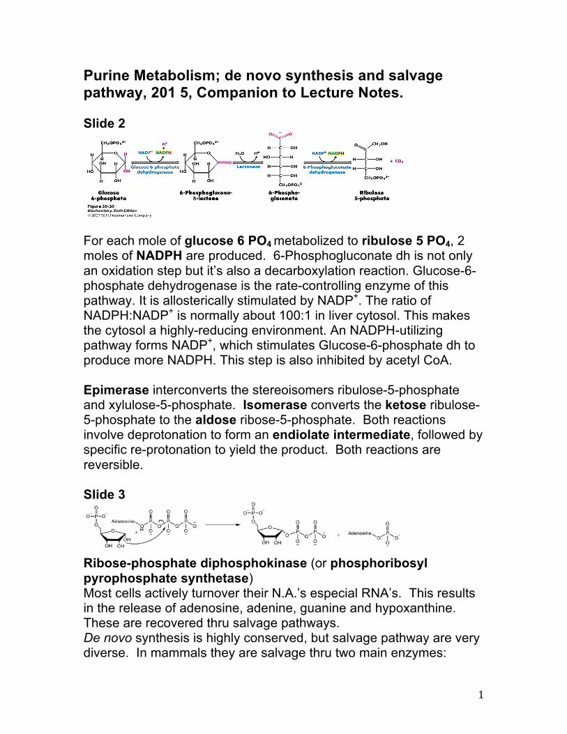
Purine Metabolism; de novo synthesis and salvage pathway, 201 5, Companion to Lecture Notes. Slide 2
For each mole of glucose 6 PO4 metabolized to ribulose 5 PO4, 2 moles of NADPH are produced. 6-Phosphogluconate dh is not only an oxidation step but it’s also a decarboxylation reaction. Glucose-6-phosphate dehydrogenase is the rate-controlling enzyme of this pathway. It is allosterically stimulated by NADP+. The ratio of NADPH:NADP+ is normally about 100:1 in liver cytosol. This makes the cytosol a highly-reducing environment. An NADPH-utilizing pathway forms NADP+, which stimulates Glucose-6-phosphate dh to produce more NADPH. This step is also inhibited by acetyl CoA. Epimerase interconverts the stereoisomers ribulose-5-phosphate and xylulose-5-phosphate. Isomerase converts the ketose ribulose-5-phosphate to the aldose ribose-5-phosphate. Both reactions involve deprotonation to form an endiolate intermediate, followed by specific re-protonation to yield the product. Both reactions are reversible. Slide 3
Ribose-phosphate diphosphokinase (or phosphoribosyl pyrophosphate synthetase) Most cells actively turnover their N.A.’s especial RNA’s. This results in the release of adenosine, adenine, guanine and hypoxanthine. These are recovered thru salvage pathways. De novo synthesis is highly conserved, but salvage pathway are very diverse. In mammals they are salvage thru two main enzymes:
2
adenine phosphoribosyltransferase (APRT), this mediates the transfer of PRPP to adenine to form AMP. Hypoxanthine-guanine phosphribosyltransferase (HGPRT), which forms IMP & GMP. The biosynthetic origins of purine ring atoms. Note that C4,C5 and N7 come from a single glycine, but all other atoms are independently acquired. The initially synthesized purine is IMP, the nucleotide base is hypoxanthine. AMP and GMP are synthesized by separate pathways. Purines are actually synthesize as ribonucleotides. The pathway is virtually identical in all living things that de novo synthesize purines. Slide 5
Synthesis of the first fully formed purine nucleotide, IMP begins with 5-phosphoaribosyl-1-pyrophosphate, PRPP. Through a series of reactions utilizing ATP, tetrahydrofolate (THF) derivatives, glutamine, glycine and aspartate this pathway yields IMP. The two indicated enzymes (A and B) are those catalyzing the ratelimiting step and the reaction necessary for the purine nucleotide cycle, respectively. In plants and micro-organisms AICA (Rxn 9) is also formed in the biosynthesis of histidine. The structure of the nucleobase of IMP (hypoxanthine). Protozoal parasites lack this pathway and do not de novo synthesize purines. They must obtain them from their host. They do have salvage pathways and are exceptional at scavenging anycompound that contains a purine ring like AdoMet or MTA, as well as adneine and adenosine all found in blood. (trypanosomes rule!).

3
Slide 7
Formed by PRPP(diphosphokinase) synthetase, the activity varies with the conc of PPi, 2,3BPG which are activiators and ADP & GDP which are inhibitors. This is a unique reaction in that ATP will directly transfer a pyrophosphate to the C1 of R5P. The glutamine PRPP aminotransferase reaction is the committed step to purine synthesis. This rxn is similar to the substrate tunneling of CPS & glutamate synthetase where the NH3 moves along a 20A path lined with non polar amino acids side chains that do not rx with the NH3 removed first from glutamine at one site and added to the ribose at another site 20A away. This enzyme is regulated by feedback inhibition from purine nucleotides. Ribose-phosphate diphosphokinase transfers the diphosphoryl group from Mg-ATP (Mg2+ coordinated to ATP) to ribose 5-phosphate. The enzymatic reaction begins with the binding of ribose 5-phosphate, followed by binding of Mg-ATP to the enzyme. In the transition state upon binding of both substrates, the diphosphate is transferred. The enzyme first releases AMP before releasing the product phosphoribosyl pyrophosphate. Experiments using oxygen 18 labelled water demonstrate that the reaction mechanism proceeds with the nucleophilic attack of the anomeric hydroxyl group of ribose 5-phosphate on the beta-phosphorus of ATP in an SN2 reaction.
4
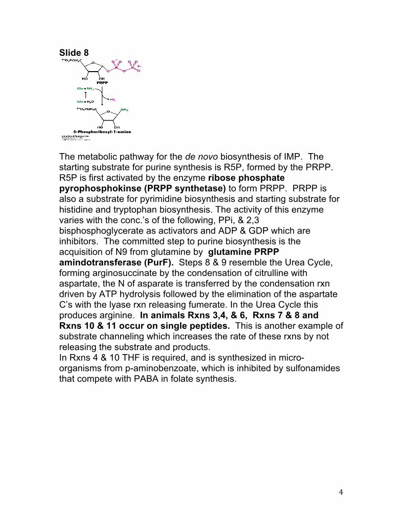
Slide 8
The metabolic pathway for the de novo biosynthesis of IMP. The starting substrate for purine synthesis is R5P, formed by the PRPP. R5P is first activated by the enzyme ribose phosphate pyrophosphokinse (PRPP synthetase) to form PRPP. PRPP is also a substrate for pyrimidine biosynthesis and starting substrate for histidine and tryptophan biosynthesis. The activity of this enzyme varies with the conc.’s of the following, PPi, & 2,3 bisphosphoglycerate as activators and ADP & GDP which are inhibitors. The committed step to purine biosynthesis is the acquisition of N9 from glutamine by glutamine PRPP amindotransferase (PurF). Steps 8 & 9 resemble the Urea Cycle, forming arginosuccinate by the condensation of citrulline with aspartate, the N of asparate is transferred by the condensation rxn driven by ATP hydrolysis followed by the elimination of the aspartate C’s with the lyase rxn releasing fumerate. In the Urea Cycle this produces arginine. In animals Rxns 3,4, & 6, Rxns 7 & 8 and Rxns 10 & 11 occur on single peptides. This is another example of substrate channeling which increases the rate of these rxns by not releasing the substrate and products. In Rxns 4 & 10 THF is required, and is synthesized in micro-organisms from p-aminobenzoate, which is inhibited by sulfonamides that compete with PABA in folate synthesis.
5
Slide 9
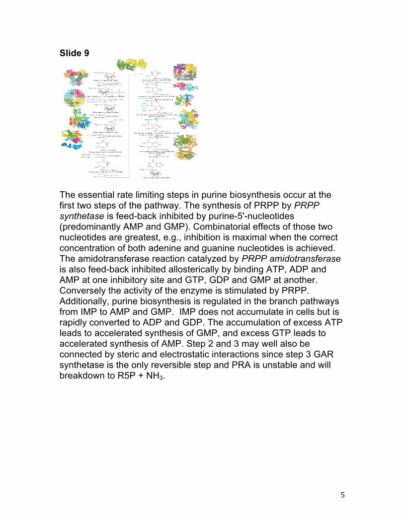
The essential rate limiting steps in purine biosynthesis occur at the first two steps of the pathway. The synthesis of PRPP by PRPP synthetase is feed-back inhibited by purine-5'-nucleotides (predominantly AMP and GMP). Combinatorial effects of those two nucleotides are greatest, e.g., inhibition is maximal when the correct concentration of both adenine and guanine nucleotides is achieved. The amidotransferase reaction catalyzed by PRPP amidotransferase is also feed-back inhibited allosterically by binding ATP, ADP and AMP at one inhibitory site and GTP, GDP and GMP at another. Conversely the activity of the enzyme is stimulated by PRPP. Additionally, purine biosynthesis is regulated in the branch pathways from IMP to AMP and GMP. IMP does not accumulate in cells but is rapidly converted to ADP and GDP. The accumulation of excess ATP leads to accelerated synthesis of GMP, and excess GTP leads to accelerated synthesis of AMP. Step 2 and 3 may well also be connected by steric and electrostatic interactions since step 3 GAR synthetase is the only reversible step and PRA is unstable and will breakdown to R5P + NH3.
6
Slide 10
Slide 11
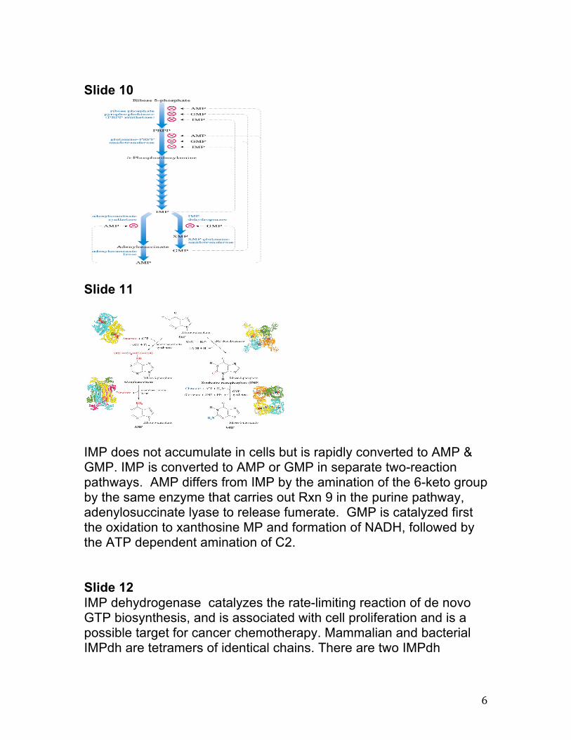
IMP does not accumulate in cells but is rapidly converted to AMP & GMP. IMP is converted to AMP or GMP in separate two-reaction pathways. AMP differs from IMP by the amination of the 6-keto group by the same enzyme that carries out Rxn 9 in the purine pathway, adenylosuccinate lyase to release fumerate. GMP is catalyzed first the oxidation to xanthosine MP and formation of NADH, followed by the ATP dependent amination of C2. Slide 12 IMP dehydrogenase catalyzes the rate-limiting reaction of de novo GTP biosynthesis, and is associated with cell proliferation and is a possible target for cancer chemotherapy. Mammalian and bacterial IMPdh are tetramers of identical chains. There are two IMPdh

7
isozymes in humans. IMP dh nearly always contains a long insertion that has two CBS domains within it. The structure of this enzyme is composed of a TIM barrel domain with two CBS domains inserted within a loop. inosine 5'-phosphate + NAD+ + H2O xanthosine 5'- phosphate + NADH + H+
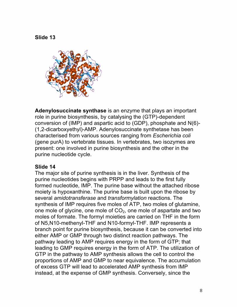
The crystal structure of adenylosuccinate synthetase from E. coli reveals that the dominant structural element of each monomer of the homodimer is a central beta-sheet of 10 strands. The first nine strands of the sheet are mutually parallel with right-handed crossover connections between the strands. The 10th strand is antiparallel with respect to the first nine strands. In addition, the enzyme has two antiparallel beta-sheets, composed of two strands and three strands each, 11 alpha-helices and two short 3/10-helices. Further, it has been suggested that the similarities in the GTP-binding domains of the synthetase and the p21ras protein are an example of convergent evolution of two distinct families of GTP-binding proteins. Structures of adenylosuccinate synthetase from Triticum aestivum and Arabidopsis thaliana when compared with the known structures from E. coli reveals that the overall fold is very similar to that of the E. coli protein.
8
Slide 13
Adenylosuccinate synthase is an enzyme that plays an important role in purine biosynthesis, by catalysing the (GTP)-dependent conversion of (IMP) and aspartic acid to (GDP), phosphate and N(6)-(1,2-dicarboxyethyl)-AMP. Adenylosuccinate synthetase has been characterised from various sources ranging from Escherichia coli (gene purA) to vertebrate tissues. In vertebrates, two isozymes are present: one involved in purine biosynthesis and the other in the purine nucleotide cycle. Slide 14 The major site of purine synthesis is in the liver. Synthesis of the purine nucleotides begins with PRPP and leads to the first fully formed nucleotide, IMP. The purine base without the attached ribose moiety is hypoxanthine. The purine base is built upon the ribose by several amidotransferase and transformylation reactions. The synthesis of IMP requires five moles of ATP, two moles of glutamine, one mole of glycine, one mole of CO2, one mole of aspartate and two moles of formate. The formyl moieties are carried on THF in the form of N5,N10-methenyl-THF and N10-formyl-THF. IMP represents a branch point for purine biosynthesis, because it can be converted into either AMP or GMP through two distinct reaction pathways. The pathway leading to AMP requires energy in the form of GTP; that leading to GMP requires energy in the form of ATP. The utilization of GTP in the pathway to AMP synthesis allows the cell to control the proportions of AMP and GMP to near equivalence. The accumulation of excess GTP will lead to accelerated AMP synthesis from IMP instead, at the expense of GMP synthesis. Conversely, since the
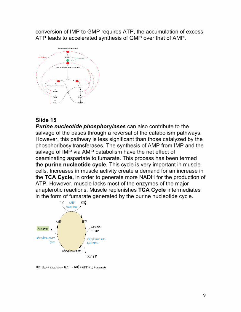
9
conversion of IMP to GMP requires ATP, the accumulation of excess ATP leads to accelerated synthesis of GMP over that of AMP.
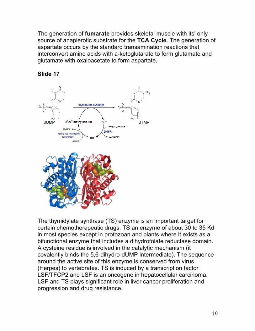
Slide 15 Purine nucleotide phosphorylases can also contribute to the salvage of the bases through a reversal of the catabolism pathways. However, this pathway is less significant than those catalyzed by the phosphoribosyltransferases. The synthesis of AMP from IMP and the salvage of IMP via AMP catabolism have the net effect of deaminating aspartate to fumarate. This process has been termed the purine nucleotide cycle. This cycle is very important in muscle cells. Increases in muscle activity create a demand for an increase in the TCA Cycle, in order to generate more NADH for the production of ATP. However, muscle lacks most of the enzymes of the major anaplerotic reactions. Muscle replenishes TCA Cycle intermediates in the form of fumarate generated by the purine nucleotide cycle.
10
The generation of fumarate provides skeletal muscle with its' only source of anaplerotic substrate for the TCA Cycle. The generation of aspartate occurs by the standard transamination reactions that interconvert amino acids with a-ketoglutarate to form glutamate and glutamate with oxaloacetate to form aspartate. Slide 17
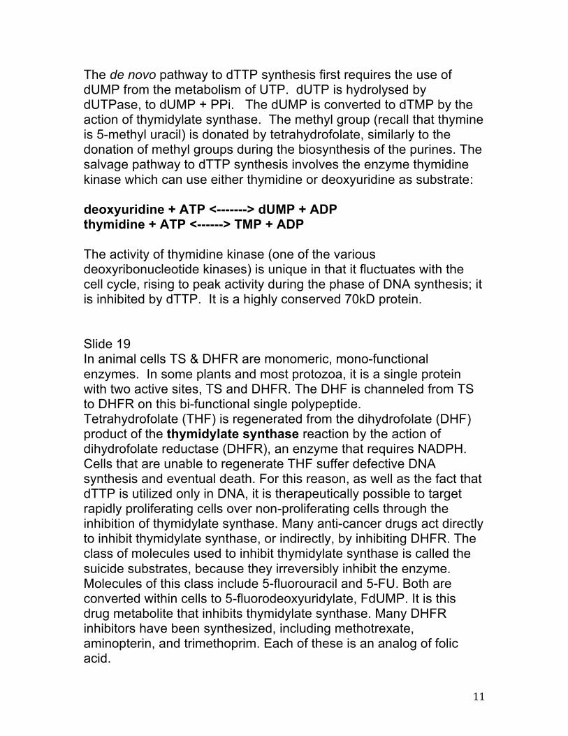
The thymidylate synthase (TS) enzyme is an important target for certain chemotherapeutic drugs. TS an enzyme of about 30 to 35 Kd in most species except in protozoan and plants where it exists as a bifunctional enzyme that includes a dihydrofolate reductase domain. A cysteine residue is involved in the catalytic mechanism (it covalently binds the 5,6-dihydro-dUMP intermediate). The sequence around the active site of this enzyme is conserved from virus (Herpes) to vertebrates. TS is induced by a transcription factor LSF/TFCP2 and LSF is an oncogene in hepatocellular carcinoma. LSF and TS plays significant role in liver cancer proliferation and progression and drug resistance.
11
The de novo pathway to dTTP synthesis first requires the use of dUMP from the metabolism of UTP. dUTP is hydrolysed by dUTPase, to dUMP + PPi. The dUMP is converted to dTMP by the action of thymidylate synthase. The methyl group (recall that thymine is 5-methyl uracil) is donated by tetrahydrofolate, similarly to the donation of methyl groups during the biosynthesis of the purines. The salvage pathway to dTTP synthesis involves the enzyme thymidine kinase which can use either thymidine or deoxyuridine as substrate: deoxyuridine + ATP <-------> dUMP + ADP thymidine + ATP <------> TMP + ADP The activity of thymidine kinase (one of the various deoxyribonucleotide kinases) is unique in that it fluctuates with the cell cycle, rising to peak activity during the phase of DNA synthesis; it is inhibited by dTTP. It is a highly conserved 70kD protein. Slide 19 In animal cells TS & DHFR are monomeric, mono-functional enzymes. In some plants and most protozoa, it is a single protein with two active sites, TS and DHFR. The DHF is channeled from TS to DHFR on this bi-functional single polypeptide. Tetrahydrofolate (THF) is regenerated from the dihydrofolate (DHF) product of the thymidylate synthase reaction by the action of dihydrofolate reductase (DHFR), an enzyme that requires NADPH. Cells that are unable to regenerate THF suffer defective DNA synthesis and eventual death. For this reason, as well as the fact that dTTP is utilized only in DNA, it is therapeutically possible to target rapidly proliferating cells over non-proliferating cells through the inhibition of thymidylate synthase. Many anti-cancer drugs act directly to inhibit thymidylate synthase, or indirectly, by inhibiting DHFR. The class of molecules used to inhibit thymidylate synthase is called the suicide substrates, because they irreversibly inhibit the enzyme. Molecules of this class include 5-fluorouracil and 5-FU. Both are converted within cells to 5-fluorodeoxyuridylate, FdUMP. It is this drug metabolite that inhibits thymidylate synthase. Many DHFR inhibitors have been synthesized, including methotrexate, aminopterin, and trimethoprim. Each of these is an analog of folic acid.
12
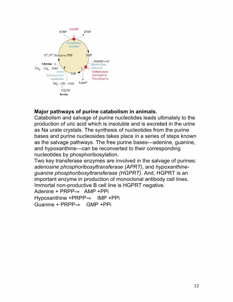
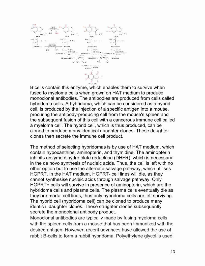
Major pathways of purine catabolism in animals. Catabolism and salvage of purine nucleotides leads ultimately to the production of uric acid which is insoluble and is excreted in the urine as Na urate crystals. The synthesis of nucleotides from the purine bases and purine nucleosides takes place in a series of steps known as the salvage pathways. The free purine bases---adenine, guanine, and hypoxanthine---can be reconverted to their corresponding nucleotides by phosphoribosylation. Two key transferase enzymes are involved in the salvage of purines: adenosine phosphoribosyltransferase (APRT), and hypoxanthine-guanine phosphoribosyltransferase (HGPRT). And, HGPRT is an important enzyme in production of monoclonal antibody cell lines. Immortal non-productive B cell line is HGPRT negative. Adenine + PRPP⇒ AMP +PPi Hypoxanthine +PRPP⇒ IMP +PPi Guanine + PRPP⇒ GMP +PPi
13
B cells contain this enzyme, which enables them to survive when fused to myeloma cells when grown on HAT medium to produce monoclonal antibodies. The antibodies are produced from cells called hybridoma cells. A hybridoma, which can be considered as a hybrid cell, is produced by the injection of a specific antigen into a mouse, procuring the antibody-producing cell from the mouse's spleen and the subsequent fusion of this cell with a cancerous immune cell called a myeloma cell. The hybrid cell, which is thus produced, can be cloned to produce many identical daughter clones. These daughter clones then secrete the immune cell product. The method of selecting hybridomas is by use of HAT medium, which contain hypoxanthine, aminopterin, and thymidine. The aminopterin inhibits enzyme dihydrofolate reductase (DHFR), which is necessary in the de novo synthesis of nucleic acids. Thus, the cell is left with no other option but to use the alternate salvage pathway, which utilises HGPRT. In the HAT medium, HGPRT- cell lines will die, as they cannot synthesise nucleic acids through salvage pathway. Only HGPRT+ cells will survive in presence of aminopterin, which are the hybridoma cells and plasma cells. The plasma cells eventually die as they are mortal cell lines, thus only hybridoma cells are left surviving. The hybrid cell (hybridoma cell) can be cloned to produce many identical daughter clones. These daughter clones subsequently secrete the monoclonal antibody product. Monoclonal antibodies are typically made by fusing myeloma cells with the spleen cells from a mouse that has been immunized with the desired antigen. However, recent advances have allowed the use of rabbit B-cells to form a rabbit hybridoma. Polyethylene glycol is used
14
to fuse adjacent plasma membranes, but the success rate is low so a selective medium in which only fused cells can grow is used. This is possible because myeloma cells have lost the ability to synthesize hypoxanthine-guanine-phosphoribosyl transferase (HGPRT), an enzyme necessary for the salvage synthesis of nucleic acids. The absence of HGPRT is not a problem for these cells unless the de novo purine synthesis pathway is also disrupted. By exposing cells to aminopterin (a folic acid analogue, which inhibits DHFR, they are unable to use the de novo pathway and become fully auxotrophic for nucleic acids requiring supplementation to survive. The selective culture medium is called HAT medium because it contains hypoxanthine, aminopterin, andthymidine. This medium is selective for fused (hybridoma) cells. Unfused myeloma cells cannot grow because they lack HGPRT, and thus cannot replicate their DNA. Unfused spleen cells cannot grow indefinitely because of their limited life span. Only fused hybrid cells, referred to as hybridomas, are able to grow indefinitely in the media because the spleen cell partner supplies HGPRT and the myeloma partner has traits that make it immortal (similar to a cancer cell). This mixture of cells is then diluted and clones are grown from single parent cells on microtitre wells. The antibodies secreted by the different clones are then assayed for their ability to bind to the antigen (with a test such as ELISA or Antigen Microarray Assay) or immuno-dot blot. The most productive and stable clone is then selected for future use. The hybridomas can be grown indefinitely in a suitable cell culture medium. They can also be injected into mice in the peritoneal cavity, surrounding the gut). There, they produce tumors secreting an antibody-rich fluid called ascites fluid.


![De Novo Purine Synthesis Nitrogen-Fixing Nodules of … · Metabolism of [8-14C]IMP (1 mM, 1,uCi) to purinebaseswasmeasuredinthesamereactionmixturesusedfor denovopurineassays.](https://img.pdfslide.net/doc/110x75/5ae4a0dd7f8b9a9e5d8b5d2d/de-novo-purine-synthesis-nitrogen-fixing-nodules-of-of-8-14cimp-1-mm-1uci.jpg)