Embed Size (px)
Citation preview
666 LETTERS
QT PROLONGATION AND RECURRENT “TORSADES DE POINTES” DURING
ERYTNROMYCIN LACTOBIONATE INFUSION
We read the report by McComb et al1 on a recurrent.ventricular tachycardia (VT) asso- ciated with QT prolongation during the intra- venous administration of erythromycin. We recently had observed a similar case with “torsades de pointes” (TP) type ventricular tachycardia [VT).
A56-year-old woman was admitted to the intensive care unit for acute respiratory fail- ure. The patient had a medical history of COT pulmonale secondary to severe kypho- scoliosis. Bronchopulmonary infection was the main factor incriminated in the acute ex- acerbation of the chronic lung disease. The serum potassium and calcium concentra- tions were normal (4.2 and 2.25 mmol/liter, respectively). The electrocardiogram(Fig. 1) showed sinus rhythm, increased P waves indicative of right auricular hypertrophy, right-axis QRS deviation, incomplete right bundle branchblockandnormaJQTinterva1 (0.38secondforaQTcorrectedforheartrate of 0.37 second]. Initial arterial blood gases at room air were: Pa02 33 mm Hg, PaCOz 70 mm Hg, pH7.16. The treatment included na- sotrachealintubationandcontrolledventiJa- tion. The arterial blood gases were normal- ized3hourslater(PaC0247mmHg,PaOz~08 mm Hg under 50% oxygen). The pulmonary infectionwastreatedwith3g/dayoferythro- mycin Jactobionate. The first 2 injections
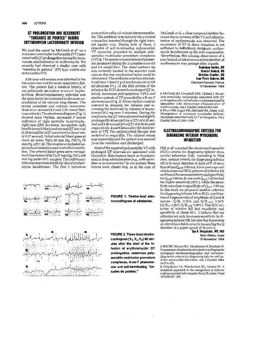
were achieved by a &minute intravenous bo- lus. The antibiotic was injected via a central venous line inserted through the right inter- nal jugular vein. During both of them, 2 episodes of self-terminating polymorphic VT occurred, preceded by multiple poly- morphic ventricular premature complexes (VPCsJ. The serum concentrations ofpotassi- urn measured during the 2 episodes were 4.6 and 4.4 mmol/Iiter. The distal catheter tip was correctly located in the superior vena cava so that any mechanical factor could be eliminated. The antibiotic was then adminis- tered over 1 hour (1 g of erythromycin in 125 ml dextrose 5%]. At the 45th minute of the infusion the ECG showed a prolonged QT in- terval, numerous polymorphous VPCs and another episode of TP preceded by a R-on-T phenomenon (Fig. 2). Sinus rhythm could be restored by stopping the infusion and in- creasing the heart rate by infusion of isopro- terenol(0.2 mg over 2 minutes]. During the next hours, theQTintervalremainedslightly prolonged (0.40 second for a QTc of0.35 sec- ond and 0.40 second for a QTc of0.36 second, respectively,4and8hoursafterthe thirdepi- sode of TP). The antimicrobial therapy was switched to ampicillin. The clinical course was uneventful and the patient was weaned from the ventilator and discharged.
Most of the acquired polymorphic VT with prolonged QT intervals are associated with electrolyte disturbances (such as hypokale- mia] or drug administration (e.g., with quini- dine or procainamide).z In our patient, these factors were absent and, as in the case of
FIGURE 1. Twelve-lead elec- trocardiogram at admission.
FIGURE 2. Three-lead electro-
cardiogram (VI, Vz, V,) 45 min-
utes after the start of the in- fusion of erythromycin: QT
prolongation, numerous poly-
morphic ventricular premature complexes, R-on-T phenome-
non and self-terminating “tor-
sades de pointes.”
McComb et al, a close temporal relation be- tween the occurrence of the VT and adminis- tration of erythromycin was observed. The recurrence of TP in three occasions is not sufficient to definitively designate erythro- mycin lactobionate as the sole responsable. Nevertheless, this echoing observation of a rare hazard of intravenous administration of erythromycin may prompt other reports.
Dominique Guelon, MD Bernard Bedock, MD
Christian Chartier, MD Jean-Pierre Haberer, MD
Clermont-Ferrand, France 12 December 1984
1. McComb’lM, Campbell NPS, &land J. Recur- rent ventricular tachycardia associated with QT prolongationaftermitraIvolve replacement and its association with intravknous administration of erythromycin. Am J Cardiol1984;54:922-923. 2. Khan MM, Logan KR, McComb JM, Adgey AAJ. Management of recurrent ventricular tachyar- rhythmias associated with Q-T prolongation. Am 1 Cardiol 1981;47:1301-1308.
ELECTROCARDIOGRAPHIC CRITERIA FOR DIAGNOSING INFERIOR MYOCARDIAL
INFARCTION
Hill et al* reported the electrocardiographic (ECG] criteria for diagnosing inferior myo- cardial infarction (MI]. According to their data, optimal criteria for diagnosing inferior MI is Q-wave duration in lead aVF of more than 30 ms (Qav~ >30 ms]. A few years ago we2 checked several ECG patterns of inferior MI andfoundthesamesensitivityandspecificity forQ,m>30ms.InourworkQ,vF>Z0mshad the higher sensitivity (92%], while his speci- ficity was close to specificity of Qav~ >30 ms. In this work we proposed another criterion fordiagnosinginferior MI on ECG: combina- tion of 2 signs as ratio of amplitude of Q and R waves-Q/RI >25% and Q/R av~ >lO% (Q/R~I X5% Q/R av~ >lO%]. This ECG cri- terion of inferior MI had sensitivity and specificity of about 95%. I believe that my criterion not only increases sensitivity in di- agnosing inferior MI, but also that it prevents an almost inevitable error in measuring the Q duration at a paper speed of 25 min/s.
llya A. Ovsyshcher, MD, PbD Beer-Sheba, Israel
15 November 1984
1. Hill NE, Warner RA, Mookhejee S, Smulyan H. ComparisonofoptimaJscoIarelectrocardiographic orthogonal electrocardiographic and vectorcar- diographic criteria for diagnosing inferior and an- terior myocordial infarction. Am J Cardiol 1984; 54274-276. 2. Ovsyshcher IA, Wanderman KL, Gueron M. A statistical approach to the recognition of inferior wall myocardial infarction by the Q/R ratios. Chest 1975:68:337-339.