Embed Size (px)
Citation preview
VOL. 88, NO. 5 NOTES, CASES, INSTRUMENTS 941
provides the necessary control signal and the proper power to operate the automatic diaphragm within its full range. Once the sensitivity of the system is set, it responds reliably to changes in illumination and adjusts itself even during changes in magnification.
Proper calibration of the circuit added to the television camera (Figs. 2 and 3) is obtained by tuning a potentiometer that is within the camera body, while observing the television monitor screen for an adequately exposed picture. Maximum illumination of a bright object using the lowest magnification should be used at the time of calibration. The system is tested by increasing the magnification and reducing light intensity. This should open the diaphragm. Because the automated diaphragm itself was not modified, it can also be used with the Beaulieu 16-mm movie camera as originally intended.
We are presently using a Hitachi HB-9017U television camera. The circuit needed for this camera is shown in Figure 2. Similar good results were obtained earlier with the Magnavox 400 and 500 cameras, although a slightly different electronic circuit was used. Any black and white or color television camera can be modified to fit the automatic diaphragm (Fig. 3).
SUMMARY
We developed an automated diaphragm control to achieve constant good exposure during televised microsurgery by using a commercially available diaphragm control for a movie camera and modifying slightly the television camera.
R E F E R E N C E
1. Machemer, R., and Parel, J.-M.: An improved mierosurgieal ceiling-mounted unit and an automated television. Am. J. Ophthalmol. 85:205, 1978.
RADIAL BUCKLING OF POSTERIOR RETINAL TEARS
J O H N D. S C O T T , F. R. C. S. Cambridge, England
AND W A L T E R H. STERN, M.D.
San Francisco, California
Posterior retinal holes are associated with myopia, branch vein occlusion, and proliferative diabetic retinopathy.1,2
Since the advent of vitrectomy and techniques for dissection of epiretinal membranes, iatrogenic posterior retinal holes have become increasingly important.3,4 In iatrogenic breaks associated with dissection of epiretinal membranes, it is possible to repair the posterior breaks by using an internal tamponade of sulfur hexa-fluoride gas-air mixture combined with either cryotherapy, diathermy, or photo-coagulation. However, in certain cases where tangential retinal traction persists as the result of epiretinal membrane formation, and cannot be adequately relieved, it is necessary to provide a posterior scleral buckle in combination with internal gas tamponade to seal these breaks effectively. We have devised a method for safely and easily placing the mattress sutures to secure a posterior radial buckle.
The placement of a posterior radial buckle is often hampered because of difficulty in visualizing the needle as well as maintaining the correct depth of the needle in the sclera. Tying the sutures posteriorly can be facilitated by passing the needle in the anteroposterior direction opposite the curvature of the globe when the needle is posterior to the equator.
From the Department of Ophthalmology, Adden-brooke's Hospital, Cambridge University, Cambridge, England, (Mr. Scott); and the Department of Ophthalmology, University of California, San Francisco, and the Veterans Administration Medical Center, San Francisco (Dr. Stern).
Reprint requests to Walter H. Stern, M.D., 400 Parnassus Ave., San Francisco, CA 94143.
942 AMERICAN JOURNAL OF OPHTHALMOLOGY NOVEMBER, 1979
Tying the posterior ends of the sutures anteriorly avoids the problem of tying sutures posteriorly. This procedure is made easier in vitrectomized eyes with a pars plana infusion line in which intraocular pressure can be decreased to achieve greater visualization of the posterior scle-ra.
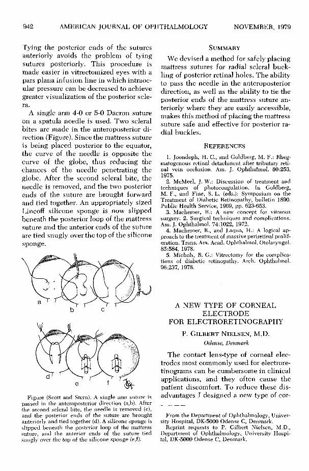
A single arm 4-0 or 5-0 Dacron suture on a spatula needle is used. Two scleral bites are made in the anteroposterior direction (Figure). Since the mattress suture is being placed posterior to the equator, the curve of the needle is opposite the curve of the globe, thus reducing the chances of the needle penetrating the globe. After the second scleral bite, the needle is removed, and the two posterior ends of the suture are brought forward and tied together. An appropriately sized Lincoff silicone sponge is now slipped beneath the posterior loop of the mattress suture and the anterior ends of the suture are tied snugly over the top of the silicone sponge.
Figure (Scott and Stern). A single arm suture is passed in the anteroposterior direction (a,b). After the second scleral bite, the needle is removed (c), and the posterior ends of the suture are brought anteriorly and tied together (d). A silicone sponge is slipped beneath the posterior loop of the mattress suture, and the anterior ends of the suture tied snugly over the top of the silicone sponge (e,f).
SUMMARY
We devised a method for safely placing mattress sutures for radial scleral buckling of posterior retinal holes. The ability to pass the needle in the anteroposterior direction, as well as the ability to tie the posterior ends of the mattress suture anteriorly where they are easily accessible, makes this method of placing the mattress suture safe and effective for posterior radial buckles.
REFERENCES 1. Joondeph, H. C , and Goldberg, M. F.: Rheg-
matogenous retinal detachment after tributary retinal vein occlusion. Am. J. Ophthalmol. 80:253, 1975.
2. McMeel, J. W.: Discussion of treatment and techniques of photocoagulation. In Goldberg, M. F., and Fine, S. L. (eds.): Symposium on the Treatment of Diabetic Retinopathy, bulletin 1890. Public Health Service, 1969, pp. 623-653.
3. Machemer, R.: A new concept for vitreous surgery. 2. Surgical techniques and complications. Am. J. Ophthalmol. 74:1022, 1972.
4. Machemer, R., and Laqua, H.: A logical approach to the treatment of massive periretinal proliferation. Trans. Am. Acad. Ophthalmol. Otolaryngol. 85:584, 1978.
5. Michels, R. G.: Vitrectomy for the complications of diabetic retinopathy. Arch. Ophthalmol. 96:237, 1978.
A NEW TYPE OF CORNEAL ELECTRODE
FOR ELECTRORETINOGRAPHY
P. G I L B E R T N I E L S E N , M.D.
Odense, Denmark
The contact lens-type of corneal electrodes most commonly used for electrore-tinograms can be cumbersome in clinical applications, and they often cause the patient discomfort. To reduce these disadvantages I designed a new type of cor-
From the Department of Ophthalmology, University Hospital, DK-5000 Odense C, Denmark.
Reprint requests to P. Gilbert Nielsen, M.D., Department of Ophthalmology, University Hospital, DK-5000 Odense C, Denmark.


![$PQZSJHIU …ousar.lib.okayama-u.ac.jp/files/public/5/56175/...rhages, retinal pigment epithelial tears, and/or chorio-capillaris atrophy [9-11]. The risk of serious complica-tions](https://img.pdfslide.net/doc/110x75/5e274ba9c8f801547e287b2d/pqzsjhiu-ousarlibokayama-uacjpfilespublic556175-rhages-retinal-pigment.jpg)