Embed Size (px)
Citation preview
Radiation Protection in Medicine: Past,
Present and Future
Madan Rehani, PhD Radiation Protection of Patients Unit, IAEA
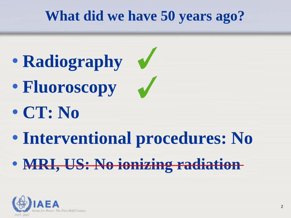
What did we have 50 years ago?
• Radiography
• Fluoroscopy
• CT: No
• Interventional procedures: No
• MRI, US: No ionizing radiation
2
Radiotherapy & Nuclear Medicine
• Mainly Cobalt-60, No Linac
• Radium for brachytherapy
• Iodine-131: Queen of nuclear medicine
3
Radiography: Past & Present
• Calcium Tungstate screens, 200 speed
• Rare earth screens: ≈ 2 times faster
• Patient dose ≈ Half or still better
• Compared to emulsions or direct
radiography- patient dose by a factor of few
tens
• Operator dose, similar proportion, depending
upon shielding
4
Radiography
5
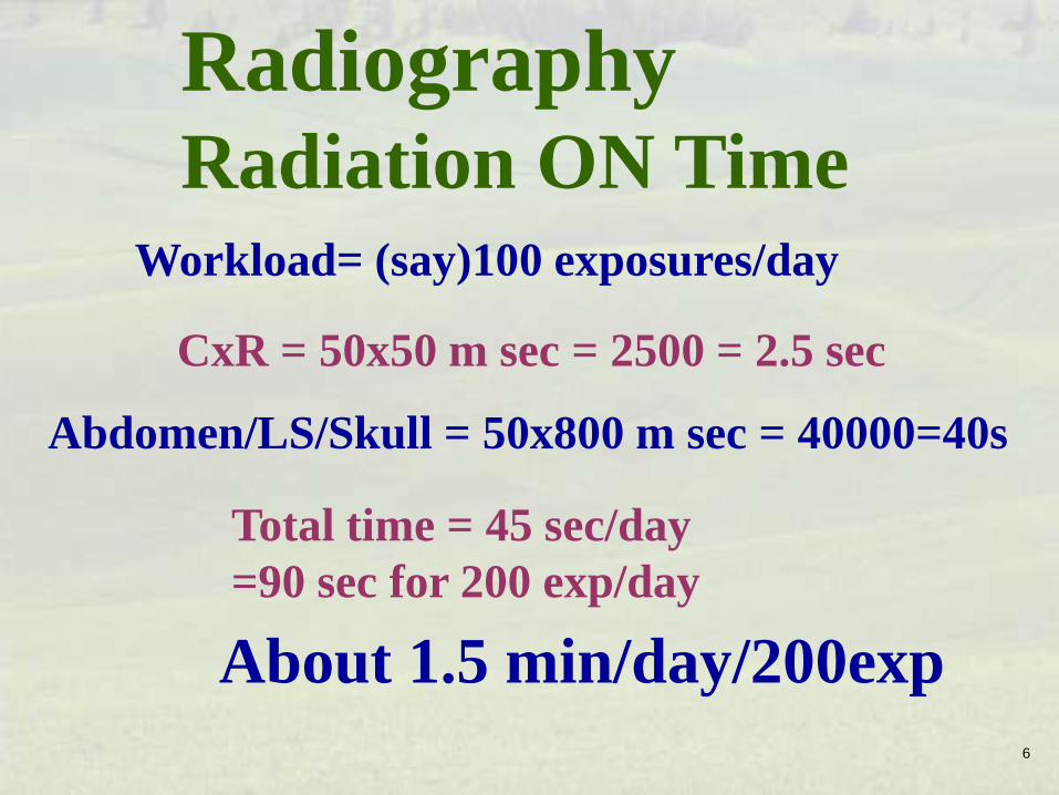
Radiography Radiation ON Time
Workload= (say)100 exposures/day
CxR = 50x50 m sec = 2500 = 2.5 sec
Abdomen/LS/Skull = 50x800 m sec = 40000=40s
Total time = 45 sec/day
=90 sec for 200 exp/day
About 1.5 min/day/200exp
6
Radiography: Present
7
Radiography
8
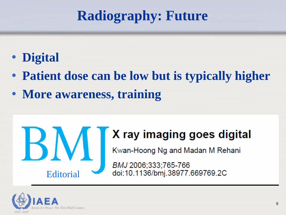
Radiography: Future
• Digital
• Patient dose can be low but is typically higher
• More awareness, training
9
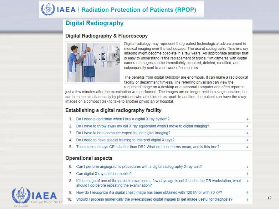
Editorial
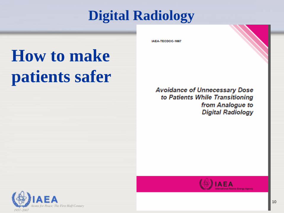
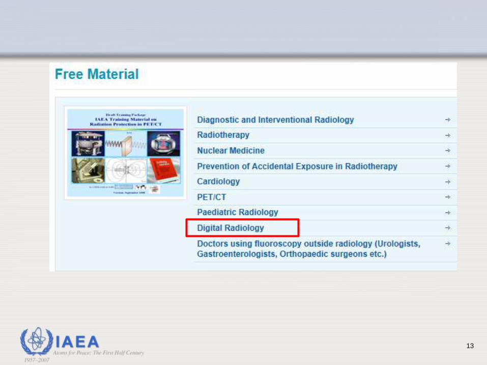
Digital Radiology
How to make
patients safer
10
12
13
14
15
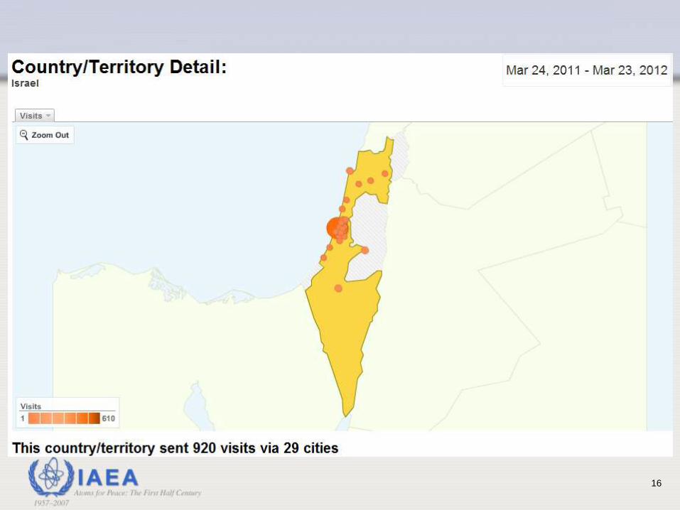
16
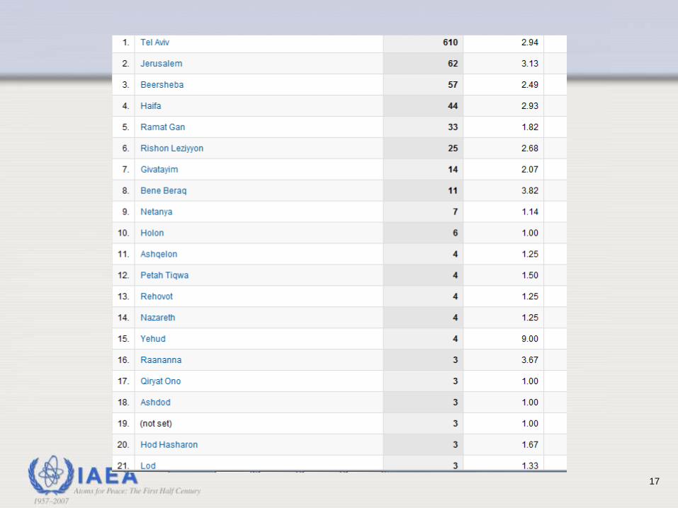
17
18
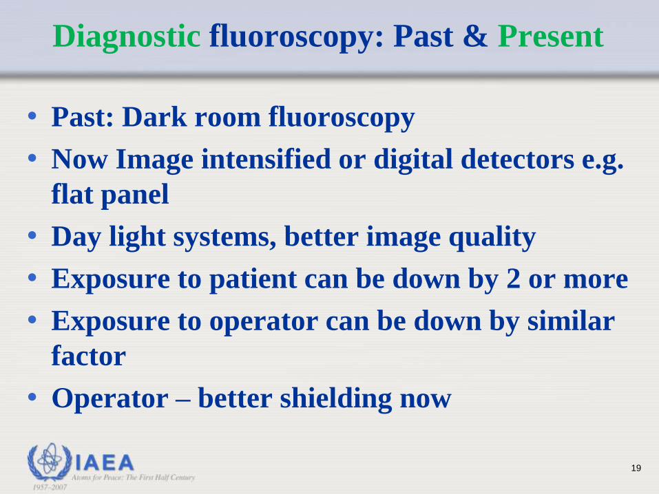
Diagnostic fluoroscopy: Past & Present
• Past: Dark room fluoroscopy
• Now Image intensified or digital detectors e.g.
flat panel
• Day light systems, better image quality
• Exposure to patient can be down by 2 or more
• Exposure to operator can be down by similar
factor
• Operator – better shielding now
19
Diagnostic Fluoroscopy: Present
20
Diagnostic Fluoroscopy: Future
• Like digital radiography
• Awareness, education and training
• Monitoring of patient dose
21
Interventional Procedures- Staff
• Potential for higher exposures to staff than in
radiology, nuclear medicine and even
radiotherapy
• Fluoroscopy times >30 min are not uncommon
• No real comparison with 50 years possible
• Technology better but safety not, mostly
because of
• better information. For same information, dose
• lack of utilization of protection means by operator
22
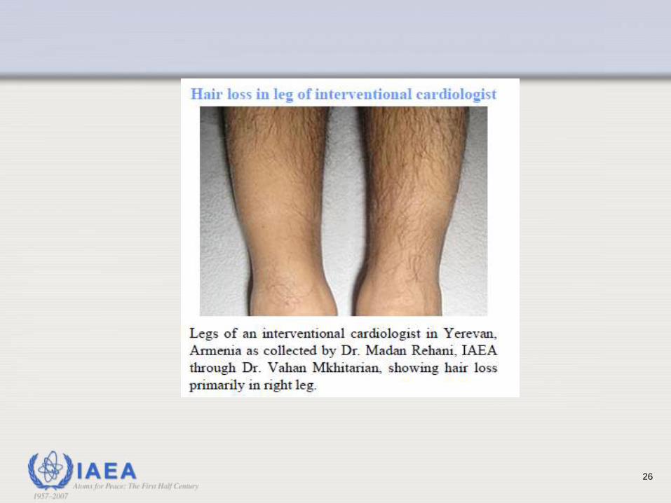
• Cataract risk substantial
• Some risks of skin injuries and hair loss
• Other risks (stochastic- carcinogenic)
can be managed through utilization of
better protection
Interventional Procedures- Staff
23
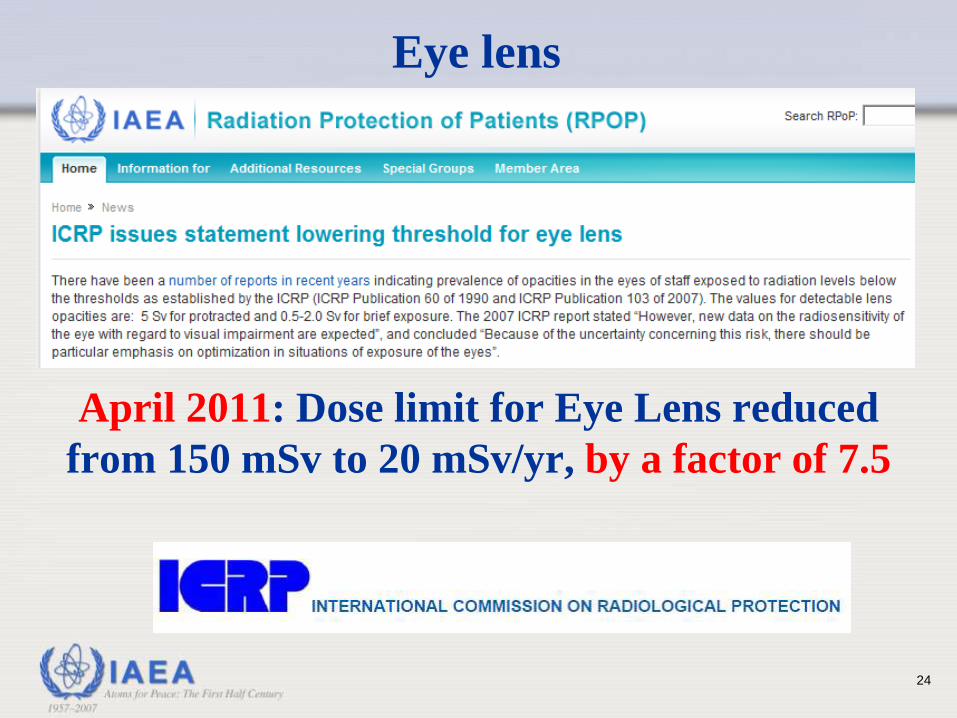
Eye lens
April 2011: Dose limit for Eye Lens reduced
from 150 mSv to 20 mSv/yr, by a factor of 7.5
24
Interventional Procedures- Staff
25
26
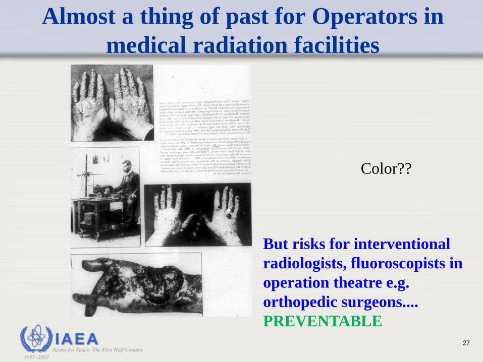
Almost a thing of past for Operators in
medical radiation facilities
Color??
But risks for interventional
radiologists, fluoroscopists in
operation theatre e.g.
orthopedic surgeons....
PREVENTABLE
27
28
29
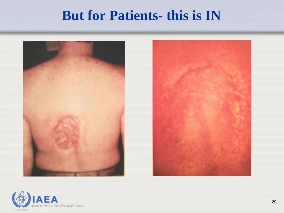
But for Patients- this is IN
29
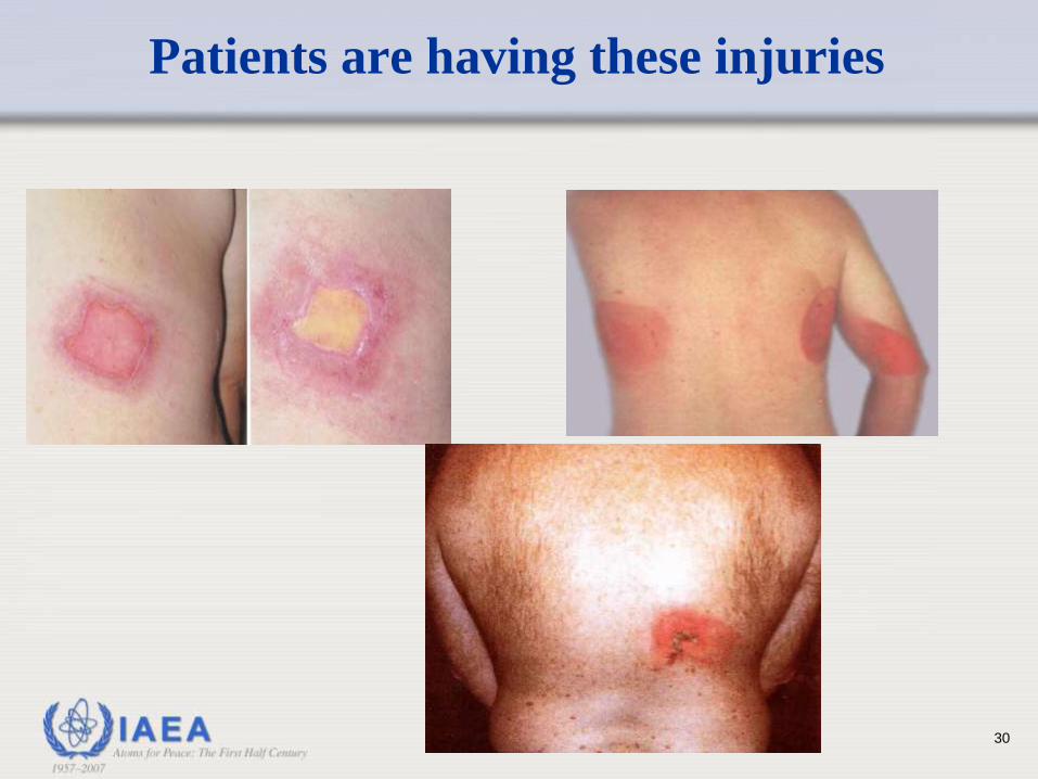
Patients are having these injuries
30
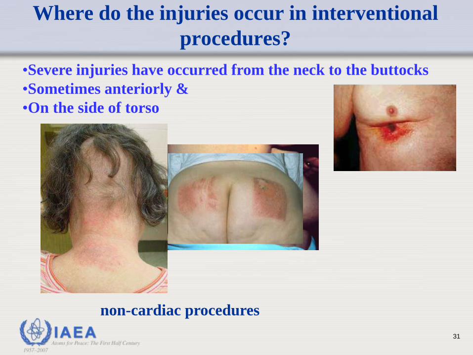
Where do the injuries occur in interventional
procedures?
•Severe injuries have occurred from the neck to the buttocks
•Sometimes anteriorly &
•On the side of torso
non-cardiac procedures
31
Current situation
A case of radiation induced skin
injury is filed in US courts every 4 to
6 weeks currently, primarily from
interventional procedures
≈10 cases/year
32
• MUCH better outcomes
• Replacement of surgery
• Risks can be substantial if not cared for
• Skin injury
• Carcinogenic risks
Interventional Procedures- Patient
33
Nuclear Medicine
• I-131 replaced by and large by Tc-99m
• Patient doses better than half
• PET/CT: 15-25 mSv
• Cardiac imaging: 10-40 mSv
• Patient doses higher but mainly because of
new applications
• Main RP issues are with hybrid imaging and
radioiodine therapy
34
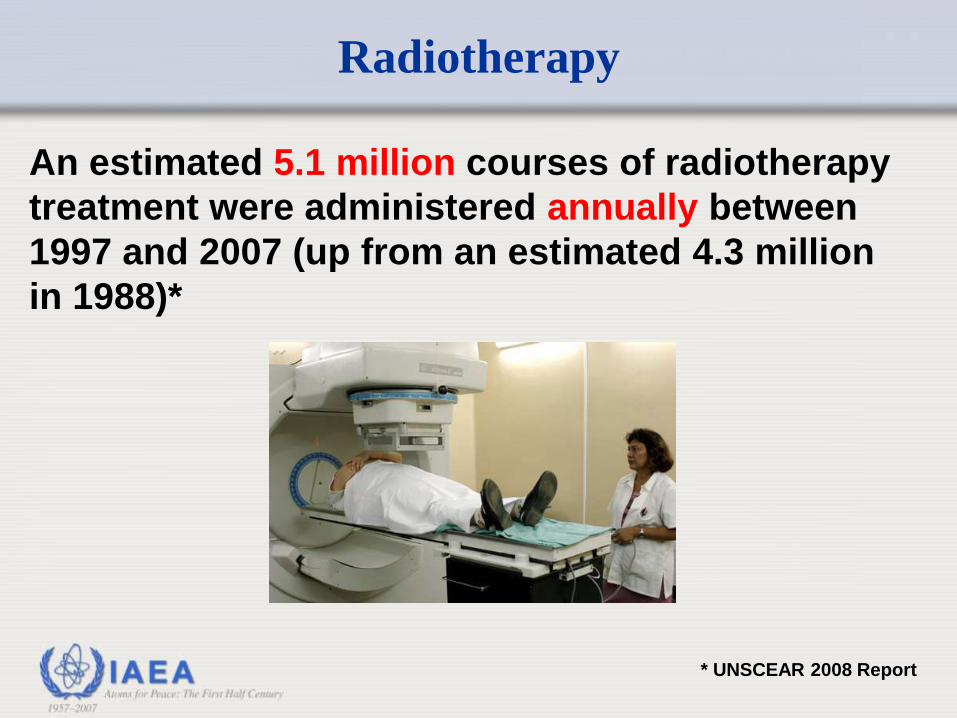
An estimated 5.1 million courses of radiotherapy
treatment were administered annually between
1997 and 2007 (up from an estimated 4.3 million
in 1988)*
* UNSCEAR 2008 Report
Radiotherapy
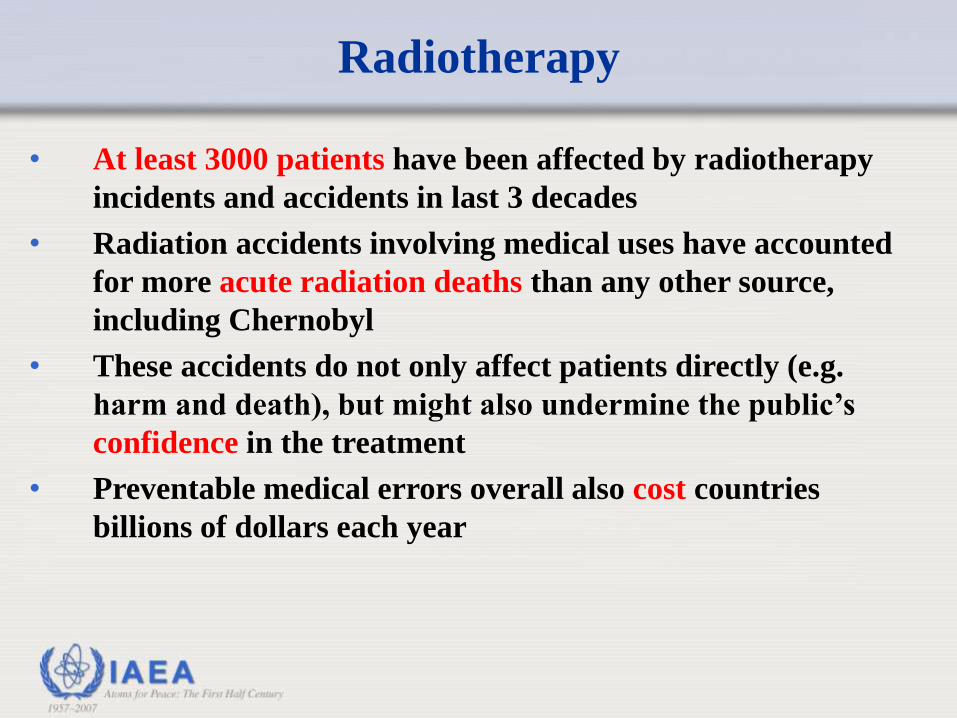
• At least 3000 patients have been affected by radiotherapy
incidents and accidents in last 3 decades
• Radiation accidents involving medical uses have accounted
for more acute radiation deaths than any other source,
including Chernobyl
• These accidents do not only affect patients directly (e.g.
harm and death), but might also undermine the public’s
confidence in the treatment
• Preventable medical errors overall also cost countries
billions of dollars each year
Radiotherapy
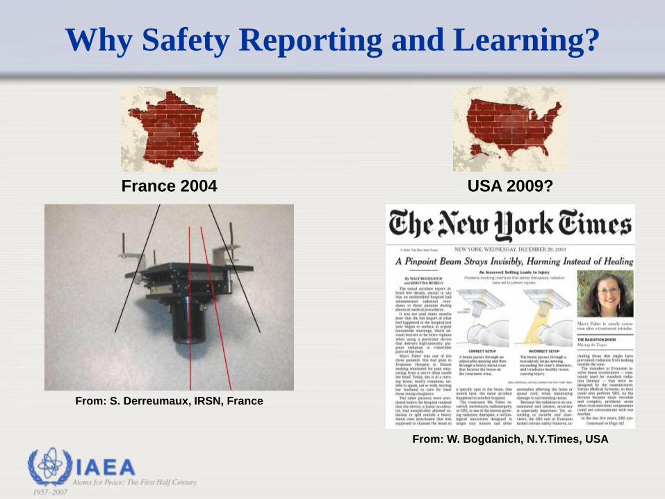
From: W. Bogdanich, N.Y.Times, USA
Why Safety Reporting and Learning?
From: S. Derreumaux, IRSN, France
France 2004 USA 2009?
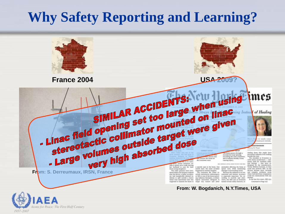
From: W. Bogdanich, N.Y.Times, USA
Why Safety Reporting and Learning?
From: S. Derreumaux, IRSN, France
France 2004 USA 2009?
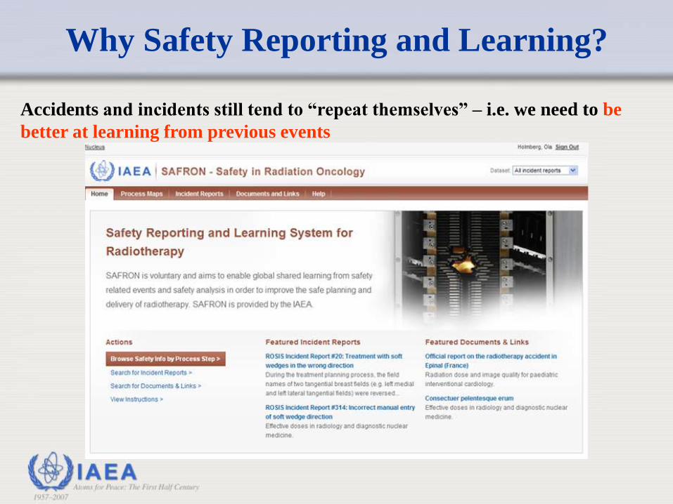
Accidents and incidents still tend to “repeat themselves” – i.e. we need to be
better at learning from previous events
Why Safety Reporting and Learning?
Computed Tomography (CT)
• Invented in 1972
• Most beneficial technique
• Technology has been improving fast
• BUT usage pattern and extent of usage has
resulted in increased risks
40
Over-exposure: 2010 Regulatory actions
41
Besides accidental exposures
Again something that has started in this decade
42
43
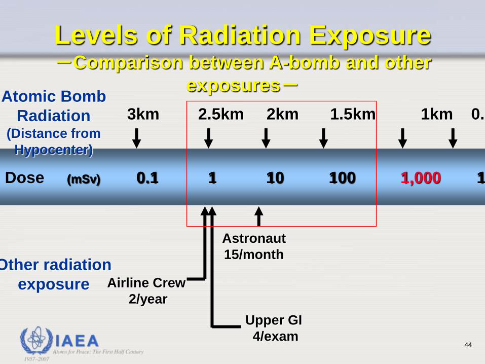
Dose (mSv) 0.1 1 10 100 1,000 10,000
Levels of Radiation Exposure -Comparison between A-bomb and other
exposures- Atomic Bomb
Radiation (Distance from
Hypocenter)
Other radiation
exposure
3km 2.5km 2km 1.5km 1km 0.5km
Astronaut
15/month
Airline Crew
2/year
Upper GI
4/exam 44
Announcement few years ago:
The era of few hundreds of Euros
for flying within Europe is gone
45
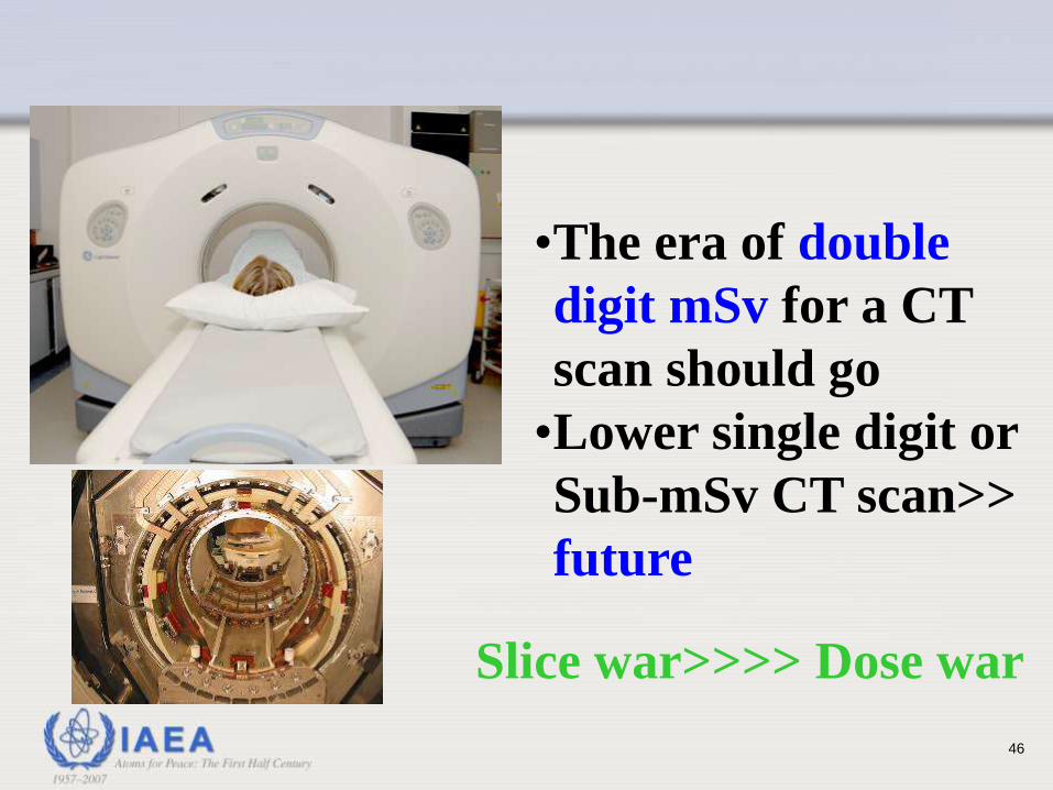
•The era of double
digit mSv for a CT
scan should go
•Lower single digit or
Sub-mSv CT scan>>
future
Slice war>>>> Dose war
46
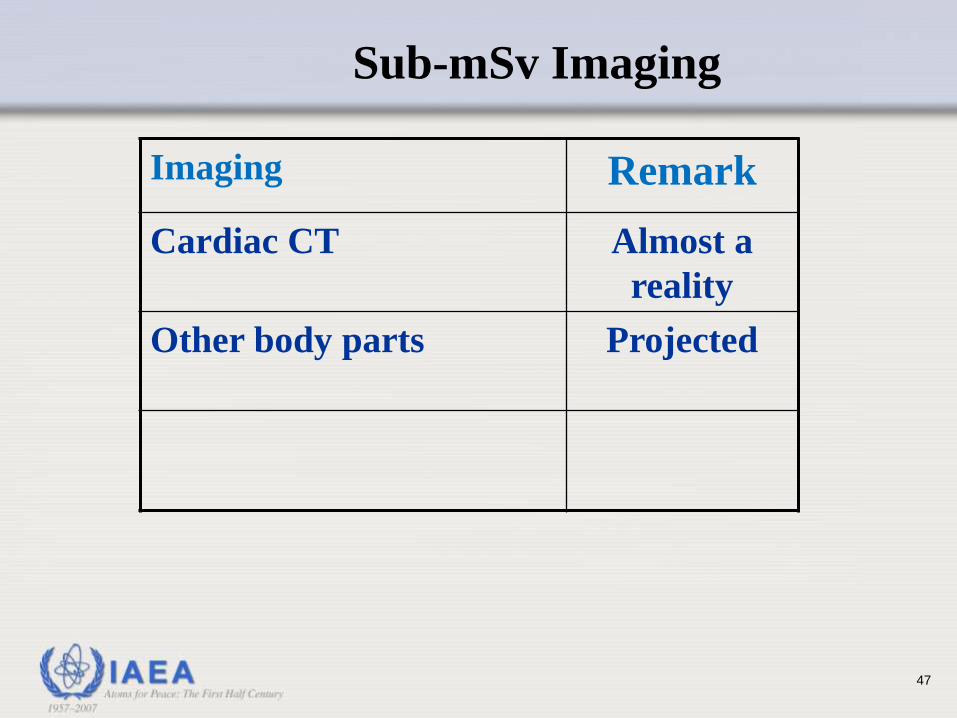
Imaging Remark
Cardiac CT Almost a
reality
Other body parts Projected
Sub-mSv Imaging
47
48
1998- ICRP
• CT usage increasing and likely to grow
• Lack of awareness among users on relatively
higher patient doses in CT
• Manufacturers not really concerned about
patient doses as hardly customers asked for it
• Most emphasis on faster and faster CT
scanners
49
Task Group of ICRP set up in 1999
• M.M. Rehani (Chairman)
• Members:
• G. Bongartz (Switzerland); S.J. Golding (UK);
L. Gordon (Sweden); W. Kalender (Germany);
T. Murakami (Japan); P. Shrimpton (UK)
• Corresponding members:
• R. Albrecht (USA) and K.Wei (China)
50
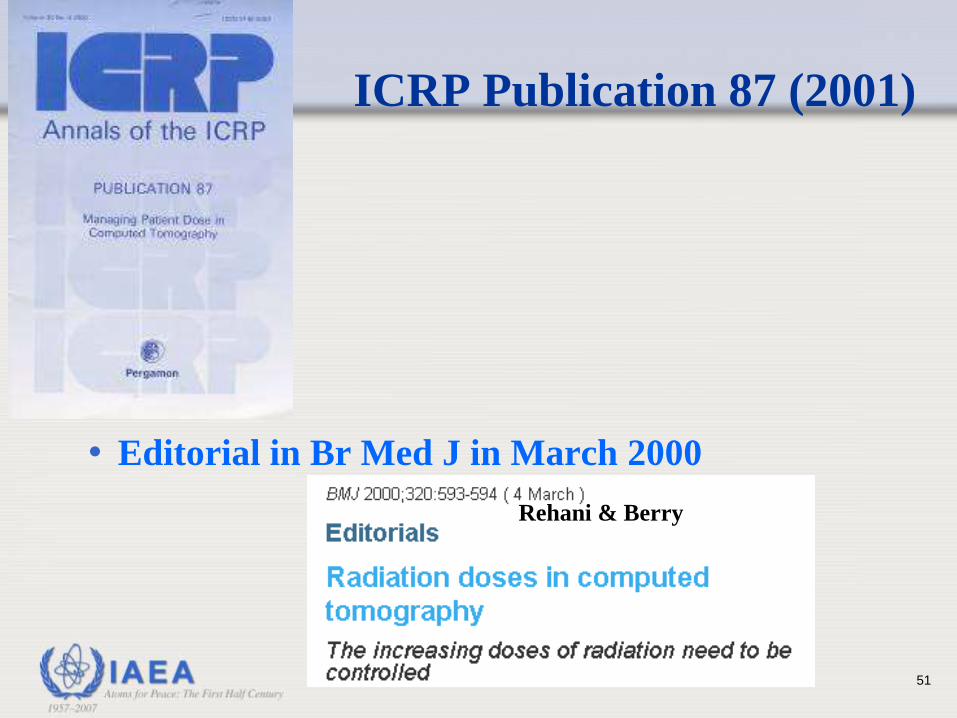
ICRP Publication 87 (2001)
• Editorial in Br Med J in March 2000
Rehani & Berry
51
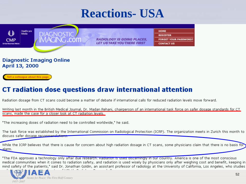
Reactions- USA
52
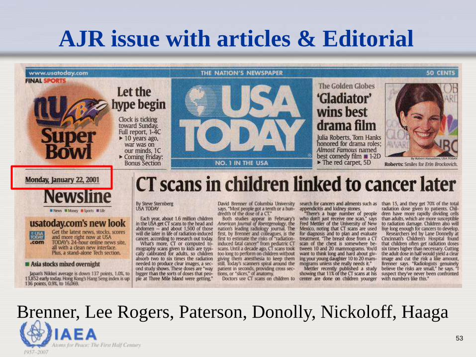
AJR issue with articles & Editorial
Brenner, Lee Rogers, Paterson, Donolly, Nickoloff, Haaga
53
CT
Turning point in the history
54
Good that it happened
55
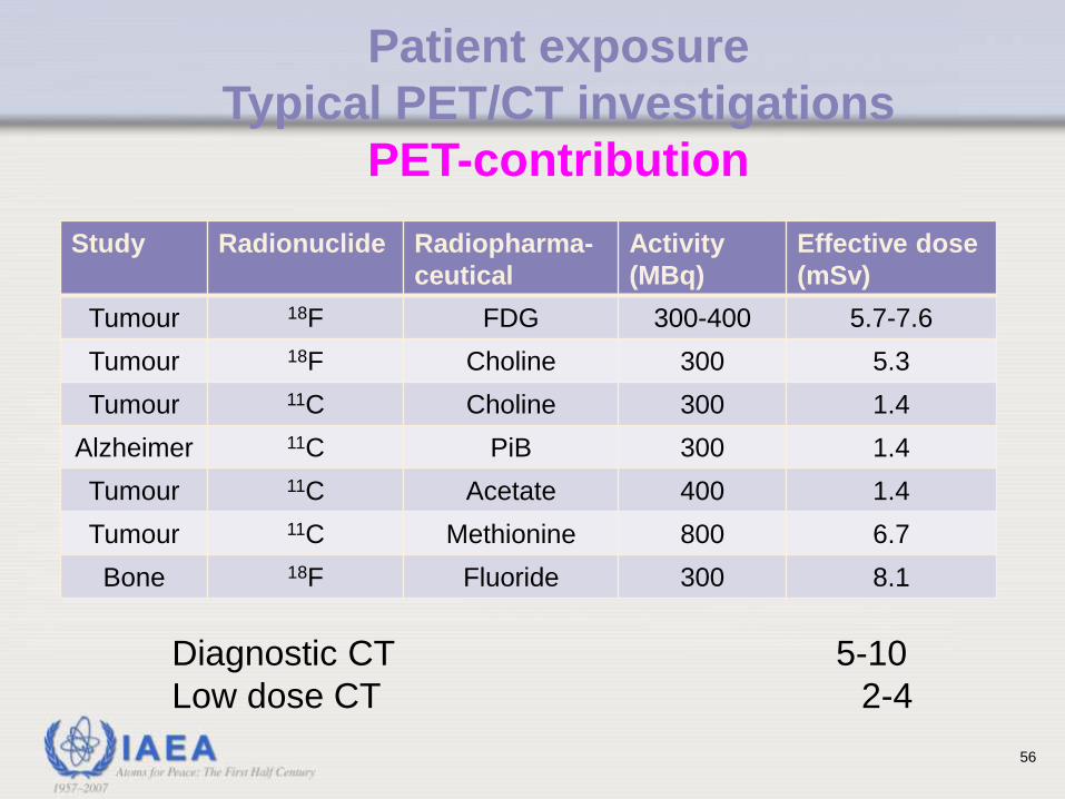
Study Radionuclide
Radiopharma-
ceutical
Activity
(MBq)
Effective dose
(mSv)
Tumour 18F FDG 300-400 5.7-7.6
Tumour 18F Choline 300 5.3
Tumour 11C Choline 300 1.4
Alzheimer 11C PiB 300 1.4
Tumour 11C Acetate 400 1.4
Tumour 11C Methionine 800 6.7
Bone 18F Fluoride 300 8.1
Patient exposure
Typical PET/CT investigations
PET-contribution
Diagnostic CT 5-10
Low dose CT 2-4
56
_
_
_
_
_
1
10
100
0.1
0.01
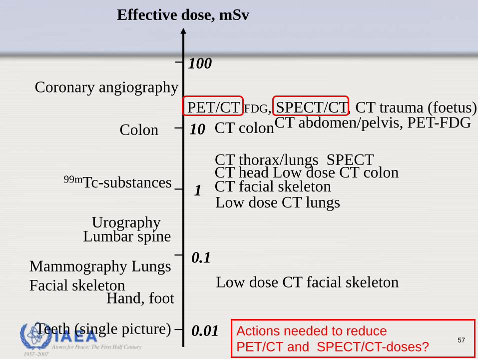
PET/CT FDG, SPECT/CT, CT trauma (foetus)
CT colon
CT thorax/lungs SPECT CT head Low dose CT colon CT facial skeleton Low dose CT lungs
Low dose CT facial skeleton
99mTc-substances
Colon
Coronary angiography
Urography Lumbar spine
Mammography Lungs
Facial skeleton Hand, foot
Teeth (single picture)
Effective dose, mSv
CT abdomen/pelvis, PET-FDG
Actions needed to reduce
PET/CT and SPECT/CT-doses? 57
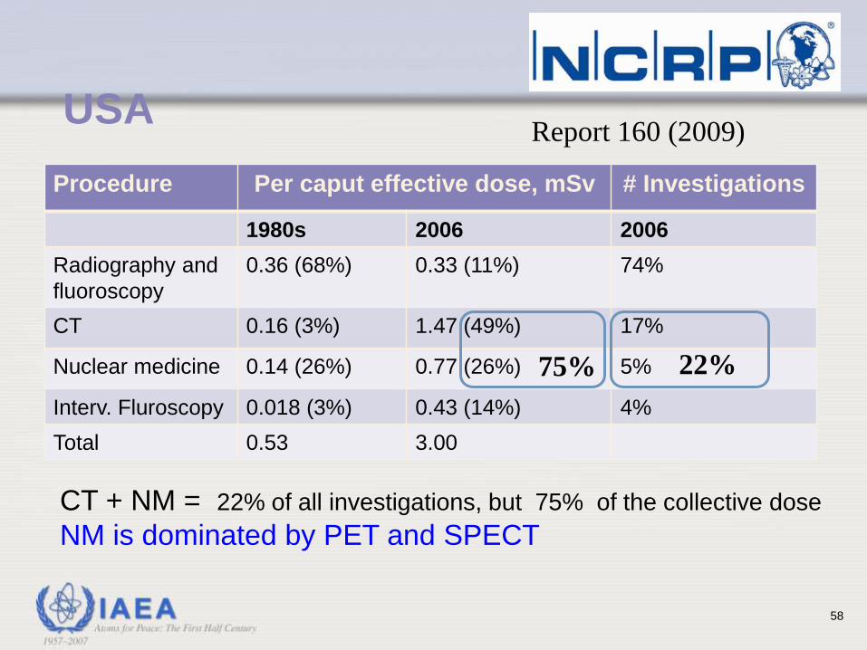
CT + NM = 22% of all investigations, but 75% of the collective dose
NM is dominated by PET and SPECT
USA Report 160 (2009)
Procedure Per caput effective dose, mSv # Investigations
1980s 2006 2006
Radiography and
fluoroscopy
0.36 (68%) 0.33 (11%) 74%
CT 0.16 (3%) 1.47 (49%) 17%
Nuclear medicine 0.14 (26%) 0.77 (26%) 5%
Interv. Fluroscopy 0.018 (3%) 0.43 (14%) 4%
Total 0.53 3.00
75% 22%
58
Dose reduction
• Technology Driven
• Operator Driven
• Who is going to drive industry: Professional
societies; Accidents & Media.
59
Recap
• Radiography: Safer for patient & staff
• Digital Radiography: Actions needed to make
it safer
• Diagnostic fluoroscopy: Safer for patient &
Staff
• Digital fluoroscopy: Actions needed
• Interventional procedures: Issues of staff (Eye
lens, hair loss) and patient protection (skin
injury, cancer risk) that cannot be ignored
60
Recap
• Nuclear Medicine: PET/CT, SPECT/CT
• Radiotherapy: Continued actions needed
• CT: Major and growing source but future
looks good
• >90% of patients and staff are safer than 50
years ago
• Smaller percentage at significant risks which
are new and challenging
61