Embed Size (px)
Citation preview
1
Johns Hopkins
Radiology and Radiological
Science
Annual Report
2016
2
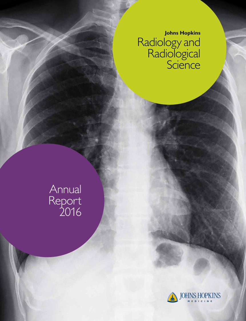
Faculty of the Department of Radiology and Radiological Science
1
FY16 was certainly a year of transitions for the Russell H. Morgan Department of Radiology and Radiological Science. With the departure of department director Jon Lewin, I was honored to have been chosen by the dean to assume the role of interim director. I feel very fortunate to have this opportunity to serve the department in this capacity. Our departmental motto for FY17 is “innovate, integrate and ignite.” Innovation has always been the cornerstone of what makes Johns Hopkins great. As a department, we continue to innovate and advance the field not only through our groundbreaking research, but also through our innovative approaches to resident education and to improving the quality and value of the care we deliver.
Integration is also increasingly crucial as we strive to integrate our research and patient care initiatives across departments, disciplines and hospitals. We are also integrating our residencies, as we plan for new, combined residencies in interventional radiology/diagnostic radiology and diagnostic radiology/molecular imaging.
This year, we are renewing our commitment to customer satisfaction, recognizing that referring physicians
as well as our patients are our “customers.” This requires a renewed focus on employee and faculty satisfaction as well. Engaged faculty and staff members result in satisfied patients and clinicians. To engage our faculty and staff members, we need to ignite our passion and compassion.
In this annual report, we have highlighted a sample of our accomplishments in all three parts of our tripartite mission of research, education and patient care. We are extremely proud of our department’s accomplishments, and we look forward to another outstanding and exciting year.
—Karen M. Horton, M.D.Professor and Interim Director
The Russell H. Morgan Department of Radiology and Radiological Science
Director’s Column
Karen M. Horton, M.D.
Radiology and Radiological Science | 1
2
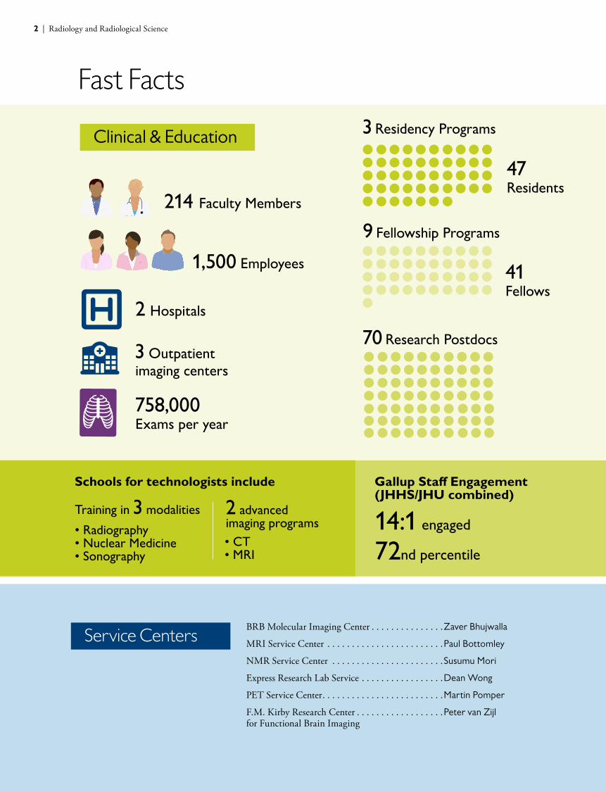
Clinical & Education
Service CentersBRB Molecular Imaging Center . . . . . . . . . . . . . . .Zaver Bhujwalla
MRI Service Center . . . . . . . . . . . . . . . . . . . . . . . .Paul Bottomley
NMR Service Center . . . . . . . . . . . . . . . . . . . . . . .Susumu Mori
Express Research Lab Service . . . . . . . . . . . . . . . . .Dean Wong
PET Service Center. . . . . . . . . . . . . . . . . . . . . . . . .Martin Pomper
F.M. Kirby Research Center . . . . . . . . . . . . . . . . . .Peter van Zijl for Functional Brain Imaging
1,500 Employees
2 Hospitals
3 Outpatient imaging centers
758,000 Exams per year
214 Faculty Members
47
Residents
3 Residency Programs
Gallup Staff Engagement (JHHS/JHU combined)
41
Fellows
9 Fellowship Programs
70 Research Postdocs
Schools for technologists include
14:1 engaged
72nd percentile
• Radiography• Nuclear Medicine• Sonography
• CT • MRI
Training in 3 modalities 2 advanced imaging programs
Fast Facts
2 | Radiology and Radiological Science
3
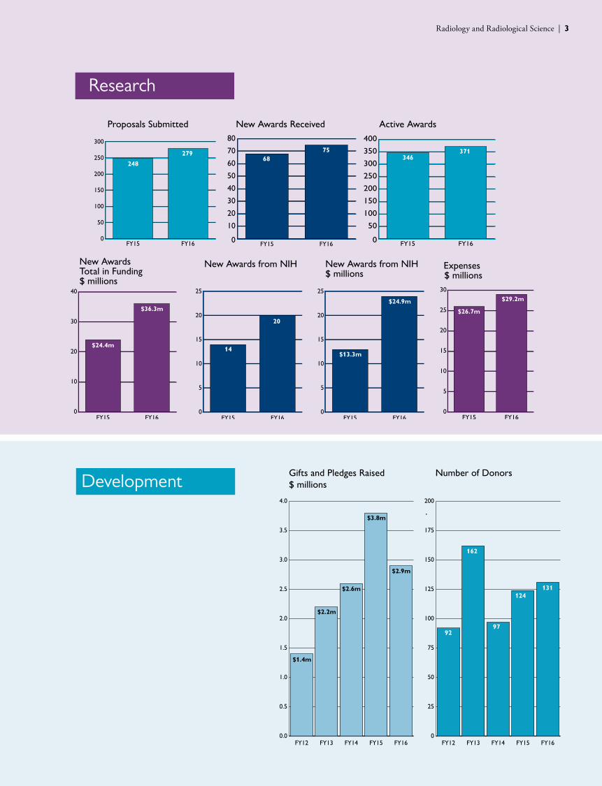
Research
Development
0
50
100
150
200
250
300
FY16FY15 0
10
20
30
40
50
60
70
80
FY16FY15
Proposals Submitted New Awards Received
0
50
100
150
200
250
300
350
400
FY16FY15
Active Awards
0
10
20
30
40
FY16FY150
5
10
15
20
25
FY16FY150
5
10
15
20
25
30
FY16FY15
New Awards Total in Funding$ millions
New Awards from NIH Expenses $ millions
0
5
10
15
20
25
FY16FY15
New Awards from NIH$ millions
248
$24.4m
$36.3m
$13.3m
$24.9m
$26.7m
$29.2m
14
20
27968
75346
371
0.0
0.5
1.0
1.5
2.0
2.5
3.0
3.5
4.0
FY16FY15FY14FY13FY120
25
50
75
100
125
150
175
200
FY16FY15FY14FY13FY12
Gifts and Pledges Raised$ millions
Number of Donors
$1.4m
$2.2m
$2.6m
$2.9m
9297
124131
162
$3.8m
Radiology and Radiological Science | 3
Breast Imaging
Quality and value have recently become buzzwords in medicine—patients and physicians alike want diagnoses and treatments that are not only more effective, efficient and safe but also cost less. It’s a vision that is a driving force for the Division of Breast Imaging, says division director Susan Harvey.“Our quality metrics are already outperforming national standards quite dramatically,” she says.
An example of this is the percentage of women who are asked to return to Johns Hopkins for additional views following a routine screening mammogram. The national standard is 10 percent, but at Johns Hopkins, it’s only six percent. The positive predictive value—the percentage of those women asked for additional views who actually have cancer—is significantly better as well. The national standard is 32 percent, but at Johns Hopkins, says Harvey, it is 52 percent.
The reason for these statistics is highly specialized staff members, Harvey says. Only breast imaging specialists, rather than general radiologists, interpret these breast imaging exams. Besides giving patients peace of mind and saving them from painful and potentially unnecessary biopsies, it is obvious that this better performance is more cost- effective for patients and their insurance companies, she adds. Because patients require fewer scans and fewer resulting procedures, the cost of care is significantly lower.
Harvey and her team are exploring an array of other options to improve quality and value. They’re currently actively
investigating a new protocol for the breast MRI scans that are often used as a screening tool for patients at particularly high risk of breast cancer, including those with genetic mutations known to predispose individuals to breast cancer or those with a strong family history of the disease. The typical scanning sequence used nationally and internationally involves at least 24 minutes of patients lying prone on an uncomfortable breast coil. The researchers’ novel protocol shortens this time to just three minutes. If ongoing research continues to show that this abbreviated protocol is as effective at revealing breast cancer as the longer one, it could change the standard of care around the world, Harvey says.
The division also uses almost entirely three-dimensional imaging, creating pictures much like a CT scan to search for potential cancers. But, unlike CT scans, these three-dimensional images are reconstructed from two-dimensional data, requiring the radiation of a single, low-dose X-ray rather than the substantially larger amount of a real CT scan. “We detect 40 percent more cancers than with a typical two-dimensional image,” Harvey says. “It really is a better tool.”
“ We detect 40 percent more cancers than with a typical two-dimensional image; it really is a better tool.”
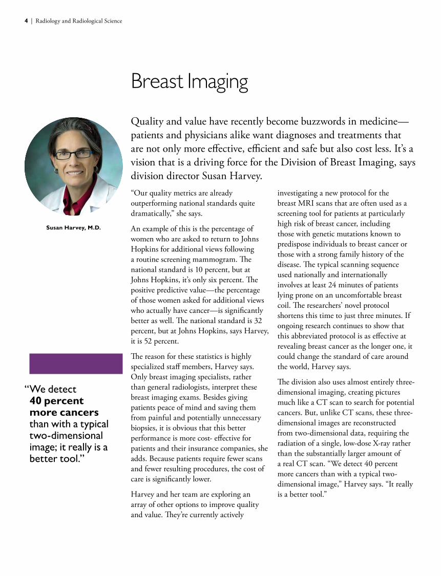
Susan Harvey, M.D.
4 | Radiology and Radiological Science
5
Beyond screening, the division’s work may eventually lead to better quality and value for breast cancer treatment as well, she adds. Harvey and her colleagues are currently investigating new treatments using cryoablation, in which extreme cold freezes tumors away. Although cryoablation has previously been used for lung, liver and renal cancers, it hasn’t been fully explored for breast cancer.
“This can be preformed in a clinic with ultrasound guidance, and women leave with an adhesive bandage. It’s hugely less expensive than surgery,” Harvey says. “My vision is that this will eventually be a preferred treatment.”
Together, she says, the division’s work will eventually change the course for the millions of women who are screened or treated for breast cancer annually, saving lives. n
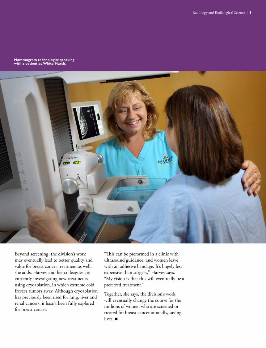
Mammogram technologist speaking with a patient at White Marsh.
Radiology and Radiological Science | 5

Cancer Imaging Research
The Division of Cancer Imaging Research is relatively small compared to other divisions in the Department of Radiology and Radiological Science, and relatively new, says division director Zaver Bhujwalla. With just eight full-time tenure-track faculty members, including Dimitri Artemov, Kristine Glunde, Michael Jacobs, Arvind Pathak, Marie-France Penet, Venu Raman and Farhad Vesuna, the 40-member division that includes research associates, fellows, technological staff and students was founded in 2012. The goal of the division is to promote the use of preclinical and clinical multimodal imaging applications to understand and effectively treat cancer. The division also operates the Miller Research Building (MRB) Molecular Imaging Service Center, supporting molecular imaging research across Johns Hopkins, and has held the NIH-funded P50 JHU ICMIC Program from 2002.
Division faculty members are highly collaborative, and pursue a wide spectrum of multimodality molecular and functional imaging research projects that span basic research to preclinical and clinical applications. Cancers investigated include breast, prostate, pancreatic, brain and ovarian. Research focus areas include understanding and targeting the tumor microenvironment and cancer-induced cachexia, and developing theranostic imaging for precision medicine. Novel targets and pathways, such as DDX3, COX-2, enzymes in choline metabolism and hypoxia targeting, are being investigated with molecular and functional imaging to understand cancer and develop effective
image-guided treatments. Novel advances are being made in mass spectrometry imaging, as well as microscopic imaging of tumor vasculature. Clinical applications include the development of novel imaging-based biomarkers to detect and stage cancer, and monitor response to treatment.
A major focus of the division is in nanotechnology to develop nanoparticles as theranostic tools. Nanoparticles have been developed to deliver siRNA, cDNA and a prodrug enzyme to target tumors under image guidance. Click chemistry-based nanoparticles are being developed for tumor-specific treatment. More recently, biomimetic nanoparticles that are coated in cancer cell membranes are being developed as decoys to disrupt stromal cell-cancer cell interactions, and to induce an immune response. Antibody-based phototherapy approaches are also being developed to specifically target cancer cells and the tumor microenvironment. “With imaging and nanotechnology, we can fight cancer with precision,” Bhujwalla says. “I think this is the core of what can be achieved with personalized medicine.” n
“ With imaging and nanotechnology, we can fight cancer with precision.”
Zaver M. Bhujwalla, M.Sc., Ph.D.
6 | Radiology and Radiological Science
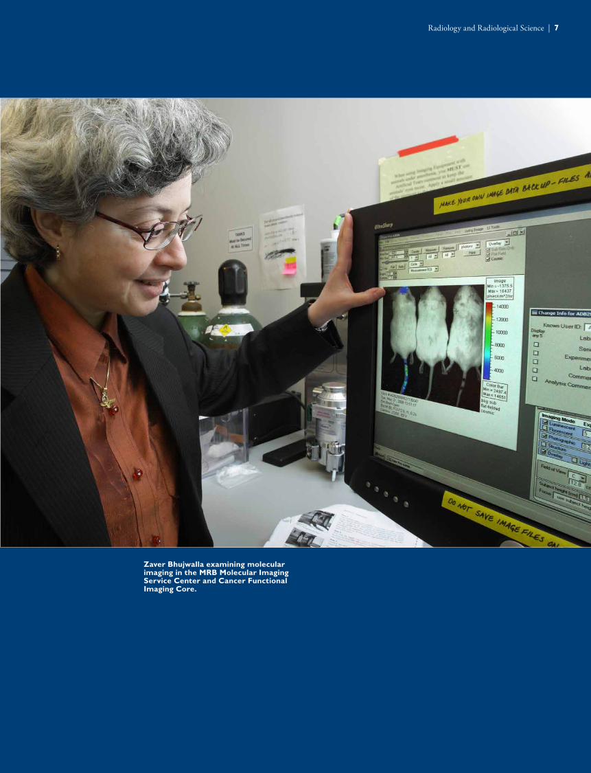
Zaver Bhujwalla examining molecular imaging in the MRB Molecular Imaging Service Center and Cancer Functional Imaging Core.
7
Radiology and Radiological Science | 7
8
Diagnostic Imaging
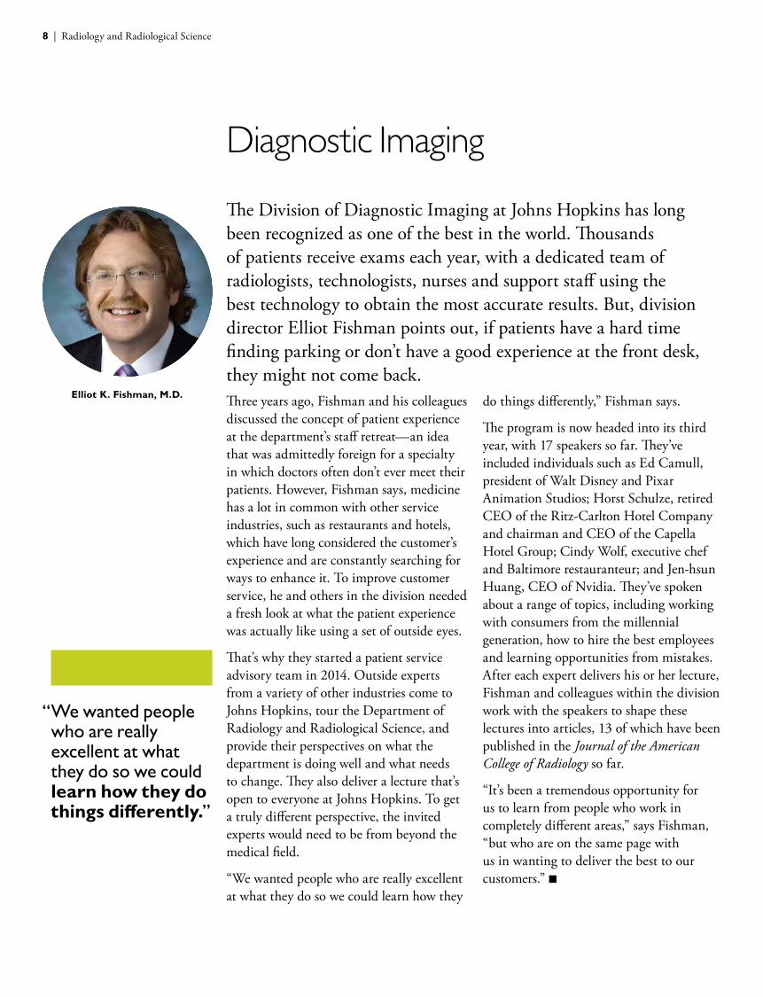
The Division of Diagnostic Imaging at Johns Hopkins has long been recognized as one of the best in the world. Thousands of patients receive exams each year, with a dedicated team of radiologists, technologists, nurses and support staff using the best technology to obtain the most accurate results. But, division director Elliot Fishman points out, if patients have a hard time finding parking or don’t have a good experience at the front desk, they might not come back.Three years ago, Fishman and his colleagues discussed the concept of patient experience at the department’s staff retreat—an idea that was admittedly foreign for a specialty in which doctors often don’t ever meet their patients. However, Fishman says, medicine has a lot in common with other service industries, such as restaurants and hotels, which have long considered the customer’s experience and are constantly searching for ways to enhance it. To improve customer service, he and others in the division needed a fresh look at what the patient experience was actually like using a set of outside eyes.
That’s why they started a patient service advisory team in 2014. Outside experts from a variety of other industries come to Johns Hopkins, tour the Department of Radiology and Radiological Science, and provide their perspectives on what the department is doing well and what needs to change. They also deliver a lecture that’s open to everyone at Johns Hopkins. To get a truly different perspective, the invited experts would need to be from beyond the medical field.
“We wanted people who are really excellent at what they do so we could learn how they
do things differently,” Fishman says.
The program is now headed into its third year, with 17 speakers so far. They’ve included individuals such as Ed Camull, president of Walt Disney and Pixar Animation Studios; Horst Schulze, retired CEO of the Ritz-Carlton Hotel Company and chairman and CEO of the Capella Hotel Group; Cindy Wolf, executive chef and Baltimore restauranteur; and Jen-hsun Huang, CEO of Nvidia. They’ve spoken about a range of topics, including working with consumers from the millennial generation, how to hire the best employees and learning opportunities from mistakes. After each expert delivers his or her lecture, Fishman and colleagues within the division work with the speakers to shape these lectures into articles, 13 of which have been published in the Journal of the American College of Radiology so far.
“It’s been a tremendous opportunity for us to learn from people who work in completely different areas,” says Fishman, “but who are on the same page with us in wanting to deliver the best to our customers.” n
“ We wanted people who are really excellent at what they do so we could learn how they do things differently.”
Elliot K. Fishman, M.D.
8 | Radiology and Radiological Science
9
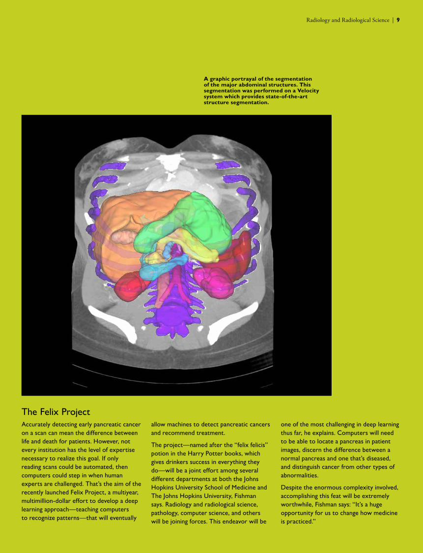
The Felix ProjectAccurately detecting early pancreatic cancer on a scan can mean the difference between life and death for patients. However, not every institution has the level of expertise necessary to realize this goal. If only reading scans could be automated, then computers could step in when human experts are challenged. That’s the aim of the recently launched Felix Project, a multiyear, multimillion-dollar effort to develop a deep learning approach—teaching computers to recognize patterns—that will eventually
allow machines to detect pancreatic cancers and recommend treatment.
The project—named after the “felix felicis” potion in the Harry Potter books, which gives drinkers success in everything they do—will be a joint effort among several different departments at both the Johns Hopkins University School of Medicine and The Johns Hopkins University, Fishman says. Radiology and radiological science, pathology, computer science, and others will be joining forces. This endeavor will be
one of the most challenging in deep learning thus far, he explains. Computers will need to be able to locate a pancreas in patient images, discern the difference between a normal pancreas and one that’s diseased, and distinguish cancer from other types of abnormalities.
Despite the enormous complexity involved, accomplishing this feat will be extremely worthwhile, Fishman says: “It’s a huge opportunity for us to change how medicine is practiced.”
A graphic portrayal of the segmentation of the major abdominal structures. This segmentation was performed on a Velocity system which provides state-of-the-art structure segmentation.
Radiology and Radiological Science | 9
10
Interventional Radiology
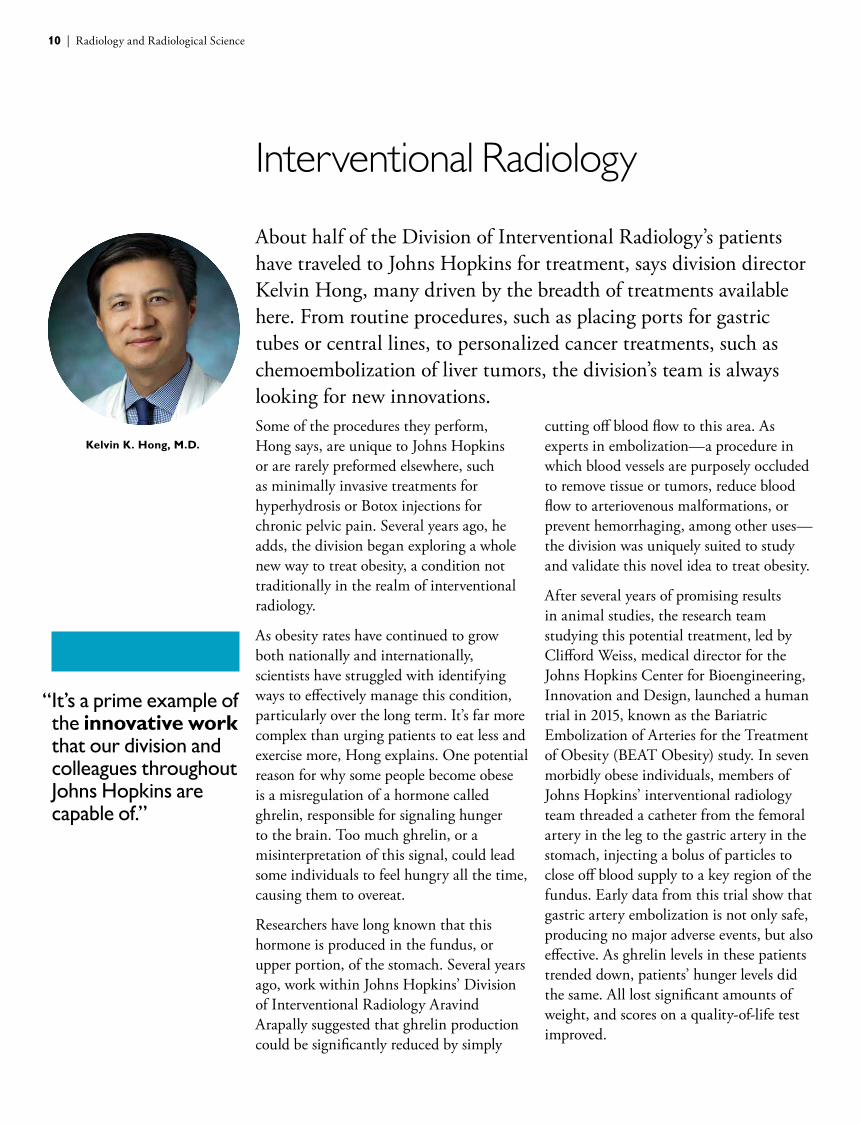
About half of the Division of Interventional Radiology’s patients have traveled to Johns Hopkins for treatment, says division director Kelvin Hong, many driven by the breadth of treatments available here. From routine procedures, such as placing ports for gastric tubes or central lines, to personalized cancer treatments, such as chemoembolization of liver tumors, the division’s team is always looking for new innovations. Some of the procedures they perform, Hong says, are unique to Johns Hopkins or are rarely preformed elsewhere, such as minimally invasive treatments for hyperhydrosis or Botox injections for chronic pelvic pain. Several years ago, he adds, the division began exploring a whole new way to treat obesity, a condition not traditionally in the realm of interventional radiology.
As obesity rates have continued to grow both nationally and internationally, scientists have struggled with identifying ways to effectively manage this condition, particularly over the long term. It’s far more complex than urging patients to eat less and exercise more, Hong explains. One potential reason for why some people become obese is a misregulation of a hormone called ghrelin, responsible for signaling hunger to the brain. Too much ghrelin, or a misinterpretation of this signal, could lead some individuals to feel hungry all the time, causing them to overeat.
Researchers have long known that this hormone is produced in the fundus, or upper portion, of the stomach. Several years ago, work within Johns Hopkins’ Division of Interventional Radiology Aravind Arapally suggested that ghrelin production could be significantly reduced by simply
cutting off blood flow to this area. As experts in embolization—a procedure in which blood vessels are purposely occluded to remove tissue or tumors, reduce blood flow to arteriovenous malformations, or prevent hemorrhaging, among other uses—the division was uniquely suited to study and validate this novel idea to treat obesity.
After several years of promising results in animal studies, the research team studying this potential treatment, led by Clifford Weiss, medical director for the Johns Hopkins Center for Bioengineering, Innovation and Design, launched a human trial in 2015, known as the Bariatric Embolization of Arteries for the Treatment of Obesity (BEAT Obesity) study. In seven morbidly obese individuals, members of Johns Hopkins’ interventional radiology team threaded a catheter from the femoral artery in the leg to the gastric artery in the stomach, injecting a bolus of particles to close off blood supply to a key region of the fundus. Early data from this trial show that gastric artery embolization is not only safe, producing no major adverse events, but also effective. As ghrelin levels in these patients trended down, patients’ hunger levels did the same. All lost significant amounts of weight, and scores on a quality-of-life test improved.
“ It’s a prime example of the innovative work that our division and colleagues throughout Johns Hopkins are capable of.”
Kelvin K. Hong, M.D.
10 | Radiology and Radiological Science
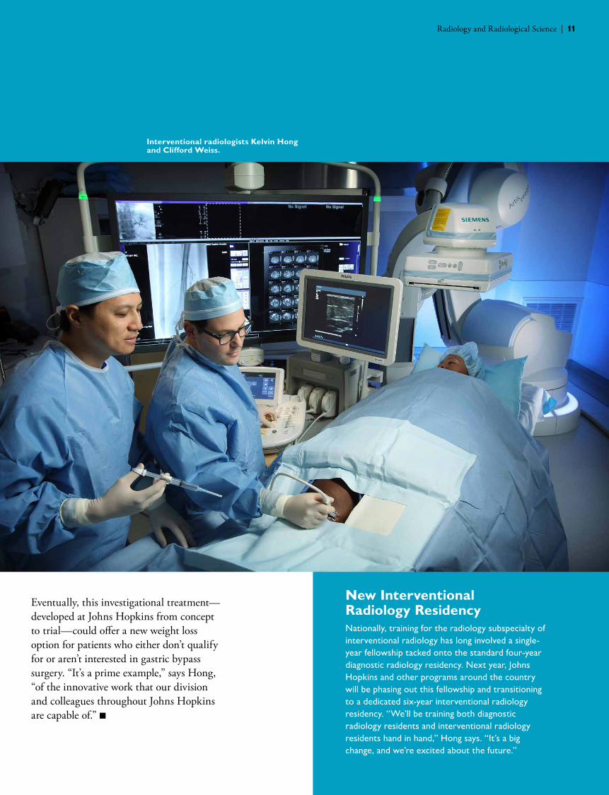
Interventional radiologists Kelvin Hong and Clifford Weiss.
11
New Interventional Radiology ResidencyNationally, training for the radiology subspecialty of interventional radiology has long involved a single-year fellowship tacked onto the standard four-year diagnostic radiology residency. Next year, Johns Hopkins and other programs around the country will be phasing out this fellowship and transitioning to a dedicated six-year interventional radiology residency. “We’ll be training both diagnostic radiology residents and interventional radiology residents hand in hand,” Hong says. “It’s a big change, and we’re excited about the future.”
Eventually, this investigational treatment—developed at Johns Hopkins from concept to trial—could offer a new weight loss option for patients who either don’t qualify for or aren’t interested in gastric bypass surgery. “It’s a prime example,” says Hong, “of the innovative work that our division and colleagues throughout Johns Hopkins are capable of.” n
Radiology and Radiological Science | 11
Medical Imaging Physics
Within medicine, radiologists have the reputation of being the “technology people,” says Division of Medical Imaging Physics director Ben Tsui. “But within radiology itself,” he adds, “medical physicists and engineers are the technology people.”The division, he explains, is composed mostly of engineers, physicists and computer scientists, who work with the goal of advancing biomedical imaging technologies that revolutionize the understanding, diagnosis and treatment of diseases for improved patient care. Since much of this work involves creating software to help enhance and expand the application of imaging technologies—including X-ray, nuclear medicine, CT, positron emission tomography (PET), single-photon emission computed tomography (SPECT) and ultrasound—this focus is a prime opportunity to interact with companies who make the devices used in these technologies. Indeed, Tsui says, many of the projects that he and his colleagues work on have been licensed to industry or have been spun off into their own companies.
For example, 30 years ago, Tsui started a side project to help him and other investigators in the field develop new methods of biomedical imaging and nuclear medicine—a software human phantom based on data gathered from a National Library of Medicine undertaking that supplied detailed scans and other information developed from a healthy cadaver. Because much of Tsui’s work at the time focused on the heart, his original phantom featured just the chest and torso. But, gradually, Tsui incorporated other parts of the body, piece by piece, until the model covered an entire human from head to toe. The simulation eventually included the motion of the heart and lungs. Currently, it incorporates
over 10,000 individual elements to provide a realistic replication of the entire body for biomedical imaging and radiation dosimetry. This phantom software is now registered with the Johns Hopkins Technology Ventures, and more than 400 users have currently purchased licenses, Tsui says.
Eric Frey, working with students and other colleagues in the division, including Tsui, has developed a 3-D SPECT image reconstruction software package for improving diagnosis of heart diseases and various cancers. That software has been licensed to GE Healthcare, and over 2,000 copies have been sold worldwide. More recently Frey, who also has an appointment in the Johns Hopkins Kimmel Cancer Center, has adapted this software for use in a project to improve the dosing of radiopharmaceutical therapeutics— drugs that carry radiation to fight cancer. Knowing exactly how much of the administered dose has ended up in normal tissues versus tumors has been a challenge for this field. The software provides significantly improved quantitative SPECT images that allow doctors to better estimate how much of the dose arrived at the target versus normal tissues, allowing them to better tailor the next dose and a patient’s overall treatment. He and a colleague in the Division of Nuclear Medicine have started a company, Radiopharmaceutical Imaging and Dosimetry LLC, to commercialize this and related technology.
“ Our work is largely about improving radiological images, which helps doctors and patients alike.”
Benjamin M.W. Tsui, Ph.D.
12 | Radiology and Radiological Science
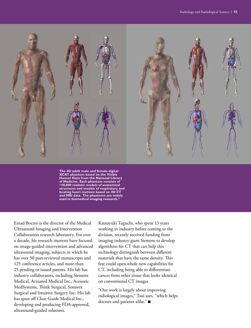
The 4D adult male and female digital XCAT phantom based on the Visible Human Data from the National Library of Medicine. Each phantom consists of ~10,000 realistic models of anatomical structures and models of respiratory and beating heart motions based on 4D CT and MRI data. The phantoms are widely used in biomedical imaging research.”
13
Emad Boctor is the director of the Medical Ultrasound Imaging and Intervention Collaboration research laboratory. For over a decade, his research interests have focused on image-guided intervention and advanced ultrasound imaging, subjects in which he has over 50 peer-reviewed manuscripts and 125 conference articles, and more than 25 pending or issued patents. His lab has industry collaborators, including Siemens Medical, Actuated Medical Inc., Acoustic MedSystems, Think Surgical, Sonavex Surgical and Intuitive Surgery Inc. His lab has spun off Clear Guide Medical Inc., developing and producing FDA-approved, ultrasound-guided solutions.
Katsuyuki Taguchi, who spent 13 years working in industry before coming to the division, recently received funding from imaging industry giant Siemens to develop algorithms for CT that can help this technology distinguish between different materials that have the same density. This feat could open whole new capabilities for CT, including being able to differentiate cancer from other tissue that looks identical on conventional CT images.
“Our work is largely about improving radiological images,” Tsui says, “which helps doctors and patients alike.” n
Radiology and Radiological Science | 13
Magnetic Resonance Research
Since publication of the first magnetic resonance (MR) images in 1973, this technology and its applications have made enormous advances. MR is now one of the major workhorses of diagnostic imaging, and it is arguably at the leading edge of imaging research as well. Nearly every hospital and community imaging center in the country has one or more scanners of their own, and the magnets and associated electronics have become progressively more powerful over the years, producing even better images, even faster. But even after nearly half a century of innovation, says Paul Bottomley, director of the Division of Magnetic Resonance Research, this technique still has room for improvement. He and the 28 faculty members in the MR Research division focus on developing novel MR methods and applying them in biomedicine. The team is organized into four sections, each with a different main focus: cardiac and interventional MR, cellular imaging, neuroscience; and neuroanatomical MR.
Bottomley himself typically does research in the cardiac and interventional MR group, led by Dara Kraitchman. One project that he is working on is the development of miniaturized MR detector coil technology, mounted on guidewire cables for producing endoscopic MRI at 50 to 100 µm resolution. The team is using this technology to develop rapid imaging of the insides of diseased blood vessels. Bottomley is also director of the MRI Service Center, which currently has two 3 Tesla and one 1.5 Tesla whole-body MRI scanners to perform research at Johns Hopkins.
Kraitchman, on the other hand, is working on MR-guided interventions for treating obesity with colleagues in the Division of
Interventional Radiology. Kraitchman is a licensed veterinarian and has established the Center for Image-Guided Animal Therapy at Johns Hopkins, which provides imaging services for pets.
The cellular imaging group is led by Jeff Bulte. One focus of this section is the development of techniques for labeling cells that render them MR-detectable—a function that may prove pivotal for monitoring the delivery and efficacy of future cellular therapies. The team is also working on methods to encapsulate cells in protective shells that preserve their viability and enable their visualization by MR, X-ray or ultrasound imaging.
A heavy focus of faculty members in the neuroscience group, led by Peter van Zijl, is a technique called chemical exchange saturation transfer, which could greatly expand the use of contrast agents that can be used for MRI to include chemicals that are already present in the body. Peter is also director of the F.M. Kirby Research Center for Functional Brain Imaging at the Kennedy Krieger Institute, which is a National Center for Biomedical Technology Research supported by the National
“ Our job isn’t developing techniques but their successful application to people and science.”
Paul A. Bottomley, Ph.D.
14 | Radiology and Radiological Science
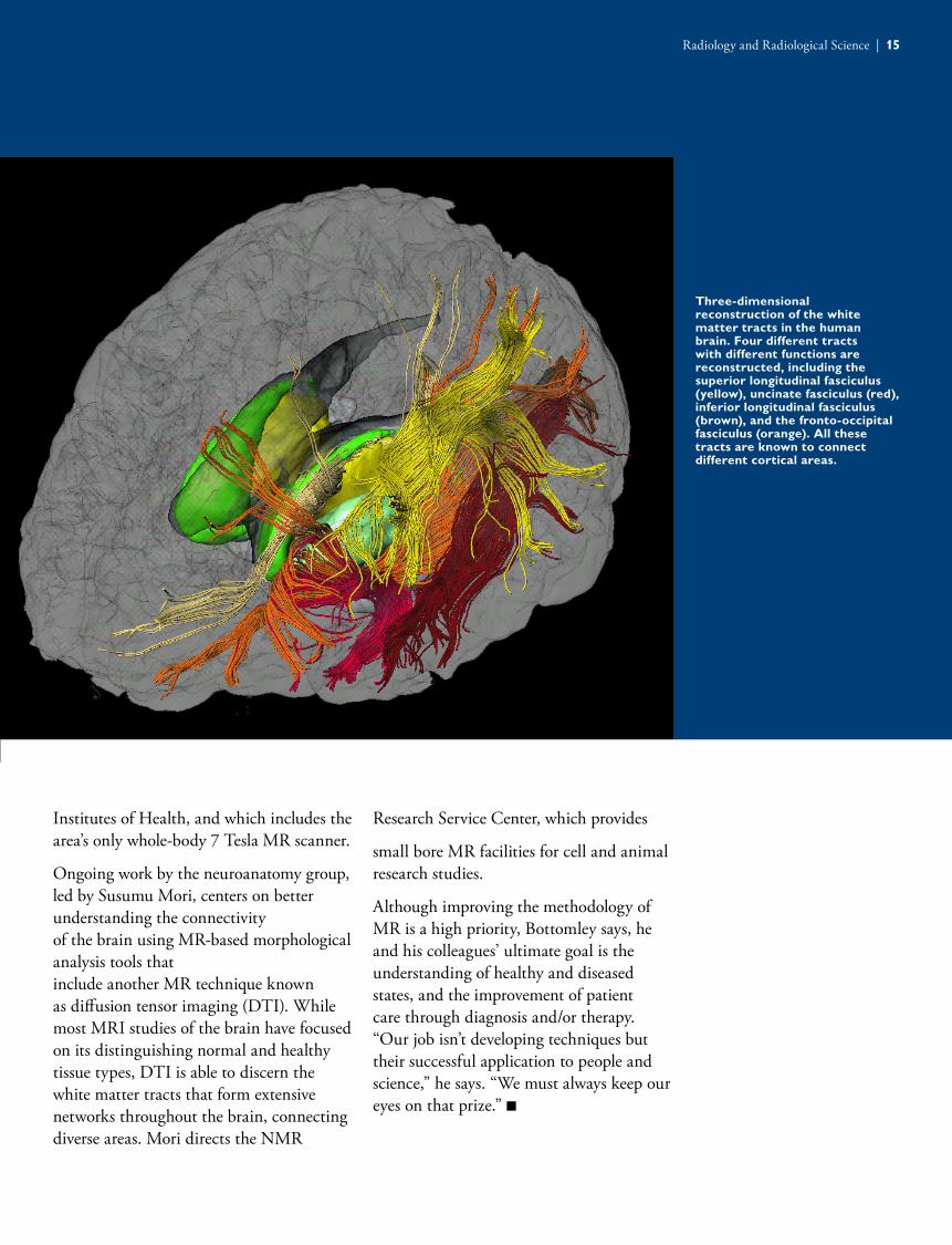
Three-dimensional reconstruction of the white matter tracts in the human brain. Four different tracts with different functions are reconstructed, including the superior longitudinal fasciculus (yellow), uncinate fasciculus (red), inferior longitudinal fasciculus (brown), and the fronto-occipital fasciculus (orange). All these tracts are known to connect different cortical areas.
15
Institutes of Health, and which includes the area’s only whole-body 7 Tesla MR scanner.
Ongoing work by the neuroanatomy group, led by Susumu Mori, centers on better understanding the connectivity of the brain using MR-based morphological analysis tools that include another MR technique known as diffusion tensor imaging (DTI). While most MRI studies of the brain have focused on its distinguishing normal and healthy tissue types, DTI is able to discern the white matter tracts that form extensive networks throughout the brain, connecting diverse areas. Mori directs the NMR
Research Service Center, which provides
small bore MR facilities for cell and animal research studies.
Although improving the methodology of MR is a high priority, Bottomley says, he and his colleagues’ ultimate goal is the understanding of healthy and diseased states, and the improvement of patient care through diagnosis and/or therapy. “Our job isn’t developing techniques but their successful application to people and science,” he says. “We must always keep our eyes on that prize.” n
Radiology and Radiological Science | 15
16
Neuroradiology
The Division of Neuroradiology has long had an outstanding reputation within the medical community for its strong clinical work—recognition that’s continued to grow as the practice has expanded beyond the main Johns Hopkins Hospital campus to practices in White Marsh, Green Spring Station and, most recently, Columbia. Referring physicians and patients alike know about the comprehensive and compassionate care that Johns Hopkins neuroradiologists deliver, offering a variety of diagnostic and interventional techniques. But behind the scenes, the division has a thriving research group as well, devoted to gaining better insight into the basic workings of the brain—knowledge that could eventually help clinicians deliver even better care.
“We have the ability to interrogate the brain in high-resolution ways that lead to a better understanding of how it works electrically, hemodynamically and chemically,” says division director David Yousem.
This research group includes a strong functional MRI (f-MRI) team, many of whom are doing cortical mapping—relating different structures in the brain to their functions. Some use task-based f-MRI, a test in which scientists look for what areas of the brain are active when individuals lying in the MRI scanner perform set actions. Recently, however, researchers within the division have been exploring a whole new way of interrogating the brain, probing function and looking for connections between different regions without the need for individuals to do any tasks—or even be awake. Such resting-
state f-MRI could eventually help doctors predict the progression of dementia, recovery from a coma or reveal the secrets of how brain networks develop in babies.
This resting-state f-MRI work mapping gray matter, led by Haris Sair, can be combined with another technology called diffusion tensor imaging that allows researchers to see the white matter tracts that connect gray matter regions of the brain. The brain is constantly active, explains Sair, even at rest. Different regions of the gray matter are continually firing. By looking for regions firing synchronously, along with mapping the white matter tracts, Sair and his colleagues are studying vast networks that stretch across the brain, connecting regions that previously seemed disparate and unrelated.
Unlike traditional f-MRI, Sair says, individuals in the scanner don’t need to be able to understand or follow instructions, or even be conscious. That opens this technique to people who are comatose, in the later stages of dementia, don’t speak the same language as the f-MRI technicians or are neonates. In addition, resting-state f-fMRI gives much more information compared to task-based f-MRI, allowing researchers to view the brain in a whole new way.
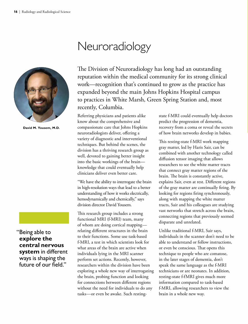
“ Being able to explore the central nervous system in different ways is shaping the future of our field.”
David M. Yousem, M.D.
16 | Radiology and Radiological Science
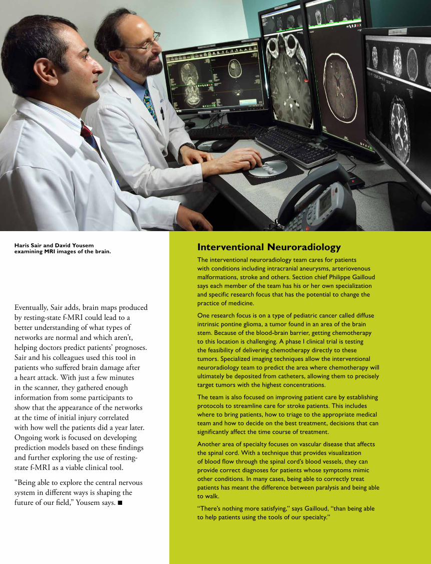
Haris Sair and David Yousem examining MRI images of the brain.
17
Interventional NeuroradiologyThe interventional neuroradiology team cares for patients with conditions including intracranial aneurysms, arteriovenous malformations, stroke and others. Section chief Philippe Gailloud says each member of the team has his or her own specialization and specific research focus that has the potential to change the practice of medicine.
One research focus is on a type of pediatric cancer called diffuse intrinsic pontine glioma, a tumor found in an area of the brain stem. Because of the blood-brain barrier, getting chemotherapy to this location is challenging. A phase I clinical trial is testing the feasibility of delivering chemotherapy directly to these tumors. Specialized imaging techniques allow the interventional neuroradiology team to predict the area where chemotherapy will ultimately be deposited from catheters, allowing them to precisely target tumors with the highest concentrations.
The team is also focused on improving patient care by establishing protocols to streamline care for stroke patients. This includes where to bring patients, how to triage to the appropriate medical team and how to decide on the best treatment, decisions that can significantly affect the time course of treatment.
Another area of specialty focuses on vascular disease that affects the spinal cord. With a technique that provides visualization of blood flow through the spinal cord’s blood vessels, they can provide correct diagnoses for patients whose symptoms mimic other conditions. In many cases, being able to correctly treat patients has meant the difference between paralysis and being able to walk.
“There’s nothing more satisfying,” says Gailloud, “than being able to help patients using the tools of our specialty.”
Eventually, Sair adds, brain maps produced by resting-state f-MRI could lead to a better understanding of what types of networks are normal and which aren’t, helping doctors predict patients’ prognoses. Sair and his colleagues used this tool in patients who suffered brain damage after a heart attack. With just a few minutes in the scanner, they gathered enough information from some participants to show that the appearance of the networks at the time of initial injury correlated with how well the patients did a year later. Ongoing work is focused on developing prediction models based on these findings and further exploring the use of resting-state f-MRI as a viable clinical tool.
“Being able to explore the central nervous system in different ways is shaping the future of our field,” Yousem says. n
18
Nuclear Medicine and Molecular Imaging
“Bench to bedside” is one of the central tenets of modern academic medicine—the idea that scientists can develop research findings from the laboratory that can be converted into novel ways to treat patients. It is a cornerstone of the Division of Nuclear Medicine and Molecular Imaging, says division director Martin Pomper. “We’re very innovative and discovery-based,” Pomper says. “We like to take basic discoveries all the way through to clinical trials.”
His division is uniquely suited for such translational research due to the nature of the materials that they work with: radiotracers and radiopharmaceuticals, compounds that mark cells and tissues or treat illnesses—or sometimes both—using radioactivity. Unlike conventional pharmaceuticals, the timeline from getting these compounds from the lab to the clinic is relatively short, Pomper explains.
For example, he and his colleagues within the Johns Hopkins University School of Medicine have taken advantage of this trait to produce numerous useful compounds over the years, with many now licensed to outside companies. For example, they’ve developed a compound called DCFPyL which has the ability to latch onto cells with a surface protein called prostate-specific membrane antigen (PSMA), which prostate cancer cells tend to express in high concentrations. Because it carries a radioactive label, this compound allows researchers to get sharp images of prostate cancers, even very small tumors.
He and his colleagues aren’t the only ones to see the utility of this tool. Progenics
Pharmaceuticals, based in New York City, recently licensed this compound. Another compound, which Pomper and his colleagues also crafted to target PSMA, carries a cancer-killing form of radioactivity that not only allows them to visualize prostate cancers, but also, fight them at the same time. That compound was recently licensed to the European pharmaceutical company Advanced Accelerator Applications.
Another innovation within the division includes a new method to distinguish benign from malignant masses within the kidney. By re-purposing sestamibi, an existing clinical radiotracer, Som Javadi and Steven Rowe have developed an immediately available approach to advise the referring physicians who are frequently confronted with this diagnostic dilemma.
Lilja Solnes continues to innovate in her new role as director of the divisional residency and fellowship programs – by building the first combined nuclear medicine/radiology residency program available on the Electronic Residency Application Service (ERAS). Notably, the Johns Hopkins Nuclear Medicine and Molecular Imaging residency program was ranked first in the United States in 2016 by Doximity.com.
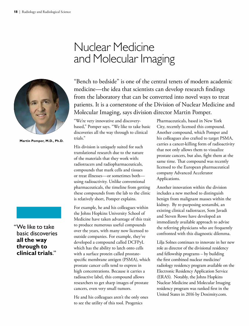
“ We like to take basic discoveries all the way through to clinical trials.”
Martin Pomper, M.D., Ph.D.
18 | Radiology and Radiological Science
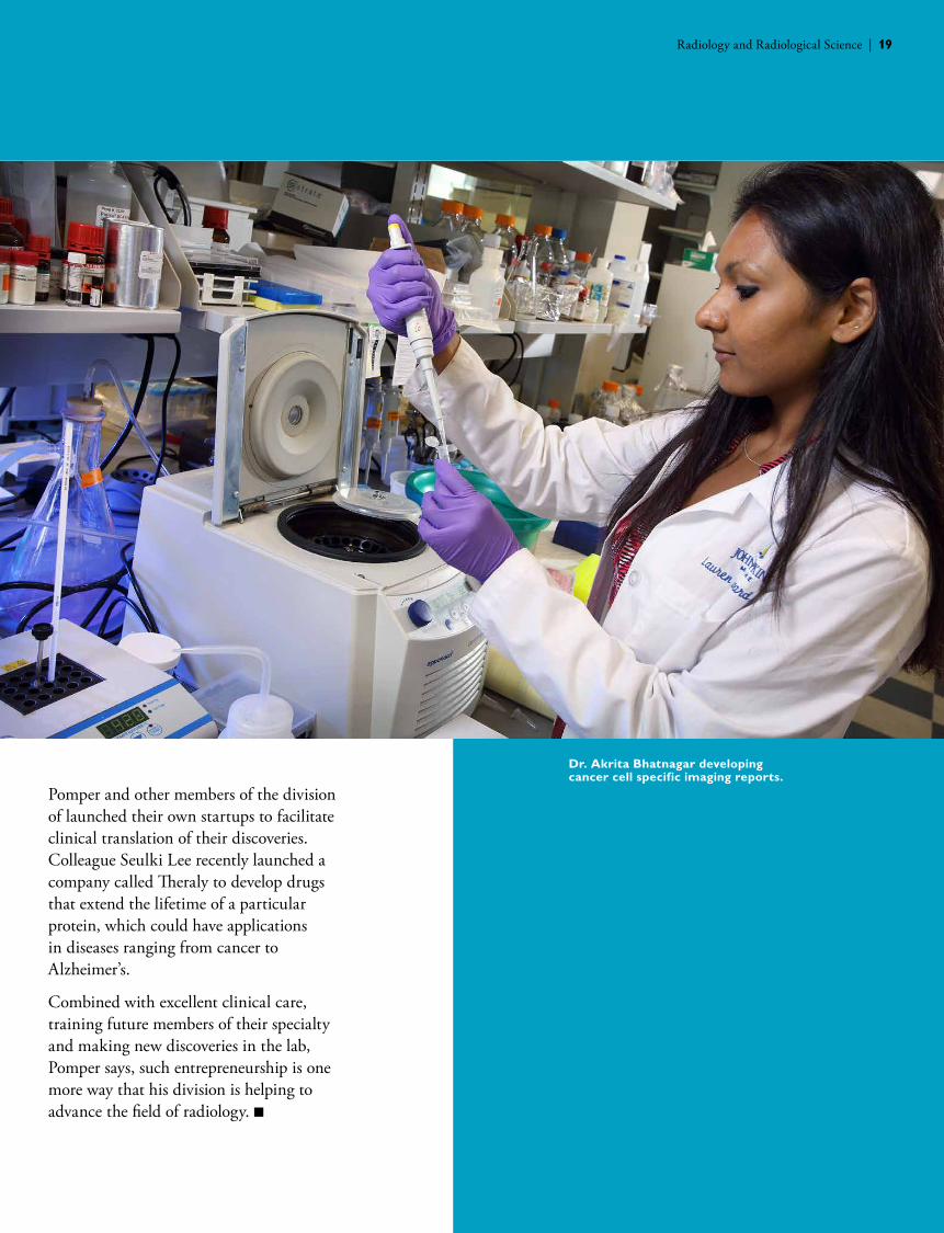
Dr. Akrita Bhatnagar developing cancer cell specific imaging reports.
19
Pomper and other members of the division of launched their own startups to facilitate clinical translation of their discoveries. Colleague Seulki Lee recently launched a company called Theraly to develop drugs that extend the lifetime of a particular protein, which could have applications in diseases ranging from cancer to Alzheimer’s.
Combined with excellent clinical care, training future members of their specialty and making new discoveries in the lab, Pomper says, such entrepreneurship is one more way that his division is helping to advance the field of radiology. n
Radiology and Radiological Science | 19
Pediatric Radiology and Pediatric Neuroradiology
Thierry Huisman, director of the Division of Pediatric Radiology and Pediatric Neuroradiology, says that he and his colleagues often refer to their young patients as “moving targets”—not just physically, but emotionally as well.” During their visit to our division, we only have a short time with these patients,” Huisman explains. “It means that when we see a child, we have to make sure that our relationship with that child and their family in that small time frame is so unique and successful that they’re not afraid to be imaged and to come back if necessary.”
That’s why he and his team have developed an incredibly detailed, multilayered approach to ease the minds of children and their families alike. For example, the division has a dedicated child life specialist to work with patients and families to help them better understand what will happen as team members gather the necessary images. They also have a mock MRI scanner to allow children to practice and become comfortable with the equipment before their real scan.
Unlike many doctors in this specialty, the division’s radiologists meet with patients and their families personally to explain the procedures and their role. To help doctors seem less imposing in these “meet and greets,” Huisman says, he and his colleagues have their baby pictures posted on the division’s website, along with a short bio, including their favorite children’s book. Each of these books is included in the library of the division’s waiting area for patients to read before their appointments.
“Our surveys show that many of our patients have seen the website before they come to see us,” Huisman says. “It opens up a discussion, a communication of trust.”
Comforting measures extend to the actual imaging, with a soothing color scheme of the examination rooms, stickers inside the scanner, explanatory graphics and texts, a coloring book specially designed for pediatric radiology, quiet rooms, and nurses and technicians speaking their patients’ language.
Making sure patients are at ease is key to getting the best images, a necessary starting point for the many daily interdisciplinary radiology conferences the division uses to discuss every patient who’s been imaged with the responsible clinical teams. Having these frequent discussions allows them to continually reevaluate care in real time, making sure any follow-up imaging is appropriate to spare patients unnecessary studies.
Each of these measures is a way for this division to deliver the best care to their young patients, Huisman says. “It’s a unique opportunity to work with future generations,” he adds. “We love our patients, and we love our jobs.” n
“ We love our patients, and we love our jobs.”
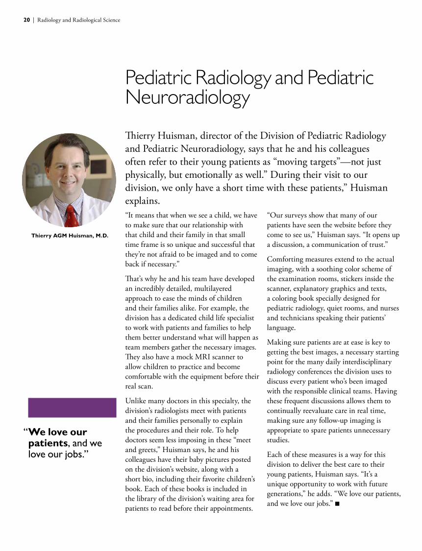
Thierry AGM Huisman, M.D.
20 | Radiology and Radiological Science
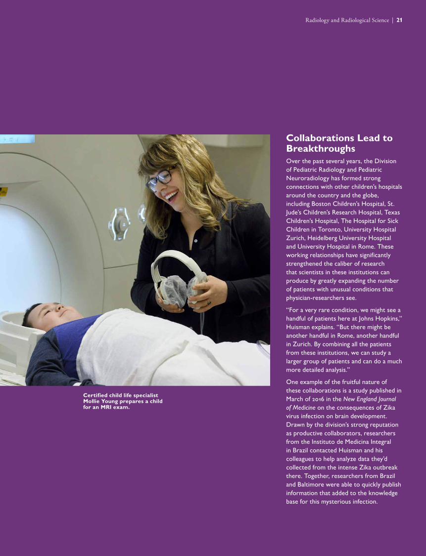
Certified child life specialist Mollie Young prepares a child for an MRI exam.
21
Collaborations Lead to BreakthroughsOver the past several years, the Division of Pediatric Radiology and Pediatric Neuroradiology has formed strong connections with other children’s hospitals around the country and the globe, including Boston Children’s Hospital, St. Jude’s Children’s Research Hospital, Texas Children’s Hospital, The Hospital for Sick Children in Toronto, University Hospital Zurich, Heidelberg University Hospital and University Hospital in Rome. These working relationships have significantly strengthened the caliber of research that scientists in these institutions can produce by greatly expanding the number of patients with unusual conditions that physician-researchers see.
“For a very rare condition, we might see a handful of patients here at Johns Hopkins,” Huisman explains. “But there might be another handful in Rome, another handful in Zurich. By combining all the patients from these institutions, we can study a larger group of patients and can do a much more detailed analysis.”
One example of the fruitful nature of these collaborations is a study published in March of 2016 in the New England Journal of Medicine on the consequences of Zika virus infection on brain development. Drawn by the division’s strong reputation as productive collaborators, researchers from the Instituto de Medicina Integral in Brazil contacted Huisman and his colleagues to help analyze data they’d collected from the intense Zika outbreak there. Together, researchers from Brazil and Baltimore were able to quickly publish information that added to the knowledge base for this mysterious infection.
Radiology and Radiological Science | 21
Radiology Education
The mission of the Radiology Residency Program at Johns Hopkins is to provide the best training in the country for this specialty, says program director Pam Johnson. “Pretty simple, right?” she adds, with a laugh. The program has a lot of advantages from the start—as part of a high-volume imaging department at one of the best academic medical centers in the world, radiology trainees here see a variety of cases and diseases that many never see over the course of their careers.
“It’s a setup for success,” Johnson says. But rather than rest on their laurels, she adds, Johnson and her colleagues are constantly working to improve the educational opportunities available to residents within the department.
For example, after many years of only offering only a four-year diagnostic radiology residency to 10 trainees, this year, the department opened an 11th spot for a molecular imaging track that provides dual board certification in diagnostic radiology and nuclear medicine. In the next year, two of the 10 diagnostic radiology spots will become part of an official interventional radiology residency.
Recognizing the talent and energy of Johns Hopkins trainees, Johnson and her colleagues developed five different special distinction programs designed to help residents become leaders in other areas important to this specialty, building on a long-standing quality improvement track.
The educator track enables residents interested in academics and teaching to develop these skills during their training. The health policy track takes advantage of the active role of the Maryland Radiology Society in policy work, and includes the opportunity for residents to lobby in Annapolis and Washington, D.C., to influence policy at the governmental level. Residents in the entrepreneur track can work with engineering students at the Johns Hopkins University Whiting School of Engineering to develop devices that can improve medical care or with the Technology Innovation Center to develop new IT platforms for health care delivery. A new consultant track allows a senior resident to actively engage with referring providers, residents and medical students about the most appropriate imaging for each case. Carrying a dedicated phone for this service, senior residents take turns being on call to provide this service.
“We’ve always taught our residents that radiology is far more than just reading an X-ray,” Johnson says. “For the residents who train at Johns Hopkins, we do everything in our power to help them excel in whatever they want to do and ensure that they become active members of the patient care team.” n
“ We’ve always taught our residents that radiology is far more than just reading an X-ray.”
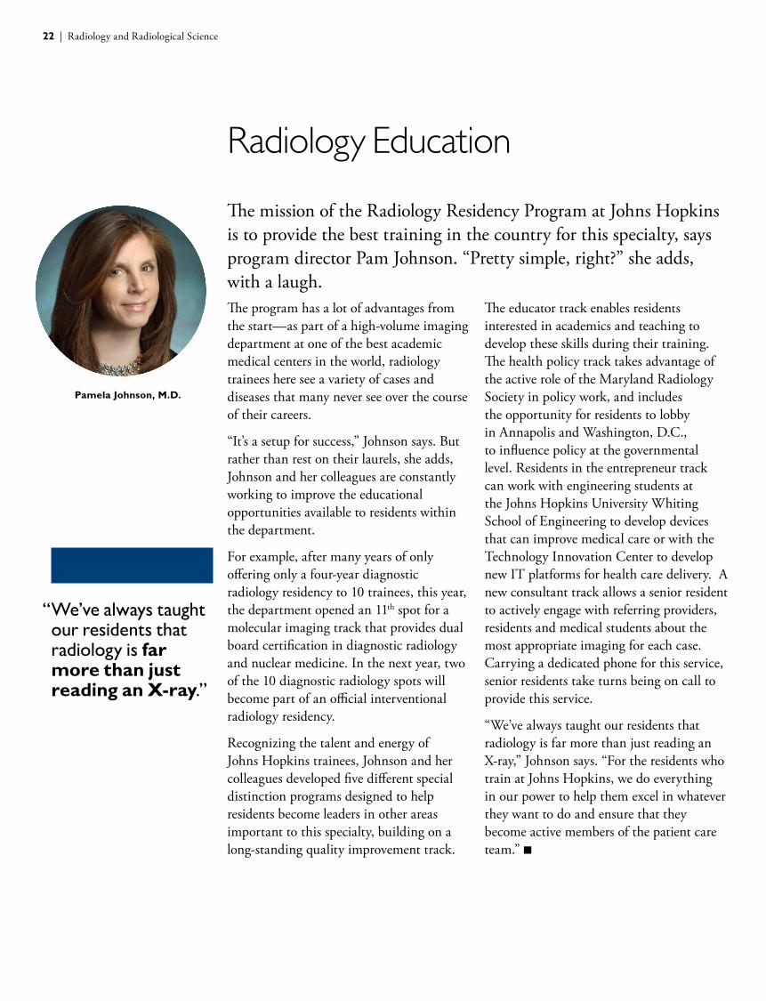
Pamela Johnson, M.D.
22 | Radiology and Radiological Science
Cross-sectional body imaging fellows.
23
Radiology and Radiological Science | 23
24
Johns Hopkins Medical Imaging
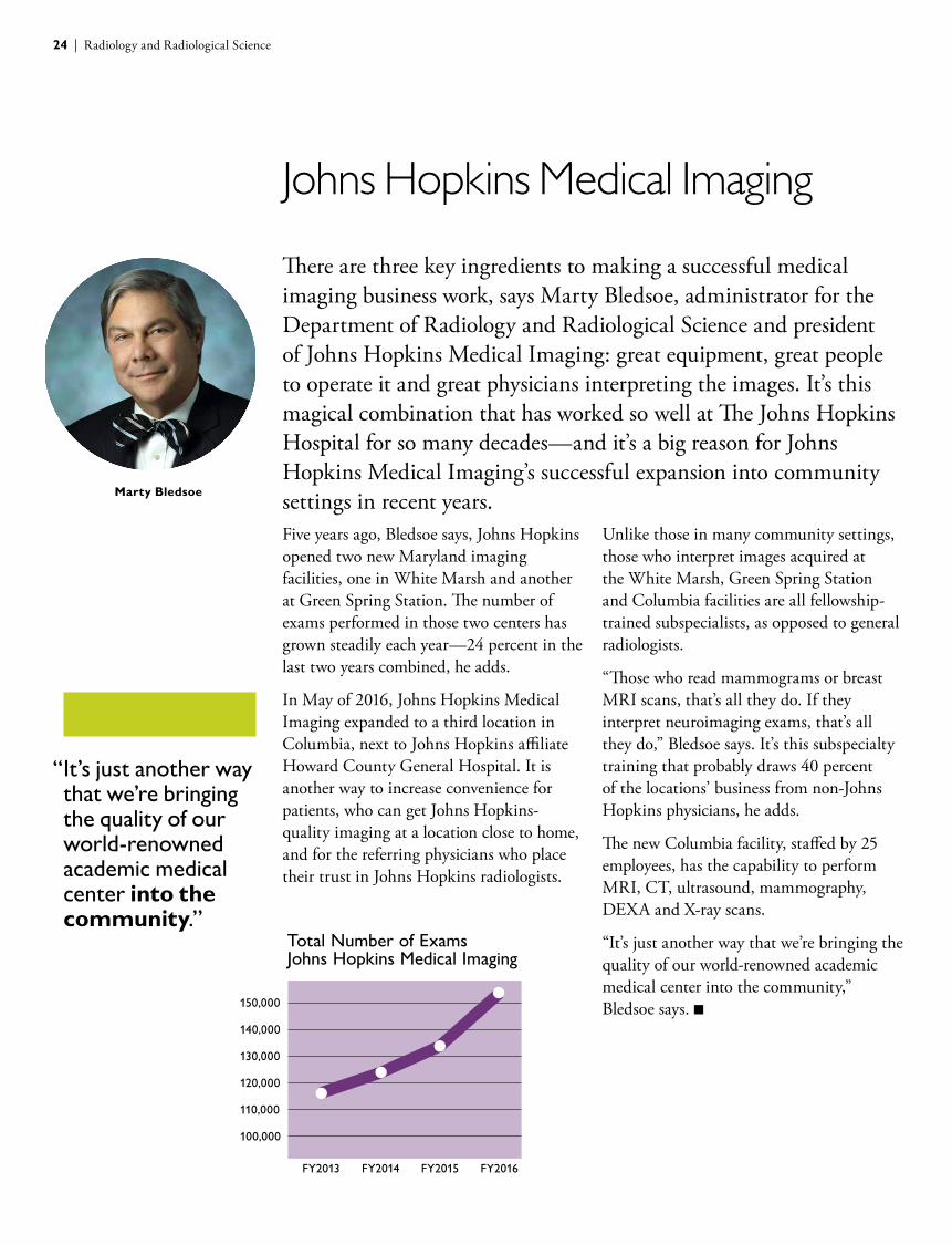
There are three key ingredients to making a successful medical imaging business work, says Marty Bledsoe, administrator for the Department of Radiology and Radiological Science and president of Johns Hopkins Medical Imaging: great equipment, great people to operate it and great physicians interpreting the images. It’s this magical combination that has worked so well at The Johns Hopkins Hospital for so many decades—and it’s a big reason for Johns Hopkins Medical Imaging’s successful expansion into community settings in recent years. Five years ago, Bledsoe says, Johns Hopkins opened two new Maryland imaging facilities, one in White Marsh and another at Green Spring Station. The number of exams performed in those two centers has grown steadily each year—24 percent in the last two years combined, he adds.
In May of 2016, Johns Hopkins Medical Imaging expanded to a third location in Columbia, next to Johns Hopkins affiliate Howard County General Hospital. It is another way to increase convenience for patients, who can get Johns Hopkins-quality imaging at a location close to home, and for the referring physicians who place their trust in Johns Hopkins radiologists.
Unlike those in many community settings, those who interpret images acquired at the White Marsh, Green Spring Station and Columbia facilities are all fellowship- trained subspecialists, as opposed to general radiologists.
“Those who read mammograms or breast MRI scans, that’s all they do. If they interpret neuroimaging exams, that’s all they do,” Bledsoe says. It’s this subspecialty training that probably draws 40 percent of the locations’ business from non-Johns Hopkins physicians, he adds.
The new Columbia facility, staffed by 25 employees, has the capability to perform MRI, CT, ultrasound, mammography, DEXA and X-ray scans.
“It’s just another way that we’re bringing the quality of our world-renowned academic medical center into the community,” Bledsoe says. n
“ It’s just another way that we’re bringing the quality of our world-renowned academic medical center into the community.”
FY2013 FY2014 FY2015 FY2016
150,000
140,000
130,000
120,000
110,000
100,000
Total Number of Exams Johns Hopkins Medical Imaging
Marty Bledsoe
24 | Radiology and Radiological Science
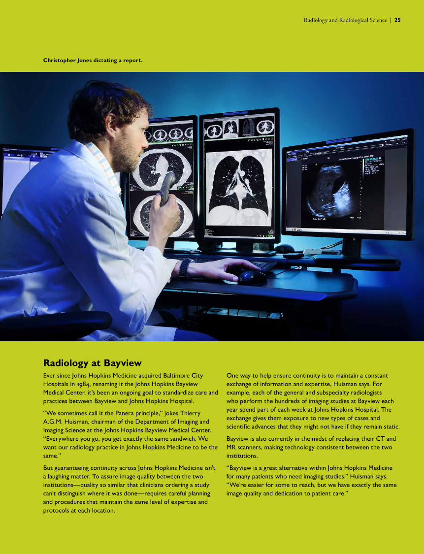
Christopher Jones dictating a report.
25
Radiology at BayviewEver since Johns Hopkins Medicine acquired Baltimore City Hospitals in 1984, renaming it the Johns Hopkins Bayview Medical Center, it’s been an ongoing goal to standardize care and practices between Bayview and Johns Hopkins Hospital.
“We sometimes call it the Panera principle,” jokes Thierry A.G.M. Huisman, chairman of the Department of Imaging and Imaging Science at the Johns Hopkins Bayview Medical Center. “Everywhere you go, you get exactly the same sandwich. We want our radiology practice in Johns Hopkins Medicine to be the same.”
But guaranteeing continuity across Johns Hopkins Medicine isn’t a laughing matter. To assure image quality between the two institutions—quality so similar that clinicians ordering a study can’t distinguish where it was done—requires careful planning and procedures that maintain the same level of expertise and protocols at each location.
One way to help ensure continuity is to maintain a constant exchange of information and expertise, Huisman says. For example, each of the general and subspecialty radiologists who perform the hundreds of imaging studies at Bayview each year spend part of each week at Johns Hopkins Hospital. The exchange gives them exposure to new types of cases and scientific advances that they might not have if they remain static.
Bayview is also currently in the midst of replacing their CT and MR scanners, making technology consistent between the two institutions.
“Bayview is a great alternative within Johns Hopkins Medicine for many patients who need imaging studies,” Huisman says. “We’re easier for some to reach, but we have exactly the same image quality and dedication to patient care.”
Radiology and Radiological Science | 25
26
Staff Engagement
What drives someone to be a great employee in the Johns Hopkins Department of Radiology and Radiological Science? What powers them to provide the best customer service, work as part of a cohesive team and have good professional relationships? According to Peg Cooper, operations administrator for the department and director of Johns Hopkins Medical Imaging, the answer is creating a positive work culture. “When you create a positive culture and people understand their purpose, all other things fall into place,” Cooper says. “They understand why they are here and how they can make a difference. They know where they fit into the bigger pieces of the puzzle for Johns Hopkins Medicine.”
Feeling one’s professional purpose is a key part of employee engagement—the passion and commitment employees feel toward their jobs and organizations and the effort they put into their work. Last year, the department ranked in the 81st percentile nationally for employee engagement compared to all health care systems that participated in the Gallup Employee Engagement Survey. According to Gallup, engagement scores at this level translate into 25 engaged employees for every one who’s not engaged. On average, only about a third of employees nationwide were considered engaged by Gallup’s analysis.
Encouraging an environment that fosters this level of engagement has been one of Cooper’s biggest goals since she joined the department a decade ago. With 12 direct reports responsible for 420 people, the key has been having a great management team.
Creating an engaged culture, she explains, starts at the top with engaged managers. One way to cultivate managers invested in their jobs is help them foster positive change within their own teams. Each month, Cooper hosts a quality improvement team meeting, in which managers meet to trouble shoot problems and develop projects that improve patient care and workflow efficiencies. Over the course of each year, they collect metrics on problem areas and improvements over time. These projects are formally presented to the team at the year’s end.
Cooper also oversees, with two managers, the Future Leaders Program, a nine-month program that teaches leadership skills to interested employees from every facet of the department. On one day each month, these employees attend classes at Johns Hopkins’ Mt. Washington facility, learning skills as diverse as public speaking, interviewing, facilitating meetings and having crucial conversations. Not only do these classes serve as a way to step out of the daily routine, says Cooper, but also for employees to meet face-to-face with others that they have only tangential relationships with, engendering respect and communication for all parts of the department.
“ When you create a positive culture and people understand their purpose, all other things fall into place.”
26 | Radiology and Radiological Science
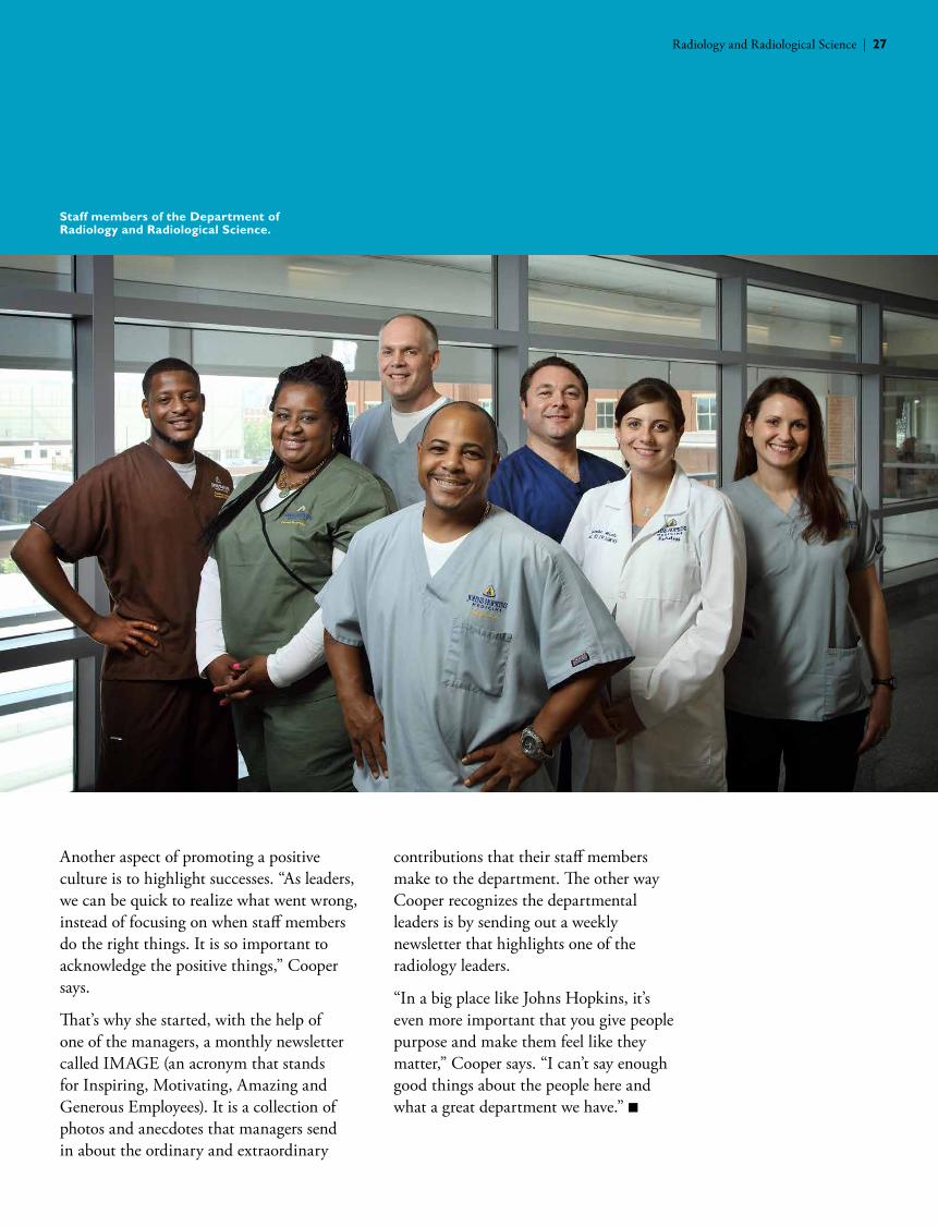
Staff members of the Department of Radiology and Radiological Science.
27
Another aspect of promoting a positive culture is to highlight successes. “As leaders, we can be quick to realize what went wrong, instead of focusing on when staff members do the right things. It is so important to acknowledge the positive things,” Cooper says.
That’s why she started, with the help of one of the managers, a monthly newsletter called IMAGE (an acronym that stands for Inspiring, Motivating, Amazing and Generous Employees). It is a collection of photos and anecdotes that managers send in about the ordinary and extraordinary
contributions that their staff members make to the department. The other way Cooper recognizes the departmental leaders is by sending out a weekly newsletter that highlights one of the radiology leaders.
“In a big place like Johns Hopkins, it’s even more important that you give people purpose and make them feel like they matter,” Cooper says. “I can’t say enough good things about the people here and what a great department we have.” n
Radiology and Radiological Science | 27
28
Faculty
Breast ImagingBrennecke, Cecilia Di Carlo, PhilipEisner, David Falomo, Eniola Harvey, SusanKhouri, Nagi Lee, Bonmyong Mullen, Lisa Myers, Kelly Woods, Ryan
Cancer Imaging Research
Artemov, DmitriBhujwalla, Zaver Chen, ZhihangGlunde, KristineJacobs, MichaelKakkad, SamataKrishnamachary, BalajiMori, NorikoPathak, ArvindPenet, Marie-FranceRaman, VenuShah, TariqVesuna, FarhadWinnard, PaulZhu, Wenlian
Diagnostic ImagingAhlawat, ShivaniAhn, HannahBerlanstein, BruceBlack Thomas, RachelChu, LindaCoquia, StephanieDemehri, ShadpourEng, JohnFayad, Laura Feigin, DavidFishman, ElliotFritz, JanFynes, MargaretGoldman, AliceHamper, UlrikeHorton, KarenHussien, AmiraJohnson, PamelaKamel, IhabKawamoto, SatomiLee, DavidLee, Nam JuLin, ChengLugo-Fagundo, CarolinaMacura, KatarzynaMagid, DonnaMalguria, NaginaRaman, SivaRamin Pour, SaraReisner, DavidSong, KarenSheth, SheilaTsai, SalinaYang, ShirleyZaheer, AtifZimmerman, Stefan
Interventional Radiology
Akman, AndrewBhagat, NikhilCarmi, LemoreGanapathy, ShanmugasundaraGeorgiades, ChristosHolly, BrianHong, KelvinJohnson, BrianLiapi, EleniMitchell, SallySrinivas, AbhishekWeiss, CliffordWerner, JohnYim, Douglas
Johns Hopkins Bayview Medical Center
Auster, MartinBurgan, ConstantineFetrat, MehrdadFradin, JoelJones, ChristopherTadimeti, HimaVerde, Franco
Medical Imaging PhysicsBoctor, EmadDu, YongFrey, EricFung, Shiu-KaiGhaly, MickelJha, Abhinav KumarLee, OkkyunLee, Taek-SooMahesh, MahadevappaStumpf, MartinTaguchi, KatsuyukiTsui, BenjaminXu, Jingyan
28 | Radiology and Radiological Science
29
MR ResearchAfework, YohannesAggarwal, ManishaBottomley, PaulBulte, JeffChan, Wai YanChoe, AnnFaria, Andreia VasconcellosFu, YingliGilad, AssafGillen, JosephHeo, Hye Young Hua, JunJanowski, MiroslawJiang, HangyiKarmarkar, ParagKedziorek, DorotaKraitchman, DaraKrimins, RebeccaLi, WenboLi, Xin Li, XuLiu, GuanshuLiu, PeiyingLu, HanzhangMcAllister, MaryMcMahon, MichaelMori, SusumuEllens, NicholasOishi, KenichiPekar, James Pelled, GalitQin, QinSchar, MichaelSolaiyappan, MeiyappanSong, XiaoleiVan Zijl, PeterWalczak, PiotrWu, DanXu, JiadiXU, XiangYadav, Nirbhay Zeng, HaifengZhang, YiZhou, Jinyuan
NeuroradiologyAhn, HyoAygun, NafiBarker, PeterBlitz, AriEdden, RichardGailloud, Philippe Gellad, FouadGong, GaryGregg, LydiaGujar, SachinHui, FerdinandIdowu, OluwatoyinIzbudak, IzlemKraut, MichaelLin, DorisLiu, LiMohamed, MonaNadgir, RohiniPearl, MonicaPillai, JayPuts, NickQiao, YeSair, HarisWasserman, BruceYousem, David
Nuclear Medicine and Molecular Imaging
Abou, DianeBora, AdrianaBrasic, JamesBrummet, MaryChen, YingCrabb, AndrewDannals, RobertDe Silva, RavindraFoss, CatherineHolt, DanielHorti, AndrewJavadi, Mehrbod SomKuwabara, HirotoLeal, JeffreyLee, SeulkiLesniak, WojciechLodge, MartinMathews, WilliamMease, RonMinn, ILNedrow, JessieNimmagadda, SridharOh, YuminPark, OgyiPomper, MartinRahmim, ArmanRavert, HaydenRay, SangeetaRousset, OlivierRowe, StevenRoy, SanchitaSchindler, ThomasSgouros, GeorgeSolnes, LiljaSzabo, ZsoltThorek, DanielWong, DeanYang, XingZhou, YunZiessman, Harvey
Pediatric Radiology and Pediatric Neuroradiology
Benson, JaneDunn, EmilyHuisman, ThierryHwang, MisunPoretti, AndreaSoares, BrunoSpevak, MelissaTekes, Aylin
Radiology Information Technology
Nagy, PaulSevinc, Gorkem
Prod
uced
by
John
s H
opki
ns M
edic
ine
Mar
keti
ng a
nd C
omm
unic
atio
ns
Radiology and Radiological Science | 29

www.hopkinsmedicine.org/radiology
RA
D16
0903
9