Embed Size (px)
DESCRIPTION
yo yo
Citation preview
CHAPTER PLANS.NO. PARTICULARS PAGE NO.
1 INTRODUCTION
2 HISTORY
3 TOOLS USED IN RDNA TECHNOLOGY
4 ENZYMES
5 CLONING METHOD
6 COLONY HYBRIDIZATION
7 PCR
8 TRANSGENICS
9 CONCLUSION
10 REFERENCES
1.INTRODUCTIO
N
Recombinant DNA technology refers to the set of techniques for recombining genes from
different sources in vitro and transferring this recombinant DNA into cells where it may be
expressed.
• These techniques were first developed around 1975 for basic research in bacterial
molecular biology, but this technology has also lead to many important discoveries in basic
eukaryotic molecular biology.
• Such discoveries resulted in the appearance of the biotechnology industry.
Biotechnology refers to the use of living organisms or their components to do practical tasks
such as:
The use of microorganisms to make wine and cheese
Selective breeding of livestock and crops
Production of antibiotics from microorganisms
Production of monoclonal antibodies
The use of recombinant DNA techniques allows modern biotechnology to be a more precise
and systematic process than earlier research methods.
It is also a powerful tool since it allows genes to be moved across the species
barrier.
Using these techniques, scientists have advanced our understanding of eukaryotic
molecular biology.
The Human Genome Project is an important application of this technology. This
project’s goal is to transcribe and translate the entire human genome in order to better
understand the human organism.
A variety of applications are possible for this technology, and the practical goal is the
improvement of human health and food production.

2.HISTORY
DNA technology makes it possible to clone genes for basic research and commercial
applications: an overview
Prior to the discovery of recombinant DNA techniques, procedures for altering the genes of
organisms were constrained by the need to find and propagate desirable mutants
Geneticists relied on either natural processes, mutagenic radiation, or chemicals to induce
mutations.
In a laborious process, each organism’s phenotype was checked to determine the presence
of the desired mutation.
Microbial geneticists developed techniques for screening mutants. For example bacteria
was cultured on media containing an antibiotic to isolate mutants which were antibiotic
resistant.
Before 1975, transferring genes between organisms was accomplished by cumbersome and
nonspecific breeding procedures. The only exception to this was the use of bacteria and their
phages.
Genes can be transferred from one bacterial strain to another by the natural processes of
transformation, conjugation and transduction.
Geneticists used these processes to carry out detailed molecular studies on the structure
and functioning of prokaryotic and phage genes.
Bacteria and phages are ideal for laboratory experiments because they are relatively
small, have simple genomes, and are easily propagated.
Although the technique was available to grow plant and animals cells in culture, the
workings of their genomes could not be examined using existing methods.
Recombinant DNA technology now makes it possible for scientists to examine the structure
and function of the eukaryotic genome, because it contains several key components:
Biochemical tools that allow construction of recombinant DNA.
Methods for purifying DNA molecules and proteins of interest.
Vectors for carrying recombinant DNA into cells and replicating it
Techniques for determining nucleotide sequences of DNA molecules.
3.TOOLS USED IN rDNA TECHNOLOGY
3.1.CLONING VECTORS
a) PLASMIDS
b) BACTERIOPHAGE λ
c) COSMID
d) PHASMID
e) SHUTTLE VECTOR
f) YAC/BAC/MAC
g) EXPRESSION VECTORS
3.2ENZYMES
a) RESTRICTION ENZYMES
b) LIGASES
CLONING VECTORS
A cloning vector is a small piece of DNA into which a foreign DNA fragment can be
inserted. The insertion of the fragment into the cloning vector is carried out by treating the
vehicle and the foreign DNA with a restriction enzyme that creates the same overhang,
then ligating the fragments together. There are many types of cloning vectors. Genetically
engineered plasmids and bacteriophages (such as phage λ) are perhaps most commonly used
for this purpose. Other types of cloning vectors include bacterial artificial
chromosomes (BACs) and yeast artificial chromosomes (YACs).
Most commercial cloning vectors have key features that have made their use in molecular
biology so widespread.
In the case of expression vectors, the main purpose of these vehicles is the controlled
expression of a particular gene inside a convenient host organism (e.g. E. coli).
Control of expression can be very important; it is usually desirable to insert the
target DNA into a site that is under the control of a particular promoter. Some
commonly used promoters are T7 promoters, lac promoters (bla promoter)
and cauliflower mosaic virus's 35s promoter (for plant vectors).
To allow for convenient and favorable insertions, most cloning vectors have had
nearly all their restriction sites engineered out of them and a synthetic multiple
cloning site (MCS) inserted that contains many restriction sites. MCSs allow for
insertions of DNA into the vector to be targeted and possibly directed in a chosen
orientation. A selectable marker, such as an antibiotic resistance [e.g. beta-
lactamase (see figure)] is often carried by the vector to allow the selection of
positively transformed cells (see Screening below). All plasmids must carry a
functional origin of replication (ORI; not shown in figure).
Some other possible features present in cloning vectors are: vir genes for plant
transformation, integrase sites for chromosomal insertion, lacZα fragment for α
complementation and blue-white selection, and/or reporter genes in frame with and
flanking the MCS to facilitate the production of recombinant proteins [e.g. fused to
the Green fluorescent protein (GFP) or to the glutathione S-transferase (see figure)].
Now, the different cloning vectors are:-
a) PLASMIDS
Plasmids are molecules of DNA that are found in bacteria separate from the bacterial
chromosome.
They are small (a few thousand base pairs)
They are usually carry only one or a few genes
They are circular
have a single origin of replication.
Plasmids are replicated by the same machinery that replicates the bacterial chromosome.
Some plasmids are copied at about the same rate as the chromosome, so a single cell is apt to
have only a single copy of the plasmid. Other plasmids are copied at a high rate and a single
cell may have 50 or more of them.
Genes on plasmids with high numbers of copies are usually expressed at high levels. In
nature, these genes often encode proteins (e.g., enzymes) that protect the bacterium from one
or more antibiotics.
Plasmids enter the bacterial cell with relative ease. This occurs in nature and may account for
the rapid spread of antibiotic resistance in hospitals and elsewhere. Plasmids can be
deliberately introduced into bacteria in the laboratory transforming the cell with the incoming
genes.
Desirable properties of plasmids
Plasmids have proven extremely useful as cloning vectors, particularly when they are
modified specifically for that purpose. A bacterial plasmid that is designed for cloning should
have some or all of the following properties:
It should be small.
The aim of most cloning experiments is to isolate the passenger DNA. The vector
only serves to carry and amplify the passenger. A small plasmid has the advantage of
contributing only a minimal amount of extraneous DNA to the plasmid/passenger
construct, thereby making it easier to prepare large amounts of the passenger DNA. In
addition, it is easier to get smaller pieces of DNA into bacteria than larger ones --
everything else being equal -- and the smaller the plasmid is, the less it contributes to
total size. Another point is that small plasmids replicate faster and require less energy
for replication than large ones. Finally, small plasmids are easier to purify than large
ones because they are less fragile.
Its DNA sequence should be known.
Knowledge of the DNA sequence of a plasmid allows the genetic engineer to
manipulate it with the full complement of recombinant DNA techniques.
It should grow to high copy number in the host cell.
In other words, relaxed replication plasmids are most often preferred to those that are
under stringent control. In most cases, having many copies makes it easier to purify the
plasmid away from the chromosomal DNA, and of course increases its yield. However, there
are circumstances when just one or a few copies of a plasmid are desirable.
It should contain a selectable marker that allows cells containing the plasmid to
be isolated.
Bacterial cells that harbor plasmids don't necessarily look or act different than those
that do not. It is only when the plasmid carries a gene that lends a special trait to the
bacteria that the molecular engineer can tell if a bacterium contains a plasmid.
Antibiotic resistance is one such trait or marker, and the genes for ampicillin
resistance and tetracycline resistance are common genes carried by frequently used
plasmids. Some plasmids are also readily lost from cells, and unless there is a
selectable gene contributed by the plasmid and the bacteria are kept under selective
conditions, the molecular engineer may end up with a plasmid-less population of cells
at the end of an experiment.
It should also contain a second selectable gene that is inactivated by insertion of
the passenger.
Imagine the following scenario. A scientist is trying to insert a passenger molecule into a
plasmid. The plasmid has only a single selectable marker. After ligation, some of the
molecules contain the passenger. Others do not. These may be plasmids that have
recircularized in the presence of DNA ligase. This mixture of plasmids is then used to
transform bacteria. Both recombinant plasmids and recircularized plasmids contain the
same marker and cannot readily be told apart genetically. How are colonies carrying a
plasmid with a passenger distinguished from those with a plasmid with no passenger?
One way is to grow many individual colonies, isolate plasmid DNA from each, run the
DNA out on an agarose gel, and identify the recombinant plasmid molecules because they
are bigger by virtue of the passenger that they carry. While this procedure works and is
very commonly carried out, it is laborious, time consuming, and expensive.
There is a better way. Suppose that a plasmid carries two different antibiotic
resistance genes, A and B. What would be the consequences of cloning the passenger
into the middle of gene B? In most cases, the foreign DNA will disrupt gene B and
not allow it to work. On the other hand, if the plasmid simply recircularizes, gene B
will be unaffected. With this arrangement it is possible to tell which cells harbor
plasmids that carry passengers. Those that contain any plasmid at all will have gene A
activity. And those that have a plasmid bearing a passenger, will lack activity from
gene B.
There should be a large number of unique restriction sites lying within one of the
two selectable markers described above.
As described above, most cloning is done by inserting a fragment of DNA that is cut
with a specific restriction endonuclease into a vector that is cut with the same enzyme
(remember, this ensures that the two have compatible ends). If the vector contains
more than one such site, it will be cut into multiple pieces by the restriction enzyme,
complicating matters unnecessarily. The presence of many unique sites allows for
maximum flexibility and ease in cloning.
Plasmid purification
Plasmids may be purified from bacteria by taking advantage of the difference between the
small, circular plasmid molecules and the large, broken (hence linear) pieces of chromosomal
DNA. (Chromosomal DNA from E. coli is also circular, but during isolation its relatively
large size (six million nucleotide pairs) and consequent fragility cause it to break easily.)
The most common method for purifying plasmid DNA involves three steps:-
First, the bacteria are broken open and the is DNA isolated.
Then the DNA is denatured.
Finally, the DNA is renatured and centrifuged.
Upon denaturation, the two circular single-stranded chains of the plasmid DNA remain
entwined and don't separate fully. When conditions are set up so that renaturation can occur,
each strand rapidly finds its complement. The chromosomal DNA, on the other hand, breaks
readily and therefore consists of noncircular pieces. Under these circumstances, the two
strands easily denature and separate. Upon renaturation, they have difficulty finding complete
copies of their complements. Often partial renaturation occurs between different sizes of
single-stranded fragments and large insoluble aggregates form. These can be simply
separated from the small circular plasmid DNA by high-speed centrifugation.
Some popular plasmids are:-
pAMP
4539 base pairs
a single replication origin
a gene conferring resistance to the antibiotic ampicillin (a relative of penicillin)
a single occurrence of the sequence
a fragment of 3755 base pairs carrying both the ampr gene and the replication origin
a fragment of 784 base pairs
both fragments have sticky ends
pBR322
The first really useful plasmid for genetic engineering, pBR322, was pieced together by
Francisco Bolivar, and others in Herbert Boyer's laboratory in the 1970s (The "B" stands for
Bolivar and the "R" for Rodriguez, another scientist in Boyer's laboratory). What makes
pBR322 useful is that it contains an ampicillin resistance gene and a tetracycline resistance
gene. In addition it has a relaxed origin of replication (indicated by the green arrow shown at
the bottom of the illustration) and accumulates to high numbers in E. coli. Its entire 4363
base-pair sequence has been determined, and 21 common enzymes are available that
recognize only a single site within it. (However, only 11 are in either of the two antibiotic
resistance genes.).
pUC18
More recently, a series of small plasmids (about 2.7 kilobase pairs) have been developed in
Joachim Messing'e laboratory that have several properties that have made them very popular
with genetic engineers. These pUC (pronounced PUCK) plasmids, exemplified by pUC18
pictured at the right, carry an ampicillin resistance gene and an origin of replication, both
from pBR322. They also bear a multiple cloning site -- a sequence of DNA that carries many
restriction sites (13, in the case of pUC18). The multiple cloning site of the pUC plasmids is
special because it also codes for a small peptide. This peptide will correct a specific mutation
in the chromosomal gene that codes for the enzyme beta-galactosidase.
When plasmids containing this sequence are cloned into a specific E. coli strain that lacks
beta-galactosidase activity, they make the peptide and thereby begin to express active
enzyme. If, however, one of the restriction endonuclease sites in the multiple cloning site is
opened and a foreign gene inserted, the peptide is no longer produced (because the protein
coding region is disturbed), and no beta-galactosidase activity appears.
Cells that harbor an active beta-galactosidase enzyme can be made to turn blue in the
presence of certain substrates. Those colonies that have a passenger inserted at the multiple
cloning site of the pUC plasmids will lack the enzyme and will be white, while those that
have simply recircularized (those that don't contain a passenger) will stain blue. In this way,
pUC plasmids containing a foreign insert of DNA can be distinguished from plasmids
without a passenger.
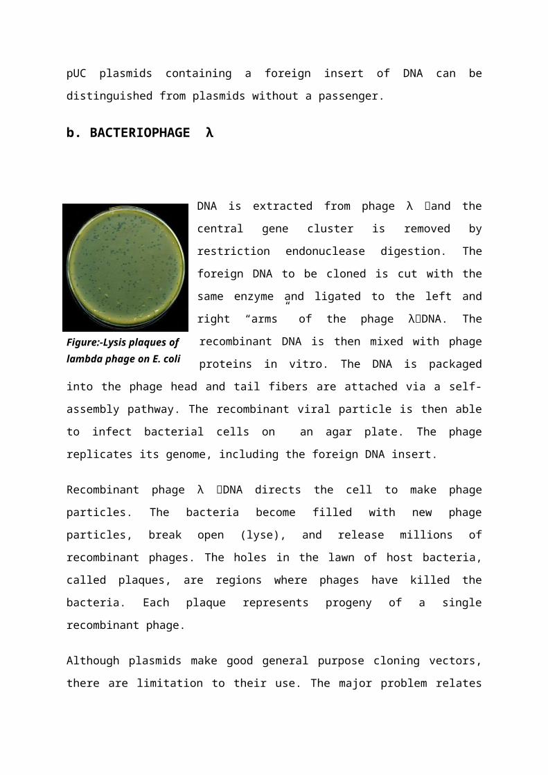
b. BACTERIOPHAGE λ
DNA is extracted from phage λ and the central gene cluster is
removed by restriction endonuclease digestion. The foreign
DNA to be cloned is cut with the same enzyme and ligated to
the left and right “arms” of the phage λDNA. The
recombinant DNA is then mixed with phage proteins in vitro.
The DNA is packaged into the phage head and tail fibers are
attached via a self-assembly pathway. The recombinant viral
particle is then able to infect bacterial cells on an agar plate.
The phage replicates its genome, including the foreign DNA
insert.
Recombinant phage λ DNA directs the cell to make phage particles. The bacteria become
filled with new phage particles, break open (lyse), and release millions of recombinant
phages. The holes in the lawn of host bacteria, called plaques, are regions where phages have
killed the bacteria. Each plaque represents progeny of a single recombinant phage.
Figure:-Lysis plaques of lambda phage on E. coli bacteria
Although plasmids make good general purpose cloning vectors, there are limitation to their
use. The major problem relates to the size of insert, which can be effectively carried into the
cell. Transformation frequencies sharply decrease as the size of the DNA circle increases. In
addition, plasmids with large inserts are unstable and give rise to smaller segregants.
Bacteriophage lambda has been widely used in recombinant DNA since engineering of
the first viral cloning vector in 1974. Phage lambda vectors are particularly useful for
preparing genomic libraries, because they can hold a larger piece of DNA than a plasmid
vector. The phage lambda has 2 modes by which it replicates itself- the 1 st is LYTIC CYCLE
which involves attachment of the phage to the bacterial cell, infection of the phage DNA and
the replication of this DNA inside the cell. After the packaging of the DAN molecule in
protein coats, the cell will LYSE, liberating the progeny phage.
The 2nd mode of replication is called as LYSOGENY, follows the
same initial path of infection, but the phage genome, rather than
independently replicating becomes integrated into the bacterial
chromosome. In this case, the cell does not lyse and the phage
genome become a silent passenger (Benign guest OR Prophage).
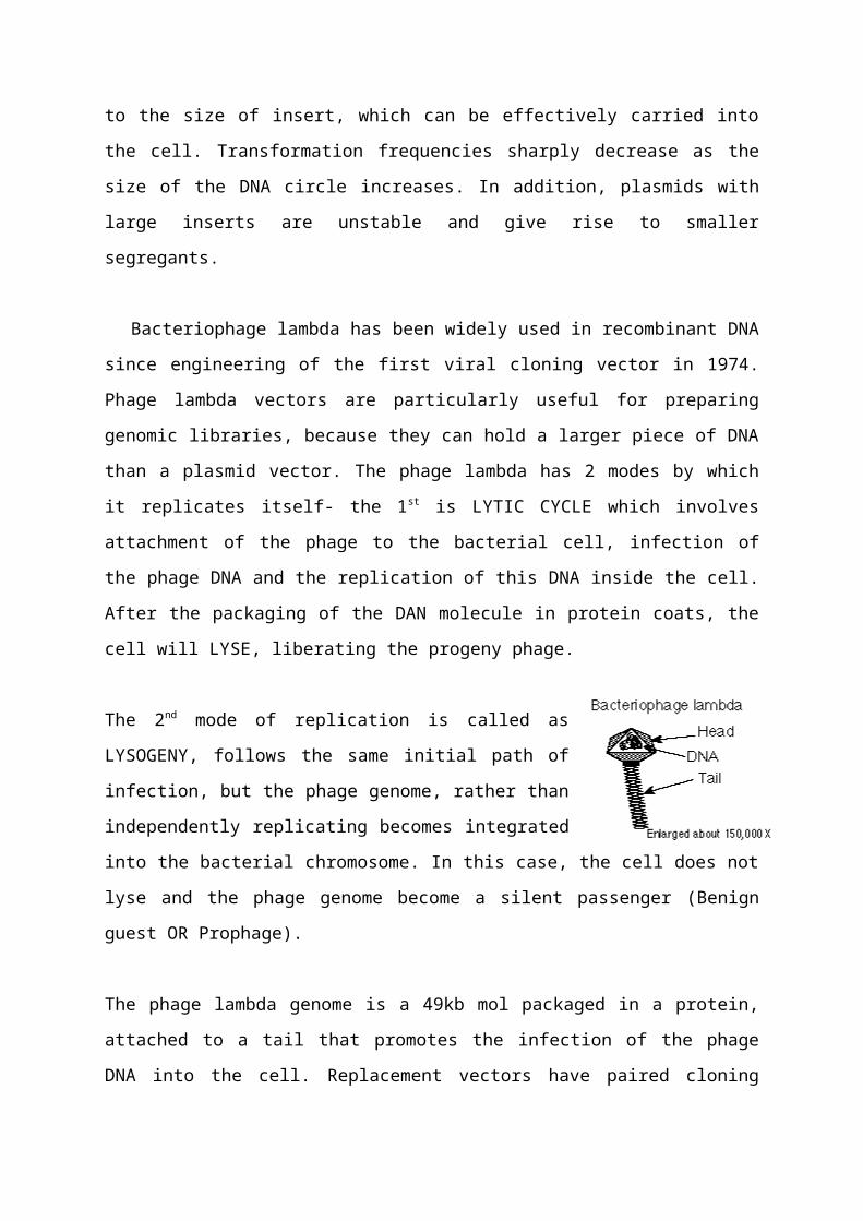
The phage lambda genome is a 49kb mol packaged in a protein, attached to a tail that
promotes the infection of the phage DNA into the cell. Replacement vectors have paired
cloning sites on either side of a central gene cluster. This central cluster contains genes for
lysogeny and recombination, which are not essential for the lytic life cycle. The central gene
cluster can be removed and foreign DNA inserted between the “arms”. All phage vectors
used as cloning vectors have been disarmed for safety and can only function in special lab
conditions. The recombinant viral particle infects bacterial host cells, in a process called
“transduction”. The host cells lyse after phage reproduction, releasing progeny virus particles.
The viral particles appear as a clear spot of lysed bacteria or “plaque” on an agar plate
containing a lawn of bacteria. Each plaque represents progeny of a single recombinant phage
and contains millions of recombinant phage particles. Most contemporary vectors carry a
lacZ’ gene allowing blue- white selection. In this type, it is possible to accommodate inserts
of up to 24kb and propagate them in a stable fashion. The great advantage of using phage
vectors is that after the foreign DNA has been ligated in the products can be packaged in the
test tube into phage coat. This process is called IN- VITRO PACKAGING- allows the
r-DNA to be added to the cell by phage infection instead of transformation/ transfection, thes
increasing the efficiency by many orders of magnitude.
Phage lambda as a vector-
There are 2 kinds of lambda vectors which differ in size, they are-
a) Lambda replacement vectors-
Contains a restriction site for phage propagation in a suitable bacterial host.
Remaining part of the lambda genome is removed and is replaced by a foreign DNA.
b) Lambda insertion vectors-
When cloning into an insertion vector, the phage DNA is cleaved with a restriction
enzyme that cut its site. No phage DNA is removed, therefore, much lower size of
DNA can be inserted.
Three reasons why bacteriophage lambda is a good cloning vehicle
1. It can accept very large pieces of foreign DNA. About 20kb of DNA can be deleted from
its central region (between the J and N genes) and elsewhere, and replaced with an equal
quantity of foreign DNA without affecting the ability of the phage to grow lytically.
2. It has been extensively reworked over the years. Genetic engineers have constructed
numerous derivatives of lambda that contain only one or two sites for a variety of
restriction enzymes.
3. Finally -- its main advantage -- bacteriophage lambda is one of the few organisms that
can be reconstituted in a test tube. By simply mixing phage DNA with a mixture of phage
proteins, an infective viral particle with the DNA inside the phage head can be produced.
This process is called in vitro packaging and it normally occurs when long tandem arrays
of phage chromosomes (they are produced in this form when they replicate in the
bacterial cell) are cleaved into monomers by certain phage proteins. The DNA is cut at a
site on the chromosome called cos, and this operation produces the cohesive ends that
were mentioned above. Ordinarily the DNA is cut into 48.5 kb fragments that are
incorporated into the head of newly formed phage particles.
c) COSMID
While plasmids and bacteriophage lambda are both highly useful as vectors, they impose two
limitations-
a) The size of the DNA fragments that can be cloned in such vectors is limited- with
plasmids, the larger the inserted fragments of foreign DNA, the lower the efficiency of
ligation and transfection, thus the cloning of DNA fragments larger than 15kb is
experimentally difficult.
In case of lambda vectors, the length of the non-essential region of the lambda DNA limits of
fragment size to 24kb or less (20kb).
b) The original lambda vectors don’t allow propagation of viable bacterial cells that carry the
inserted DNA fragments. The insert is propagated as a part of a virus that lyse the cell.
So Cosmids were developed as vectors specifically designed for the cloning of large
fragments of DNA (45kb). Cosmids combine the properties of plasmids and bacteriophage
lambda vectors. For example, the commonly used cosmid pLFR-5 has two cos sites (cos
ends) from bacteriophage lambda separated by a ScaI restriction endonuclease site, a multiple
cloning site with six unique recognition sites (HindIII, PstI, SalI, BamHI, SmaI and EcoRI),
an origin of DNA replication and a tetracycline resistance (Tetr) gene. The pLFR-5 DNA
(6kb) is cleaved initially with ScaI and then with BamHI. The final 2 DNA samples are
mixed and ligated. Some of the ligated products will have a 45kb DNA piece inserted
between the 2 fragments that are derived from the digestions of the pLFR-5 DNA. These
molecules will be about 50kb long and have cos sequences that are about 50kb apart.
Consequently, these DNA constructs are successfully packaged into bacteriophage lambda
heads in vitro. After assembly of bacteriophage particles, the DNA is delivered by infection
into E.coli, once inside the host cell, the cos ends, which were cleaved during the in vitro
packaging, base pair and enable the linear DNA to circularize. Moreover, the tetracycline
resistance gene allows colonies that carry the cosmid to grow in the presence of tetracycline.
Non- transformed cells are sensitive to tetracycline and die.
d) PHASMIDS
A 2nd combination of plasmid and phage lambda sequence has been devised to exploit the
viruses of each type of vectors. This combination consists of a plasmid vector carrying a
lambda attachment site. The plasmid may insert into a phage lambda genome (by means of
site- specific recombination/ lytic cycle) generating a phage genome containing one or more
plasmid molecule (depending on the length of the plasmid). These novel genetic combination
are called “phasmids”. They contain functional origins of replication of the plasmids and of
lambda and may be propagated as a plasmid or as a phasmid in appropriate E.coli strains.
Phage particles are easy to store, they have an effectively infinite shell- life, screening
phage plaques by molecular hybridization often gives clean results than screening bacterial
colonies.
e) SHUTTLE VECTORS
That can replicate in different organisms. They contain sequences from E.coli plasmid and a
particular region of the yeast genome.
Essential Features are-
a) Two replication origins (origin active in yeast and one in E.coli)
b) Two selective markers (trp detectable in yeast and ampicilin resistance detectable in
E.coli).
c) Restriction sites next to a yeast promoter, such a vector can be cleaved and a yeast
DNA fragment can be inserted but most important, the hybrid vector will transform trp yeast
cells, producing trp+ cells.
f) YAC
Large DNA sequence can be cloned in Yeast Artificial Chromosome (YAC). YACs are linear
DNA segments that contain all the molecular components required for replication in yeast.
YAC contains-
A replication origin called an autonomously replicating sequence (ARE),
A centromere sequence,
The telomeres (the ends of linear chromosome)
They are also called Mini Chromosome. DNAs of several 100kb (200- 400kb) can be
introduced into YACs and successfully cloned.
YAC vectors are maintained as a circle prior to inserting foreign DNA. After cutting with
restriction endonucleases BamHI and EcoRI, the left arm and right arm become linear, with
the end sequences forming the telomeres. Foreign DNA is cleaved with EcoRI and the YAC
arms and foreign DNA are ligated and then transferred into yeast host cells. The yeast host
cells are maintained as spheroplasts (lacking yeast cell wall). Yeast cells are grown on
selective nutrient regeneration plates that lack uracil and tryptophan, to select for molecules
in which the arms are joined bringing together the URA3 and TRP1 genes.
g) EXPRESSION VECTOR
The primary function of a vector in r-DNA technology is to carry a gene of interest into a
recipient cell. When such a transfer is successful, the foreign gene should be expressed in the
recipient cell. Sometimes the foreign gene in the recipient cell may not be expressed. This
may be due to the following reasons-
a) Transcription of the gene didn’t occur due to the absence of an effective promoter.
b) A termination sequence to terminate transcription may be absent.
c) The initiation codon for a particular codon is absent.
Hence, the vectors are constructed in such a way that they contain suitable
expression signals. Such vectors are called Expression vectors. A foreign gene carried by
expression vector into the recipient cell will have complete expression, i.e. the gene will be
transcribed into mRNA and then translated into the protein (gene product).
Such vectors that can produce only the proteins encoded by the foreign gene are
constructed by linking a suitable and strong prokaryotic promoter, a bacterial S-D sequence
and a start codon in front of derived structural gene. This will allow the synthesis of the
corresponding gene product as a pure fused protein.
4.ENZYME
s
Two major categories of enzymes are important tools in the isolation of DNA and the
preparation of recombinant DNA: restriction endonucleases and DNA ligases. Restriction
endonucleases recognize a specific, rather short, nucleotide sequence on a double-stranded
DNA molecule, called a restriction site, and cleave the DNA at this recognition site or
elsewhere, depending on the type of enzyme. DNA ligase joins two pieces of DNA by
forming phosphodiester bonds.
4.1. RESTRICTION ENDONUCLEASE
Restriction enzymes are major tools in recombinant DNA technology.
First discovered in the late 1960s, these enzymes occur naturally in bacteria where they
protect the bacterium against intruding DNA from other organisms.
An "endonuclease" is an enzyme that cuts duplex DNA in the middle, not at an end (for
exonuclease). Different species of bacteria have evolved different restriction endonucleases,
each to cut foreign DNA that gets into their cells by mistake. To be cut, the DNA has to lack
their own pattern of protective methylation. There are well over a hundred restriction
enzymes, each cutting in a very precise way a specific base sequence of the DNA molecule.
A restriction endonuclease cuts DNA only at a specific site, usually containing 4-6 base
pairs. The enzyme has to cut the DNA backbone twice, recognizing the same type of site;
therefore, the site "reads" the same way backwards as forwards--a palindrome.
Their grouping is based on the types of sequences recognized, the nature of the cut made in
the DNA, and the enzyme structure. Type I and III restriction endonucleases are not useful
for gene cloning because they cleave DNA at sites other than the recognition sites and thus
cause random cleavage patterns. In contrast, type II endonucleases are widely used for
mapping and reconstructing DNA in vitro because they recognize specific sites and cleave
just at these sites In addition, the type II endonuclease and methylase activities are usually
separate, single subunit enzymes.Although the two enzymes recognize the same target
sequence, they can be purified separately from each other. Some type II restriction
endonucleases do not conform to this narrow definition, making it necessary to define further
subdivisions. The discussion here will focus on the “orthodox” type II restriction
endonucleases that are commonly used in molecular biology research.
This "sticky ends" from two different DNA molecules can hybridize together; then the nicks
are sealed using ligase. (Where does ligase come from? What is its natural function?) The
result is recombinant DNA. When this recombinant vector is inserted into E. coli, the cell
will be able to process the instructions to assemble the amino acids for insulin production.
More importantly, the new instructions are passed along to the next generation of E. coli cells
in the process known as gene cloning.
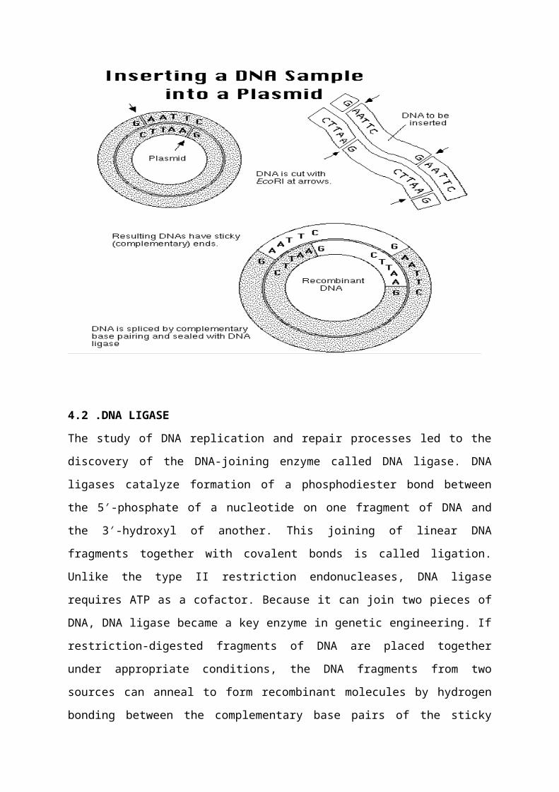
4.2 .DNA LIGASE
The study of DNA replication and repair processes led to the discovery of the DNA-joining
enzyme called DNA ligase. DNA ligases catalyze formation of a phosphodiester bond
between the 5′-phosphate of a nucleotide on one fragment of DNA and the 3′-hydroxyl of
another. This joining of linear DNA fragments together with covalent bonds is called ligation.
Unlike the type II restriction endonucleases, DNA ligase requires ATP as a cofactor. Because
it can join two pieces of DNA, DNA ligase became a key enzyme in genetic engineering. If
restriction-digested fragments of DNA are placed together under appropriate conditions, the
DNA fragments from two sources can anneal to form recombinant molecules by hydrogen
bonding between the complementary base pairs of the sticky ends. However, the two strands
are not covalently bonded by phosphodiester bonds. DNA ligase is required to seal the gaps,
covalently bonding the two strands and regenerating a circular molecule. The DNA ligase
most widely used in the lab is derived from the bacteriophage T4. T4 DNA ligase will also
ligate fragments with blunt ends, but the reaction is less efficient and higher concentrations of
the enzyme are usually required in vitro. To increase the efficiency of the reaction,
researchers often use the enzyme terminal deoxynucleotidyl transferase to modify the blunt
ends. For example, if a single-stranded poly(dA) tail is added to DNA fragments from one
source, and a single stranded poly (dT) tail is added to DNA from another source, the
complementary tails can hydrogen bond.
Recombinant DNA molecules can then be created by ligation.
5.CLONING
MeTHOD
5.1.Genomic Library:
A genomic library contains DNA fragments that represent the entire genome of an organism.
The first step in creating a genomic library is to break the DNA into manageable size pieces
usually by partial restriction endonuclease digest. Under limiting conditions, any particular
restriction site is cleaved only occasionally, so not all sites are cleaved in any particular DNA
molecule. This generates a continuum of overlapping fragments. The second step is to purify
fragments of optimal size by gel electrophoresis or centrifugation techniques. With an
average insert size of 20 kb, the number of random fragments to ensure with high probability
(95–99%) that every sequence is represented is approximately 106 clones for humans.
5.2.cDNA Library:
The principle behind cDNA cloning is that an mRNA population isolated from a specific
tissue, cell type, or developmental stage (e.g. embryo mRNA) should contain mRNAs
specific for any protein expressed in that cell type or during that stage, along with
“housekeeping” mRNAs that encode essential proteins such as the ribosomal proteins, and
other mRNAs common to many cell types or stages of development. Thus, if mRNA can be
isolated, a small subset of all the genes in a genome can be studied. mRNA cannot be cloned
directly, but a cDNA copy of the mRNA can be cloned. Because a cDNA library is derived
from mRNA, the library contains the coding region of expressed genes only, with no introns
or regulatory regions. This latter point becomes important for applications of recombinant
DNA technology to the production of transgenic animals and for human gene therapy
6.COLONY
HYBRIDIZATION

Hybrid molecular complexes (usually called hybrids) of the following types can exist (other
combinations are possible):
DNA-DNA. A single-stranded DNA molecule can form a double-stranded, base-
paired hybrid with a ssDNA target if the probe sequence is the reverse complement of
the target sequence. A radiolabeled DNA probe can be applied to DNA from a gel
transferred to a membrane, called a Southern Blot (named for its inventor).
DNA-RNA. A single-stranded DNA (ssDNA) probe molecule can form a double-
stranded, base-paired hybrid with an RNA (RNA is usually a single-strand) target if
the probe sequence is the reverse complement of the target sequence. An RNA can be
radiolabeled to probe a Southern Blot; or, a ssDNA probe can be applied to
membrane-bound RNA, called a Northern Blot (name is a pun on Southern.)
Protein-Protein. An antibody probe molecule (antibodies are proteins) can form a
complex with a target protein molecule if the antibody's antigen-binding site can bind
to an epitope (small antigenic region) on the target protein. In this case, the hybrid is
called an 'antigen-antibody complex' or 'complex' for short. A radiolabeled antibody
can probe membrane-bound proteins, called a Western Blot (an even worse pun.)
There are two important features of hybridization:
Hybridization reactions are specific - the probes will only bind to targets with
Scomplimentary sequence (or, in the case of antibodies, sites with the correct 3-d
shape).
Hybridization reactions will occur in the presence of large quantities of molecules similar but
not identical to the target. That is, a probe can find one molecule of target in a mixture of
zillions of related but non-complementary molecules
7.PCR
(POLYMERASE
CHAIN
REACTION)
PCR (POLYMERASE CHAIN REACTION)
In PCR, a heat-stable DNA polymerase is used, most commonly Taq Polymerase from the
thermophilic microbe Thermus aquaticus. Thomas Brock discovered T. aquaticus from a hot
spring at Yellowstone National Park.
More recently, an even more heat-resistant polymerase has been developed from a
hyperthermophilic microbe growing at 110 degrees C in hydrothermal vent ecosystems in the
deep ocean; it's called "Vent Polymerase." The Taq Polymerase is put with the DNA to be
amplified, plus all four NTPs, plus two primers facing each other, about 200 - 6000 kb
apart.The primers are selected based on the DNA region you want to amplify. The tube is
placed in a thermal cycler. DNA gets synthesized from each primer, for about 2 minutes.
Then the temperature is raised to 95 degrees C -- enough to denature (split apart) the DNA
base pairs. But the Taq Polymerase remains intact, because it comes from an organism that
evolved to grow at this temperature. Now the temperature is decreased again, and primers
again can hybridize to the DNA--both the old AND the newly synthesized strands. Again,
Taq Polymerase extends new DNA strands. Again, the temperature is raised. After repeated
cycles, the amount of DNA sequence between the two primers increases exponentially. First
2 strands, then 4, 8, 16, up to about a million. Thus, in a couple of hours, you can get
million-fold amplification of a DNA sequence.
PCR has replaced cloning for many purposes, particularly the sequencing of DNA. It is
faster and requires no vectors, which can mutate as they reproduce. It can be used
forensically, to amplify tiny amounts of DNA from criminal evidence; or clinically, to detect
DNA sequences linked to inherited disorders.
8.TRANSGENICS
Cloning in Animals.
Animals have two different classes of cells: germ line and somatic. Only alterations in the germ cells can be transmitted to future generations. However, some forms of somatic cell gene therapy can be useful in treating patients. For example, people with cystic fibrosis can receive the cloned CFTR gene in a nasal spray, which then infects the lining of their lungs and improves lung function. The infected (transformed) epithelial cells eventually are lost, however, during normal processes of tissue growth, so the treatment needs to be repeated.
Germ-line cloning. There are two basic ways to clone a gene in the mammalian germ line.
Inject a DNA fragment containing your gene into the nucleus of a fertilized egg (germ-line gene cloning) or of body tissues in a mature host (somatic gene cloning). The DNA gets taken up at random somewhere in one of the chromosomes. An example of germ-line cloning is the injection of angiopoietin cDNA into the fertilized mouse egg. (An example of somatic cloning is gene therapy for cystic fibrosis: inhale a vector containing the CF gene, which gets incorporated into the DNA of cells lining the lungs. Somatic cloned genes are not inherited by offspring.)
Put a transgene containing positive and negative selective markers into an embryonic stem cell tissue culture. The ES cells take up the gene by homologous recombination, replacing the host allele. The ES cells now are injected into a blastula of an unrelated host, and some of the next generation progeny arise from the ES cells.
But when you create a clone, you need to know:
DNA: Did the transgene really incorporate into the genome? Where? RNA: Is the RNA expressed? (Or prevented from expression, by a null allele?) Protein: Is a desired protein expressed
Cloning in Plants.
Plants don't have separate somatic and germ cells, so creating transgenic plants are easier than creating transgenic animals. Techniques include the use of microprojectile bombardment (the "gene gun") and the use of Agrobacterium tumefaciens.