Embed Size (px)
Citation preview
User Manual
Corporate HeadquartersInvitrogen Corporation1600 Faraday AvenueCarlsbad, CA 92008T: 1 760 603 7200F: 1 760 602 6500E: [email protected]
For country-specific contact information visit our web site at www.invitrogen.com
Rat Primary Cortical Astrocytes Catalog no. N7745-100
Rev. date: 14 May 2009 Manual part no. A11231 MAN0001664

ii
iii
Contents
Contents and Storage........................................................................................... iv
Additional Products.............................................................................................. v
Introduction ........................................................................................ 1
Rat Primary Cortical Astrocytes ..........................................................................1
Methods............................................................................................... 3
Handling Rat Primary Cortical Astrocytes........................................................3
Media Requirements..............................................................................................4
Thawing Rat Primary Cortical Astrocytes .........................................................5
Expanding Rat Primary Cortical Astrocytes .....................................................7
Characterizing Phenotype of Rat Primary Cortical Astrocytes......................9
Phenotype Marker Expression of Rat Primary Cortical Astrocytes ............11
Troubleshooting ...................................................................................................12
Appendix ........................................................................................... 13
Recipes ...................................................................................................................13
Technical Support ................................................................................................14
Purchaser Notification.........................................................................................15
References..............................................................................................................17
Contents and Storage
Shipping and Storage
Rat Primary Cortical Astrocytes are shipped on dry ice. Upon receipt, store the cells in liquid nitrogen.
Contents Amount supplied: One vial containing 1 × 10⁶ viable cells.
Composition: 1 mL of cells in freezing medium.* *Freezing medium: 90% Astrocyte growth medium (85% Dulbecco’s Modified Eagle medium containing 4.5 g/L glucose, and 15% Fetal Bovine Serum) plus 10% DMSO.
Handle cells as potentially biohazardous material under at least Biosafety Level 1 (BL-1) containment. This product contains Dimethyl Sulfoxide (DMSO), a hazardous material. Review the Material Safety Data Sheet (MSDS) before handling. Material Safety Data Sheets (MSDSs) are available on our website at www.invitrogen.com/msds.
iv
v
Additional Products
Additional Products
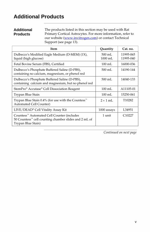
The products listed in this section may be used with Rat Primary Cortical Astrocytes. For more information, refer to our website (www.invitrogen.com) or contact Technical Support (see page 13).
Item Quantity Cat. no.
Dulbecco’s Modified Eagle Medium (D-MEM) (1X), liquid (high glucose)
500 mL 1000 mL
11995-06511995-040
Fetal Bovine Serum (FBS), Certified 100 mL 16000-036
Dulbecco’s Phosphate Buffered Saline (D-PBS), containing no calcium, magnesium, or phenol red
500 mL 14190-144
Dulbecco’s Phosphate Buffered Saline (D-PBS), containing calcium and magnesium, but no phenol red
500 mL 14040-133
StemPro® Accutase® Cell Dissociation Reagent 100 mL A11105-01
Trypan Blue Stain 100 mL 15250-061
Trypan Blue Stain 0.4% (for use with the Countess™ Automated Cell Counter)
2 1 mL T10282
LIVE/DEAD® Cell Vitality Assay Kit 1000 assays L34951
Countess™ Automated Cell Counter (includes 50 Countess™ cell counting chamber slides and 2 mL of Trypan Blue Stain)
1 unit C10227
Continued on next page
vi
Additional Products, continued
Products for Marker Analysis
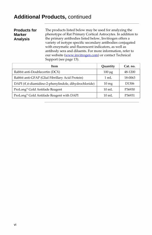
The products listed below may be used for analyzing the phenotype of Rat Primary Cortical Astrocytes. In addition to the primary antibodies listed below, Invitrogen offers a variety of isotype specific secondary antibodies conjugated with enzymatic and fluorescent indicators, as well as antibody sera and diluents. For more information, refer to our website (www.invitrogen.com) or contact Technical Support (see page 13).
Item Quantity Cat. no.
Rabbit anti-Doublecortin (DCX) 100 μg 48-1200
Rabbit anti-GFAP (Glial Fibrillary Acid Protein) 1 mL 18-0063
DAPI (4,6-diamidino-2-phenylindole, dihydrochloride) 10 mg D1306
ProLong® Gold Antifade Reagent 10 mL P36930
ProLong® Gold Antifade Reagent with DAPI 10 mL P36931
Introduction
Rat Primary Cortical Astrocytes
Introduction Astrocytes are by far the most numerous cell type in the
central nervous system (CNS), outnumbering their neuronal counterparts by approximately tenfold, and have critical roles in adult CNS homeostasis (Pekny & Nilsson, 2005). They provide biochemical and nutritional support of neurons and endothelial cells which form the blood-brain barrier, perform the vast majority of synaptic glutamate uptake, and maintain extracellular potassium levels (Rothstein et al., 1996; Rothstein et al., 1994). Astroglial dysfunction has been implicated in a number of CNS pathologies including amyotrophic lateral sclerosis (ALS) and ischemic neuronal death (Maragakis & Rothstein, 2006; Takano et al., 2009), and transplantation-based astrocyte replacement therapy has been shown to be a promising therapeutic strategy against neuronal death (Lepore et al., 2008). Although there are few known differences between cortical and hippocampal astrocytes, it has been reported that astrocytes from different regions of the brain show a differential sensitivity to ischemic injury (Xu et al., 2001; Zhao & Flavin, 2000).
Source of Rat Primary Cortical Astrocytes
Rat Primary Cortical Astrocytes are isolated from the cortices of fetal Sprague-Dawley rats at embryonic day 19 (E19) of gestation. The cells are isolated from tissue under sterile conditions, placed through one round of enzymatic dissociation and expansion in astrocyte growth medium (85% Dulbecco’s Modified Eagle medium containing 4.5 g/L glucose, and 15% Fetal Bovine Serum). The cells are cryopreserved at passage 1 (P1) in 90% astrocyte growth medium plus 10% DMSO. Each vial of Rat Primary Cortical Astrocytes contains 1 × 10⁶ cells/mL that can be expanded in culture for at least one passage.
Continued on next page
Rat Primary Cortical Astrocytes, continued
Characteristics of Rat Primary Cortical Astrocytes
Isolated from the brain cortex of fetal Sprague-Dawley rats at embryonic day 19 (E19) of gestation
Exhibit 70% viability upon thawing
Stain > 80% positive for the astrocyte specific marker, glial fibrillary acid protein (GFAP)
Stain 10% positive for neuron and oligodendrocyte specific markers galactocerebroside (GalC) and doublecortin (DCX)
Exhibit a doubling time of approximately 9 days at P2
Can be expanded in culture for at least one passage
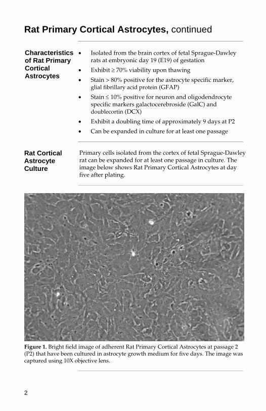
Rat Cortical Astrocyte Culture
Primary cells isolated from the cortex of fetal Sprague-Dawley rat can be expanded for at least one passage in culture. The image below shows Rat Primary Cortical Astrocytes at day five after plating.
Figure 1. Bright field image of adherent Rat Primary Cortical Astrocytes at passage 2 (P2) that have been cultured in astrocyte growth medium for five days. The image was captured using 10X objective lens.
2
Methods
Handling Rat Primary Cortical Astrocytes
As with other mammalian cells, when working with Rat Primary Cortical Astrocytes, handle as potentially biohazardous material under at least Biosafety Level 1 (BL-1) containment. For more information on BL-1 guidelines, refer to Biosafety in Microbiological and Biomedical Laboratories, 5th ed., published by the Centers for Disease Control, or see the following website: www.cdc.gov/od/ohs/biosfty/bmbl5/bmbl5toc.htm
Guidelines for Culturing Rat Primary Cortical Astrocytes
Follow the general guidelines below to grow and maintain Rat Primary Cortical Astrocytes.
All solutions and equipment that come in contact with the cells must be sterile. Always use proper aseptic technique and work in a laminar flow hood.
For consistent results in your experiments, we recommend using cells below passage 3 (P3). If you expand Rat Primary Cortical Astrocytes beyond P3, we recommend that you perform another round of characterization prior to further experiments.
For general maintenance of Rat Primary Cortical Astrocytes as an adherent culture, passage cells when they reach 100% confluency at a seeding density of 20,000 cells/cm2.
When thawing or subculturing cells, transfer cells into pre-warmed medium.
Standard physical growth conditions for Rat Primary Cortical Astrocytes are 37°C in a humidified atmosphere of 5% CO2 in air.
��������
We recommend that you use Rat Primary Cortical Astrocytes right after recovery. After thawing Rat Primary Cortical Astrocytes, expand the cells once to have a 1.5 to 2-fold increase in their number, and harvest them to use in your experiments (e.g., transplantation experiments, metabolic studies).
Continued on next page
3
4
Media Requirements
Astrocyte Growth Medium
You can grow Rat Primary Cortical Astrocytes as an adherent culture on uncoated, tissue culture treated vessels. For optimal growth and expansion of these cells, we recommend using astrocyte growth medium consisting of 85% Dulbecco’s Modified Eagle medium (containing 4.5 g/L glucose) and 15% Fetal Bovine Serum. This medium is designed to support isolation and growth of astrocytes derived from cortical tissue of fetal rats.
Prepare your growth medium prior to use.
When thawing or subculturing cells, transfer cells into pre-warmed medium at 37°C.
We recommend that you aliquot astrocyte growth medium into required working amounts to avoid exposing it to 37°C multiple times.
Component Cat. no. Amount
Dulbecco’s Modified Eagle medium (high glucose)
11995-065 85%
Fetal Bovine Serum 16000-036 15%
Thawing Rat Primary Cortical Astrocytes
Materials Needed
The following materials are required (see page v for ordering information).
Rat Primary Cortical Astrocytes, stored in liquid nitrogen
Ethanol or 70% isopropanol
Astrocyte growth medium (see page 4); pre-warmed to 37°C
Disposable, sterile 15-mL tubes
Flame-polished and autoclaved glass Pasteur pipettes, or plastic Pasteur pipettes pre-rinsed with growth medium
37°C water bath
37°C incubator with a humidified atmosphere of 5% CO2
Microcentrifuge
Tissue-culture treated flasks, plates, or Petri dishes (uncoated)
Hemacytometer, cell counter and Trypan Blue, LIVE/DEAD® Cell Vitality Assay Kit, or the Countess™ Automated Cell Counter
The Countess™ Automated Cell Counter is a benchtop instrument designed to measure cell count and viability (live, dead, and total cells) accurately and precisely in less than a minute per sample, using the standard Trypan Blue uptake technique (see page v for ordering information).
Using the same amount of sample that you currently use with the hemocytometer, the Countess™ Automated Cell Counter takes less than a minute per sample for a typical cell count and is compatible with a wide variety of eukaryotic cells.
��������
Rat Primary Cortical Astrocytes readily stick to the plastic used in cell culture dishes and centrifuge tubes. Prior to use, rinse all material that will come in contact with the cells with medium to prevent cells from sticking to the plastic. To thaw and establish Rat Primary Cortical Astrocytes, follow the procedure on the next page.
Continued on next page
5
6
Thawing Rat Primary Cortical Astrocytes, continued
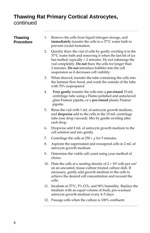
Thawing Procedure
1. Remove the cells from liquid nitrogen storage, and immediately transfer the cells to a 37°C water bath to prevent crystal formation.
2. Quickly thaw the vial of cells by gently swirling it in the 37°C water bath and removing it when the last bit of ice has melted, typically < 2 minutes. Do not submerge the vial completely. Do not thaw the cells for longer than 2 minutes. Do not introduce bubbles into the cell suspension as it decreases cell viability.
3. When thawed, transfer the tube containing the cells into the laminar flow hood, and wash the outside of the tube with 70% isopropanol.
4. Very gently transfer the cells into a pre-rinsed 15-mL centrifuge tube using a Flame-polished and autoclaved glass Pasteur pipette, or a pre-rinsed plastic Pasteur pipette.
5. Rinse the vial with 1 mL of astrocyte growth medium, and dropwise add to the cells in the 15-mL centrifuge tube (one drop/second). Mix by gentle swirling after each drop.
6. Dropwise add 8 mL of astrocyte growth medium to the cell solution and mix gently.
7. Centrifuge the cells at 250 g for 5 minutes.
8. Aspirate the supernatant and resuspend cells in 2 mL of astrocyte growth medium.
9. Determine the viable cell count using your method of choice.
10. Plate the cells at a seeding density of 2 104 cells per cm2 on an uncoated, tissue-culture treated culture dish. If necessary, gently add growth medium to the cells to achieve the desired cell concentration and recount the cells.
11. Incubate at 37°C, 5% CO2, and 90% humidity. Replace the medium with an equal volume of fresh, pre-warmed astrocyte growth medium every 4–5 days.
12. Passage cells when the culture is 100% confluent.
Expanding Rat Primary Cortical Astrocytes
Introduction You may expand Rat Primary Cortical Astrocytes as an
adherent culture on uncoated, tissue-culture treated flasks, plates or dishes. Subculture your cells when 100% confluent.
Note: We recommend that you use Rat Primary Cortical Astrocytes right after recovery. After thawing Rat Primary Cortical Astrocytes, expand the cells once to have a 1.5 to 2-fold increase in their number, and harvest them to use in your experiments (e.g., transplantation experiments, metabolic studies).
Materials Needed
The following materials are required for passaging Rat Primary Cortical Astrocytes (see page v for ordering information).
Culture vessels containing Rat Primary Cortical Astrocytes (100% confluent)
Uncoated, tissue-culture treated flasks, plates, or Petri dishes
Astrocyte growth medium (see page 4), pre-warmed to 37°C
Disposable, sterile 15-mL or 50-mL conical tubes, pre-rinsed with medium
37°C incubator with humidified atmosphere of 5% CO2
Dulbecco’s Phosphate Buffered Saline (D-PBS), containing no calcium, magnesium, or phenol red
StemPro® Accutase® Cell Dissociation Reagent (see page v), pre-warmed to 37°C
Hemacytometer, cell counter and Trypan Blue, LIVE/DEAD® Cell Vitality Assay Kit, or the Countess™ Automated Cell Counter
��������
Rat Primary Cortical Astrocytes readily stick to the plastic used in cell culture dishes and centrifuge tubes. Prior to use, rinse all material that will come in contact with the cells with medium to prevent cells from sticking to the plastic. To passage Rat Primary Cortical Astrocytes, follow the procedure on the next page.
Continued on next page
7
8
Expanding Rat Primary Cortical Astrocytes, continued
Passaging Rat Primary Cortical Astrocytes
1. Remove the spent growth medium from the culture dish containing the cells, and store in a sterile tube to use as a washing solution.
2. Rinse the surface of the cell layer once with D-PBS without Ca2+ and Mg2+ (approximately 2 mL D-PBS per 10 cm2 culture surface area) by adding the D-PBS to the side of the vessel opposite the attached cell layer, and rocking back and forth several times.
3. Aspirate the D-PBS and discard.
4. To detach the cells, add 3 mL of pre-warmed StemPro® Accutase® Cell Dissociation Reagent per T75 flask; adjust volume accordingly for culture dishes of other sizes.
5. Incubate for up to 20 minutes at 37°C. Rock the cells every 5 minutes, and check for cell detachment and dissociation toward single cell under the microscope.
6. Once you observe cell detachment, gently pipette up and down to break clumps into a single cell suspension. Stop the cell dissociation reaction by an adding equal volume of the spent medium from step 1. Disperse the medium by pipetteting over the cell layer surface several times.
7. Transfer the cells to a new 15-mL or 50-mL pre-rinsed conical tube, and centrifuge at 250 g for 5 minutes at room temperature. Aspirate and discard the supernatant.
8. Gently resuspend the cell pellet in pre-warmed astrocyte growth medium and remove a sample for counting.
9. Determine the total number of cells and percent viability using your method of choice. If necessary, add astrocyte growth medium to the cells to achieve the desired cell concentration and recount the cells.
10. Plate cells in an uncoated tissue-culture treated flask, plate, or Petri dish at a seeding density of 2 104 cells per cm2.
11. Incubate cells at 37°C, 5% CO2, and 90% humidity, and change growth medium every 4–5 days.
9
Characterizing Phenotype of Rat Primary Cortical Astrocytes
Phenotypic Markers
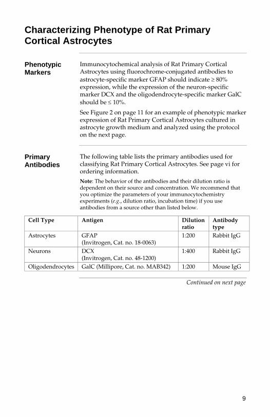
Immunocytochemical analysis of Rat Primary Cortical Astrocytes using fluorochrome-conjugated antibodies to astrocyte-specific marker GFAP should indicate 80% expression, while the expression of the neuron-specific marker DCX and the oligodendrocyte-specific marker GalC should be 10%.
See Figure 2 on page 11 for an example of phenotypic marker expression of Rat Primary Cortical Astrocytes cultured in astrocyte growth medium and analyzed using the protocol on the next page.
Primary Antibodies
The following table lists the primary antibodies used for classifying Rat Primary Cortical Astrocytes. See page vi for ordering information.
Note: The behavior of the antibodies and their dilution ratio is dependent on their source and concentration. We recommend that you optimize the parameters of your immunocytochemistry experiments (e.g., dilution ratio, incubation time) if you use antibodies from a source other than listed below.
Cell Type Antigen Dilution ratio
Antibody type
Astrocytes GFAP (Invitrogen, Cat. no. 18-0063)
1:200 Rabbit IgG
Neurons DCX (Invitrogen, Cat. no. 48-1200)
1:400 Rabbit IgG
Oligodendrocytes GalC (Millipore, Cat. no. MAB342) 1:200 Mouse IgG
Continued on next page
10
Characterizing Phenotype of Rat Primary Cortical Astrocytes, continued
Immunocyto-chemistry
Fixing Cells:
1. Remove culture medium and gently rinse the cells once with D-PBS without dislodging.
2. Fix the cells with 4% fresh Paraformaldehyde Fixing Solution (PFA; see Appendix, page 13 for recipe) at room temperature for 15 minutes.
3. Rinse 3X with D-PBS containing Ca2+ and Mg2+.
4. Check for presence of cells after fixing.
5. Proceed to staining on the next page. You may also store slides for up to 3–4 weeks in D-PBS at 4°C. Do not allow slides to dry.
Staining Cells:
1. Incubate cells for 30–60 minutes in blocking buffer (5% serum of the secondary antibody host species, 1% BSA, 0.1% Triton-X in D-PBS with Ca2+ and Mg2+). Note: If you are using a surface antigen such as GalC, omit Triton-X in blocking buffer.
2. Remove the blocking buffer and incubate cells overnight at 4°C with primary antibody diluted in D-PBS containing 5% serum. Ensure that the cell surfaces are covered uniformly with the antibody solution.
3. Wash the cells 3X for 5 minutes with D-PBS containing Ca2+ and Mg2+ (if using a slide, use a staining dish with a magnetic stirrer).
4. Incubate the cells with fluorescence-labeled secondary antibody (5% serum in D-PBS with Ca2+ and Mg2+) in the dark at 37°C for 30–45 minutes.
5. Wash the cells 3X with D-PBS containing Ca2+ and Mg2+, and in the last wash counter stain with DAPI solution (3 ng/mL) for 5 minutes, and rinse with D-PBS.
6. If desired, mount with 3 drops of ProLong® Gold antifade reagent per slide and seal with the cover slip (see page vi for ordering information). You may store the slides in the dark at 4°C.
Phenotype Marker Expression of Rat Primary Cortical Astrocytes
Astrocyte-specific Marker Expression
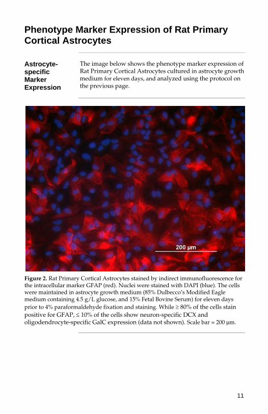
The image below shows the phenotype marker expression of Rat Primary Cortical Astrocytes cultured in astrocyte growth medium for eleven days, and analyzed using the protocol on the previous page.
Figure 2. Rat Primary Cortical Astrocytes stained by indirect immunofluorescence for the intracellular marker GFAP (red). Nuclei were stained with DAPI (blue). The cells were maintained in astrocyte growth medium (85% Dulbecco’s Modified Eagle medium containing 4.5 g/L glucose, and 15% Fetal Bovine Serum) for eleven days prior to 4% paraformaldehyde fixation and staining. While 80% of the cells stain positive for GFAP, 10% of the cells show neuron-specific DCX and oligodendrocyte-specific GalC expression (data not shown). Scale bar = 200 μm.
11
12
Troubleshooting
Culturing Cells
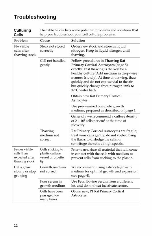
The table below lists some potential problems and solutions that help you troubleshoot your cell culture problems.
Problem Cause Solution
Stock not stored correctly
Order new stock and store in liquid nitrogen. Keep in liquid nitrogen until thawing.
Follow procedures in Thawing Rat Primary Cortical Astrocytes (page 5) exactly. Fast thawing is the key for a healthy culture. Add medium in drop-wise manner (slowly). At time of thawing, thaw quickly and do not expose vial to the air but quickly change from nitrogen tank to 37°C water bath.
Obtain new Rat Primary Cortical Astrocytes.
Use pre-warmed complete growth medium, prepared as described on page 4.
Cell not handled gently
Generally we recommend a culture density of 2 104 cells per cm2 at the time of recovery.
No viable cells after thawing stock
Thawing medium not correct
Rat Primary Cortical Astrocytes are fragile; treat your cells gently, do not vortex, bang the flasks to dislodge the cells, or centrifuge the cells at high speeds.
Fewer viable cells than expected after thawing stock
Cells sticking to plastic culture vessel or pipette tip
Prior to use, rinse all material that will come in contact with the cells with medium to prevent cells from sticking to the plastic.
Growth medium not correct
We recommend using astrocyte growth medium for optimal growth and expansion (see page 4).
Poor serum in growth medium
Use Fetal Bovine Serum from a different lot, and do not heat inactivate serum.
Cells grow slowly or stop growing
Cells have been passaged too many times
Obtain new, P1 Rat Primary Cortical Astrocytes.
13
Appendix
Recipes
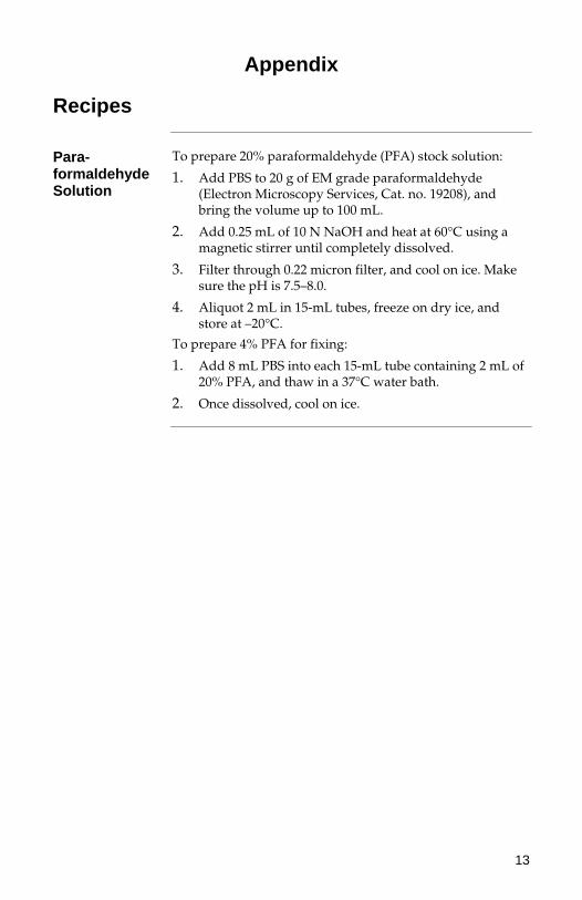
Para-formaldehyde Solution
To prepare 20% paraformaldehyde (PFA) stock solution:
1. Add PBS to 20 g of EM grade paraformaldehyde (Electron Microscopy Services, Cat. no. 19208), and bring the volume up to 100 mL.
2. Add 0.25 mL of 10 N NaOH and heat at 60°C using a magnetic stirrer until completely dissolved.
3. Filter through 0.22 micron filter, and cool on ice. Make sure the pH is 7.5–8.0.
4. Aliquot 2 mL in 15-mL tubes, freeze on dry ice, and store at –20°C.
To prepare 4% PFA for fixing:
1. Add 8 mL PBS into each 15-mL tube containing 2 mL of 20% PFA, and thaw in a 37°C water bath.
2. Once dissolved, cool on ice.

Technical Support
Web Resources
Visit the Invitrogen website at www.invitrogen.com for:
Technical resources, including manuals, vector maps and sequences, application notes, MSDSs, FAQs, formulations, citations, handbooks, etc.
Complete Technical Support contact information
Access to the Invitrogen Online Catalog
Additional product information and special offers
Contact Us For more information or technical assistance, call, write, fax,
or email. Additional international offices are listed on our website (www.invitrogen.com).
Corporate Headquarters: 5791 Van Allen Way Carlsbad, CA 92008 USA Tel: 1 760 603 7200 Tel (Toll Free): 1 800 955 6288 Fax: 1 760 602 6500 E-mail: [email protected]
Japanese Headquarters: LOOP-X Bldg. 6F 3-9-15, Kaigan Minato-ku, Tokyo 108-0022 Tel: 81 3 5730 6509 Fax: 81 3 5730 6519 E-mail: [email protected]
European Headquarters: Inchinnan Business Park 3 Fountain Drive Paisley PA4 9RF, UK Tel: 44 (0) 141 814 6100 Tech Fax: 44 (0) 141 814 6117 E-mail: [email protected]
Material Safety Data Sheets (MSDSs)
MSDSs (Material Safety Data Sheets) are available on our website at www.invitrogen.com/msds.
Certificate of Analysis
The Certificate of Analysis provides detailed quality control information for each product. Certificates of Analysis are available on our website. Go to www.invitrogen.com/support and search for the Certificate of Analysis by product lot number, which is printed on the box.
14
15
Purchaser Notification
Limited Warranty
Invitrogen (a part of Life Technologies Corporation) is committed to providing our customers with high-quality goods and services. Our goal is to ensure that every customer is 100% satisfied with our products and our service. If you should have any questions or concerns about an Invitrogen product or service, contact our Technical Support Representatives. All Invitrogen products are warranted to perform according to specifications stated on the certificate of analysis. The Company will replace, free of charge, any product that does not meet those specifications. This warranty limits the Company’s liability to only the price of the product. No warranty is granted for products beyond their listed expiration date. No warranty is applicable unless all product components are stored in accordance with instructions. The Company reserves the right to select the method(s) used to analyze a product unless the Company agrees to a specified method in writing prior to acceptance of the order. Invitrogen makes every effort to ensure the accuracy of its publications, but realizes that the occasional typographical or other error is inevitable. Therefore the Company makes no warranty of any kind regarding the contents of any publications or documentation. If you discover an error in any of our publications, please report it to our Technical Support Representatives. Life Technologies Corporation shall have no responsibility or liability for any special, incidental, indirect or consequential loss or damage whatsoever. The above limited warranty is sole and exclusive. No other warranty is made, whether expressed or implied, including any warranty of merchantability or fitness for a particular purpose.
16
Purchaser Notification, continued
Limited Use Label License No. 5: Invitrogen Technology
The purchase of this product conveys to the buyer the non-transferable right to use the purchased amount of the product and components of the product in research conducted by the buyer (whether the buyer is an academic or for-profit entity). The buyer cannot sell or otherwise transfer (a) this product (b) its components or (c) materials made using this product or its components to a third party or otherwise use this product or its components or materials made using this product or its components for Commercial Purposes. The buyer may transfer information or materials made through the use of this product to a scientific collaborator, provided that such transfer is not for any Commercial Purpose, and that such collaborator agrees in writing (a) not to transfer such materials to any third party, and (b) to use such transferred materials and/or information solely for research and not for Commercial Purposes. Commercial Purposes means any activity by a party for consideration and may include, but is not limited to: (1) use of the product or its components in manufacturing; (2) use of the product or its components to provide a service, information, or data; (3) use of the product or its components for therapeutic, diagnostic or prophylactic purposes; or (4) resale of the product or its components, whether or not such product or its components are resold for use in research. For products that are subject to multiple limited use label licenses, the terms of the most restrictive limited use label license shall control. Life Technologies Corporation will not assert a claim against the buyer of infringement of patents owned or controlled by Life Technologies Corporation which cover this product based upon the manufacture, use or sale of a therapeutic, clinical diagnostic, vaccine or prophylactic product developed in research by the buyer in which this product or its components was employed, provided that neither this product nor any of its components was used in the manufacture of such product. If the purchaser is not willing to accept the limitations of this limited use statement, Life Technologies is willing to accept return of the product with a full refund. For information on purchasing a license to this product for purposes other than research, contact Licensing Department, Life Technologies Corporation, 5791 Van Allen Way, Carlsbad, California 92008. Phone (760) 603-7200. Fax (760) 602-6500. Email: [email protected].
17
References
Lepore, A. C., Rauck, B., Dejea, C., Pardo, A. C., Rao, M. S., Rothstein, J. D., and
Maragakis, N. J. (2008) Focal transplantation-based astrocyte replacement is neuroprotective in a model of motor neuron disease. Nat Neurosci 11, 1294-1301
Maragakis, N. J., and Rothstein, J. D. (2006) Mechanisms of Disease: astrocytes in neurodegenerative disease. Nat Clin Pract Neurol 2, 679-689
Pekny, M., and Nilsson, M. (2005) Astrocyte activation and reactive gliosis. Glia 50, 427-434
Rothstein, J. D., Dykes-Hoberg, M., Pardo, C. A., Bristol, L. A., Jin, L., Kuncl, R. W., Kanai, Y., Hediger, M. A., Wang, Y., Schielke, J. P., and Welty, D. F. (1996) Knockout of glutamate transporters reveals a major role for astroglial transport in excitotoxicity and clearance of glutamate. Neuron 16, 675-686
Rothstein, J. D., Martin, L., Levey, A. I., Dykes-Hoberg, M., Jin, L., Wu, D., Nash, N., and Kuncl, R. W. (1994) Localization of neuronal and glial glutamate transporters. Neuron 13, 713-725
Takano, T., Oberheim, N., Cotrina, M. L., and Nedergaard, M. (2009) Astrocytes and ischemic injury. Stroke 40, S8-12
Xu, L., Sapolsky, R. M., and Giffard, R. G. (2001) Differential sensitivity of murine astrocytes and neurons from different brain regions to injury. Exp Neurol 169, 416-424
Zhao, G., and Flavin, M. P. (2000) Differential sensitivity of rat hippocampal and cortical astrocytes to oxygen-glucose deprivation injury. Neurosci Lett 285, 177-180
©2009 Life Technologies Corporation. All rights reserved. For research use only. Not intended for any animal or human therapeutic or diagnostic use.
User Manual
Corporate HeadquartersInvitrogen Corporation5791 Van Allen WayCarlsbad, CA 92008T: 1 760 603 7200F: 1 760 602 6500E: [email protected]
For country-specific contact information, visit our web site at www.invitrogen.com