Embed Size (px)
Citation preview
Vol. 54, No. 12
Reconstitution of the Gastrointestinal Microflora ofLactobacillus-Free Mice
GERALD W. TANNOCK,1t* CAROLINE CRICHTON,' GJALT W. WELLING,2 JOOP P. KOOPMAN,3AND TORE MIDTVEDT4
Department of Microbiology, University of Otago, Dunedin, New Zealand'; Department of Medical Microbiology, StateUniversity, Groningen,2 and Central Animal Laboratory, Catholic University, Nijmegen,3 The Netherlands; and
Department of Medical Microbial Ecology, Karolinska Institute, Stockholm, Sweden4
Received 20 June 1988/Accepted 22 September 1988
A colony of mice that do not harbor lactobacilli in their digestive tracts but whose intestinal microflora isotherwise functionally similar to that of conventional animals was derived. Methods used to reconstitute theintestinal microflora of the mice included inoculation of the animals with cultures of specific microbes,noncultivable microbes attached to epithelial cells, and cecal contents from conventional mice treated withchloramphenicol. Twenty-six microflora-associated characteristics were monitored by using relatively simpletests to determine the microflora status of the mice.
Lactobacilli are numerically predominant members of thenormal microflora of the proximal digestive tract of severalanimal species (15). Historically, microbiological interest ingastrointestinal lactobacilli has centered on the supposedbeneficial effects conferred on the animal host by thesemicrobes (20). Modern scientific investigation of lactobacil-lus-host interactions requires the development of an exper-imental animal model with which microbial influences on thehost can be observed under microbiologically constant con-ditions. Gnotobiotic animal technology can be used to main-tain rodents under such conditions, but gnotobiotes monoas-sociated with lactobacilli do not provide a satisfactory modelwith which to study the microecology of these bacteria. Thisis because the colonization of the gastrointestinal tract bylactobacilli is influenced by other members of the normalmicroflora (24). The other members of the microflora alsoinfluence the host in ways that may be critical to theevaluation of the importance of the lactobacilli as inhabitantsof the digestive tract (5). There is a need, therefore, to deriveexperimental animals, housed under gnotobiotic conditions,that do not harbor lactobacilli in their gastrointestinal tractsyet retain a microflora functionally equivalent to that of theirconventional counterparts. Lactobacillus-free (LF) animalsand animals colonized by selected strains of lactobacillicould then be compared to determine the influence of themicrobes on the host.A colony of LF mice was derived previously by using a
combination of gnotobiotic technology and antibiotic treat-ment (24). Briefly, conventional mice were administeredpenicillin in the drinking water and transferred betweensterile germfree isolators until lactobacilli had been elimi-nated from the environment. The descendants of theseanimals have been maintained in an LF state by usinggnotobiotic methodology, in the absence of penicillin admin-istration, for approximately 4 years. Although still harboringa complex collection of microbes, the LF mice lacked some
other members of the gastrointestinal tract microflora inaddition to lactobacilli; filamentous segmented bacteria in-habiting the small bowel of conventional mice were absent,
* Corresponding author.t Present address: AFRC Institute for Food Research, Reading
Laboratory, Shinfield, Reading RG2 9AT, United Kingdom.
as were enterococci from the gastrointestinal tract. The ceca
of LF mice were larger than those of conventional mice,which pointed to the absence of a complete microflora (5,24).We report in this paper the methods used to restore a
conventionally functioning intestinal microflora to LF micewhile maintaining the LF status of the mice. Relativelysimple methods by which the status of the microflora can bemonitored are also described. Particular emphasis was
placed on reconstitution of the large-bowel microflora, sincethis site harbors the largest numbers and greatest diversity ofindigenous microbes inhabiting the gastrointestinal tract(15).
MATERIALS AND METHODS
Mice. All animals (LF, partially reconstituted LF [PRLF],reconstituted LF [RLF], and conventional) in this studywere maintained under identical conditions in flexible plasticisolators (Standard Safety Equipment Co., Palatine, Ill.), fedgamma radiation-sterilized food (pellets B [1]) and auto-claved water, and provided with autoclaved paper bedding(13). LF BALB/c mice were derived as reported previously(24). Conventional BALB/c mice were introduced into asterile isolator and maintained thereafter as if they were
gnotobiotes. PRLF and RLF mice were derived by inocu-lating lightly anesthetized LF or PRLF mice intragastricallywith appropriate preparations as listed below (13). Forexamination of organs, mice were killed by carbon dioxideanesthesia followed by cervical dislocation.Enumeration of gastrointestinal microbes. Lactobacilli, en-
terococci, and coliforms were enumerated by culturing ster-ile distilled water homogenates of gastrointestinal organs on
selective media as described in previous publications (13,19). Fusiform populations were calculated from dilutions oforgan homogenates observed in counting chambers byphase-contrast microscopy (13). Bacteroides were enumer-
ated by culturing brain heart infusion broth dilutions ofhomogenates on brain heart infusion agar plates (9). Homo-genates, dilutions, and cultures were made in an anaerobicglove box (Forma Scientific, Marietta, Ohio).
Observation of spiral-shaped and mucosa-associated mi-crobes. Spiral-shaped bacteria were observed by transmis-sion electron microscopy in negatively stained washings
2971
APPLIED AND ENVIRONMENTAL MICROBIOLOGY, Dec. 1988, p. 2971-29750099-2240/88/122971-05$02.00/0Copyright © 1988, American Society for Microbiology
on June 7, 2020 by guesthttp://aem
.asm.org/
Dow
nloaded from
2972 TANNOCK ET AL.
prepared from the colon mucosa of mice. A length ofproximal colon was tied onto a glass rod and everted. Afterremoval of the bulk of any attached colon contents from themucosal surface, the tissue was agitated vigorously in 10-mlportions of 0.1 M phosphate-buffered saline (pH 7.2) until acloudy suspension was obtained. The washings were centri-fuged at 5,000 x g for 10 min, and the pellet was suspendedin 3 drops of 2% phosphotungstic acid (pH 6.0) and exam-ined by transmission electron microscopy. Epithelium-asso-ciated filamentous segmented microbes inhabiting the smallbowel were observed in specimens by scanning electronmicroscopy as described previously (24). Layers of microbesassociated with the colon mucosa were demonstrated bylight microscopy of sections prepared with a microtome-cryostat and Gram stained (21).
Size, pH, and dry-matter content of the cecum. The cecumwas removed after recording the body weight of the animal.Cecal size was expressed as cecal weight (as a percentage oftotal body weight). Dry-matter content was obtained bylyophilizing preweighed amounts of cecal contents for 24 h.The dry weight of the specimens was determined and ex-pressed as a percentage of the wet weight. In other experi-ments, cecal contents were homogenized in sterile Milli-Qwater (Millipore S.A., Molsheim, France) to make a 10-folddilution (wt/vol). The pH of pooled suspensions from threemice was determined by using a pH meter.
Indole concentration, 0-glucuronidase activity, and volatilefatty acids in cecal contents. Indole concentration in cecalcontents was determined by the method of Whitt and De-moss (28). P-Glucuronidase activity in ultrasonically disin-tegrated cecal contents was determined by the method ofRod and Midtvedt (14). Volatile fatty acids (acetic, propio-nic, and butyric) were detected by gas chromatographicanalysis of distillates from cecal homogenates (13).
,I-Aspartylglycine. Lyophilized cecal contents (5 mg) werereconstituted in 200 RIul of demineralized water and homoge-nized in an ultrasonic bath for 5 min. The homogenate wascentrifuged for 15 min in an Eppendorf centrifuge (11,430 xg), and the supernatant was used to assay for ,B-aspartylgly-cine by high-voltage electrophoresis (27).
Deconjugation of taurodeoxycholic acid by cecal contents.Lyophilized cecal contents (25 mg) were homogenized in 1ml of demineralized water containing 0.1% Triton X-100 byusing an Ultraturrax. The suspension was centrifuged for 15min in an Eppendorf centrifuge (11,430 x g), and theresulting supernatant was then ultracentrifuged (210,000 xg) for 60 min. The supernatant was dialyzed overnightagainst phosphate-buffered saline (pH 7.2) to remove low-molecular-weight substances, including taurine. The reten-tate was used as a cecal enzyme preparation. A 40-pJl sampleof preparation was added to 20 ,ul of taurodeoxycholatesodium salt (2.5 mg/ml of phosphate-buffered saline) andincubated overnight at 37°C. A 45-,lI amount was applied in5-pI portions to 3MM chromatography paper (Whatman,Inc., Clifton, N.J.). A 15-pI portion of a taurine referencesolution (0.6 mg/ml) and a control incubation mixture con-taining 20 RI of phosphate-buffered saline instead of tauro-deoxycholate were also applied. High-voltage paper electro-phoresis was performed as described previously (27). Theamount of taurine liberated from taurodeoxycholate by cecalenzymatic action was semiquantitatively determined by vi-sual comparison with the intensity of the reference solutionof taurine.
Urobilinogen and coprostanol. Urobilinogen was detectedin concentrated cecal homogenates by using Bilugen-Teststrips (Boehringer GmbH, Mannheim, Federal Republic of
Germany). Cecal contents from five mice were pooled anddiluted 10-fold in sterile distilled water. The contents werecentrifuged (12,718 x g for 3 min); the supernatant waslyophilized and then suspended in 1/10th the original volumeof water. Concentrated cecal contents (20-pul volumes) wereapplied to the test strips, and appropriate color reactionswere recorded according to the instructions of the manufac-turer. Coprostanol was detected in cecal homogenates bygas-liquid chromatography according to the method of Carl-stedt-Duke and colleagues (2).
Electrophoresis of cecal contents. Electrophoretic patternsof cecal mucins and proteins were detected by using a gelGNA-100 apparatus (Pharmacia, Uppsala, Sweden) in amanner similar to that described by Gustafsson and Carl-stedt-Duke (6). Agarose gels (0.8%) were prepared in 0.1 Mbarbitone buffer (pH 8.6) on glass plates. Electrophoresis ofconcentrated cecal contents (see above) was carried out byusing barbitone buffer (0.2 M) for 30 min at 20 V/cm. Gelswere pressed between paper towels for about 1 h to expressbuffer from the gel matrix and then dried overnight beforebeing stained with either periodic acid-Schiff reagent, 0.05%toluidine blue stain, or Coomassie brilliant blue R stain (6).API ZYM galleries. ,-Glucuronidase, a-galactosidase, ,B-
galactosidase, P-glucosidase, N-acetyl-p-glucosaminidase,and trysin activities in cecal contents were assayed semi-quantitatively by using API ZYM galleries (API SystemS. A., La Balme les Grottes, France). Cecal contents fromfive mice were pooled and homogenized in sterile saline togive a 10-fold dilution (wt/vol). The homogenates werecentrifuged at 700 x g for 2 min, and the supernatant wasused to inoculate API ZYM galleries (65 pA per cupule). Thegalleries were incubated at 37°C for 4 h, after which theresults were read according to the instructions of the manu-facturer. These enzyme activities were assayed becauselittle if any galactosidase, glucuronidase, glucosidase, orglucosaminidase activity can be detected in the cecal con-tents of germfree mice examined with API ZYM galleries.Trypsin activity, by contrast, is always high in germfreecontents but low in conventional cecal contents (D. D.Whitt, D. C. Savage, and G. W. Tannock, unpublishedobservations).
Proteolytic activity in cecal contents. Cecal contents werehomogenized in 0.05 M Tris buffer (pH 7.8) to give a 10-folddilution (wt/vol). Volumes of 100 and 10 1.l of supernatantobtained from centrifugation of the homogenates (6,647 x gfor 10 min) were added to 5-ml volumes of buffer containing20 mg of hide powder azure (Sigma Chemical Co., St. Louis,Mo.) according to the method of Rinderknecht and col-leagues (12). The suspensions were incubated at 37°C for 30min with frequent agitation. Proteolytic activity was mea-sured colorimetrically at a 595-nm wavelength of light and byreference to a standard curve prepared from trypsin (Sigma).
Reconstitution of the enterococcal component of the intesti-nal microflora. The cecum of each of four conventional micewas removed, homogenized in sterile distilled water to givea 10-fold dilution (wt/vol), and diluted in 10-fold steps to10-6. Then a 100-pul volume of each dilution was spreadplated on methylene blue agar plates, selective for entero-cocci, and incubated aerobically for 24 h at 37°C (17).Bacterial growth on plates containing 25 to 30 colonies waswashed from the agar surface with 5 ml of sterile distilledwater. The resulting suspensions were diluted and culturedas before but on brain heart infusion agar and on medium 10agar (selective for lactobacilli [18]). The colonies growing onbrain heart infusion agar inoculated with the 106 dilutionwere verified by Gram stain to be gram-positive cocci. The
APPL. ENVIRON. MICROBIOL.
on June 7, 2020 by guesthttp://aem
.asm.org/
Dow
nloaded from
RECONSTITUTION OF MURINE MICROFLORA 2973
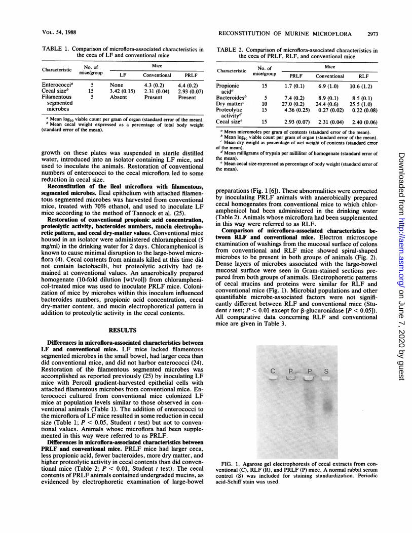
TABLE 1. Comparison of microflora-associated characteristics inthe ceca of LF and conventional mice
Characteristic No. of Micemice/group LF Conventional PRLF
Enterococcia 5 None 4.3 (0.2) 4.4 (0.2)Cecal sizeb 15 3.42 (0.15) 2.31 (0.04) 2.93 (0.07)Filamentous S Absent Present Presentsegmentedmicrobesa Mean log1o viable count per gram of organ (standard error of the mean).b Mean cecal weight expressed as a percentage of total body weight
(standard error of the mean).
growth on these plates was suspended in sterile distilledwater, introduced into an isolator containing LF mice, andused to inoculate the animals. Restoration of conventionalnumbers of enterococci to the cecal microflora led to somereduction in cecal size.
Reconstitution of the Beal microflora with filamentous,segmented microbes. Iteal epithelium with attached filamen-tous segmented microbes was harvested from conventionalmice, treated with 70% ethanol, and used to inoculate LFmice according to the method of Tannock et al. (25).
Restoration of conventional propionic acid concentration,proteolytic activity, bacteroides numbers, mucin electropho-retic pattern, and cecal dry-matter values. Conventional micehoused in an isolator were administered chloramphenicol (5mg/ml) in the drinking water for 2 days. Chloranmphenicol isknown to cause minimal disruption to the large-bowel micro-flora (4). Cecal contents from animals killed at this time didnot contain lactobacilli, but proteolytic activity had re-mained at conventional values. An anaerobically preparedhomogenate (10-fold dilution [wt/vol]) from chlorampheni-col-treated mice was used to inoculate PRLF mice. Coloni-zation of mice by microbes within this inoculum influencedbacteroides numbers, propionic acid concentration, cecaldry-matter content, and mucin electrophoretical pattern inaddition to proteolytic activity in the cecal contents.
RESULTS
Differences in microflora-associated characteristics betweenLF and conventional mice. LF mice lacked filamentoussegmented microbes in the small bowel, had larger ceca thandid conventional mice, and did not harbor enterococci (24).Restoration of the filamentous segmented microbes wasaccomplished as reported previously (25) by inoculating LFmice with Percoll gradient-harvested epithelial cells withattached filamentous microbes from conventional mice. En-terococci cultured from conventional mice colonized LFmice at population levels similar to those observed in con-ventional animals (Table 1). The addition of enterococci tothe microflora ofLF mice resulted in some reduction in cecalsize (Table 1; P < 0.05, Student t test) but not to conven-tional values. Animals whose microflora had been supple-mented in this way were referred to as PRLF.
Differences in microflora-associated characteristics betweenPRLF and conventional mice. PRLF mice had larger ceca,less propionic acid, fewer bacteroides, more dry matter, andhigher proteolytic activity in cecal contents than did conven-tional mice (Table 2; P < 0.01, Student t test). The cecalcontents ofPRLF animals contained undergraded mucins, asevidenced by electrophoretic examination of large-bowel
TABLE 2. Comparison of microflora-associated characteristics inthe ceca of PRLF, RLF, and conventional mice
Characteristic No. of Micemice/group PRLF Conventional RLF
Propionic 15 1.7 (0.1) 6.9 (1.0) 10.6 (1.2)acida
Bacteroidesb 5 7.4 (0.2) 8.9 (0.1) 8.5 (0.1)Dry matterc 10 27.0 (0.2) 24.4 (0.6) 25.5 (1.0)Proteolytic 15 4.36 (0.25) 0.27 (0.02) 0.22 (0.08)
activitydCecal sizee 15 2.93 (0.07) 2.31 (0.04) 2.40 (0.06)
a Mean micromoles per gram of contents (standard error of the mean).b Mean log1o viable count per gram of organ (standard error of the mean).c Mean dry weight as percentage of wet weight of contents (standard error
of the mean).d Mean milligrams of trypsin per milliliter of homogenate (standard error of
the mean).e Mean cecal size expressed as percentage of body weight (standard error of
the mean).
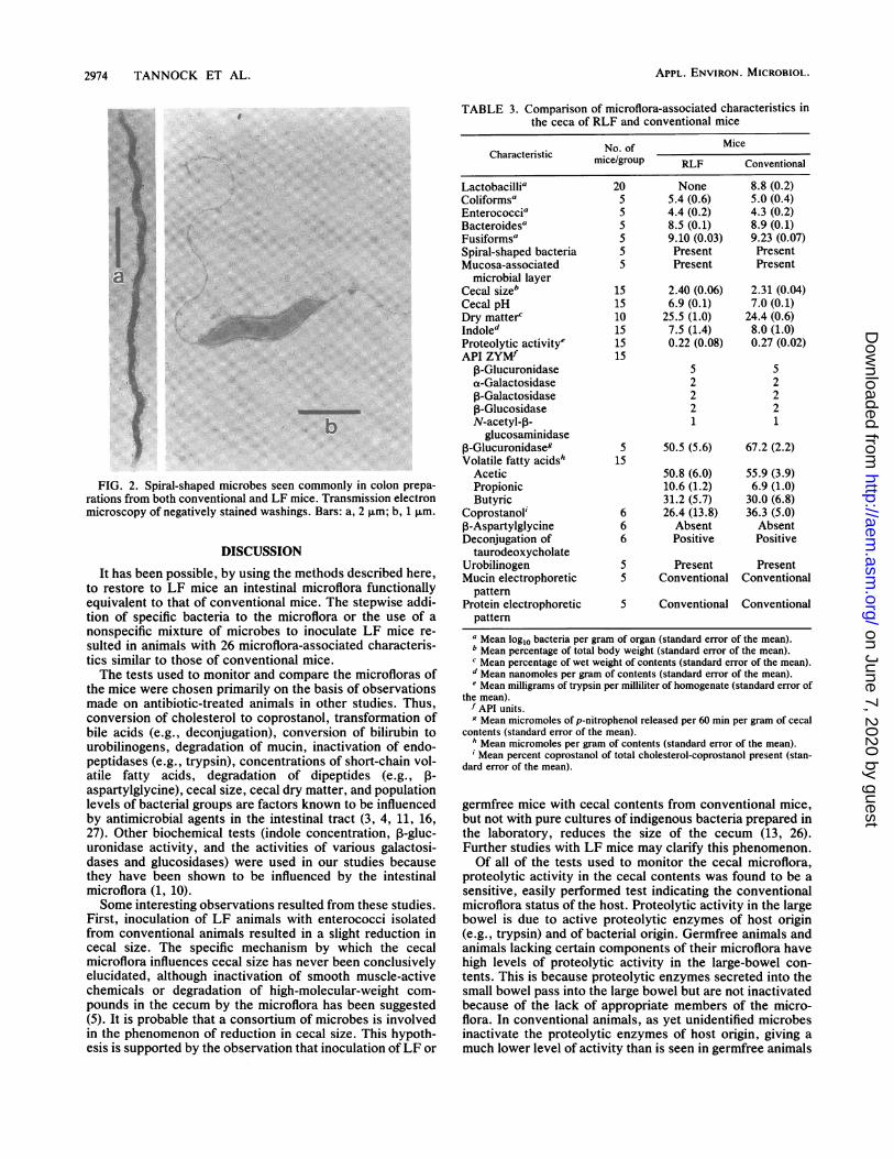
preparations (Fig. 1 [6]). These abnormalities were correctedby inoculating PRLF animals with anaerobically preparedcecal homogenates from conventional mice to which chlor-amphenicol had been administered in the drinking water(Table 2). Animals whose microflora had been supplementedin this way were referred to as RLF.Comparison of microflora-associated characteristics be-
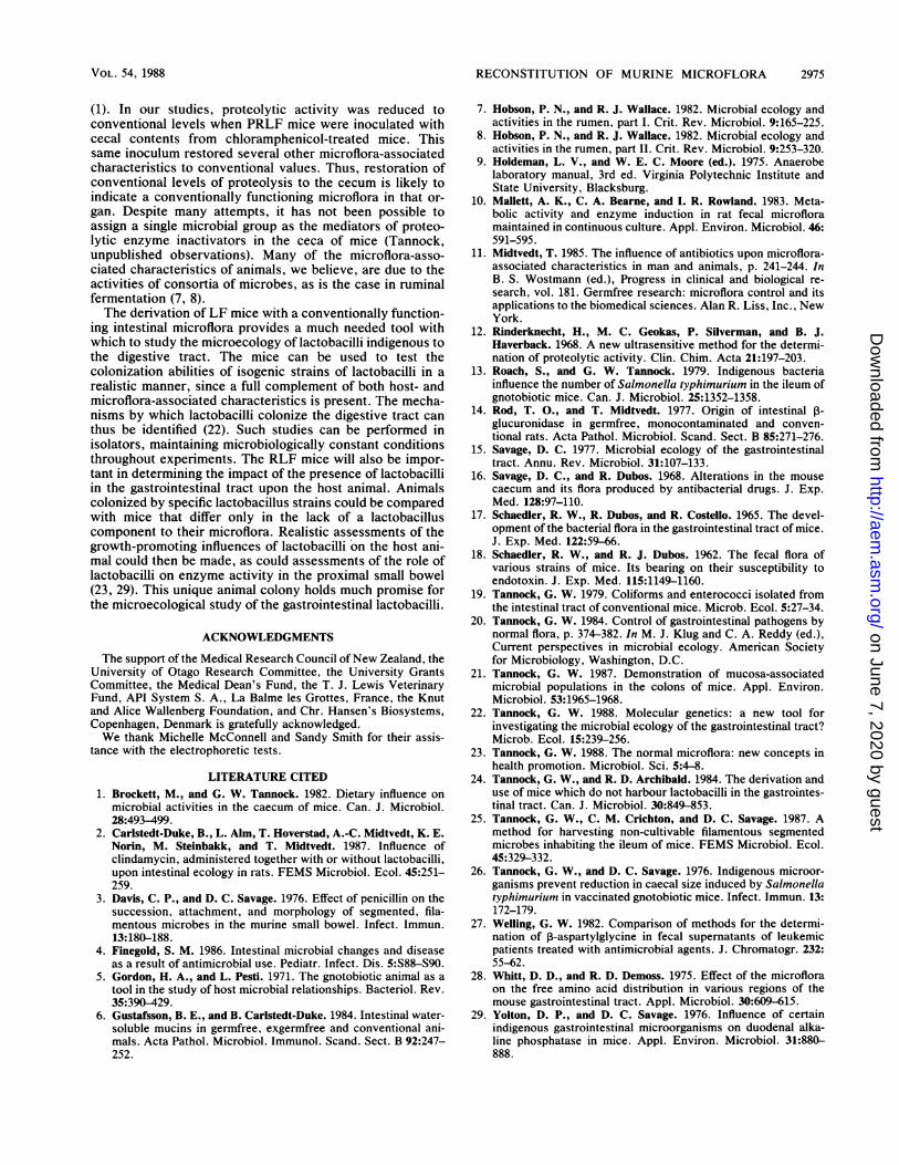
tween RLF and conventional mice. Electron microscopeexamination of washings from the mucosal surface of colonsfrom conventional and RLF mice showed spiral-shapedmicrobes to be present in both groups of animals (Fig. 2).Dense layers of microbes associated with the large-bowelmucosal surface were seen in Gram-stained sections pre-pared from both groups of animals. Electrophoretic patternsof cecal mucins and proteins were similar for RLF andconventional mice (Fig. 1). Microbial populations and otherquantifiable microbe-associated factors were not signifi-cantly different between RLF and conventional mice (Stu-dent t test; P < 0.01 except for P-glucuronidase [P < 0.05]).All comparative data concerning RLF and conventionalmice are given in Table 3.
FIG. 1. Agarose gel electrophoresis of cecal extracts from con-ventional (C), RLF (R), and PRLiF (P) mice. A normal rabbit serumcontrol (5) was incluided for staining standardization. Periodicacid-Schiff stain was used.
VOL. 54, 1988
on June 7, 2020 by guesthttp://aem
.asm.org/
Dow
nloaded from
2974 TANNOCK ET AL.
FIG. 2. Spiral-shaped microbes seen commonly in colon prepa-rations from both conventional and LF mice. Transmission electronmicroscopy of negatively stained washings. Bars: a, 2 ,um; b, 1 ,um.
DISCUSSION
It has been possible, by using the methods described here,to restore to LF mice an intestinal microflora functionallyequivalent to that of conventional mice. The stepwise addi-tion of specific bacteria to the microflora or the use of anonspecific mixture of microbes to inoculate LF mice re-sulted in animals with 26 microflora-associated characteris-tics similar to those of conventional mice.The tests used to monitor and compare the microfloras of
the mice were chosen primarily on the basis of observationsmade on antibiotic-treated animals in other studies. Thus,conversion of cholesterol to coprostanol, transformation ofbile acids (e.g., deconjugation), conversion of bilirubin tourobilinogens, degradation of mucin, inactivation of endo-peptidases (e.g., trypsin), concentrations of short-chain vol-atile fatty acids, degradation of dipeptides (e.g., P-aspartylglycine), cecal size, cecal dry matter, and populationlevels of bacterial groups are factors known to be influencedby antimicrobial agents in the intestinal tract (3, 4, 11, 16,27). Other biochemical tests (indole concentration, P-gluc-uronidase activity, and the activities of various galactosi-dases and glucosidases) were used in our studies becausethey have been shown to be influenced by the intestinalmicroflora (1, 10).Some interesting observations resulted from these studies.
First, inoculation of LF animals with enterococci isolatedfrom conventional animals resulted in a slight reduction incecal size. The specific mechanism by which the cecalmicroflora influences cecal size has never been conclusivelyelucidated, although inactivation of smooth muscle-activechemicals or degradation of high-molecular-weight com-
pounds in the cecum by the microflora has been suggested(5). It is probable that a consortium of microbes is involvedin the phenomenon of reduction in cecal size. This hypoth-esis is supported by the observation that inoculation ofLF or
TABLE 3. Comparison of microflora-associated characteristics inthe ceca of RLF and conventional mice
No.of ~~MiceCharacteristic No.cofgMicemice/grriticoupof RLF Conventional
Lactobacillia 20 None 8.8 (0.2)Coliformsa 5 5.4 (0.6) 5.0 (0.4)Enterococcia 5 4.4 (0.2) 4.3 (0.2)Bacteroidesa 5 8.5 (0.1) 8.9 (0.1)Fusiformsa 5 9.10 (0.03) 9.23 (0.07)Spiral-shaped bacteria S Present PresentMucosa-associated 5 Present Present
microbial layerCecal size' 15 2.40 (0.06) 2.31 (0.04)Cecal pH 15 6.9 (0.1) 7.0 (0.1)Dry matterc 10 25.5 (1.0) 24.4 (0.6)Indoled 15 7.5 (1.4) 8.0 (1.0)Proteolytic activitye 15 0.22 (0.08) 0.27 (0.02)API ZYMf 15
P-Glucuronidase 5 5a-Galactosidase 2 213-Galactosidase 2 2P-Glucosidase 2 2N-acetyl-p- 1 1
glucosaminidase,B-Glucuronidaseg 5 50.5 (5.6) 67.2 (2.2)Volatile fatty acidsh 15
Acetic 50.8 (6.0) 55.9 (3.9)Propionic 10.6 (1.2) 6.9 (1.0)Butyric 31.2 (5.7) 30.0 (6.8)
Coprostanoli 6 26.4 (13.8) 36.3 (5.0)P-Aspartylglycine 6 Absent AbsentDeconjugation of 6 Positive Positive
taurodeoxycholateUrobilinogen 5 Present PresentMucin electrophoretic 5 Conventional Conventional
patternProtein electrophoretic 5 Conventional Conventional
patterna Mean log10 bacteria per gram of organ (standard error of the mean).b Mean percentage of total body weight (standard error of the mean).c Mean percentage of wet weight of contents (standard error of the mean).d Mean nanomoles per gram of contents (standard error of the mean).Mean milligrams of trypsin per milliliter of homogenate (standard error of
the mean).f API units.g Mean micromoles of p-nitrophenol released per 60 min per gram of cecal
contents (standard error of the mean).h Mean micromoles per gram of contents (standard error of the mean).Mean percent coprostanol of total cholesterol-coprostanol present (stan-
dard error of the mean).
germfree mice with cecal contents from conventional mice,but not with pure cultures of indigenous bacteria prepared inthe laboratory, reduces the size of the cecum (13, 26).Further studies with LF mice may clarify this phenomenon.Of all of the tests used to monitor the cecal microflora,
proteolytic activity in the cecal contents was found to be asensitive, easily performed test indicating the conventionalmicroflora status of the host. Proteolytic activity in the largebowel is due to active proteolytic enzymes of host origin(e.g., trypsin) and of bacterial origin. Germfree animals andanimals lacking certain components of their microflora havehigh levels of proteolytic activity in the large-bowel con-tents. This is because proteolytic enzymes secreted into thesmall bowel pass into the large bowel but are not inactivatedbecause of the lack of appropriate members of the micro-flora. In conventional animals, as yet unidentified microbesinactivate the proteolytic enzymes of host origin, giving amuch lower level of activity than is seen in germfree animals
APPL. ENVIRON. MICROBIOL.
on June 7, 2020 by guesthttp://aem
.asm.org/
Dow
nloaded from
RECONSTITUTION OF MURINE MICROFLORA 2975
(1). In our studies, proteolytic activity was reduced toconventional levels when PRLF mice were inoculated withcecal contents from chloramphenicol-treated mice. Thissame inoculum restored several other microflora-associatedcharacteristics to conventional values. Thus, restoration ofconventional levels of proteolysis to the cecum is likely toindicate a conventionally functioning microflora in that or-gan. Despite many attempts, it has not been possible toassign a single microbial group as the mediators of proteo-lytic enzyme inactivators in the ceca of mice (Tannock,unpublished observations). Many of the microflora-asso-ciated characteristics of animals, we believe, are due to theactivities of consortia of microbes, as is the case in ruminalfermentation (7, 8).The derivation of LF mice with a conventionally function-
ing intestinal microflora provides a much needed tool withwhich to study the microecology of lactobacilli indigenous tothe digestive tract. The mice can be used to test thecolonization abilities of isogenic strains of lactobacilli in arealistic manner, since a full complement of both host- andmicroflora-associated characteristics is present. The mecha-nisms by which lactobacilli colonize the digestive tract canthus be identified (22). Such studies can be performed inisolators, maintaining microbiologically constant conditionsthroughout experiments. The RLF mice will also be impor-tant in determining the impact of the presence of lactobacilliin the gastrointestinal tract upon the host animal. Animalscolonized by specific lactobacillus strains could be comparedwith mice that differ only in the lack of a lactobacilluscomponent to their microflora. Realistic assessments of thegrowth-promoting influences of lactobacilli on the host ani-mal could then be made, as could assessments of the role oflactobacilli on enzyme activity in the proximal small bowel(23, 29). This unique animal colony holds much promise forthe microecological study of the gastrointestinal lactobacilli.
ACKNOWLEDGMENTS
The support of the Medical Research Council of New Zealand, theUniversity of Otago Research Committee, the University GrantsCommittee, the Medical Dean's Fund, the T. J. Lewis VeterinaryFund, API System S. A., La Balme les Grottes, France, the Knutand Alice Wallenberg Foundation, and Chr. Hansen's Biosystems,Copenhagen, Denmark is gratefully acknowledged.We thank Michelle McConnell and Sandy Smith for their assis-
tance with the electrophoretic tests.
LITERATURE CITED1. Brockett, M., and G. W. Tannock. 1982. Dietary influence on
microbial activities in the caecum of mice. Can. J. Microbiol.28:493-499.
2. Carlstedt-Duke, B., L. Alm, T. Hoverstad, A.-C. Midtvedt, K. E.Norin, M. Steinbakk, and T. Midtvedt. 1987. Influence ofclindamycin, administered together with or without lactobacilli,upon intestinal ecology in rats. FEMS Microbiol. Ecol. 45:251-259.
3. Davis, C. P., and D. C. Savage. 1976. Effect of penicillin on thesuccession, attachment, and morphology of segmented, fila-mentous microbes in the murine small bowel. Infect. Immun.13:180-188.
4. Finegold, S. M. 1986. Intestinal microbial changes and diseaseas a result of antimicrobial use. Pediatr. Infect. Dis. 5:S88-S90.
5. Gordon, H. A., and L. Pesti. 1971. The gnotobiotic animal as atool in the study of host microbial relationships. Bacteriol. Rev.35:390-429.
6. Gustafsson, B. E., and B. Carlstedt-Duke. 1984. Intestinal water-soluble mucins in germfree, exgermfree and conventional ani-mals. Acta Pathol. Microbiol. Immunol. Scand. Sect. B 92:247-252.
7. Hobson, P. N., and R. J. Wallace. 1982. Microbial ecology andactivities in the rumen, part I. Crit. Rev. Microbiol. 9:165-225.
8. Hobson, P. N., and R. J. Wallace. 1982. Microbial ecology andactivities in the rumen, part II. Crit. Rev. Microbiol. 9:253-320.
9. Holdeman, L. V., and W. E. C. Moore (ed.). 1975. Anaerobelaboratory manual, 3rd ed. Virginia Polytechnic Institute andState University, Blacksburg.
10. Mallett, A. K., C. A. Bearne, and I. R. Rowland. 1983. Meta-bolic activity and enzyme induction in rat fecal microfloramaintained in continuous culture. Appl. Environ. Microbiol. 46:591-595.
11. Midtvedt, T. 1985. The influence of antibiotics upon microflora-associated characteristics in man and animals, p. 241-244. InB. S. Wostmann (ed.), Progress in clinical and biological re-search, vol. 181. Germfree research: microflora control and itsapplications to the biomedical sciences. Alan R. Liss, Inc., NewYork.
12. Rinderknecht, H., M. C. Geokas, P. Silverman, and B. J.Haverback. 1968. A new ultrasensitive method for the determi-nation of proteolytic activity. Clin. Chim. Acta 21:197-203.
13. Roach, S., and G. W. Tannock. 1979. Indigenous bacteriainfluence the number of Salmonella typhimurium in the ileum ofgnotobiotic mice. Can. J. Microbiol. 25:1352-1358.
14. Rod, T. O., and T. Midtvedt. 1977. Origin of intestinal ,B-glucuronidase in germfree, monocontaminated and conven-tional rats. Acta Pathol. Microbiol. Scand. Sect. B 85:271-276.
15. Savage, D. C. 1977. Microbial ecology of the gastrointestinaltract. Annu. Rev. Microbiol. 31:107-133.
16. Savage, D. C., and R. Dubos. 1968. Alterations in the mousecaecum and its flora produced by antibacterial drugs. J. Exp.Med. 128:97-110.
17. Schaedler, R. W., R. Dubos, and R. Costello. 1965. The devel-opment of the bacterial flora in the gastrointestinal tract of mice.J. Exp. Med. 122:59-66.
18. Schaedler, R. W., and R. J. Dubos. 1962. The fecal flora ofvarious strains of mice. Its bearing on their susceptibility toendotoxin. J. Exp. Med. 115:1149-1160.
19. Tannock, G. W. 1979. Coliforms and enterococci isolated fromthe intestinal tract of conventional mice. Microb. Ecol. 5:27-34.
20. Tannock, G. W. 1984. Control of gastrointestinal pathogens bynormal flora, p. 374-382. In M. J. Klug and C. A. Reddy (ed.),Current perspectives in microbial ecology. American Societyfor Microbiology, Washington, D.C.
21. Tannock, G. W. 1987. Demonstration of mucosa-associatedmicrobial populations in the colons of mice. Appl. Environ.Microbiol. 53:1965-1968.
22. Tannock, G. W. 1988. Molecular genetics: a new tool forinvestigating the microbial ecology of the gastrointestinal tract?Microb. Ecol. 15:239-256.
23. Tannock, G. W. 1988. The normal microflora: new concepts inhealth promotion. Microbiol. Sci. 5:4-8.
24. Tannock, G. W., and R. D. Archibald. 1984. The derivation anduse of mice which do not harbour lactobacilli in the gastrointes-tinal tract. Can. J. Microbiol. 30:849-853.
25. Tannock, G. W., C. M. Crichton, and D. C. Savage. 1987. Amethod for harvesting non-cultivable filamentous segmentedmicrobes inhabiting the ileum of mice. FEMS Microbiol. Ecol.45:329-332.
26. Tannock, G. W., and D. C. Savage. 1976. Indigenous microor-ganisms prevent reduction in caecal size induced by Salmonellatyphimurium in vaccinated gnotobiotic mice. Infect. Immun. 13:172-179.
27. Welling, G. W. 1982. Comparison of methods for the determi-nation of P-aspartylglycine in fecal supematants of leukemicpatients treated with antimicrobial agents. J. Chromatogr. 232:55-62.
28. Whitt, D. D., and R. D. Demoss. 1975. Effect of the microfloraon the free amino acid distribution in various regions of themouse gastrointestinal tract. Appl. Microbiol. 30:609-615.
29. Yolton, D. P., and D. C. Savage. 1976. Influence of certainindigenous gastrointestinal microorganisms on duodenal alka-line phosphatase in mice. Appl. Environ. Microbiol. 31:880-888.
VOL. 54, 1988
on June 7, 2020 by guesthttp://aem
.asm.org/
Dow
nloaded from