Embed Size (px)
Citation preview
Review
Red blood cells (RBCs), epoxyeicosatrienoic
acids (EETs) and adenosine triphosphate (ATP)
Houli Jiang, Gail D. Anderson, John C. McGiff
���������� ������ ���� ��� ��� ���� �� ������� ��������� �� ������ � !
Correspondence: "�� �# � $�� �%����& '��(� ��)��� #��*
Abstract:
In addition to serving as carriers of O�, red blood cells (RBCs) regulate vascular resistance and the distribution of microvascular per-
fusion by liberating adenosine triphosphate (ATP) and epoxyeicosatrienoic acids (EETs) upon exposure to a low O� environment.
Therefore, RBCs act as sensors that respond to low pO� by releasing millimolar amounts of ATP, a signaling molecule, and lipid me-
diators (EETs). The release of EETs occurs by a mechanism that is activated by ATP stimulation of P2X� receptors coupled to ATP
transporters, which should greatly amplify the circulatory response to ATP. RBCs are reservoirs of EETs and the primary sources of
plasma EETs, which are esterified to the phospholipids of lipoproteins. Levels of free EETs in plasma are low, about 3% of circulat-
ing EETs. RBC EETs are produced by direct oxidation of arachidonic acid (AA) esterified to glycerophospholipids and the
monooxygenase-like activity of hemoglobin. On release, EETs affect vascular tone, produce profibrinolysis and dampen inflamma-
tion. A soluble epoxide hydrolase (sEH) regulates the concentrations of RBC and vascular EETs by metabolizing both cis- and
trans-EETs to form dihydroxyeicosatrienoic acids (DHETs). The function and pathophysiological roles of trans-EETs and
erythro-DHETs has yet to be integrated into a physiological and pathophysiological context.
Key words:
epoxyeicosatrienoic acids, red blood cells, arachidonic acid, soluble epoxide hydrolase, glycerophospholipids
Abbreviations: 20-HETE – 20-hydroxyeicosatetraenoic acid,
AA – arachidonic acid, cPLA� – cytoplasmic phospholipase
A��, DHETs – dihydroxyeicosatrienoic acids, EETs – epoxyei-
cosatrienoic acids, PPAR – peroxisome proliferator-activated
receptor, RBCs – red blood cells, sEH – soluble epoxide hy-
drolase, t-BHP – tert-butyl-hydroperoxide
Introduction
Epoxyeicosatrienoic acids (EETs) are arachidonic
acid (AA)-derived lipid mediators that dilate arteri-
oles by activating the large conductance Ca2+ acti-
vated K+ channel. EETs are opposed by 20-hydroxy-
eicosatetraenoic acid (20-HETE) at this site [24, 29].
EETs were first identified as products of the rat liver
microsomal cytochrome P450 pathway of AA me-
tabolism [8]. Red blood cells (RBCs) have been
shown to serve as reservoirs for EETs, which are
abundant and incorporated into the sn-2 position of
the RBC membrane phospholipids [22]. Stored EETs
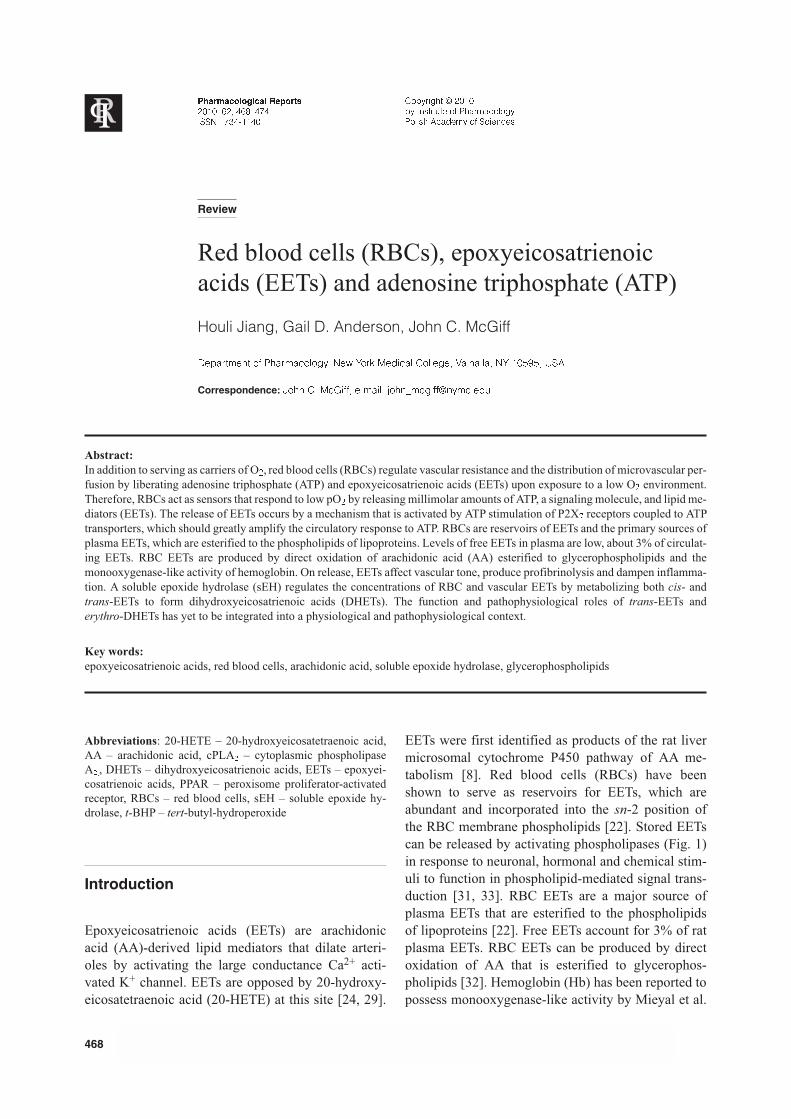
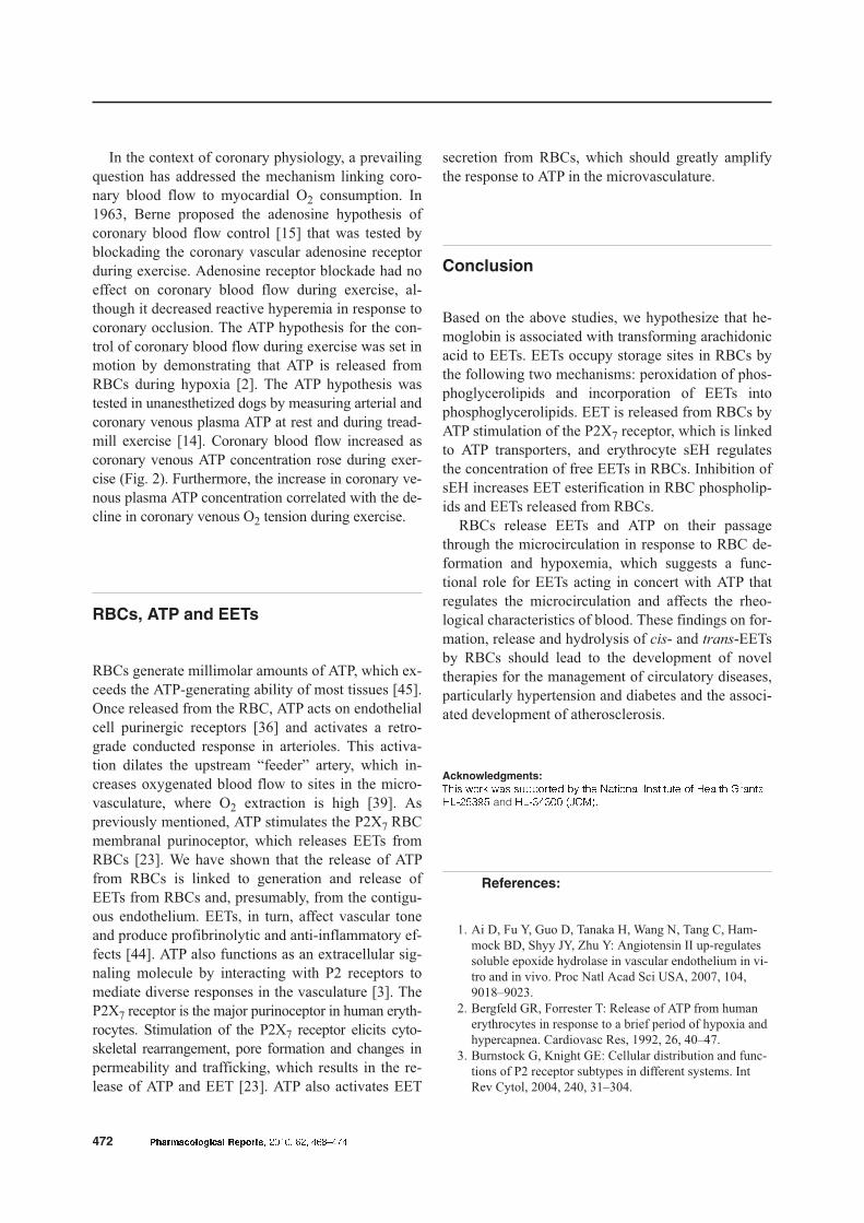
can be released by activating phospholipases (Fig. 1)
in response to neuronal, hormonal and chemical stim-
uli to function in phospholipid-mediated signal trans-
duction [31, 33]. RBC EETs are a major source of
plasma EETs that are esterified to the phospholipids
of lipoproteins [22]. Free EETs account for 3% of rat
plasma EETs. RBC EETs can be produced by direct
oxidation of AA that is esterified to glycerophos-
pholipids [32]. Hemoglobin (Hb) has been reported to
possess monooxygenase-like activity by Mieyal et al.
468 ������������� �� ����� ����� ��� ������
������������� �� ����
����� ��� ������
��� � ������
��������� � ����
�� �������� �� �� �! "�#���
��#��� $" %�!� �� �"���"��
[46]. The monooxygenase-like activity of Hb can be
activated by nicotinic acid and riboflavin, which serve
as electron-carrier cofactors and divert AA peroxida-
tion to form EETs. This process obviates the need for
NADPH reductase. The de novo production of EETs
produced by RBCs occurs via AA transformation by
the monooxygenase-like activity of Hb, which in-
volves a calcium dependent cytoplasmic phospholi-
pase A2 (rPLA2) [41] that cleaves free AA from phos-
pholipids. In addition to their vasoactivity and effects
on renal tubular salt and water metabolism [29], EETs
exert potent anti-inflammatory effects by activating
transcription of the endothelial peroxisome prolifera-
tor activated receptor (PPAR) [27, 29, 34]. The result-
ing inhibition of nuclear factor �B-mediated expres-
sion of adhesion molecules and endothelin synthesis
counters the development or progression of vascular
wall damage and atherogenesis [4, 27]. The wide
range of actions of EETs is further exhibited by their
ability to activate fibrinolysis by inducing the expres-
sion of tissue plasminogen activator [34].
Metabolism of EETs by soluble epoxide
hydrolase
Soluble epoxide hydrolase (sEH) regulates the con-
centration of vascular EETs by metabolizing them to
form dihydroxyeicosatrienoic acids (DHETs) [13, 35].
sEH demonstrates regioselectivity and stereoselectiv-
ity because the rates of EET hydration are highest for
14,15-EET, and it prefers 14(R),15(S)-EET [49]. In
contrast, hydration of 11,12-EET is not stereoselec-
tive. DHETs are considered to be inactive, although
exceptions have been reported, such as the effect of
sEH on metabolism of 11,12-EET, which did not lose
its coronary arteriolar dilator activity upon hydrolysis
by sEH to form 11,12-DHET [24]. In contrast, the
coronary vasodilator actions of 8,9- and 14,15-EETs
are eradicated upon their conversion to DHETs. EETs
and DHETs reduce cellular cholesterol levels as effec-
tively as simvastatin in the human hepatoma (HepG2)
cell line [11]. The cholesterol-lowering response was
found only for 11,12- and 14,15-DHETs, which showed
similar potency to 11,12- and 14,15-EETs. Inhibition
of sEH also increases EET incorporation into phospholip-
ids [48], which potentiates endothelium-dependent va-
sorelaxation to bradykinin mediated by 11,12-EET
acting as an endothelial-derived hyperpolarizing fac-
tor (EDHF) [16]. Indeed, the renal vasodilator re-
sponse to intra-arterial administration of bradykinin
has been shown to produce a prostaglandin, a nitric
oxide (NO) and a CYP450 AA metabolite [29]. The
renal vasodilator response to bradykinin could be po-
tentiated by increasing EET levels by either prevent-
ing their degradation through inhibiting sEH or by
adding EETs to vascular tissues [48]. Increased sEH activ-
ity has been associated with diabetes [37, 47] and hyper-
tension [1, 20], whereas elevated EET levels, in response
to inhibition of sEH, lowered blood pressure in hyperten-
������������� �� ����� ����� ��� ������ 469
RBC, EETs and ATP
����� ���� � ���
Control PLA2
Time
EE
T(n
g/1
09
RB
Cs
)
0.60
0.50
0.40
0.30
0.20
0.10
0.00
Control PLA2
Time
EE
T(n
g/1
09
RB
Cs
)
0.60
0.50
0.40
0.30
0.20
0.10
0.00
-
-
-
-
-
-
-
m/z 481
m/z 489
4.00 4.40 4.80
Fig. 1. ������� �� ���� � ���� ���� ��� � �� ���� ������ � ����� ���������� ��� ���� ��������� ���� ���������� � ����� �� ���� �� ���� ����� ��� � ! "# $ % ���&'� (����# )�*+,����-��� �� ��� �������� � ������������ ����� ��� ���� &#./cis/ ��� &#./trans/� 0��# ���� $���������1
sive states [9, 18, 20]. sEH has become a therapeutic
target for controlling blood pressure [28, 42, 50], vas-
cular inflammation [10, 38, 43] and cancer progres-
sion [30] as well as providing cardiac [25, 40] and re-
nal protection [19] by raising tissue and plasma levels
of EETs.
Due to the cardiovascular effects of substituted
urea-derived sEH inhibitors (CUDA, AUDA) [12], it
is possible that sEH inhibitors are not very specific
and do more than inhibit EET metabolism. CUDA and
AUDA activate PPAR� directly by functioning as
a PPAR� ligand, which is similar to 11,12-EET ([29].
Thus, some of the actions of EETs, such as their abil-
ity to stimulate PPAR� can be duplicated by this class
of sEH inhibitors, which activate PPAR� through an
EET-independent mechanism.
trans-EETs and sEH
RBC sEH prefers hydrolysis of trans-EETs to cis-EETs
because the Vmax of trans-EET hydrolysis by rat RBCs
is about 3 times that of cis-EETs [22]. Therefore, inhi-
bition of sEH will have a greater effect on trans-EET
concentration in tissues and plasma than cis-EET con-
centration. On giving a sEH inhibitor, the increased
levels of trans-EETs and erythro-DHETs are thought
to account for the blood pressure-lowering effects of
sEH inhibition in the spontaneously hypertensive rat
(SHR), a rat genetic model of hypertension. Addition-
ally, the greater vasodilator potency of trans-EETs
with regard to cis-EETs and the retention of vasoac-
tivity of the erythro-DHETs contribute to the antihy-
pertensive effects of inhibition of sEH [21].
EETs formed by oxidation of AA
esterified to glycerophospholipids
Isoprostanes and isoleukotrienes are produced by free
radical oxidation of AA and demonstrate potent bio-
logical activity, which is the case with EETs arising
from non-enzymatic oxidation of esterified AA [22].
Nakamura, Bratton and Murphy [32] have proposed
a non-enzymatic pathway for generating EETs that is
induced by free radical-based peroxidation of phos-
pholipids, which is an alternative pathway to cyto-
chrome P450 epoxygenases and activated Hb produc-
tion of EETs. They developed a model system in
which tert-butyl-hydroperoxide (t-BHP), a lipophilic
agent, was used to increase hydroperoxide tone in cel-
lular membranal glycerophospholipids. Thus, in re-
sponse to the oxidizing agent, t-BHP, EETs were pro-
duced in situ by oxidizing AA that was already incor-
porated in the membranal phospholipid bilayer, not by
re-acylation of lysophospholipid precursors. This pro-
cess increased EETs esterified in glycerophospho-
ethanolamine, -serine and -choline by 30- to 60-fold.
EETs arising enzymatically or by an Hb-catalyzed
mechanism can be esterified in the fatty acyl chains of
glycerophospholipids via an acyl-CoA synthase (CoA)-
dependent mechanism [48], which is the dominant
mechanism responsible for incorporating EETs into
human platelet phospholipids [51]. During activation
of platelets with either thrombin or platelet activating
factor, EETs were released from phospholipids and
reached an estimated intracellular concentration of
about 1 μmol/L, which has been shown to inhibit
platelet aggregation by affecting platelet cyclooxyge-
nase activity and calcium entry [51]. Cobra venom
PLA2 was also used to release AA metabolites esteri-
fied in the sn-2 position of platelet phospholipids. Gas
chromatrograpy/mass spectrometry (GC/MS) analy-
ses showed endogenous EETs and 20-HETE. The amount
of 20-HETE was 200-fold lower than the amount of
EETs. Unbound EETs and 20-HETE were not de-
tected. More than 60% of the EETs were esterified in
platelet phosphatidylinositol, which suggests involve-
ment in signal transduction. Platelet EETs were
thought to be produced enzymatically by the cyto-
chrome P450 system because chiral analysis showed
only a single enantiomer, which is an indicator of en-
zymatic origin. In a study of EETs extracted from he-
patic phospholipids, Capdevila and colleagues [6]
traced EET incorporation into phospholipid pools.
Given the magnitude of the EET reservoir capacity
of RBCs and the RBC-EET releasing mechanism,
which is activated by ATP stimulation of the erythro-
cyte P2X7 receptor [23], RBC EETs are postulated to
be the chief source of free EETs in plasma (3%) and
EETs esterified in circulating phospholipids (95% of
circulating EETs). The disposition of intracellular
EETs and DHETs shows remarkable diversity [44].
Unlike EETs that can be released from RBCs by
a purinoceptor mechanism, DHETs are extruded from
cells. Cellular retention of EETs likely reflects bind-
470 ������������� �� ����� ����� ��� ������
ing to cytosolic fatty acid binding proteins (FABP),
which possibly act as transport proteins for EETs. In
contrast, DHETs show weak binding to FABP [44].
We have analyzed RBC EETs with electrospray ioni-
zation tandem mass spectrometry and found that both
cis- and trans-EETs increase when rat RBCs are ex-
posed to t-BHP, which oxidizes esterified AA [22]. In-
corporation of newly formed free EETs into RBC
phospholipids by an acyl CoA dependent mechanism
also occurs [48]. The plasma levels of cis- and
trans-EETs of RBC origin are similar in the rat
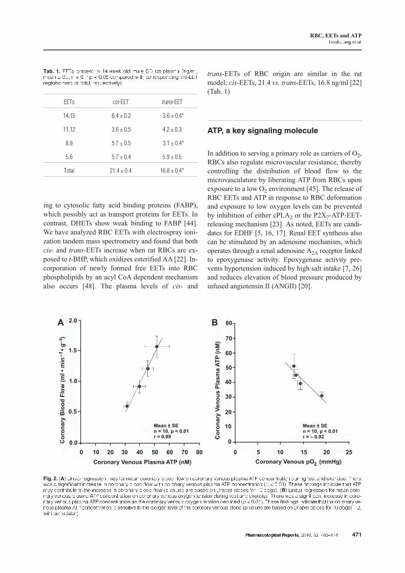
model; cis-EETs, 21.4 vs. trans-EETs, 16.8 ng/ml [22]
(Tab. 1)
ATP, a key signaling molecule
In addition to serving a primary role as carriers of O2,
RBCs also regulate microvascular resistance, thereby
controlling the distribution of blood flow to the
microvasculature by liberating ATP from RBCs upon
exposure to a low O2 environment [45]. The release of
RBC EETs and ATP in response to RBC deformation
and exposure to low oxygen levels can be prevented
by inhibition of either cPLA2 or the P2X7-ATP-EET-
releasing mechanism [23]. As noted, EETs are candi-
dates for EDHF [5, 16, 17]. Renal EET synthesis also
can be stimulated by an adenosine mechanism, which
operates through a renal adenosine A2A receptor linked
to epoxygenase activity. Epoxygenase activity pre-
vents hypertension induced by high salt intake [7, 26]
and reduces elevation of blood pressure produced by
infused angiotensin II (ANGII) [20].
������������� �� ����� ����� ��� ������ 471
RBC, EETs and ATP
����� ���� � ���
Tab. 1. ���� ������� � �� ��� ��� ���� �� ��� ������ ����������� � ��� � � �� � � � � �! "������� �# "����������� cis�������������� �� ������ �����"�$��%&
EETs cis-EET trans-EET
14,15 6.4 ± 0.2 3.6 ± 0.4*
11,12 3.6 ± 0.5 4.2 ± 0.3
8,9 5.7 ± 0.5 3.1 ± 0.4*
5,6 5.7 ± 0.4 5.9 ± 0.5
Total 21.4 ± 0.4 16.8 ± 0.4*
Mean ± SEn = 10, p < 0.01r = – 0.92
0 5 10 15 20 25
80
70
60
50
40
30
20
10
0
Mean ± SEn = 10, p < 0.01r = 0.99
80
1.5
706050403020100
2.0
1.0
0.5
0.0
Coronary Venous Plasma ATP (nM)
Co
ron
ary
Ven
ou
sP
lasm
aA
TP
(nM
)
A B
Co
ron
ary
Blo
od
Flo
w(m
l•
min
•g
)–
1–
1
Coronary Venous pO (mmHg)2
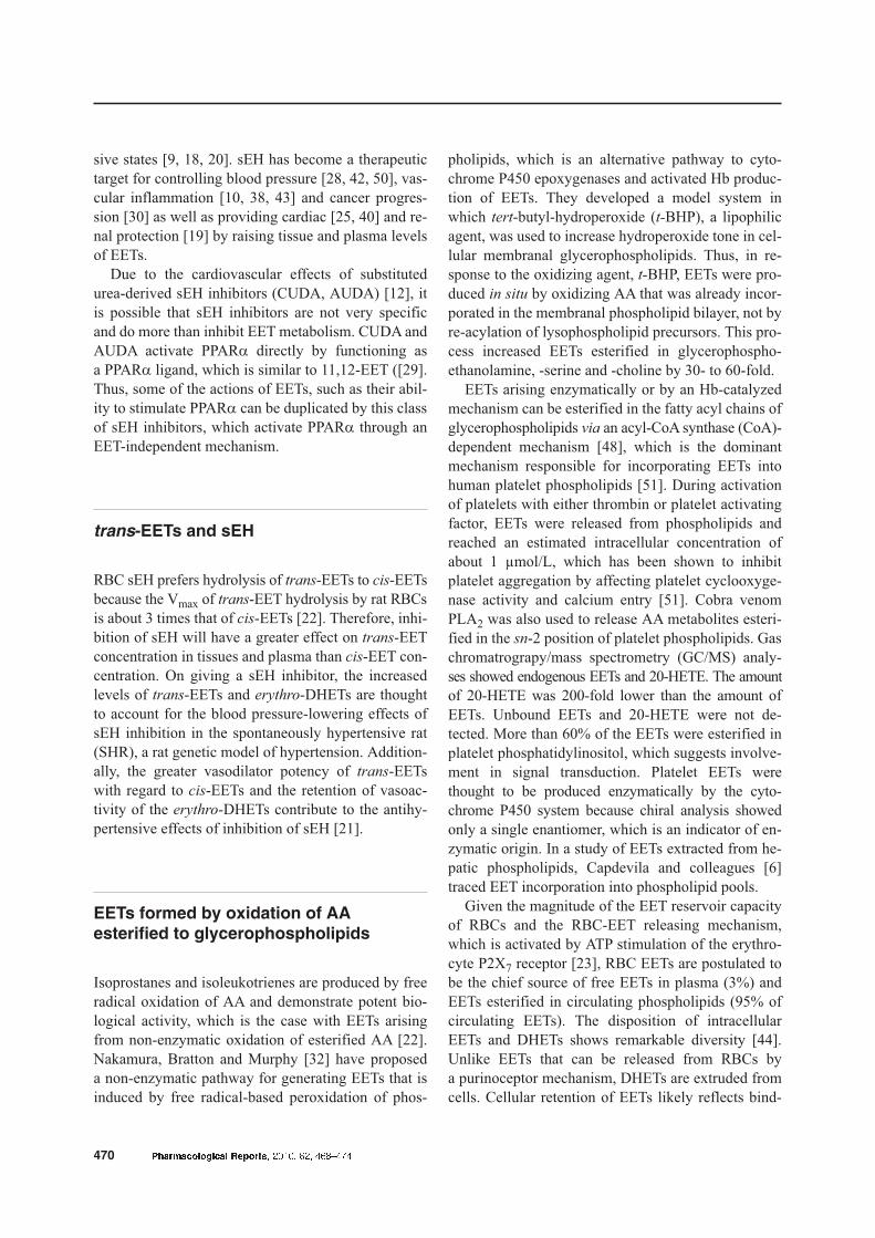
Fig. 2. (A) '���� ��������� ���� (�� ���� "������% )���� (�� �� "������% $���*� ������ +�, "��"�������� �*��� ���� ��� �-��"�� �#��� �� � ���("��� �"����� � "������% )���� (�� �# "������% $���*� ������ +�, "��"�������� �� � � �& �#��� (����� ��"��� �#�� +�,��% "����)*�� �� �#� �"����� � "������% )���� (�� �� $��*�� ��� )���� �� ������ ������ (�� � ����& (B) '���� ��������� (�� ���� "�������% $���*� ������ +�, "��"�������� �� "������% $���*� �-%��� ������ �*��� ���� ��� �-��"�� �#��� �� � ���("��� �"����� � "�������% $���*� ������ +�, "��"�������� �� �#� "������% $���*� �-%��� ������ ��"���� �� � � �& �#��� (����� ��"��� �#�� �#� "������% $����*� ������ +�, "��"�������� � �����$� �� �#� �-%��� ��$�� �( �#� "������% $���*� )���� �� $��*�� ��� )���� �� ������ ������ (�� � ����& ./� �# ��������0
In the context of coronary physiology, a prevailing
question has addressed the mechanism linking coro-
nary blood flow to myocardial O2 consumption. In
1963, Berne proposed the adenosine hypothesis of
coronary blood flow control [15] that was tested by
blockading the coronary vascular adenosine receptor
during exercise. Adenosine receptor blockade had no
effect on coronary blood flow during exercise, al-
though it decreased reactive hyperemia in response to
coronary occlusion. The ATP hypothesis for the con-
trol of coronary blood flow during exercise was set in
motion by demonstrating that ATP is released from
RBCs during hypoxia [2]. The ATP hypothesis was
tested in unanesthetized dogs by measuring arterial and
coronary venous plasma ATP at rest and during tread-
mill exercise [14]. Coronary blood flow increased as
coronary venous ATP concentration rose during exer-
cise (Fig. 2). Furthermore, the increase in coronary ve-
nous plasma ATP concentration correlated with the de-
cline in coronary venous O2 tension during exercise.
RBCs, ATP and EETs
RBCs generate millimolar amounts of ATP, which ex-
ceeds the ATP-generating ability of most tissues [45].
Once released from the RBC, ATP acts on endothelial
cell purinergic receptors [36] and activates a retro-
grade conducted response in arterioles. This activa-
tion dilates the upstream “feeder” artery, which in-
creases oxygenated blood flow to sites in the micro-
vasculature, where O2 extraction is high [39]. As
previously mentioned, ATP stimulates the P2X7 RBC
membranal purinoceptor, which releases EETs from
RBCs [23]. We have shown that the release of ATP
from RBCs is linked to generation and release of
EETs from RBCs and, presumably, from the contigu-
ous endothelium. EETs, in turn, affect vascular tone
and produce profibrinolytic and anti-inflammatory ef-
fects [44]. ATP also functions as an extracellular sig-
naling molecule by interacting with P2 receptors to
mediate diverse responses in the vasculature [3]. The
P2X7 receptor is the major purinoceptor in human eryth-
rocytes. Stimulation of the P2X7 receptor elicits cyto-
skeletal rearrangement, pore formation and changes in
permeability and trafficking, which results in the re-
lease of ATP and EET [23]. ATP also activates EET
secretion from RBCs, which should greatly amplify
the response to ATP in the microvasculature.
Conclusion
Based on the above studies, we hypothesize that he-
moglobin is associated with transforming arachidonic
acid to EETs. EETs occupy storage sites in RBCs by
the following two mechanisms: peroxidation of phos-
phoglycerolipids and incorporation of EETs into
phosphoglycerolipids. EET is released from RBCs by
ATP stimulation of the P2X7 receptor, which is linked
to ATP transporters, and erythrocyte sEH regulates
the concentration of free EETs in RBCs. Inhibition of
sEH increases EET esterification in RBC phospholip-
ids and EETs released from RBCs.
RBCs release EETs and ATP on their passage
through the microcirculation in response to RBC de-
formation and hypoxemia, which suggests a func-
tional role for EETs acting in concert with ATP that
regulates the microcirculation and affects the rheo-
logical characteristics of blood. These findings on for-
mation, release and hydrolysis of cis- and trans-EETs
by RBCs should lead to the development of novel
therapies for the management of circulatory diseases,
particularly hypertension and diabetes and the associ-
ated development of atherosclerosis.
Acknowledgments:
���� ���� �� ������ � �� �� ������ ������� �� � ��� �����
�������� and �������� !"#$%
References:
1. Ai D, Fu Y, Guo D, Tanaka H, Wang N, Tang C, Ham-
mock BD, Shyy JY, Zhu Y: Angiotensin II up-regulates
soluble epoxide hydrolase in vascular endothelium in vi-
tro and in vivo. Proc Natl Acad Sci USA, 2007, 104,
9018–9023.
2. Bergfeld GR, Forrester T: Release of ATP from human
erythrocytes in response to a brief period of hypoxia and
hypercapnea. Cardiovasc Res, 1992, 26, 40–47.
3. Burnstock G, Knight GE: Cellular distribution and func-
tions of P2 receptor subtypes in different systems. Int
Rev Cytol, 2004, 240, 31–304.
472 ������������� �� ����� ����� ��� ������
4. Campbell WB: New role for epoxyeicosatrienoic acids
as anti-inflammatory mediators. Trends Pharmacol Sci,
2000, 21, 125–127.
5. Campbell WB, Falck JR: Arachidonic acid metabolites
as endothelium-derived hyperpolarizing factors. Hyper-
tension, 2007, 49, 590–596.
6. Capdevila JH, Kishore V, Dishman E, Blair IA: A novel
pool of rat liver inositol and ethanolamine phospholipids
contains epoxyeicosatrienoic acids (EETs). Biochem
Biophys Res Commun, 1987, 146, 638–644.
7. Capdevila JH, Wei S, Yan J, Karara A, Jacobson HR,
Falck JR, Guengerich FP, DuBois RN: Cytochrome
P-450 arachidonic acid epoxygenase. Regulatory control
of the renal epoxygenase by dietary salt loading. J Biol
Chem, 1992, 267, 21720–21726.
8. Chacos N, Falck JR, Wixtrom C, Capdevila J: Novel ep-
oxides formed during the liver cytochrome P-450 oxida-
tion of arachidonic acid. Biochem Biophys Res Com-
mun, 1982, 104, 916–922.
9. Chiamvimonvat N, Ho CM, Tsai HJ, Hammock BD: The
soluble epoxide hydrolase as a pharmaceutical target for hy-
pertension. J Cardiovasc Pharmacol, 2007, 50, 225–237.
10. Davis BB, Thompson DA, Howard LL, Morisseau C,
Hammock BD, Weiss RH: Inhibitors of soluble epoxide
hydrolase attenuate vascular smooth muscle cell prolif-
eration. Proc Natl Acad Sci USA, 2002, 99, 2222–2227.
11. Enayetallah A, Cao L, Grant DF: Novel role of soluble
epoxide hydrolase in regulating cholesterol in mammal-
ian cells. The Open Drug Metab J, 2007, 1, 1–6.
12. Fang X, Hu S, Watanabe T, Weintraub NL, Snyder GD,
Yao J, Liu Y, et al.: Activation of peroxisome
proliferator-activated receptor � by substituted urea-
derived soluble epoxide hydrolase inhibitors. J Pharma-
col Exp Ther, 2005, 314, 260–270.
13. Fang X, Weintraub NL, McCaw RB, Hu S, Harmon SD,
Rice JB, Hammock BD, Spector AA: Effect of soluble
epoxide hydrolase inhibition on epoxyeicosatrienoic acid
metabolism in human blood vessels. Am J Physiol Heart
Circ Physiol, 2004, 287, H2412–H2420.
14. Farias III M, Gorman MW, Savage MV, Feigl EO:
Plasma ATP during exercise: possible role in regulation
of coronary blood flow. Am J Physiol Heart Circ
Physiol, 2005, 288, H1586–H1590.
15. Feigl EO: Berne’s adenosine hypothesis of coronary
blood flow control. Am J Physiol Heart Circ Physiol,
2004, 287, H1891–1894.
16. Fisslthaler B, Popp R, Kiss L, Potente M, Harder DR,
Fleming I, Busse R: Cytochrome P450 2C is an EDHF
synthase in coronary arteries. Nature, 1999, 401, 493–497.
17. Fulton D, McGiff JC, Quilley J: Pharmacological evalua-
tion of an epoxide as the putative hyperpolarizing factor
mediating the nitric oxide-independent vasodilator effect
of bradykinin in the rat heart. J Pharmacol Exp Ther,
1998, 287, 497–503.
18. Ghosh S, Chiang PC, Wahlstrom JL, Fujiwara H, Selbo
JG, Roberds SL: Oral delivery of 1,3-dicyclohexylurea
nanosuspension enhances exposure and lowers blood
pressure in hypertensive rats. Basic Clin Pharmacol
Toxicol, 2008, 102, 453–458.
19. Imig JD: Epoxide hydrolase and epoxygenase metabo-
lites as therapeutic targets for renal diseases. Am
J Physiol Renal Physiol, 2005, 289, F496–F503.
20. Imig JD, Zhao X, Capdevila JH, Morisseau C, Hammock
BD: Soluble epoxide hydrolase inhibition lowers arterial
blood pressure in angiotensin II hypertension. Hyperten-
sion, 2002, 39, 690–694.
21. Jiang H, McGiff JC, Quilley J, Sacerdoti D, Reddy LM,
Falck JR, Zhang F et al.: Identification of 5,6-trans-
epoxyeicosatrienoic acid in the phospholipids of red
blood cells. J Biol Chem, 2004, 279, 36412–36418.
22. Jiang H, Quilley J, Manmohan-Reddy L, Falck JR, Wong
PYK, McGiff JC: Red blood cells: reservoirs of cis- and
trans-epoxyeicosatrienoic acids. Prostaglandins Other
Lipid Mediat, 2005, 75, 65–78.
23. Jiang H, Zhu AG, Mamczur M, Falck JR, Lerea KM,
McGiff JC: Stimulation of rat erythrocyte P2X� receptor
induces the release of epoxyeicosatrienoic acids. Br
J Pharmacol, 2007, 151, 1033–1040.
24. Larsen BT, Miura H, Hatoum OS, Campbell WB, Ham-
mock BD, Zeldin DC, Falck JR, Gutterman DD: Ep-
oxyeicosatrienoic and dihydroxyeicosatrienoic acids di-
late human coronary arterioles via BK�� channels: impli-
cations of soluble epoxide hydrolase inhibition. Am
J Physiol Heart Circ Physiol, 2006, 290, H491–H499.
25. Lee CR, North KE, Bray MS, Fornage M, Seubert JM,
Newman JW, Hammock BD et al.: Genetic variation in
soluble epoxide hydrolase (EPHX2) and risk of coronary
heart disease: The Atherosclerosis Risk in Communities
(ARIC) study. Hum Mol Genet, 2006, 15, 1640–1649.
26. Liclican EL, McGiff JC, Falck JR, Carroll MA: Failure
to upregulate the adenosine 2A receptor-
epoxyeicosatrienoic acid pathway contributes to the de-
velopment of hypertension in Dahl salt-sensitive rats.
Am J Physiol Renal Physiol, 2008, 295, F1696–F1704.
27. Liu Y, Zhang Y, Schmelzer K, Lee TS, Fang X, Zhu Y,
Spector AA et al.: The antiinflammatory effect of lami-
nar flow: the role of PPARgamma, epoxyeicosatrienoic
acids, and soluble epoxide hydrolase. Proc Natl Acad Sci
USA, 2005, 102, 16747–16752.
28. Loch D, Hoey A, Morisseau C, Hammock BO, Brown L:
Prevention of hypertension in DOCA-salt rats by an in-
hibitor of soluble epoxide hydrolase. Cell Biochem Bio-
phys, 2007, 47, 87–98.
29. McGiff JC, Ferreri NF: Eicosanoid and the kidney. In:
Seldin and Giebisch’s The Kidney. Eds. Alpern RJ, He-
bert SC, Elsevier, Boston, 2008, 359–384.
30. Morisseau C, Hammock BD: Epoxide hydrolases:
mechanisms, inhibitor designs, and biological roles.
Annu Rev Pharmacol Toxicol, 2005, 45, 311–333.
31. Munzenmaier DH, Harder DR: Cerebral microvascular
endothelial cell tube formation: role of astrocytic ep-
oxyeicosatrienoic acid release. Am J Physiol Heart Circ
Physiol, 2000, 278, H1163–H1167.
32. Nakamura T, Bratton DL, Murphy RC: Analysis of ep-
oxyeicosatrienoic and monohydroxyeicosatetraenoic ac-
ids esterified to phospholipids in human red blood cells
by electrospray tandem mass spectrometry. J Mass Spec-
trom, 1997, 32, 888–896.
33. Negro-Vilar A, Snyder GD, Falck JR, Manna S, Chacos
N, Capdevila J: Involvement of eicosanoids in release of
������������� �� ����� ����� ��� ������ 473
RBC, EETs and ATP
����� ���� � ���
oxytocin and vasopressin from the neural lobe of the rat
pituitary. Endocrinology, 1985, 116, 2663–2668.
34. Node K, Ruan XL, Dai J, Yang SX, Graham L, Zeldin
DC, Liao JK: Activation of G�� mediates induction of
tissue-type plasminogen activator gene transcription by
epoxyeicosatrienoic acids. J Biol Chem, 2001, 276,
15983–15989.
35. Ota K, Hammock BD: Cytosolic and microsomal epox-
ide hydrolases: differential properties in mammalian
liver. Science, 1980, 207, 1479–1481.
36. Ralevic V, Burnstock G: Receptors for purines and py-
rimidines. Pharmacol Rev, 1998, 50, 413–492.
37. Rodriguez M, Clare-Salzler M: Eicosanoid imbalance in
the NOD mouse is related to a dysregulation in soluble
epoxide hydrolase and 15-PGDH expression. Ann NY
Acad Sci, 2006, 1079, 130–134.
38. Schmelzer KR, Kubala L, Newman JW, Kim IH, Eis-
erich JP, Hammock BD: Soluble epoxide hydrolase is
a therapeutic target for acute inflammation. Proc Natl
Acad Sci USA, 2005, 102, 9772–9777.
39. Segal SS, Duling BR: Flow control among microvessels
coordinated by intercellular conduction. Science, 1986,
234, 868–870.
40. Seubert JM, Sinal CJ, Graves J, Degraff LM, Bradbury
JA, Lee CR, Goralski K et al.: Role of soluble epoxide
hydrolase in postischemic recovery of heart contractile
function. Circ Res, 2006, 99, 442–450.
41. Shin HS, Chin MR, Kim JS, Chung JH, Ryu CK, Jung
SY, Kim DK: Purification and characterization of a cyto-
solic, 42-kDa and Ca��-dependent phospholipase A2
from bovine red blood cells: its involvement in Ca��-
dependent release of arachidonic acid from mammalian
red blood cells. J Biol Chem, 2002, 277, 21086–21094.
42. Sinal CJ, Miyata M, Tohkin M, Nagata K, Bend JR,
Gonzalez FJ: Targeted disruption of soluble epoxide hy-
drolase reveals a role in blood pressure regulation. J Biol
Chem, 2000, 275, 40504–40510.
43. Smith KR, Pinkerton KE, Watanabe T, Pedersen TL, Ma
SJ, Hammock BD: Attenuation of tobacco smoke-
induced lung inflammation by treatment with a soluble
epoxide hydrolase inhibitor. Proc Natl Acad Sci USA,
2005, 102, 2186–2191.
44. Spector AA, Fang X, Snyder GD, Weintraub NL: Ep-
oxyeicosatrienoic acids (EETs): metabolism and bio-
chemical function. Prog Lipid Res, 2004, 43, 55–90.
45. Sprague RS, Stephenson AH, Ellsworth ML: Red not
dead: signaling in and from erythrocytes. Trends Endo-
crinol Metab, 2007, 18, 350–355.
46. Starke DW, Blisard KS, Mieyal JJ: Substrate specificity
of the monooxygenase activity of hemoglobin. Mol
Pharmacol, 1984, 25, 467–475.
47. Thomas H, Schladt L, Knehr M, Oesch F: Effect of dia-
betes and starvation on the activity of rat liver epoxide hy-
drolases, glutathione S-transferases and peroxisomal beta-
oxidation. Biochem Pharmacol, 1989, 38, 4291–4297.
48. Weintraub NL, Fang X, Kaduce TL, VanRollins M,
Chatterjee P, Spector AA: Epoxide hydrolases regulate
epoxyeicosatrienoic acid incorporation into coronary en-
dothelial phospholipids. Am J Physiol, 1999, 277,
H2098–H2108.
49. Zeldin DC, Kobayashi J, Falck JR, Winder BS, Ham-
mock BD, Snapper JR, Capdevila JH: Regio- and enan-
tiofacial selectivity of epoxyeicosatrienoic acid hydration
by cytosolic epoxide hydrolase. J Biol Chem, 1993, 268,
6402–6407.
50. Zhang W, Koerner IP, Noppens R, Grafe M, Tsai HJ,
Morisseau C, Luria A et al.: Soluble epoxide hydrolase:
a novel therapeutic target in stroke. J Cereb Blood Flow
Metab, 2007, 27, 1931–1940.
51. Zhu Y, Brand-Schieber E, McGiff J, Balazy M: Identifi-
cation of arachidonate P-450 metabolites in human plate-
let phospholipids. Hypertension, 1995, 25, 854–859.
Received:
��������� � ��� � �� ������� ����� ����� �� �����
474 ������������� �� ����� ����� ��� ������


![Triphosphate Tunnel Metalloenzyme Function in Senescence ... · Triphosphate Tunnel Metalloenzyme Function in Senescence Highlights a Biological Diversification of This Protein Superfamily1[OPEN]](https://img.pdfslide.net/doc/110x75/5e1eadbfbc21573d060be539/triphosphate-tunnel-metalloenzyme-function-in-senescence-triphosphate-tunnel.jpg)