Embed Size (px)
Citation preview
Reexpansion Pulmonary Edema Following ThoracentesisRASHA ALQADI, MD; CAROLINA FONSECA-VALENCIA, MD; MICHAEL VISCUSI, SYED R. LATIF, MD
INTRODUCTION
Thoracentesis is a common procedure performed for both diagnostic and therapeutic purposes. As with any procedure there are potential com-plications associated with removing pleural fluid from the intrathoracic space. Among these is the phenome-non known as reexpansion pulmonary edema (RPE).
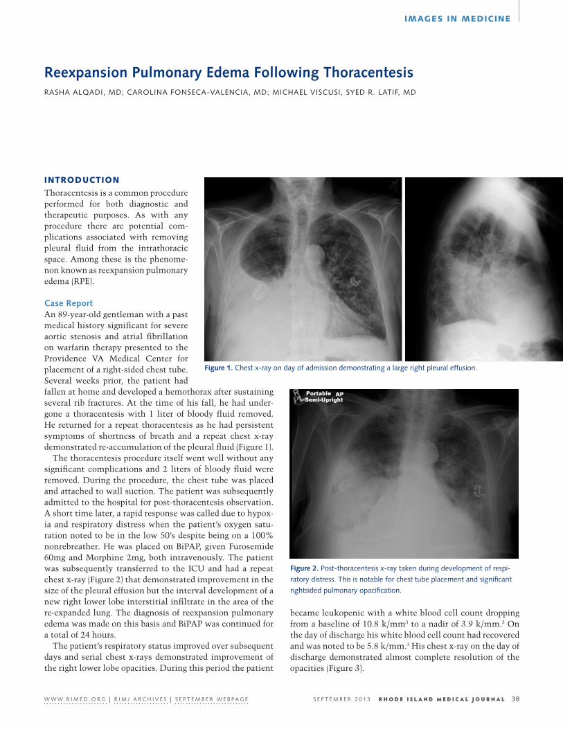
Case ReportAn 89-year-old gentleman with a past medical history significant for severe aortic stenosis and atrial fibrillation on warfarin therapy presented to the Providence VA Medical Center for placement of a right-sided chest tube. Several weeks prior, the patient had fallen at home and developed a hemothorax after sustaining several rib fractures. At the time of his fall, he had under-gone a thoracentesis with 1 liter of bloody fluid removed. He returned for a repeat thoracentesis as he had persistent symptoms of shortness of breath and a repeat chest x-ray demonstrated re-accumulation of the pleural fluid (Figure 1).
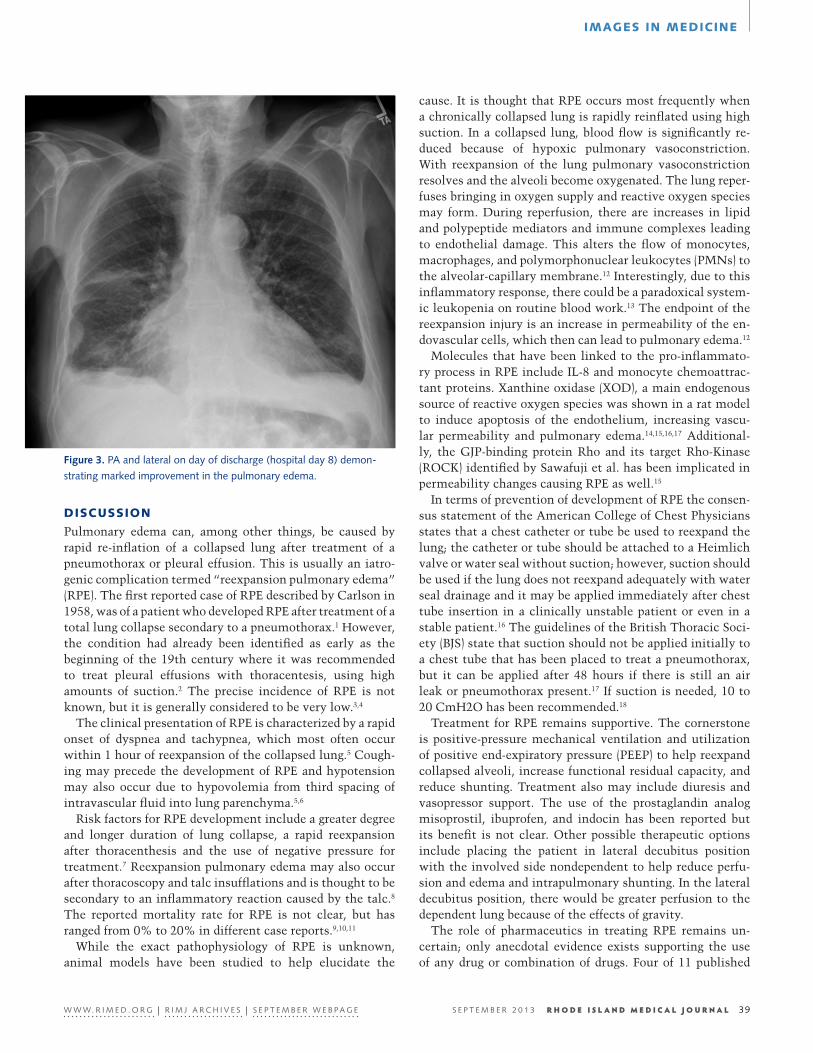
The thoracentesis procedure itself went well without any significant complications and 2 liters of bloody fluid were removed. During the procedure, the chest tube was placed and attached to wall suction. The patient was subsequently admitted to the hospital for post-thoracentesis observation. A short time later, a rapid response was called due to hypox-ia and respiratory distress when the patient’s oxygen satu-ration noted to be in the low 50’s despite being on a 100% nonrebreather. He was placed on BiPAP, given Furosemide 60mg and Morphine 2mg, both intravenously. The patient was subsequently transferred to the ICU and had a repeat chest x-ray (Figure 2) that demonstrated improvement in the size of the pleural effusion but the interval development of a new right lower lobe interstitial infiltrate in the area of the re-expanded lung. The diagnosis of reexpansion pulmonary edema was made on this basis and BiPAP was continued for a total of 24 hours.
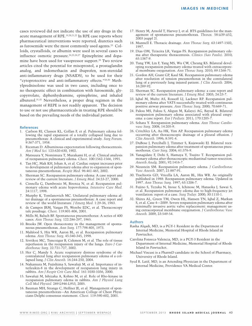
The patient’s respiratory status improved over subsequent days and serial chest x-rays demonstrated improvement of the right lower lobe opacities. During this period the patient
Figure 1. Chest x-ray on day of admission demonstrating a large right pleural effusion.
Figure 2. Post-thoracentesis x-ray taken during development of respi-
ratory distress. This is notable for chest tube placement and significant
rightsided pulmonary opacification.
became leukopenic with a white blood cell count dropping from a baseline of 10.8 k/mm3 to a nadir of 3.9 k/mm.3 On the day of discharge his white blood cell count had recovered and was noted to be 5.8 k/mm.3 His chest x-ray on the day of discharge demonstrated almost complete resolution of the opacities (Figure 3).
R H O D E I S L A N D M E D I C A L J O U R N A L W W W. R I M E D . O R G | R I M J A R C H I V E S | S E P T E M B E R W E B P A G E 38S E P T E M B E R 2 0 1 3
IMAGES IN MEDICINE
27
28
EN
DISCUSSION
Pulmonary edema can, among other things, be caused by rapid re-inflation of a collapsed lung after treatment of a pneumothorax or pleural effusion. This is usually an iatro-genic complication termed “reexpansion pulmonary edema” (RPE). The first reported case of RPE described by Carlson in 1958, was of a patient who developed RPE after treatment of a total lung collapse secondary to a pneumothorax.1 However, the condition had already been identified as early as the beginning of the 19th century where it was recommended to treat pleural effusions with thoracentesis, using high amounts of suction.2 The precise incidence of RPE is not known, but it is generally considered to be very low.3,4
The clinical presentation of RPE is characterized by a rapid onset of dyspnea and tachypnea, which most often occur within 1 hour of reexpansion of the collapsed lung.5 Cough-ing may precede the development of RPE and hypotension may also occur due to hypovolemia from third spacing of intravascular fluid into lung parenchyma.5,6
Risk factors for RPE development include a greater degree and longer duration of lung collapse, a rapid reexpansion after thoracenthesis and the use of negative pressure for treatment.7 Reexpansion pulmonary edema may also occur after thoracoscopy and talc insufflations and is thought to be secondary to an inflammatory reaction caused by the talc.8 The reported mortality rate for RPE is not clear, but has ranged from 0% to 20% in different case reports.9,10,11
While the exact pathophysiology of RPE is unknown, animal models have been studied to help elucidate the
cause. It is thought that RPE occurs most frequently when a chronically collapsed lung is rapidly reinflated using high suction. In a collapsed lung, blood flow is significantly re-duced because of hypoxic pulmonary vasoconstriction. With reexpansion of the lung pulmonary vasoconstriction resolves and the alveoli become oxygenated. The lung reper-fuses bringing in oxygen supply and reactive oxygen species may form. During reperfusion, there are increases in lipid and polypeptide mediators and immune complexes leading to endothelial damage. This alters the flow of monocytes, macrophages, and polymorphonuclear leukocytes (PMNs) to the alveolar-capillary membrane.12 Interestingly, due to this inflammatory response, there could be a paradoxical system-ic leukopenia on routine blood work.13 The endpoint of the reexpansion injury is an increase in permeability of the en-dovascular cells, which then can lead to pulmonary edema.12
Molecules that have been linked to the pro-inflammato-ry process in RPE include IL-8 and monocyte chemoattrac-tant proteins. Xanthine oxidase (XOD), a main endogenous source of reactive oxygen species was shown in a rat model to induce apoptosis of the endothelium, increasing vascu-lar permeability and pulmonary edema.14,15,16,17 Additional-ly, the GJP-binding protein Rho and its target Rho-Kinase (ROCK) identified by Sawafuji et al. has been implicated in permeability changes causing RPE as well.15
In terms of prevention of development of RPE the consen-sus statement of the American College of Chest Physicians states that a chest catheter or tube be used to reexpand the lung; the catheter or tube should be attached to a Heimlich valve or water seal without suction; however, suction should be used if the lung does not reexpand adequately with water seal drainage and it may be applied immediately after chest tube insertion in a clinically unstable patient or even in a stable patient.16 The guidelines of the British Thoracic Soci-ety (BJS) state that suction should not be applied initially to a chest tube that has been placed to treat a pneumothorax, but it can be applied after 48 hours if there is still an air leak or pneumothorax present.17 If suction is needed, 10 to 20 CmH2O has been recommended.18
Treatment for RPE remains supportive. The cornerstone is positive-pressure mechanical ventilation and utilization of positive end-expiratory pressure (PEEP) to help reexpand collapsed alveoli, increase functional residual capacity, and reduce shunting. Treatment also may include diuresis and vasopressor support. The use of the prostaglandin analog misoprostil, ibuprofen, and indocin has been reported but its benefit is not clear. Other possible therapeutic options include placing the patient in lateral decubitus position with the involved side nondependent to help reduce perfu-sion and edema and intrapulmonary shunting. In the lateral decubitus position, there would be greater perfusion to the dependent lung because of the effects of gravity.
The role of pharmaceutics in treating RPE remains un-certain; only anecdotal evidence exists supporting the use of any drug or combination of drugs. Four of 11 published
Figure 3. PA and lateral on day of discharge (hospital day 8) demon-
strating marked improvement in the pulmonary edema.
R H O D E I S L A N D M E D I C A L J O U R N A L W W W. R I M E D . O R G | R I M J A R C H I V E S | S E P T E M B E R W E B P A G E 39S E P T E M B E R 2 0 1 3
IMAGES IN MEDICINE
cases reviewed did not indicate the use of any drugs in the acute management of RPE.19,20,21,22 In RPE case reports where pharmaceutical interventions were reported, diuretics such as furosemide were the most commonly used agents.23 Col-loids, crystalloids, or albumin were used in several cases to influence osmotic pressure.24,25,26,27 Epinephrine and dopa-mine have been used for vasopressor support.28 Two review articles cited the potential for misoprostol, a prostaglandin analog, and indomethacin and ibuprofen, non-steroidal anti-inflammatory drugs (NSAIDS), to be used for their “cytoprotective and anti-inflammatory effects.”29,30 Meth-ylprednisolone was used in two cases, including once to no therapeutic effect in combination with furosemide, gly-copyrrolate, diphenhydramine, epinephrine, and inhaled albuterol.31,32 Nevertheless, a proper drug regimen in the management of REPE is not readily apparent. The decision to use or not use pharmaceuticals in treating RPE should be based on the prevailing needs of the individual patient.
References1. Carlson RI, Classen KL, Gollan F, et al. Pulmonary edema fol-
lowing the rapid expansion of a totally collapsed lung due to pneumothorax: A clinical and experimental study. Surg Forum. 9:367-371, 1958.
2. Riesman D. Albuminous expectoration following thoracentesis. Am J Med Sci. 123:620-630, 1902.
3. Matsuura Y, Nomimura T, Murakami H, et al. Clinical analysis of reexpansion pulmonary edema. Chest. 100:1562-1566, 1991.
4. Tan HC, Mak KH, Johan A, et al. Cardiac output increases prior to development of pulmonary edema after re-expansion of spon-taneous pneumothorax. Respir Med. 96:461-465, 2002.
5. Sherman SC. Reexpansion pulmonary edema: A case report and review of the current literature. J Emerg Med. 24:23-27, 2000.
6. Cinnella G, Dambrosio M, Birenza N, et al. Reexpansion pul-monary edema with acute hypovolemia. Intensive Care Med. 24:1117, 1998.
7. Murphy K, Tomlanovich MC. Unilateral pulmonary edema af-ter drainage of a spontaneous pneumothorax: A case report and review of the world literature. J Emerg Med. 1:29-36, 1983.
8. de Campos JRM, Vargas FS, Werebe EDC, et al. Thoracoscopy talc poudrage. Chest. 119:801-806, 2001.
9. Mills M, Balsch BF. Spontaneous pneumothorax: A series of 400 cases. Ann Thorac Surg. 122:286-297, 1965.
10. Brooks JW. Open thoracotomy in the management of sponta-neous pneumothorax. Ann Surg. 177:798-805, 1973.
11. Mahfood S, Hix WR, Aaron BL, et al. Reexpansion pulmonary edema. Ann Thorac Surg. 45:340-345, 1998.
12. Sivrikoz MC, Tuncozgur B, Cekmen M, et al. The role of tissue reperfusion in the reexpansion injury of the lungs. Euro J Car-diothorac Surg. 22:721-727, 2002.
13. Her C, Mandy S. Acute respiratory distress syndrome of the contralateral lung after reexpansion pulmonary edema of a col-lapsed lung. J Clin Anesth. 16:244-250, 2004.
14. Nakamura M, Fujishima S, Sawafuji M, et al. Importance of in-terleukin-8 in the development of reexpansion lung injury in rabbits. Am J Respir Crit Care Med. 161:1030-1036, 2000.
15. Sawafuji M, Ishizaka A, Kohno M, et al. Role of Rho-kinase in reexpansion pulmonary edema in rabbits. Am J Physiol Lung Cell Mol Physiol. 289:L946-L953, 2005.
16. Bauman MH, Strange C, Heffner JE, et al. Management of spon-taneous pneumothorax—An American College of Chest Physi-cians Delphi consensus statement. Chest. 119:590-602, 2001.
17. Henry M, Arnold T, Harvey J, et al. BTS guidelines for the man-agement of spontaneous pneumothorax. Thorax. 58:ii39-ii52, 2003 (suppl 2).
18. Munnell E. Thoracic drainage. Ann Thorac Surg. 63:1497-1502, 1997.
19. Dias OM, Teixeira LR, Vargas FS. Reexpansion pulmonary ede-ma after therapeutic thoracentesis. Clinics (Sao Paulo). 2010; 65:1387-9.
20. Tung YW, Lin F, Yang MS, Wu CW, Cheung KS. Bilateral devel-oping reexpansion pulmonary edema treated with extracorpore-al membrane oxygenation. Ann Thorac Surg. 2010; 89:1268-71.
21. Gordon AH, Grant GP, Kaul SK. Reexpansion pulmonary edema after resolution of tension pneumothorax in the contralateral lung of a previously lung injured patient. J Clin Anesth. 2004; 16:289-92.
22. Sherman SC. Reexpansion pulmonary edema: a case report and review of the current literature. J Emerg Med. 2003; 24:23-7.
23. Iqbal M, Multz AS, Rossoff LJ, Lackner RP. Reexpansion pul-monary edema after VATS successfully treated with continuous positive airway pressure. Ann Thorac Surg. 2000; 70:669-71.
24. Paksu MS, Paksu S, Akgun M, Kalayci AG, Baysal K. Bilateral reexpansion pulmonary edema associated with pleural empy-ema: a case report. Eur J Pediatr. 2011; 170:1205-7.
25. Sohara Y. Reexpansion pulmonary edema. Ann Thorac Cardio-vasc Surg. 2008; 14:205-9.
26. Critchley LA, Au HK, Yim AP. Reexpansion pulmonary edema occurring after thoracoscopic drainage of a pleural effusion. J Clin Anesth. 1996; 8:591-4.
27. DuBose J, Perciballi J, Timmer S, Kujawaski EJ. Bilateral reex-pansion pulmonary edema after treatment of spontaneous pneu-mothorax. Curr Surg. 2004; 61:376-9.
28. Yanagidate F, Dohi S, Hamaya Y, Tsujito T. Reexpansion pul-monary edema after thoracoscopic mediastinal tumor resection. Anesth Analg. 2001; 92:1416-7.
29. Neustein SM. Reexpansion pulmonary edema. J Cardiothorac Vasc Anesth. 2007; 21:887-91.
30. Trachiotis GD, Vricella LA, Aaron BL, Hix WR. As originally published in 1988: Reexpansion pulmonary edema. Updated in 1997. Ann Thorac Surg. 1997; 63:1206-7.
31. Fujino S, Tezuka N, Inoue S, Ichinose M, Hanaoka J, Sawai S, et al. Reexpansion pulmonary edema due to high-frequency jet ventilation: report of a case. Surg Today. 2000; 30:1110-1.
32. Shires AL, Green TM, Owen HL, Hansen TN, Iqbal Z, Markan S, et al. Case 4—2009. Severe rexpansion pulmonary edema after minimally invasive aortic valve replacement: management us-ing extracorporeal membrane oxygenation. J Cardiothorac Vasc Anesth. 2009; 23:549-54.
AuthorsRasha Alqadi, MD, is a PGY-1 Resident in the Department of
Internal Medicine, Memorial Hospital of Rhode Island in Pawtucket.
Carolina Fonseca-Valencia, MD, is a PGY-3 Resident in the Department of Internal Medicine, Memorial Hospital of Rhode Island in Pawtucket.
Michael Viscusi is a PharmD candidate in the School of Pharmacy, University of Rhode Island.
Syed R. Latif, MD, is an Attending Physician in the Department of Internal Medicine, Providence VA Medical Center.
R H O D E I S L A N D M E D I C A L J O U R N A L W W W. R I M E D . O R G | R I M J A R C H I V E S | S E P T E M B E R W E B P A G E 40S E P T E M B E R 2 0 1 3
IMAGES IN MEDICINE