Embed Size (px)
Citation preview
www.jpis.org
Journal of Periodontal& Implant ScienceJPIS
pISSN 2093-2278eISSN 2093-2286
Copyright © 2012 Korean Academy of PeriodontologyThis is an Open Access article distributed under the terms of the Creative Commons Attribution Non-Commercial License (http://creativecommons.org/licenses/by-nc/3.0/).
Reliability of two different presurgical preparation methods for implant dentistry based on
panoramic radiography and cone-beam computed tomography in cadavers
Kyung-Seok Hu1, Da-Yae Choi1, Won-Jae Lee1, Hee-Jin Kim1, Ui-Won Jung2, Sungtae Kim3,* 1Division in Anatomy and Histology, Department of Oral Biology, Yonsei University College of Dentistry, Seoul, Korea
2Department of Periodontology, Yonsei University College of Dentistry, Seoul, Korea3Department of Periodontology, Seoul National University School of Dentistry, Seoul, Korea
Purpose: Special care is necessary to avoid invading important anatomic structures during surgery when presurgical plan-ning is made based on radiographs. However, none of these types of radiography represents a perfect modality. The purpose of this study was to determine the reliability of presurgical planning based on the use of two types of radiographic image (digital panoramic radiography [DPR] and cone-beam computed tomography [CBCT]) by beginner dentists to place implants, and to quantify differences in measurements between radiographic images and real specimens. Methods: Ten fresh cadavers without posterior teeth were used, and twelve practitioners who had no experience of implant surgery performed implant surgery after 10 hours of basic instruction using conventional surgical guide based on CBCT or DPR. Two types of measurement error were evaluated: 1) the presurgical measurement error, defined as that between the pre-surgical and postsurgical measurements in each modality of radiographic analysis, and 2) the measurement error between postsurgical radiography and the real specimen.Results: The mean presurgical measurement error was significantly smaller for CBCT than for DPR in the maxillary region, whereas it did not differ significantly between the two imaging modalities in the mandibular region. The mean measurement error between radiography and real specimens was significantly smaller for CBCT than for DPR in the maxillary region, but did not differ significantly in the mandibular region. Conclusions: Presurgical planning can be performed safely using DPR in the mandible; however, presurgical planning using CBCT is recommended in the maxilla when a structure in a buccolingual location needs to be evaluated because this imaging modality supplies buccolingual information that cannot be obtained from DPR.
Keywords: Complications, Cone-beam computed tomography, Dental Implantation, Panoramic radiography.
J Periodontal Implant Sci 2012;42:39-44 • http://dx.doi.org/10.5051/jpis.2012.42.2.39
Research Article
INTRODUCTION
Dental implants have been successfully used for replacing missing teeth with reported success rates of more than 90%
[1,2]. Improvements in the surface and design of dental im-plants have contributed to this remarkable success rates. Re-cent advances in the radiographic techniques used in im-plant dentistry have increased the accuracy of presurgical
Received: Jan. 26, 2012; Accepted: Mar. 8, 2012*Correspondence: Sungtae KimDepartment of Periodontology, Seoul National University School of Dentistry, 101 Daehak-ro, Seoul 110-749, KoreaE-mail: [email protected], Tel: +82-2-2072-4712, Fax: +82-2-744-0051
Journal of Periodontal& Implant ScienceJPISReliability of panoramic radiography and cone-beam computed tomography
for implantation 40
planning [3,4]. However, there exist the risks of nerve injury and maxillary sinus perforation originated from inappropri-ate presurgical planning due to innate discrepancy between radiographic measurements and real dimensions. Precise ra-diographic assessment of available alveolar bone and identi-fication of characteristic bone morphology are indispensible for dental implant placement. When posterior maxillary and mandibular sites are considered, the location of important anatomic structures such as the mandibular canal and maxil-lary sinus should be identified carefully for proper implant site selection and length selection.
Pain or paresthesia caused by inferior alveolar nerve injury is one of major complications. Another major complication is maxillary sinus inferior wall perforation which is known to be correlated with the incidence of infection followed by fail-ure of a dental implant [5,6]. Therefore, special care should be taken not to invade important anatomic structures during surgery when presurgical planning is made based on radio-graphs. More precise radiographic techniques and corre-sponding proper computer programs to guide precise sur-gery would reduce the complication rate [4]. Presurgical plan-ning is mostly made with radiographs such as panoramic, lateral cephalometric, and periapical radiographs, and com-puted tomography (CT) [7,8]. However, none of these types of radiography represents a perfect modality [9,10]. More accu-rate radiographic technique is required, especially for presur-gical planning in areas where important anatomic structures are located. Finding an appropriate location and measuring the available bone based on panoramic radiograph is one of the most frequently used modalities for presurgical planning. A panoramic radiograph displays the body of the mandible and maxilla, mandibular canal, and maxillary sinus on a sin-gle image. The length and mesiodistal angulation of an im-plant are usually determined in the panoramic radiograph. However, in the anterior area where more distortion exists than posterior area, the reliability of panoramic radiographs for presurgical planning of an implant is questionable [11-13].
The necessity of cross-sectional imaging for a dental im-plant has been emphasized [14,15]. According to the recom-mendation from American Academy of Oral and Maxillofa-cial radiology in 2000, cross-sectional images including con-ventional spiral tomography, linear tomography, or CT should be used for presurgical planning [16]. These cross-sectional imaging techniques exhibit various accuracies. Linear tomog-raphy is reported to be significantly less accurate than spiral tomography in detecting mandibular canal [17]. However, it was reported that measuring available bone volume only us-ing spiral tomography may lead to a dangerous situation be-cause the available vertical bone height is exaggerated in spi-ral tomography relative to panoramic radiography [12]. CT
provides less magnification than the other types of cross-sec-tional tomography. The magnification in CT corresponds to 0 to 4% [18]. CT can offer direct volumetric reconstruction. Faster and easier data transformation into three-dimensional analysis is possible. In addition, convenient interpretation is also possible without overlapping of images [17]. However, one major disadvantage of CT is a high radiation dose [19], and therefore cone-beam CT (CBCT) was developed to over-come this limitation [20,21]. CBCT images are known to have higher quality than CT images with 1/400 radiation dose of conventional CT [19,20].
Each type of radiograph has its own advantages and disad-vantages. Therefore, the radiation dose, magnification rate, and specific indications need to be considered when select-ing the type of radiographic images to use in presurgical planning. The purpose of this study was to determine the re-liability of presurgical planning based on the use of two types of radiographic image (digital panoramic radiography [DPR] and CBCT) by beginner dentists to place implants.
MATERIALS AND METHODS
Study designTen fresh cadavers without posterior tooth were used to ex-
amine the accuracy of CBCT and DPR measurements. These cadavers were from persons who donated their bodies for re-search or teaching purposes. The posterior teeth were ex-tracted when there is not any missing posterior tooth. Twelve dentists who never had any experience of implant surgery were participated in implant surgery after 10 hours of basic instruction not including hands-on training. Thereafter, each participant performed implant surgery using a conventional surgical guide based on CBCT or DPR on their own. The sites for implant surgery were randomly assigned for the two in-vestigated presurgical planning methods: 31 implants were placed with the surgical guide based on DPR and the corre-sponding computer program (Starpacs, Infinitt Co., Seoul, Korea), while 32 implants were placed with the surgical guide based on CBCT and the corresponding computer program (Ondemend 3D, Cybermed Inc., Seoul, Korea).
Two types of measurement error were evaluated in this study: 1) The presurgical measurement error: difference be-tween the presurgical and postsurgical measurements in ra-diographs. 2) The measurement error: difference of measured distances between postsurgical radiographs and the cadaver specimens.
Measurement errors for CBCTVirtual planning to determine the appropriate length of the
implant was first performed using CBCT and the correspond-
Journal of Periodontal& Implant ScienceJPIS Kyung-Seok Hu et al. 41
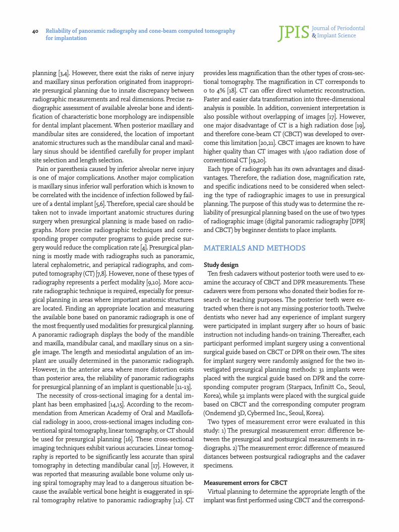
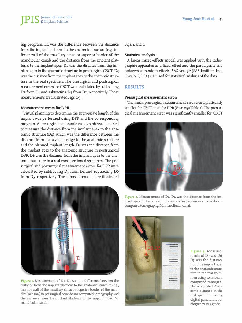
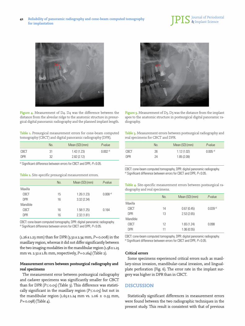
ing program. D1 was the difference between the distance from the implant platform to the anatomic structure (e.g., in-ferior wall of the maxillary sinus or superior border of the mandibular canal) and the distance from the implant plat-form to the implant apex. D2 was the distance from the im-plant apex to the anatomic structure in postsurgical CBCT. D3 was the distance from the implant apex to the anatomic struc-ture in the real specimen. The presurgical and postsurgical measurement errors for CBCT were calculated by subtracting D2 from D1 and subtracting D3 from D2, respectively. These measurements are illustrated Figs. 1-3.
Measurement errors for DPRVirtual planning to determine the appropriate length of the
implant was performed using DPR and the corresponding program. A presurgical panoramic radiograph was obtained to measure the distance from the implant apex to the ana-tomic structure (D4), which was the difference between the distance from the alveolar ridge to the anatomic structure and the planned implant length. D5 was the distance from the implant apex to the anatomic structure in postsurgical DPR. D6 was the distance from the implant apex to the ana-tomic structure in a real cross-sectioned specimen. The pre-surgical and postsurgical measurement errors for DPR were calculated by subtracting D5 from D4 and subtracting D6 from D5, respectively. These measurements are illustrated
Figs. 4 and 5.
Statistical analysisA linear mixed-effects model was applied with the radio-
graphic apparatus as a fixed effect and the participants and cadavers as random effects. SAS ver. 9.2 (SAS Institute Inc., Cary, NC, USA) was used for statistical analysis of the data.
RESULTS
Presurgical measurement errors The mean presurgical measurement error was significantly
smaller for CBCT than for DPR (P≤0.05) (Table 1). The presur-gical measurement error was significantly smaller for CBCT
Figure 1. Measurement of D1. D1 was the difference between the distance from the implant platform to the anatomic structure (e.g., inferior wall of the maxillary sinus or superior border of the man-dibular canal) in presurgical cone-beam computed tomography and the distance from the implant platform to the implant apex. M: mandibular canal.
Figure 2. Measurement of D2. D2 was the distance from the im-plant apex to the anatomic structure in postsurgical cone-beam computed tomography. M: mandibular canal.
Figure 3. Measure-ments of D3 and D6. D3 was the distance from the implant apex to the anatomic struc-ture in the real speci-men using cone-beam computed tomogra-phy as a guide. D6 was same distance in the real specimen using digital panoramic ra-diography as a guide.
Journal of Periodontal& Implant ScienceJPISReliability of panoramic radiography and cone-beam computed tomography
for implantation 42
(1.26±1.23 mm) than for DPR (3.32±2.34 mm, P=0.008) in the maxillary region, whereas it did not differ significantly between the two imaging modalities in the mandibular region (1.58±1.25 mm vs. 2.32±1.81 mm, respectively, P=0.164) (Table 2).
Measurement errors between postsurgical radiography and real specimens
The measurement error between postsurgical radiography and cadaver specimens was significantly smaller for CBCT than for DPR (P≤0.05) (Table 3). This difference was statisti-cally significant in the maxillary region (P≤0.05) but not in the mandibular region (1.65±1.24 mm vs. 1.06 ± 0.55 mm, P=0.098) (Table 4).
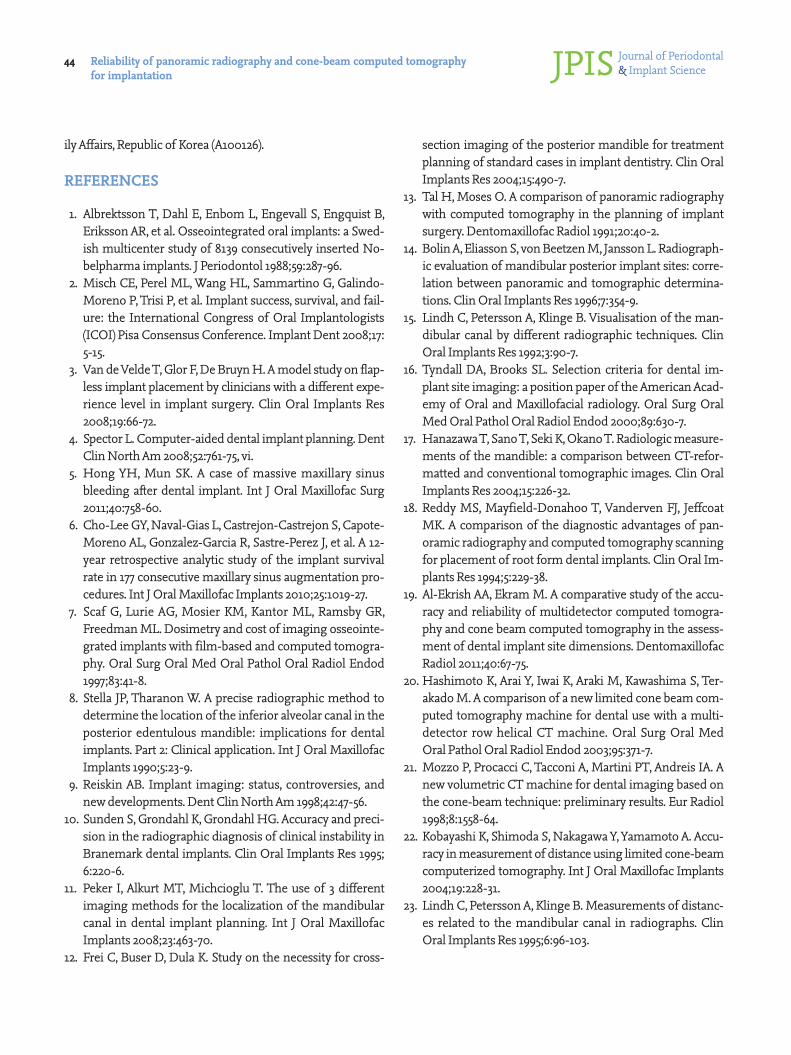
Critical errorsSome specimens experienced critical errors such as maxil-
lary-sinus invasion, mandibular-canal invasion, and lingual-plate perforation (Fig. 6). The error rate in the implant sur-gery was higher in DPR than in CBCT.
DISCUSSION
Statistically significant differences in measurement errors were found between the two radiographic techniques in the present study. This result is consistent with that of previous
Figure 4. Measurement of D4. D4 was the difference between the distance from the alveolar ridge to the anatomic structure in presur-gical digital panoramic radiography and the planned implant length.
Figure 5. Measurement of D5. D5 was the distance from the implant apex to the anatomic structure in postsurgical digital panoramic ra-diography.
Table 1. Presurgical measurement errors for cone-beam computed tomography (CBCT) and digital panoramic radiography (DPR).
No. Mean (SD) (mm) P-value
CBCT 31 1.42 (1.23) 0.002 a)
DPR 32 2.82 (2.12)
a) Significant difference between errors for CBCT and DPR, P<0.05.
Table 2. Site-specific presurgical measurement errors.
No. Mean (SD) (mm) P-value
Maxilla CBCT 15 1.26 (1.23) 0.008 a)
DPR 16 3.32 (2.34)Mandible CBCT 16 1.58 (1.25) 0.164 DPR 16 2.32 (1.81)
CBCT: cone-beam computed tomography, DPR: digital panoramic radiography.a) Significant difference between errors for CBCT and DPR, P<0.05.
Table 3. Measurement errors between postsurgical radiography and real specimens for CBCT and DPR.
No. Mean (SD) (mm) P-value
CBCT 26 1.12 (1.02) 0.005 a)
DPR 24 1.85 (2.09)
CBCT: cone-beam computed tomography, DPR: digital panoramic radiography.a) Significant difference between errors for CBCT and DPR, P<0.05.
Table 4. Site-specific measurement errors between postsurgical ra-diography and real specimens.
No. Mean (SD) (mm) P-value
Maxilla CBCT 14 0.67 (0.45) 0.039 a)
DPR 13 2.53 (2.65)Mandible CBCT 12 1.65 (1.24) 0.098 DPR 11 1.06 (0.55)
CBCT: cone-beam computed tomography, DPR: digital panoramic radiography.a) Significant difference between errors for CBCT and DPR, P<0.05.

Journal of Periodontal& Implant ScienceJPIS Kyung-Seok Hu et al. 43
studies [22,23]. The errors were greater for DPR than CBCT because presurgical plans are made on a two-dimensional plane for DPR. CBCT should be used for presurgical planning and postoperative evaluation, especially when dentists with limited experiences place implants, considering the reduced errors in three-dimensional presurgical planning.
The measurement errors in the maxilla were significantly lower for CBCT than DPR. It can be attributed to the difficul-ty of identifying the exact location of the inferior wall of the maxillary sinus in presurgical planning when using DPR. Various levels of the inferior border of the maxillary sinus are overlapped in DPR. To the contrary, the exact levels of the in-ferior border of the maxillary sinus can be identified using CBCT because the view in the specific plane where the im-plant will be placed is used for presurgical planning. These data suggest that presurgical planning using DPR for implant placement on the maxillary premolar or molar area is not sufficiently reliable. Even though limitation of DPR can be compensated by abundant clinical experiences, more accu-rate and precise methods are recommended for preventing unexpected complications.
In mandible, the measurement error in CBCT didn’t show any statistically significant difference from that in DPR. This result is in accordance with that of one previous study [11]. This shows that there will be fewer errors when presurgical plans are made using DPR in mandible than maxilla. Identify-ing the superior border of the mandibular canal is easier than the inferior border of the maxillary sinus because the man-dibular canal is easy to identify in most cases, except those with thick cortical bone or a high proportion of trabecular bone. This convenient detection of the mandibular canal can allow dentists to place implants in the posterior mandibular area without any critical complications as long as the bucco-lingual width is measured carefully. Direct measurement us-ing calipers is recommended intraorally or extraorally in a study cast. In summary, presurgical planning in the mandible
A B C
Figure 6. Some critical errors. (A) Maxillary-sinus invasion, (B) mandibular-canal inva-sion, and (C) lingual-plate perforation.
can be performed safely using DPR by dentists with sufficient experience and skill, whereas presurgical planning using CBCT is strongly recommended when a buccolingual loca-tion of the mandibular canal needs to be evaluated.
Radiographic images do not always display anatomic struc-tures accurately. In maxilla, it was revealed that less measure-ment errors between postsurgical radiographic images and real specimens were found in CBCT than DPR. More accu-rate detection of the inferior wall of the maxillary sinus was possible using CBCT.
Both positive and negative presurgical measurement errors were obtained, whereas only positive measurement errors were obtained between postsurgical radiographs and real specimens. It appears that the distance from the implant apex to anatomic structures was always greater in a postsurgical radiograph than in the corresponding real specimen.
This study has revealed the best radiographic methods to use in order to reduce errors by beginner dentists during dental implantation. Future studies should evaluate the va-lidity of computer-assisted implant surgery with a surgical guide fabricated based on CBCT. In addition, it is necessary to evaluate the measurement errors when experienced den-tists are placing implants.
CONFLICT OF INTEREST
No potential conflict of interest relevant to this article was reported.
ACKNOWLEDGEMENTS
This research was supported by Basic Science Research Pro-gram through the National Research Foundation of Korea (NRF) funded by the Ministry of Education, Science and Tech-nology (2010-0007829) and by a grant of the Korean Health Technology R&D Project, Ministry for Health, Welfare & Fam-
Journal of Periodontal& Implant ScienceJPISReliability of panoramic radiography and cone-beam computed tomography
for implantation 44
ily Affairs, Republic of Korea (A100126).
REFERENCES
1. Albrektsson T, Dahl E, Enbom L, Engevall S, Engquist B, Eriksson AR, et al. Osseointegrated oral implants: a Swed-ish multicenter study of 8139 consecutively inserted No-belpharma implants. J Periodontol 1988;59:287-96.
2. Misch CE, Perel ML, Wang HL, Sammartino G, Galindo-Moreno P, Trisi P, et al. Implant success, survival, and fail-ure: the International Congress of Oral Implantologists (ICOI) Pisa Consensus Conference. Implant Dent 2008;17: 5-15.
3. Van de Velde T, Glor F, De Bruyn H. A model study on flap-less implant placement by clinicians with a different expe-rience level in implant surgery. Clin Oral Implants Res 2008;19:66-72.
4. Spector L. Computer-aided dental implant planning. Dent Clin North Am 2008;52:761-75, vi.
5. Hong YH, Mun SK. A case of massive maxillary sinus bleeding after dental implant. Int J Oral Maxillofac Surg 2011;40:758-60.
6. Cho-Lee GY, Naval-Gias L, Castrejon-Castrejon S, Capote-Moreno AL, Gonzalez-Garcia R, Sastre-Perez J, et al. A 12-year retrospective analytic study of the implant survival rate in 177 consecutive maxillary sinus augmentation pro-cedures. Int J Oral Maxillofac Implants 2010;25:1019-27.
7. Scaf G, Lurie AG, Mosier KM, Kantor ML, Ramsby GR, Freedman ML. Dosimetry and cost of imaging osseointe-grated implants with film-based and computed tomogra-phy. Oral Surg Oral Med Oral Pathol Oral Radiol Endod 1997;83:41-8.
8. Stella JP, Tharanon W. A precise radiographic method to determine the location of the inferior alveolar canal in the posterior edentulous mandible: implications for dental implants. Part 2: Clinical application. Int J Oral Maxillofac Implants 1990;5:23-9.
9. Reiskin AB. Implant imaging: status, controversies, and new developments. Dent Clin North Am 1998;42:47-56.
10. Sunden S, Grondahl K, Grondahl HG. Accuracy and preci-sion in the radiographic diagnosis of clinical instability in Branemark dental implants. Clin Oral Implants Res 1995; 6:220-6.
11. Peker I, Alkurt MT, Michcioglu T. The use of 3 different imaging methods for the localization of the mandibular canal in dental implant planning. Int J Oral Maxillofac Implants 2008;23:463-70.
12. Frei C, Buser D, Dula K. Study on the necessity for cross-
section imaging of the posterior mandible for treatment planning of standard cases in implant dentistry. Clin Oral Implants Res 2004;15:490-7.
13. Tal H, Moses O. A comparison of panoramic radiography with computed tomography in the planning of implant surgery. Dentomaxillofac Radiol 1991;20:40-2.
14. Bolin A, Eliasson S, von Beetzen M, Jansson L. Radiograph-ic evaluation of mandibular posterior implant sites: corre-lation between panoramic and tomographic determina-tions. Clin Oral Implants Res 1996;7:354-9.
15. Lindh C, Petersson A, Klinge B. Visualisation of the man-dibular canal by different radiographic techniques. Clin Oral Implants Res 1992;3:90-7.
16. Tyndall DA, Brooks SL. Selection criteria for dental im-plant site imaging: a position paper of the American Acad-emy of Oral and Maxillofacial radiology. Oral Surg Oral Med Oral Pathol Oral Radiol Endod 2000;89:630-7.
17. Hanazawa T, Sano T, Seki K, Okano T. Radiologic measure-ments of the mandible: a comparison between CT-refor-matted and conventional tomographic images. Clin Oral Implants Res 2004;15:226-32.
18. Reddy MS, Mayfield-Donahoo T, Vanderven FJ, Jeffcoat MK. A comparison of the diagnostic advantages of pan-oramic radiography and computed tomography scanning for placement of root form dental implants. Clin Oral Im-plants Res 1994;5:229-38.
19. Al-Ekrish AA, Ekram M. A comparative study of the accu-racy and reliability of multidetector computed tomogra-phy and cone beam computed tomography in the assess-ment of dental implant site dimensions. Dentomaxillofac Radiol 2011;40:67-75.
20. Hashimoto K, Arai Y, Iwai K, Araki M, Kawashima S, Ter-akado M. A comparison of a new limited cone beam com-puted tomography machine for dental use with a multi-detector row helical CT machine. Oral Surg Oral Med Oral Pathol Oral Radiol Endod 2003;95:371-7.
21. Mozzo P, Procacci C, Tacconi A, Martini PT, Andreis IA. A new volumetric CT machine for dental imaging based on the cone-beam technique: preliminary results. Eur Radiol 1998;8:1558-64.
22. Kobayashi K, Shimoda S, Nakagawa Y, Yamamoto A. Accu-racy in measurement of distance using limited cone-beam computerized tomography. Int J Oral Maxillofac Implants 2004;19:228-31.
23. Lindh C, Petersson A, Klinge B. Measurements of distanc-es related to the mandibular canal in radiographs. Clin Oral Implants Res 1995;6:96-103.