Embed Size (px)
Citation preview
RENAL MACROPHAGES IN AUTOSOMALRECESSIVE POLYCYSTIC KIDNEY DISEASE
Michal Mrug,1,3 Juling Zhou,1 Lisa MGuay-Woodford4 and Lesley E Smythies2, 1Division ofNephrology, Nephrology Research and Training Center and2Division of Gastroenterology, Department of Medicine,University of Alabama at Birmingham, 3Department ofVeterans Affairs Medical Center, Birmingham, Alabama,and 4Children’s National Medical Center and School ofMedicine and Health Sciences, George WashingtonUniversity, Washington DC, USA
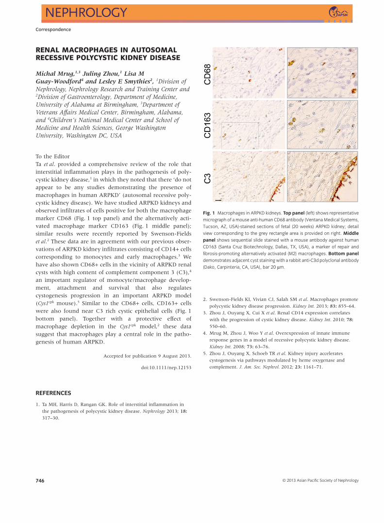
To the EditorTa et al. provided a comprehensive review of the role thatinterstitial inflammation plays in the pathogenesis of poly-cystic kidney disease,1 in which they noted that there ‘do notappear to be any studies demonstrating the presence ofmacrophages in human ARPKD’ (autosomal recessive poly-cystic kidney disease). We have studied ARPKD kidneys andobserved infiltrates of cells positive for both the macrophagemarker CD68 (Fig. 1 top panel) and the alternatively acti-vated macrophage marker CD163 (Fig. 1 middle panel);similar results were recently reported by Swenson-Fieldset al.2 These data are in agreement with our previous obser-vations of ARPKD kidney infiltrates consisting of CD14+ cellscorresponding to monocytes and early macrophages.3 Wehave also shown CD68+ cells in the vicinity of ARPKD renalcysts with high content of complement component 3 (C3),4
an important regulator of monocyte/macrophage develop-ment, attachment and survival that also regulatescystogenesis progression in an important ARPKD model(Cys1cpk mouse).5 Similar to the CD68+ cells, CD163+ cellswere also found near C3 rich cystic epithelial cells (Fig. 1bottom panel). Together with a protective effect ofmacrophage depletion in the Cys1cpk model,2 these datasuggest that macrophages play a central role in the patho-genesis of human ARPKD.
Accepted for publication 9 August 2013.
doi:10.1111/nep.12153
REFERENCES
1. Ta MH, Harris D, Rangan GK. Role of interstitial inflammation in
the pathogenesis of polycystic kidney disease. Nephrology 2013; 18:317–30.
2. Swenson-Fields KI, Vivian CJ, Salah SM et al. Macrophages promote
polycystic kidney disease progression. Kidney Int. 2013; 83: 855–64.
3. Zhou J, Ouyang X, Cui X et al. Renal CD14 expression correlates
with the progression of cystic kidney disease. Kidney Int. 2010; 78:550–60.
4. Mrug M, Zhou J, Woo Y et al. Overexpression of innate immune
response genes in a model of recessive polycystic kidney disease.
Kidney Int. 2008; 73: 63–76.
5. Zhou J, Ouyang X, Schoeb TR et al. Kidney injury accelerates
cystogenesis via pathways modulated by heme oxygenase and
complement. J. Am. Soc. Nephrol. 2012; 23: 1161–71.
Fig. 1 Macrophages in ARPKD kidneys. Top panel (left) shows representative
micrograph of a mouse anti-human CD68 antibody (Ventana Medical Systems,
Tucson, AZ, USA)-stained sections of fetal (20 weeks) ARPKD kidney; detail
view corresponding to the grey rectangle area is provided on right. Middlepanel shows sequential slide stained with a mouse antibody against human
CD163 (Santa Cruz Biotechnology, Dallas, TX, USA), a marker of repair and
fibrosis-promoting alternatively activated (M2) macrophages. Bottom paneldemonstrates adjacent cyst staining with a rabbit anti-C3d polyclonal antibody
(Dako, Carpinteria, CA, USA), bar 20 μm.
bs_bs_banner
Correspondence
© 2013 Asian Pacific Society of Nephrology746
![Clinical manifestations of autosomal recessive polycystic kidney ... · viduals to survive the perinatal period [ 8, 10]. Pulmonaryhypoplasia,aserio uscomplicationthatgenerally occurs](https://img.pdfslide.net/doc/110x75/5f09f6827e708231d4295907/clinical-manifestations-of-autosomal-recessive-polycystic-kidney-viduals-to.jpg)