Embed Size (px)
Citation preview
Renal Physiology
Dr. Meg-angela Christi Amores
The Kidney
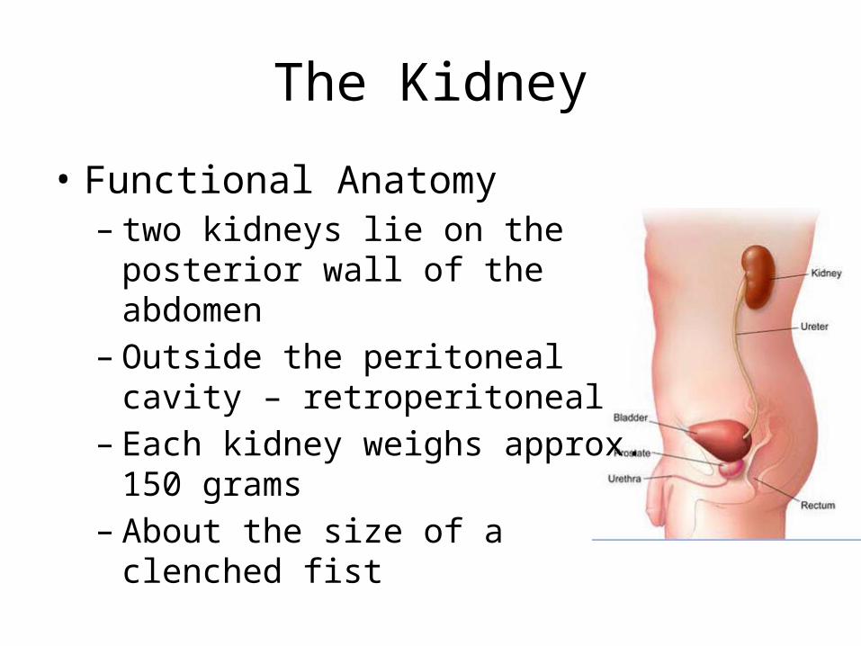
• Functional Anatomy– two kidneys lie on the posterior wall of
the abdomen– Outside the peritoneal cavity –
retroperitoneal– Each kidney weighs approx. 150 grams – About the size of a clenched fist
The kidney
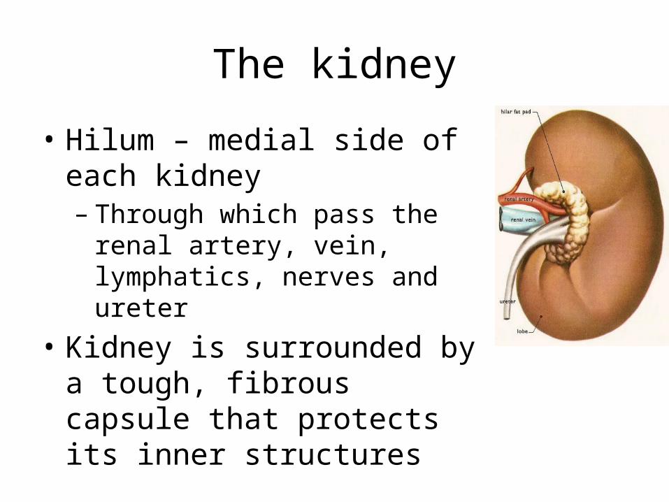
• Hilum – medial side of each kidney– Through which pass the renal artery,
vein, lymphatics, nerves and ureter• Kidney is surrounded by a tough,
fibrous capsule that protects its inner structures
The Kidney
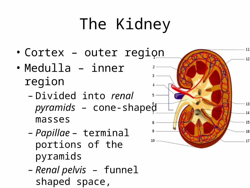
• Cortex – outer region• Medulla – inner region– Divided into renal pyramids –
cone-shaped masses– Papillae – terminal portions of
the pyramids– Renal pelvis – funnel shaped
space, continuous with the ureter
The Nephron
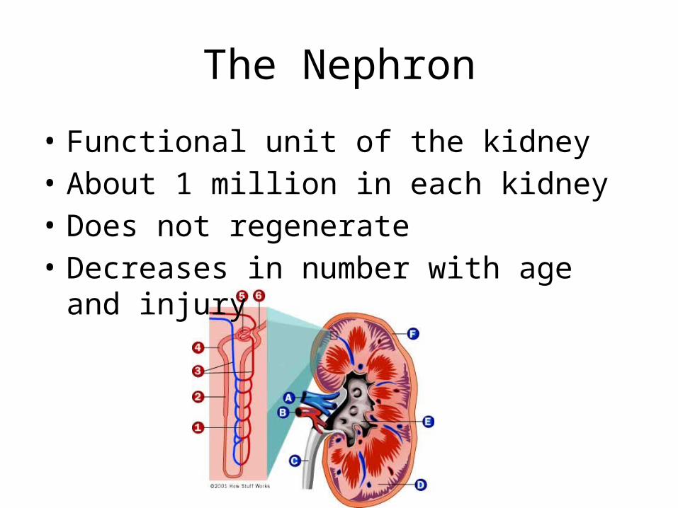
• Functional unit of the kidney• About 1 million in each kidney• Does not regenerate• Decreases in number with age and injury
The nephron
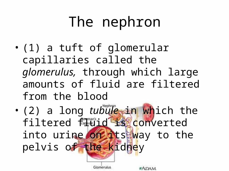
• (1) a tuft of glomerular capillaries called the glomerulus, through which large amounts of fluid are filtered from the blood
• (2) a long tubule in which the filtered fluid is converted into urine on its way to the pelvis of the kidney
The nephron
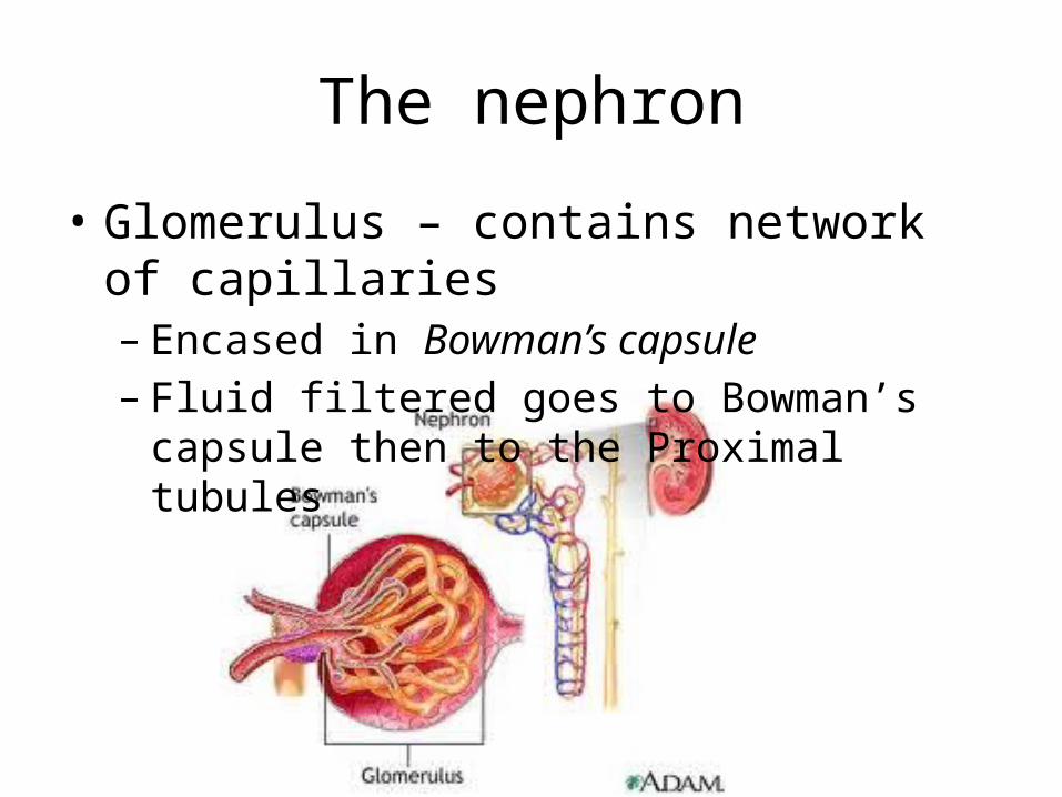
• Glomerulus – contains network of capillaries– Encased in Bowman’s capsule– Fluid filtered goes to Bowman’s capsule then to
the Proximal tubules
The Nephron
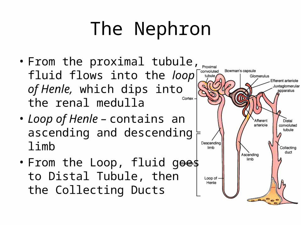
• From the proximal tubule, fluid flows into the loop of Henle, which dips into the renal medulla
• Loop of Henle – contains an ascending and descending limb
• From the Loop, fluid goes to Distal Tubule, then the Collecting Ducts
The kidney
• urine excreted by the kidneys• multiple mechanisms that control the rate of
urine excretion• most important means by which the body
maintains a balance between water intake and output and electrolytes is by controlling rate of their excretion
• adjusting the excretion rate of water and electrolytes to match precisely the intake
Urine Formation
• (1) glomerular filtration• (2) reabsorption of substances from the renal
tubules into the blood• (3) secretion of substances from the blood
into the renal tubules
• Urinary excretion rate = Filtration rate – Reabsorption rate + Secretion rate
Urine Formation
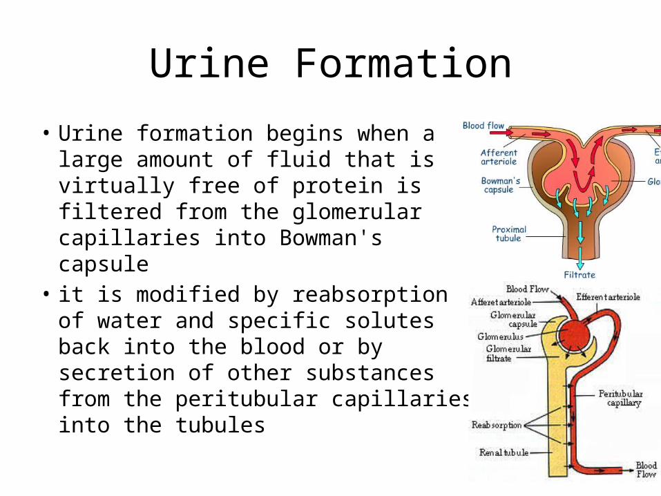
• Urine formation begins when a large amount of fluid that is virtually free of protein is filtered from the glomerular capillaries into Bowman's capsule
• it is modified by reabsorption of water and specific solutes back into the blood or by secretion of other substances from the peritubular capillaries into the tubules
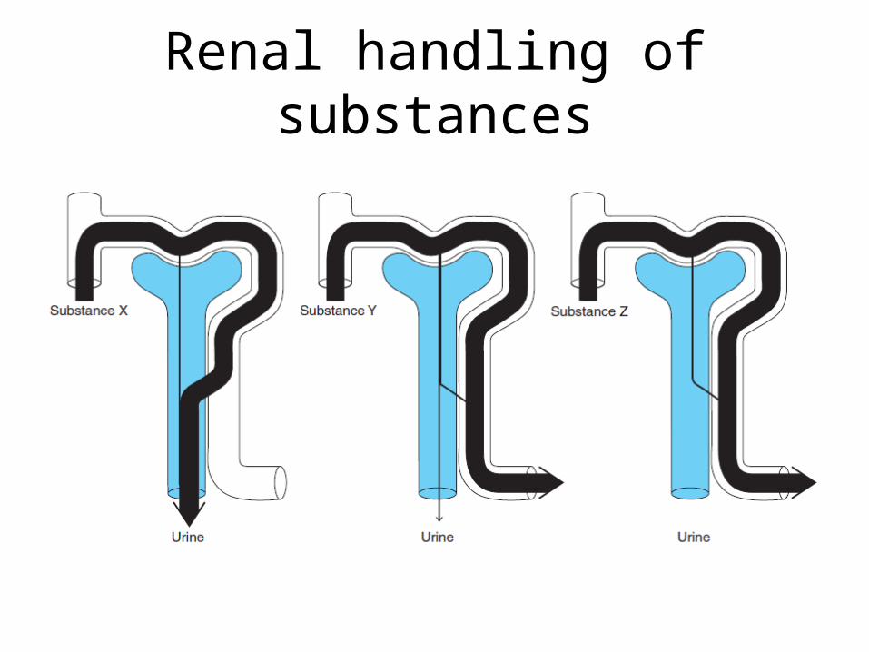
Renal handling of substances
Urine Formation
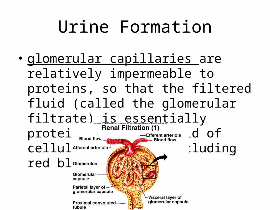
• glomerular capillaries are relatively impermeable to proteins, so that the filtered fluid (called the glomerular filtrate) is essentially protein-free and devoid of cellular elements, including red blood cells
Urine Formation
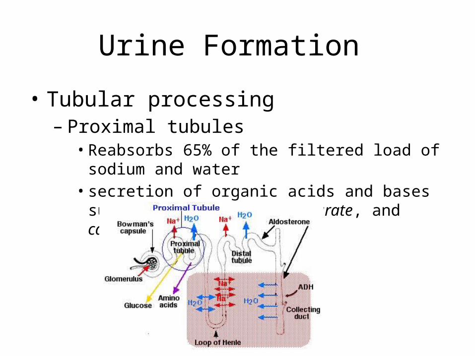
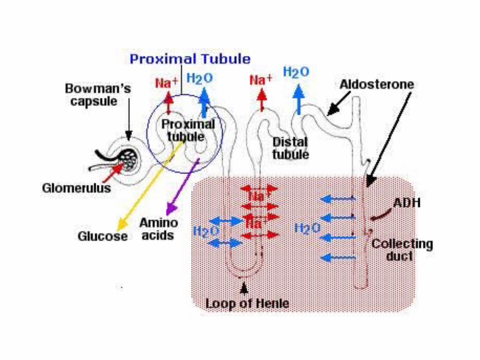
• Tubular processing– Proximal tubules• Reabsorbs 65% of the filtered load of sodium and water• secretion of organic acids and bases such as bile salts,
oxalate, urate, and catecholamines
Urine Formation
• Loop of Henle– Reabsorbs 20% of the filtered water, 25%of the
filtered loads – ascending limb is virtually impermeable to water– Thick ascending - active reabsorption of sodium,
chloride, and potassium – thick ascending limb of the loop of Henle is the
site of action of the powerful "loop" diuretics furosemide
Urine Formation
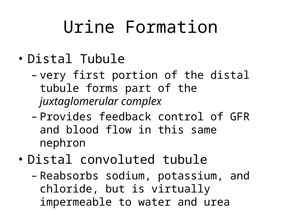
• Distal Tubule– very first portion of the distal tubule forms part of
the juxtaglomerular complex– Provides feedback control of GFR and blood flow
in this same nephron• Distal convoluted tubule– Reabsorbs sodium, potassium, and chloride, but is
virtually impermeable to water and urea– Diluting segment
Urine Formation
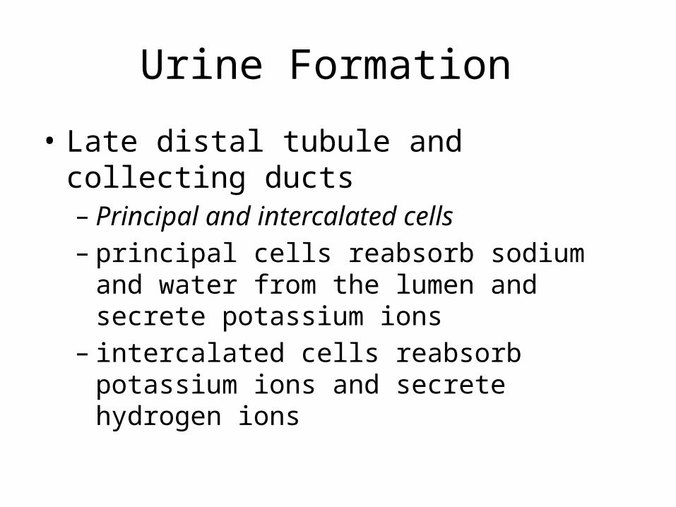
• Late distal tubule and collecting ducts– Principal and intercalated cells– principal cells reabsorb sodium and water from
the lumen and secrete potassium ions– intercalated cells reabsorb potassium ions and
secrete hydrogen ions
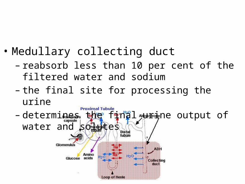
• Medullary collecting duct– reabsorb less than 10 per cent of the filtered water and
sodium– the final site for processing the urine– determines the final urine output of water and solutes
Micturition Reflex
• Micturition - the process by which the urinary bladder empties when it becomes filled
• the bladder fills progressively until the tension in its walls rises above a threshold level
• this elicits the second step, which is a nervous reflex called the micturition reflex that empties the bladder or, if this fails, at least causes a conscious desire to urinate
Micturition Reflex
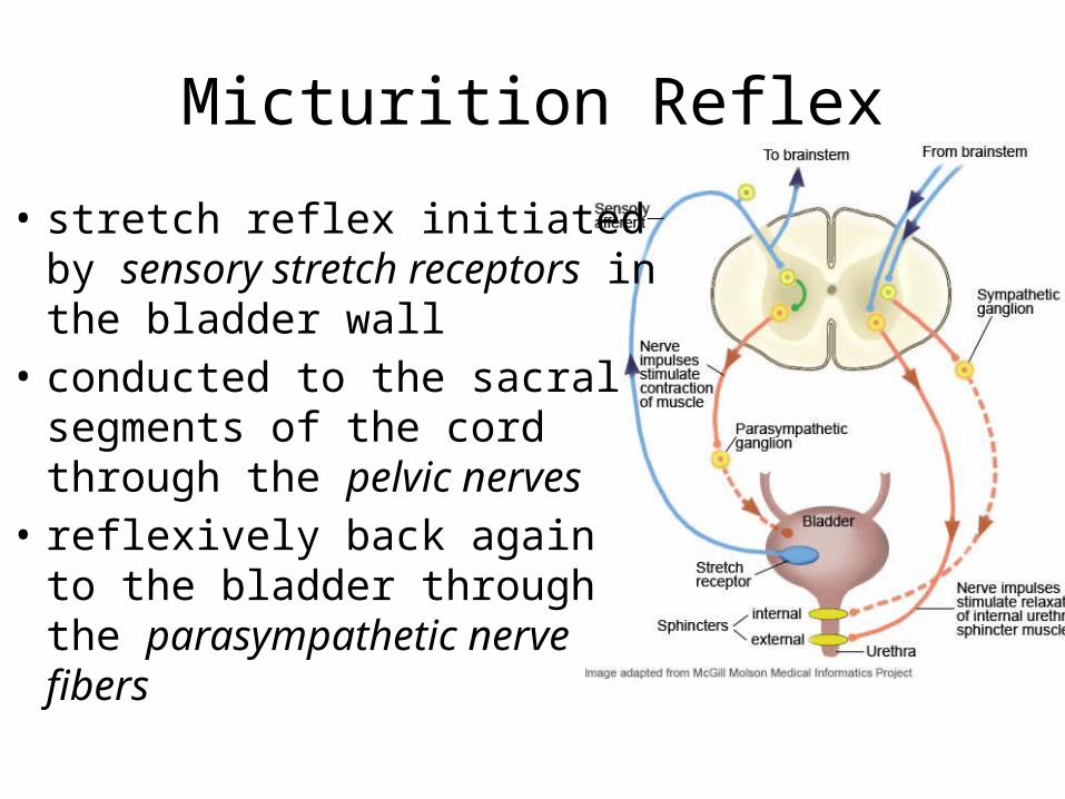
• stretch reflex initiated by sensory stretch receptors in the bladder wall
• conducted to the sacral segments of the cord through the pelvic nerves
• reflexively back again to the bladder through the parasympathetic nerve fibers
Micturition Reflex
• Once a micturition reflex begins, it is "self-regenerative.“
• initial contraction of the bladder activates the stretch receptors to cause a greater increase in sensory impulses to the bladder and posterior urethra, which causes a further increase in reflex contraction of the bladder
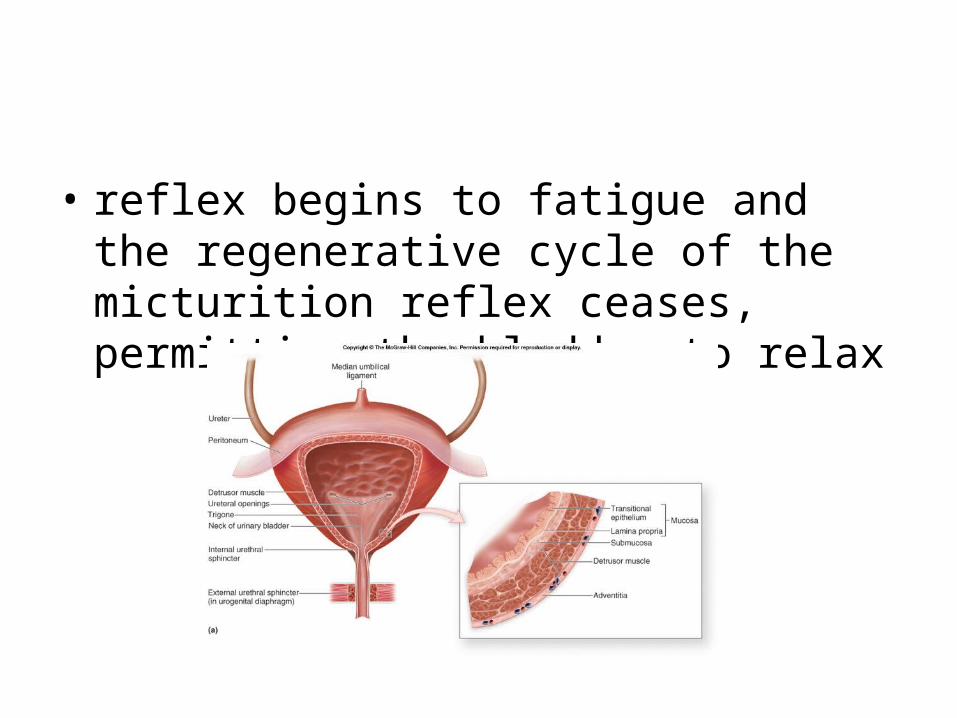
• reflex begins to fatigue and the regenerative cycle of the micturition reflex ceases, permitting the bladder to relax
Micturition reflex
• micturition reflex is a single complete cycle of – (1) progressive and rapid increase of pressure, – (2) a period of sustained pressure, and– (3) return of the pressure to the basal tone of the
bladder
• Next meeting: Read on Body Fluid composition, Acid base disturbances
• Chapters: 25, 30 . Guyton’s Textbook of Medical Physiology 11th edition