Embed Size (px)
Citation preview
8/11/2019 Renton T..pdf
http://slidepdf.com/reader/full/renton-tpdf 1/5
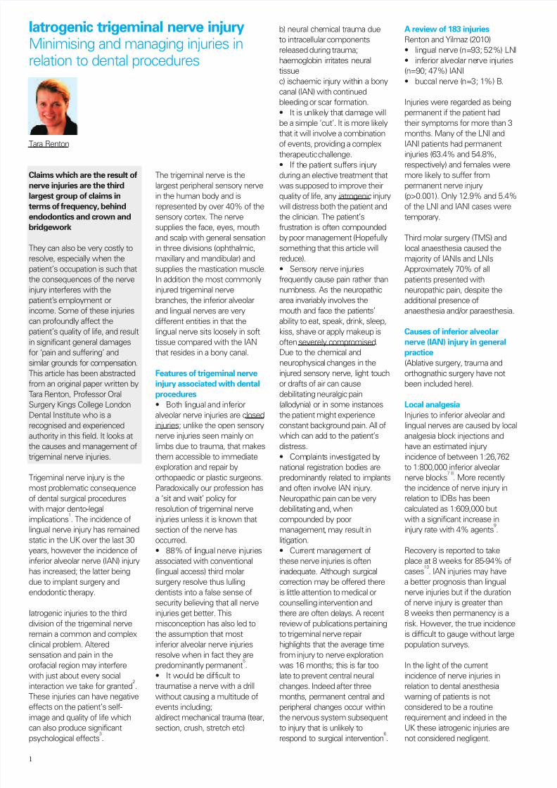
1
Iatrogenic trigeminal nerve injuryMinimising and managing injuries inrelation to dental procedures
The trigeminal nerve is the
largest peripheral sensory nerve
in the human body and is
represented by over 40% of the
sensory cortex. The nerve
supplies the face, eyes, mouth
and scalp with general sensation
in three divisions (ophthalmic,
maxillary and mandibular) and
supplies the mastication muscle.
In addition the most commonlyinjured trigeminal nerve
branches, the inferior alveolar
and lingual nerves are very
different entities in that the
lingual nerve sits loosely in soft
tissue compared with the IAN
that resides in a bony canal.
Features of trigeminal nerve
injury associated with dental
procedures
• Both lingual and inferior
alveolar nerve injuries are closedinjuries; unlike the open sensory
nerve injuries seen mainly on
limbs due to trauma, that makes
them accessible to immediate
exploration and repair by
orthopaedic or plastic surgeons.
Paradoxically our profession has
a ‘sit and wait’ policy for
resolution of trigeminal nerve
injuries unless it is known that
section of the nerve has
occurred.
• 88% of lingual nerve injuriesassociated with conventional
(lingual access) third molar
surgery resolve thus lulling
dentists into a false sense of
security believing that all nerve
injuries get better. This
misconception has also led to
the assumption that most
inferior alveolar nerve injuries
resolve when in fact they are
predominantly permanent5
.
• It would be difficult to
traumatise a nerve with a drill
without causing a multitude of
events including;
a)direct mechanical trauma (tear,
section, crush, stretch etc)
b) neural chemical trauma due
to intracellular components
released during trauma;
haemoglobin irritates neural
tissue
c) ischaemic injury within a bony
canal (IAN) with continued
bleeding or scar formation.
• It is unlikely that damage will
be a simple ‘cut’. It is more likely
that it will involve a combination
of events, providing a complex
therapeutic challenge.
• If the patient suffers injury
during an elective treatment that
was supposed to improve their
quality of life, any iatrogenic injury
will distress both the patient and
the clinician. The patient’s
frustration is often compounded
by poor management (Hopefully
something that this article will
reduce).
• Sensory nerve injuries
frequently cause pain rather thannumbness. As the neuropathic
area invariably involves the
mouth and face the patients’
ability to eat, speak, drink, sleep,
kiss, shave or apply makeup is
often severely compromised.
Due to the chemical and
neurophysical changes in the
injured sensory nerve, light touch
or drafts of air can cause
debilitating neuralgic pain
(allodynia) or in some instances
the patient might experienceconstant background pain. All of
which can add to the patient’s
distress.
• Complaints investigated by
national registration bodies are
predominantly related to implants
and often involve IAN injury.
Neuropathic pain can be very
debilitating and, when
compounded by poor
management, may result in
litigation.
• Current management ofthese nerve injuries is often
inadequate. Although surgical
correction may be offered there
is little attention to medical or
counselling intervention and
there are often delays. A recent
review of publications pertaining
to trigeminal nerve repair
highlights that the average time
from injury to nerve exploration
was 16 months; this is far too
late to prevent central neural
changes. Indeed after three
months, permanent central and
peripheral changes occur within
the nervous system subsequent
to injury that is unlikely to
respond to surgical intervention6
.
A review of 183 injuries
Renton and Yilmaz (2010)
• lingual nerve (n=93; 52%) LNI
• inferior alveolar nerve injuries
(n=90; 47%) IANI
• buccal nerve (n=3; 1%) B.
Injuries were regarded as being
permanent if the patient had
their symptoms for more than 3
months. Many of the LNI and
IANI patients had permanent
injuries (63.4% and 54.8%,
respectively) and females were
more likely to suffer from
permanent nerve injury
(p>0.001). Only 12.9% and 5.4%
of the LNI and IANI cases were
temporary.
Third molar surgery (TMS) and
local anaesthesia caused the
majority of IANIs and LNIs
Approximately 70% of all
patients presented withneuropathic pain, despite the
additional presence of
anaesthesia and/or paraesthesia.
Causes of inferior alveolar
nerve (IAN) injury in general
practice
(Ablative surgery, trauma and
orthognathic surgery have not
been included here).
Local analgesia
Injuries to inferior alveolar andlingual nerves are caused by local
analgesia block injections and
have an estimated injury
incidence of between 1:26,762
to 1:800,000 inferior alveolar
nerve blocks7 8
. More recently
the incidence of nerve injury in
relation to IDBs has been
calculated as 1:609,000 but
with a significant increase in
injury rate with 4% agents9
.
Recovery is reported to takeplace at 8 weeks for 85-94% of
cases10
. IAN injuries may have
a better prognosis than lingual
nerve injuries but if the duration
of nerve injury is greater than
8 weeks then permanency is a
risk. However, the true incidence
is difficult to gauge without large
population surveys.
In the light of the current
incidence of nerve injuries in
relation to dental anesthesia
warning of patients is not
considered to be a routine
requirement and indeed in the
UK these iatrogenic injuries are
not considered negligent.
Claims which are the result of
nerve injuries are the third
largest group of claims in
terms of frequency, behind
endodontics and crown and
bridgework
They can also be very costly to
resolve, especially when the
patient’s occupation is such that
the consequences of the nerveinjury interferes with the
patient’s employment or
income. Some of these injuries
can profoundly affect the
patient’s quality of life, and result
in significant general damages
for ‘pain and suffering’ and
similar grounds for compensation.
This article has been abstracted
from an original paper written by
Tara Renton, Professor Oral
Surgery Kings College London
Dental Institute who is arecognised and experienced
authority in this field. It looks at
the causes and management of
trigeminal nerve injuries.
Trigeminal nerve injury is the
most problematic consequence
of dental surgical procedures
with major dento-legal
implications1
. The incidence of
lingual nerve injury has remained
static in the UK over the last 30
years, however the incidence ofinferior alveolar nerve (IAN) injury
has increased; the latter being
due to implant surgery and
endodontic therapy.
Iatrogenic injuries to the third
division of the trigeminal nerve
remain a common and complex
clinical problem. Altered
sensation and pain in the
orofacial region may interfere
with just about every social
interaction we take for granted2
.
These injuries can have negative
effects on the patient’s self-
image and quality of life which
can also produce significant
psychological effects3
.
Tara Renton
8/11/2019 Renton T..pdf
http://slidepdf.com/reader/full/renton-tpdf 2/5
2
However there is increasing
evidence that higher
concentration local anaesthetic
agents may be associated with
increased rates of neuropathy
resulting in increasing litigation in
the USA. A recent settlement of
U$1.4 million dollars (Main USA)
for lingual nerve injury caused by
local analgesic inferior alveolar
nerve block highlights the
associated disability and social
repercussions of these injuries.
Causes of damage
• Direct mechanical trauma by
the needle
• Chemical nerve injury due to
LA components.
The resultant nerve injury may
be a combination of neural
haemorrhage, inflammation and
scarring resulting in
demyelination.
Articaine
This amide analgesic was
introduced to dentistry in 1998,
however lignocaine (also an
amide analgesic) remains the
gold standard in the UK.
Articaine is the most widely
used local analgesic in many
countries for over 20 years11 12
.
Articaine is said to have a
number of advantages, namely;
low toxicity subsequent to
inadvertent intravascular injectionwhich may be due to the rapid
breakdown to an inactive
metabolite (Articainic acid), rapid
onset of surgical analgesia (2.5
=/-1.1 minutes) compared with
conventional Lignocaine and
better diffusion through soft and
hard tissue. The conclusion
drawn is that Articaine is a safe
and effective local anaesthetic
for use in clinical dentistry13
.
There is, however, someconcern with regard using
Articaine for inferior alveolar and
lingual nerve blocks. Prolonged
paraesthesia and altered
sensation may be due to the
high concentration of the local
anaesthetic; however, the
technique cannot be excluded
as the cause for nerve injury
(Haas and Lennon, 1995)14
.
Another report suggests that it
is the type of anaesthetic that
dictates the degree of
inflammatory reaction to local
anaesthetic; Lidocaine being the
least irritant followed by
Articaine, Mepivicaine and
Bupivicaine.
Longstanding altered sensation
or nerve pain associated with
Articaine inferior alveolar nerve
blocks for routine dentistry has
been reported even though the
product information sheet states
that resolution usually takes place
within 2 weeks
(www.septodont.co.uk). As a
result of these concerns inferior
alveolar nerve blocks using
Lignocaine remains standard care.
Alternatives to inferior alveolar
nerve blocks with Articaine have
been suggested for implant
surgery and it is becoming routine
practice for orthodontic extraction
of premolars and restorative
treatment of premolars and
molars in adults using Articaine
local analgesic infiltrations rather
than inferior alveolar nerve
blocks15
.
Preventing problems
• Avoid multiple blocks where
possible
• Avoid IAN blocks by using
Articaine infiltrations only
• Avoid high concentration LA for
ID blocks (use 2% Lidocaine as
standard)
• Document any unusual patient
reaction during local analgesic
blocks (such as sharp pain or an
electrical shock–like sensation).
Management
The clinician need to be be
sympathetic to the patient’s
concerns which can lead to
distrust of future dental treatment
and a real fear of similar problems
arising on the contralateral side.
Management usually involves
counselling and medication for any
pain if present. In addition the
patient needs to be reassured and
given realistic expectations of
recovery. An explanation of whythey were not warned of this
complication may also be required.
If injury persists more than 6
weeks with more than 50% of
the dermatome affected, recovery
is unlikely.
Implants
The literature shows that the
incidence of implant related
inferior alveolar nerve (IAN) nerve
injuries varies from 0-40%. In
addition the 25% incidence of
edentulous patients presenting
with a degree of altered IAN
function, reinforces the need for a
preoperative neurosensory
evaluation.
Great care must be taken when
selecting the patient and possible
sites for implant placement.
Appropriate radiographic
evaluation of the implant site is a
given. Cone beam CT Scanning,
now introduced to many
specialist practises and dental
hospitals, provides lower
radiation dosage compared with
conventional CT and improved
imaging for planning implant
treatment. However several
papers have drawn attention to
the weakness of CT evaluation in
identifying the IAN canal when
compared with a panoramic
radiograph.
Many practitioners use software
to assist in the planning of
implants and for the identification
of the IAN canal position, with
the specific aim to place the
implants with a safety zone ofmore than 2mm from the IAN
canal16
. Because it is the clinician
who ‘draws in’ the IAN canal, the
assessment will be subjective.
Increasingly practitioners in the
USA now recommend a safety
zone of a minimum of 4mm.
More recently17
the necessity for
cross-sectional imaging, even for
surgical procedures in the
symphyseal region, has been
recommended to prevent
unforeseen nerve injuries. Mostcases of iatrogenic paraesthesia
can be prevented. However,
when this problem occurs,
follow-up must be initiated
quickly, since the first few
months significantly influence
the degree of nerve healing.
A sudden ‘give’ during
preparation may be indicative of
protrusion through the lingual or
buccal plate but may also be
associated with fracturing of the
IAN canal roof which will
increase the risk of haemorrhage
into the canal and subsequent
compression of the nerve. It will,
furthermore, increase the
likelihood of extrusion of
preparation debris or alkaline
solutions being introduced into
the canal causing potential harm
to the nerve.
If there is an inferior alveolar
arterial or venous bleed it may
be advisable not to place the
implant and to wait 2-3 days to
ensure that no nerve damage
has occurred before placing the
implant in granulation tissue.
Whilst this should not
compromise success there is,as yet, no evidence to support
this practice.
If a nerve injury is suspected, the
clinician should perform a basic
neurosensory examination and
ascertain whether the patient
experiences pain, altered
sensation or numbness and
document the results later that
day when the effects of the
anaesthetic should have worn
off. A phone call six hours postsurgery will enable the surgeon
to ascertain this.
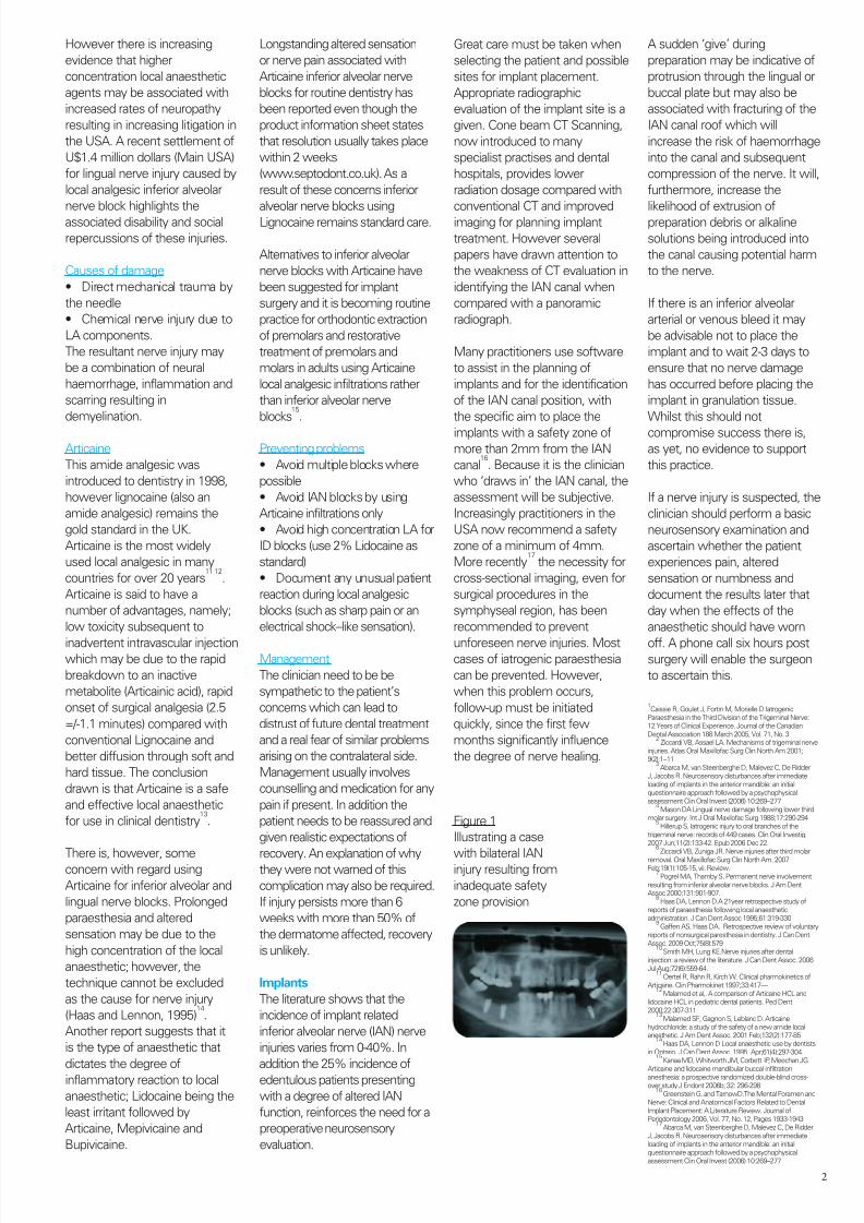
Figure 1
Illustrating a case
with bilateral IANinjury resulting from
inadequate safety
zone provision
1Caissie R, Goulet J, Fortin M, Morielle D Iatrogenic
Paraesthesia in the Third Division of the Trigeminal Nerve:12 Years of Clinical Experience. Journal of the CanadianDental Association 188 March 2005, Vol. 71, No. 3
2Ziccardi VB, Assael LA. Mechanisms of trigeminal nerve
injuries. Atlas Oral Maxillofac Surg Clin North Am 2001;9(2):1–11
3Abarca M, van Steenberghe D, Malevez C, De Ridder
J, Jacobs R. Neurosensory disturbances after immediateloading of implants in the anterior mandible: an initialquestionnaire approach followed by a psychophysicalassessment Clin Oral Invest (2006) 10:269–277
4Mason DA Lingual nerve damage following lower third
molar surgery. Int J Oral Maxilofac Surg 1988;17:290-2945
Hillerup S. Iatrogenic injury to oral branches of thetrigeminal nerve: records of 449 cases. Clin Oral Investig.2007 Jun;11(2):133-42. Epub 2006 Dec 22.
6Ziccardi VB, Zuniga JR. Nerve injuries after third molar
removal. Oral Maxillofac Surg Clin North Am. 2007Feb;19(1):105-15, vii. Review.
7Pogrel MA, Thamby S. Permanent nerve involvement
resulting from inferior alveolar nerve blocks. J Am DentAssoc 2000;131:901-907.
8Haas DA, Lennon D.A 21year retrospective study of
reports of paraesthesia following local anaestheticadministration. J Can Dent Assoc 1995;61:319-330
9Gaffen AS, Haas DA. Retrospective review of voluntary
reports of nonsurgical paresthesia in dentistry. J Can DentAssoc. 2009 Oct;75(8):579
10Smith MH, Lung KE.Nerve injuries after dental
injection: a review of the literature. J Can Dent Assoc. 2006Jul-Aug;72(6):559-64.
11Oertel R, Rahn R, Kirch W. Clinical pharmokinetics of
Articaine. Clin Pharmokinet 1997;33:417—12
Malamed et al,. A comparison of Articaine HCL andlidocaine HCL in pediatric dental patients. Ped Dent2000;22:307-311
13Malamed SF, Gagnon S, Leblanc D. Articaine
hydrochloride: a study of the safety of a new amide localanesthetic. J Am Dent Assoc. 2001 Feb;132(2):177-85
14Haas DA, Lennon D Local anaesthetic use by dentists
in Ontario. J Can Dent Assoc. 1995 Apr;61(4):297-30415
Kanaa MD, Whitworth JM, Corbett IP, Meechan JG
Articaine and lidocaine mandibular buccal infiltrationanesthesia: a prospective randomized double-blind cross-over study J Endont 2006b; 32: 296-298
16Greenstein G. and TarnowD.The Mental Foramen and
Nerve: Clinical and Anatomical Factors Related to DentalImplant Placement: A Literature Review. Journal ofPeriodontology 2006, Vol. 77, No. 12, Pages 1933-1943
17Abarca M, van Steenberghe D, Malevez C, De Ridder
J, Jacobs R. Neurosensory disturbances after immediateloading of implants in the anterior mandible: an initialquestionnaire approach followed by a psychophysicalassessment Clin Oral Invest (2006) 10:269–277
8/11/2019 Renton T..pdf
http://slidepdf.com/reader/full/renton-tpdf 3/5
3
Preventing problems
• Adequate work up and
planning >4mm safety zone.
• Do not place implant if there
is a bleed during implant bed
preparation – place the implant
2-3 days later.
• If an implant is potentially
violating the canal (a sudden give
experienced during preparation)
then its depth in the bone couldbe decreased by unscrewing it a
few turns ‘back up’. However if
there is haemorrhage the nerve
will will be compressed within
the bony canal and removal is
preferred. Back up may also
leave excessive implant exposed
coronally thus preferably remove
the implant, check for
haemorrhage and if none
replace with a shorter implant.
• Document all unusual patient
reactions occurring duringimplant bed preparation or
placement (such as sharp pain
or an electrical shock–like
sensation) and IAN vessel bleed.
• Routinely check on patient
early post operatively at 6 hours.
• If patient has neuropathy
immediately after local analgesia
has worn off:
– Consider removing the
implant in less than 24 hours
– Steroids and NSAIDS
– Refer to specialist (anappropriately trained micro-
neurosurgeon if necessary).
Late removal of implant
To optimise neural recovery the
potential harmful implant must
be removed very early on if
there is persistent neuropathy
after the LA has worn off. Even
doing this within 36 hours may
still be too late.
Endodontics
• The risk factors for
endodontic inferior alveolar
nerve injury include;
• Proximity of the tooth to the
mandibular canal
• Over instrumentation
• Overfill
• Chemical nerve injury (including
sodium hypochlorite)
Similar to extracting mandibular
teeth proximal to the IAN canal,
root treatment of these teeth has
the same potential for nerve injury.If the apex of the tooth is adjacent
or intrudes into the IAN canal,
any material leakage or overfilling
may compromise the nerve.
Assessment of the proximity of
the tooth apex to the IAN canal
has become significantly improved
with cone beam CT scanning
(CBCT). However the risk of
additional radiation and may not
provide significantly more
information than a standard long
cone radiograph.
Any tooth requiring endodontic
therapy that sits in close proximity
to the IAN canal needs special
care. If the canal is over-prepared
and the apex opened, then
chemical nerve injuries from
irrigation of canal medicaments
are possible. In addition physical
injury precipitated by overfilling
using pressurised thermal filling
techniques can occur. Postoperative
radiographs must be arranged onthe day of completion and the
presence of any materials in the
vicinity of the IAN canal should
be reviewed carefully. If nerve
function is compromised after the
local anaesthetic has worn off
then immediate arrangements
should be made to remove the
over-fill.
The optimum pH of an endodontic
medicament is as close as
possible to that of body fluids, (pH
7.35.) If the pH is higher or lower
cellular necrosis is possible for any
tissue in direct contact with the
medicament.
Commonly used endodontic
medicaments
• Formocresol
pH 12.45 +/- 0.02
• Sodium hypochlorite
pH 11-12
• Calcium hydroxide (Calyxl).
pH 10-14
• Antibiotic-corticosteroid paste
(Ledermix)
pH 8.13 +/- 0.01• Neutral
pH 7.35-7.45
• Eugenol
pH 4.34 +/- 0.05
• Iodoform paste
pH 2.90 +/- 0.02
Chemical nerve injuries are
commonly permanent and can
cause severe neuropathic pain.
If the patient is suffering from
neuropathy after the LA has
worn off and the postoperativeradiographs confirm that there is
no radio-opaque material in the
canal, chemical nerve injury may
be presumed. Because the
injury is likely to be irreversible,
any subsequent removal of the
obturation or extraction of the
tooth extraction is unlikely to
reverse the damage.
Preventing problems
• Preoperatively identify teeth
proximal to the IAN and takespecial care in preventing apical
breech with over
instrumentation.
• Recognise and record certain
events during treatment
including;
– Pain during irrigation
– Pain during preparation and
filling
– If Inferior alveolar vessel
bleeds during preparation, delay
filling.
• If nerve injury is suspected,
the postoperative radiograph
must be scrutinised for evidence
of breach of apex and deposition
of endodontic material into the
IAN canal. The patient should be
informed that the material, apex
and or tooth must be removed
within 48 hours of placement in
order to maximise recovery from
nerve injury.
• Routinely contact patients
postoperatively to ensure patient
is comfortable once local
analgesia has worn off. If nerveinjury is suspected at this stage,
the clinician must inform the
patient and also consider
removing any overfill of
endodontic material, or apicect
or extract the tooth within 48
hours.
Iatrogenictrigeminal nerveinjury
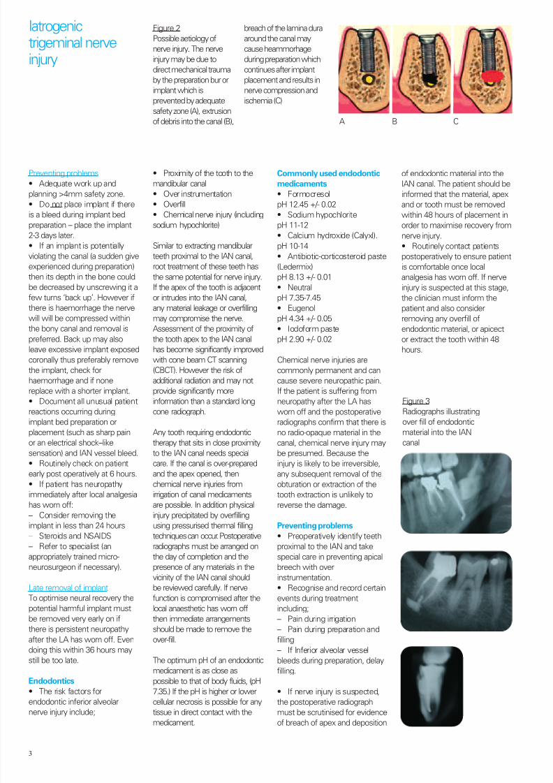
Figure 2
Possible aetiology of
nerve injury. The nerve
injury may be due to
direct mechanical trauma
by the preparation bur or
implant which is
prevented by adequate
safety zone (A), extrusion
of debris into the canal (B),
Figure 3
Radiographs illustratingover fill of endodontic
material into the IAN
canal
breach of the lamina dura
around the canal may
cause heammorhage
during preparation which
continues after implant
placement and results in
nerve compression and
ischemia (C)
A B C
8/11/2019 Renton T..pdf
http://slidepdf.com/reader/full/renton-tpdf 4/5
4
• If pain persists and no
evidence of overfill is present,
residual symptoms could result
from;
– apical inflammation (neuritis)
confirmed by prescription of
antibiotics
– chemical nerve injury from
irrigant or endo material
– thermal damage.
In any event once neuropathy is
identified the clinician should
reassure the patient, prescribe
steroids (Prednisolone step
down 15mg/ 5 days, 10mg/ 5
days and 5mg/ 5 days and high
dose NSAIDs, 600mg Ibuprofen
QDS if not contraindicated) and
make a timely referral to an
appropriately trained specialist if
necessary.
Lower third molarsThe risks for lingual nerve injury
during mandibular third molar
surgery are well established.
A recent systematic review
concluded18
that there was a
significant increase in lingual
nerve injury when using a lingual
split technique with lingual flap
or a lingual flap on its own when
compared with a buccal
approach. The paper concludes
that lingual flap ‘protection’ of
the lingual nerve is notnecessary and also potentially
harmful.
Prevention of lingual nerve
injuries may be possible if
undergraduate and postgraduate
training promoted the use of the
buccal technique. There is still a
reluctance to change from
traditional techniques (lingual
split/lingual retraction) as in
some experienced hands these
techniques involve minimal
morbidity. However, if the buccaltechnique is adhered to there is
no risk to the lingual nerve.
Avoidance of the envelope flap
minimises the necessity of a
long distal extension of the flap
which exposes the distal bone
adjacent to the third molar and
thus may ‘tempt’ the surgeon to
remove distal bone which would
compromise the lingual nerve.
Preventing problems
• Identify ‘high risk’ teeth. Pre-operative x-rays are essential to
assess the proximity of the IAN
canal. Radiographic signs (Figure
4) indicative of possible IAN risk
include;
• Diversion of the canal
• Darkening of the root
• Interruption of the canal LD
• Juxta-apical area (Figure 5)
• Narrowing of the roots or canal
The patient must be informed
about any elevated risk andshould be offered a suitable
referral if this is thought
necessary. If the tooth is high risk
with these radiographic features
the risk is elevated from 2%
temporary to 20% temporary and
from 0,5% permanent to 2%
permanent ID nerve injury.
• The clinician must identify
mandibular teeth at high risk of
IAN injury based on radiographic
features. If deemed at high risk
the patient must be made aware
of the increased nerve injury
incidence and perhaps offered
alternative procedures that may in
course reduce the risk of injury.
• If the tooth is in closeproximity to the IAN on plain film
then cone beam CT scanning
may further elucidate the
relationship between IAN and
tooth roots. If the tooth is non
vital, or pathology associated with
it, then tooth removal has to take
place and the roots should be
sectioned appropriately to
minimise trauma to the adjacent
IAN.
Other teethIf a mandibular tooth (lower
8,7,6,5 or 4) crosses the IAN
canal, and displays radiographic
signs associated with an
increased risk of IAN injury if the
(if the tooth was removed) then
the patient must be assessed
accordingly, consented and
treated in a similar manner to a
patient with a high risk lower third
molar.
Socket medications
There is limited availability of
information on the relative
alkalinity or acidity of various
dental compounds used for
socket medication including;
Alvolgyl, Whiteheads varnish,
Corsodyl and Surgicel. However,
a previous study highlighted the
relative neurotoxicity of Carnoy’s
solution, Surgicel, Whiteheadsvarnish and bismuth iodoform
paraffin paste (BIPP) reporting
that Carnoys is likely to cause
permanent nerve damage and
Surgicel along with Whiteheads
varnish can cause temporary
sensory disturbances. BIPP was
the least neurotoxic19
. Bone wax
is a neutral pH however
excessive packing or pressure
can lead to nerve compression
and injury.
Management of trigeminal
nerve injuries
We now know that up to 70%
of patients with iatrogenic
trigeminal nerve injuries present
with post traumatic nerve
neuropathy which is painful. This
is reflected by the wide variety
of functional problems that can
arise from nerve damage
created by a dental intervention
helps to explain why the claims
and helps to explain why theclaims that arise from
thesesometimes be extremely
high.
Figure 4
DPT radiographs
illustrating 2 cases of
‘high risk’ mandibular
third molars.
Figure 5
Juxta-apical area
In both cases the lower
third molar is crossing
the IAN canal completely,
there is darkening of the
tooth roots and loss of
lamina dura of the canal
roof and banding
18Pichler JW and Beirne OR .Lingual flap retraction and
prevention of lingual nerve damage associated with thirdmolar surgery: a systematic review of the literature. OralSurg Oral Med Oral Pathol Oral Radiol Endod 2001; 91(4):395-401
19Loescher.A and Robinson P. The effect of surgical
medicaments on peripheral nerve function. Br J OralMaxillofac Surg. 1998 Oct;36(5):327-32.
8/11/2019 Renton T..pdf
http://slidepdf.com/reader/full/renton-tpdf 5/5