Embed Size (px)
Citation preview
Ultrasound
Reproducible EF in seconds
Philips EPIQ 7 HeartModelA.I.
You’ve told us that 3D transthoracic echocardiography, while important, is sometimes seen as too complex and time-consuming for mainstream practice. With one-button simplicity, Philips HeartModelA.I. helps overcome that barrier, bringing robust 3D quantification to everyday clinical practice. This anatomically intelligent premier cardiac application automatically detects, segments, and quantifies the left ventricle (LV) and left atrium (LA) from a Live 3D volume. With HeartModelA.I., you have automated 2D views and reproducible quantification across users and over time, with the workflow efficiency to facilitate faster exams for the robust measurement of cardiac function necessary for management of patients with chronic disease.
Please visit www.philips.com/AnatomicalIntelligence
Printed in The Netherlands.4522 991 13061 * AUG 2015
The print quality of this copy is not an accurate representation of the original.
2
Quantify LA volume using the same 3D acquisition
HeartModelA.I., as well as providing LV quantification, is the only validated tool to provide simultaneous LA volumes. It allows easy characterization of LA volume to gain additional clinical information with no additional time or steps. LA volume has been shown to be an indicator of cardiovascular outcomes.
Internal university clinical evaluation demonstrates that HeartModelA.I. quantification results correlate well to expert manual tracing in 3D. The algorithm and its intelligent and innovative editing tools are capable of performing under a wide range of conditions to aid in reducing variability among users and across time when automatically detecting, segmenting, and quantifying LV and LA from 3D volume.1
Bring 3D to every dayCardiac dysfunction has many causes and so accurate quantification of left ventricle (LV) and left atrial (LA) function is an important component of cardiac exams. HeartModelA.I. assesses LV and LA global volumes, which are shown to be important in characterizing a wide range of conditions, such as:• Infarction• Ischemia • Dialated cardiomyopathy• Chemotherapy induced cardiomyopathy• Mitral and aortic regurgitation effects on LV and LA function
Robust reproducibility
HeartModelA.I. is a 3D tool that can provide robust, reproducible ejection fraction (EF) in just seconds. This intuitive and validated application is designed to deliver the confidence of cardiac quantification that fits into everyday workflow. HeartModelA.I. offers easy and fast 3D cardiac chamber quantification, simultaneously computing the LV and LA volumes from a single volume loop.
Chronic disease populations are growing, requiring the need to effectively and efficiently measure small changes in chamber volume to successfully plan therapy for more patients in less time.1
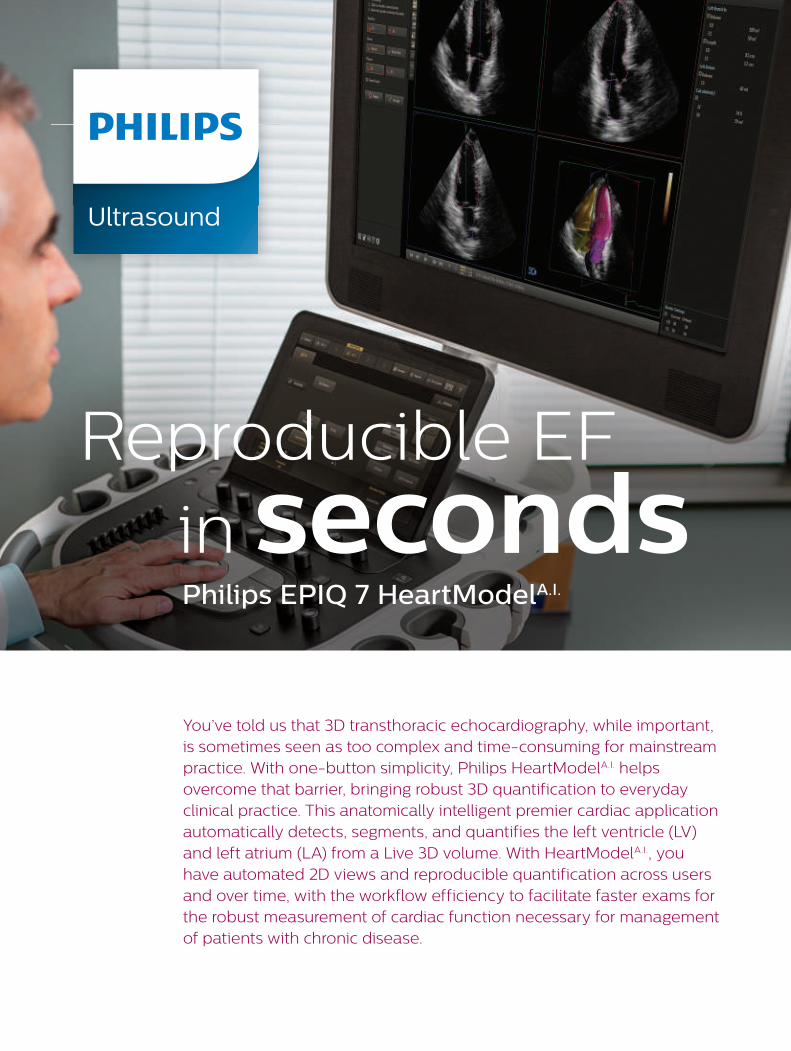
Broad clinical application
The HeartModelA.I. algorithm has been studied at several leading centers, and results show that the algorithm accurately adapts to a wide variety of heart sizes and shapes.* HeartModelA.I. is designed to be useful in echocardiographic evaluation and patient management across an extensive range of adult patients.
* HeartModelA.I. has only been validated on adult patient with infarction, ischemia, dilated cardiomyopathy, mitral and aortic regurgitation, and those undergoing chemotherapy
The print quality of this copy is not an accurate representation of the original.
3
Helps overcome limitations of 2D echo
Industry guidelines have documented the issues of frequently foreshortened apexes and geometric assumptions that are encountered with 2D EF calculations. 3D EF overcomes these issues by avoiding foreshortening and geometric assumptions.1
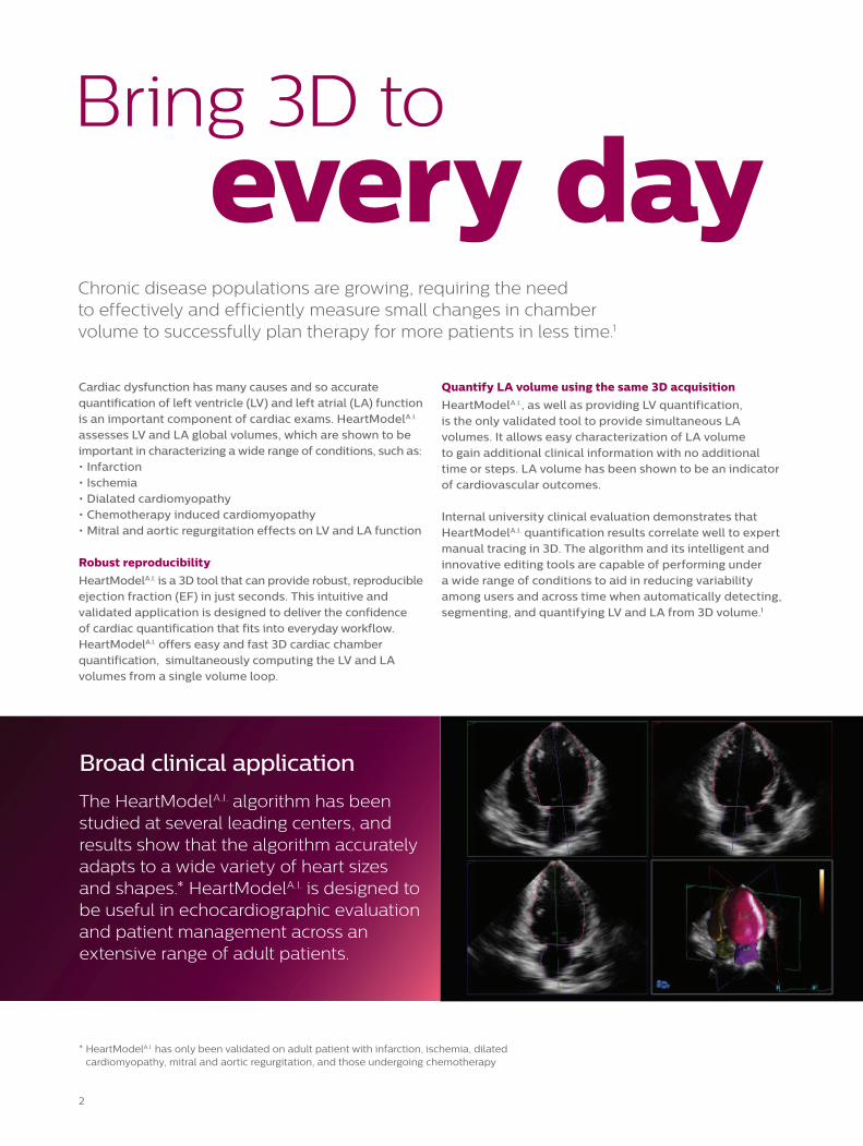
Offers automated 2D views and reproducible quantification with one-button simplicity.
Enhanced workflow that’s fast and easy
HeartModelA.I. automatically derives routine 2D apical views from a 3D volume. Features such as easy-to-edit borders are designed to bring 3D cardiac echo into mainstream practice. The user has the flexibility to easily set the border where desired for the end-diastolic (ED) and end-systolic (ES) cardiac phases. HeartModelA.I. finds the shape of the heart chambers and displays the chamber border in ASE/ESE views for the user to accept, reject, or edit. New, innovative global editing capabilities allow edits to be completed within seconds and results easily exported in DICOM-SR.
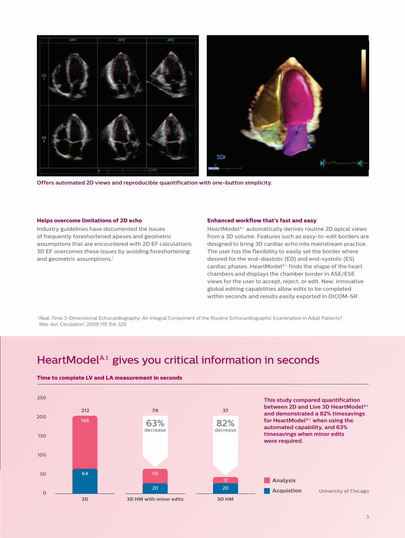
HeartModelA.I. gives you critical information in seconds
This study compared quantification between 2D and Live 3D HeartModelA.I. and demonstrated a 82% timesavings for HeartModelA.I. when using the automated capability, and 63% timesavings when minor edits were required.
250
200
150
100
50
02D 3D HM with minor edits
Time to complete LV and LA measurement in seconds
3D HM
Analysis
Acquistion
64
20 20
148 63%decrease
82%decrease
5917
1 Real-Time 3-Dimensional Echocardiography: An Integral Component of the Routine Echocardiographic Examination in Adult Patients? Mor-Avi. Circulation. 2009;119:314-329
University of Chicago
3779212
The print quality of this copy is not an accurate representation of the original.
© 2015 Koninklijke Philips N.V. All rights are reserved.
Philips Healthcare reserves the right to make changes in specifications and/or to discontinue any product at any time without notice or obligation and will not be liable for any consequences resulting from the use of this publication.
Please visit www.philips.com/AnatomicalIntelligence
Printed in The Netherlands.4522 991 13061 * AUG 2015
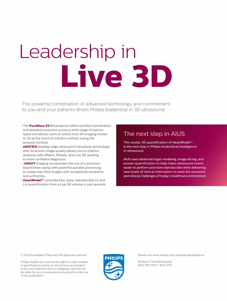
The next step in AIUS The cardiac 3D quantification of HeartModelA.I. is the next step in Philips Anatomical Intelligence in ultrasound.
AIUS uses advanced organ modeling, image slicing, and proven quantification to help make ultrasound exams easier to perform and more reproducible while delivering new levels of clinical information to meet the economic and clinical challenges of today’s healthcare environment.
Leadership in
Live 3D• The PureWave X5-1 transducer offers excellent penetration
and detailed resolution across a wide range of patient types and allows users to switch from 2D imaging modes to 3D at the touch of a button without losing the acoustic window
• xMATRIX leading-edge ultrasound transducer technology with its proven image quality allows you to explore anatomy with xPlane, iRotate, and Live 3D, leading to more confident diagnoses
• nSIGHT imaging incorporates the use of a precision beamformer along with powerful parallel processing to create real-time images with exceptional resolution and uniformity
• HeartModelA.I. provides fast, easy, reproducible LV and LA quantification from a Live 3D volume in just seconds
The powerful combination of advanced technology and commitment to you and your patients drives Philips leadership in 3D ultrasound.
The print quality of this copy is not an accurate representation of the original.