Embed Size (px)
Citation preview
METHODS & TECHNIQUES
Reprogrammable CRISPR/Cas9-based system for inducing site-specific DNA methylationJames I. McDonald1,*, Hamza Celik2,*, Lisa E. Rois1, Gregory Fishberger3, Tolison Fowler1, Ryan Rees3,Ashley Kramer2, Andrew Martens2, John R. Edwards1,‡ and Grant A. Challen2,4,‡
ABSTRACTAdvances in sequencing technology allow researchers tomap genome-wide changes in DNA methylation in development and disease.However, there is a lack of experimental tools to site-specificallymanipulate DNA methylation to discern the functional consequences.We developed a CRISPR/Cas9 DNA methyltransferase 3A (DNMT3A)fusion to induce DNA methylation at specific loci in the genome. Weinduced DNA methylation at up to 50% of alleles for targeted CpGdinucleotides. DNA methylation levels peaked within 50 bp of the shortguide RNA (sgRNA) binding site and between pairs of sgRNAs. Weused our approach to target methylation across the entire CpG island atthe CDKN2A promoter, three CpG dinucleotides at the ARF promoter,and the CpG island within theCdkn1a promoter to decrease expressionof the target gene. These tools permit mechanistic studies of DNAmethylation and its role in guiding molecular processes that determinecellular fate.
KEY WORDS: CRISPR/Cas9-based system, CpG dinucleotides,DNA methylation
INTRODUCTIONDNA methylation of CpG dinucleotides is a prominent epigeneticmodification of the mammalian genome that can influence geneexpression, and aberrant distribution of DNA methylation isassociated with a spectrum of human disorders including cancers(Egger et al., 2004). Despite intensive study, it remains unclearwhich CpG dinucleotides must change methylation state in order toalter transcription. Genome-wide analyses have found associationsbetween DNA methylation and reduced gene expression that occurboth in the proximal promoter and downstream of the gene’stranscription start site (TSS) (Bell et al., 2011; Bock et al., 2012;Lou et al., 2014; VanderKraats et al., 2013; Lund et al., 2014).However, evidence supports both that DNAmethylation can cause a
loss of expression, and that expression changes can alter DNAmethylation patterns (Bestor et al., 2015; Busslinger et al., 1983).Here, we sought to develop tools for locus-specific epigeneticremodeling to directly address the role of DNA methylation inregulating gene expression.
Targeted DNA methylation approaches have been attempted byfusing DNA methyltransferase enzymes (DNMTs) to DNA-binding proteins such as zinc finger proteins (ZFPs) (Siddiqueet al., 2013), and transcriptional activator-like effector (TALE)(Bernstein et al., 2015). However, engineering custom proteins foreach targeted sequence is laborious and requires specializedexpertise. Moreover, in these studies, induced DNA methylation ofthe targeted loci was relatively poor, with substantial off-targetactivity. An engineered form of the clustered, regularlyinterspaced, short palindromic repeat (CRISPR) system hasemerged as an alternative for achieving site-specific DNAtargeting (Jinek et al., 2012). Here, the Cas9 endonuclease isdirected to genomic targets by engineered short guide RNAs(sgRNAs) (Jinek et al., 2012). Because the sgRNA is the DNAsequence-specific component of the system, it allows for efficienttargeting of multiple regions due to the ease of design andsynthesis of new sgRNAs (relative to engineering new customproteins for each target site). A Cas9 mutant (D10A and H840A;henceforth referred to as dCas9) that lacks endonuclease activitybut can still be recruited by sgRNA(s) (Jinek et al., 2012) hasrecently been used to target genes in mammalian cells fortranscriptional activation (Perez-Pinera et al., 2013a,b; Maederet al., 2013; Mali et al., 2013). Here, we demonstrate an easilyreprogrammable CRISPR/dCas9 DNMT fusion capable ofinducing site-specific DNA methylation.
RESULTSTo design a flexible system to target DNA methylation, we fuseddCas9 to the catalytic domain of the de novo DNAmethyltransferase DNMT3A (Fig. 1A). To test this system wetargeted DNA methylation to the tumor suppressor gene CDKN2A(cyclin dependent kinase 2A), which inhibits progression throughthe cell cycle (Liggett and Sidransky, 1998). CDKN2A is one of themost frequently hypermethylated genes in The Cancer GenomeAtlas (Ciriello et al., 2013), and numerous clinical studies show anegative correlation between CDKN2A methylation and expressionin colorectal cancer (Shima et al., 2011). While it is generallyassumed that CDKN2A methylation induces gene silencing, it hasalso been suggested that DNA methylation occurs after the loss ofexpression (Hinshelwood et al., 2009). From a literature search, weidentified 17 publications that associate CDKN2A methylation withexpression and/or cancer (Fig. S1.1). Overwhelmingly, these papersstudied the differentially methylated region (cancer DMR, cDMR)on the 3′ end of the CpG island that overlapped the first exon ofCDKN2A (Fig. 1B).Received 14 April 2016; Accepted 18 April 2016
1Center for Pharmacogenomics, Department of Medicine, Washington University inSt. Louis School of Medicine, St. Louis, MO, USA. 2Section of Stem Cell Biology,Division of Oncology, Department of Medicine, Washington University in St. LouisSchool of Medicine, St. Louis, MO 63110, USA. 3College of Arts and Science,Washington University in St. Louis, St. Louis, MO 63130, USA. 4Developmental,Regenerative and Stem Cell Biology Program, Division of Biology and BiomedicalSciences, Washington University in St. Louis School of Medicine, St. Louis, MO63110, USA.*These authors contributed equally to this work
‡Authors for correspondence ( [email protected];[email protected])
G.A.C., 0000-0003-4669-8814
This is an Open Access article distributed under the terms of the Creative Commons AttributionLicense (http://creativecommons.org/licenses/by/3.0), which permits unrestricted use,distribution and reproduction in any medium provided that the original work is properly attributed.
866
© 2016. Published by The Company of Biologists Ltd | Biology Open (2016) 5, 866-874 doi:10.1242/bio.019067
BiologyOpen
by guest on May 25, 2020http://bio.biologists.org/Downloaded from
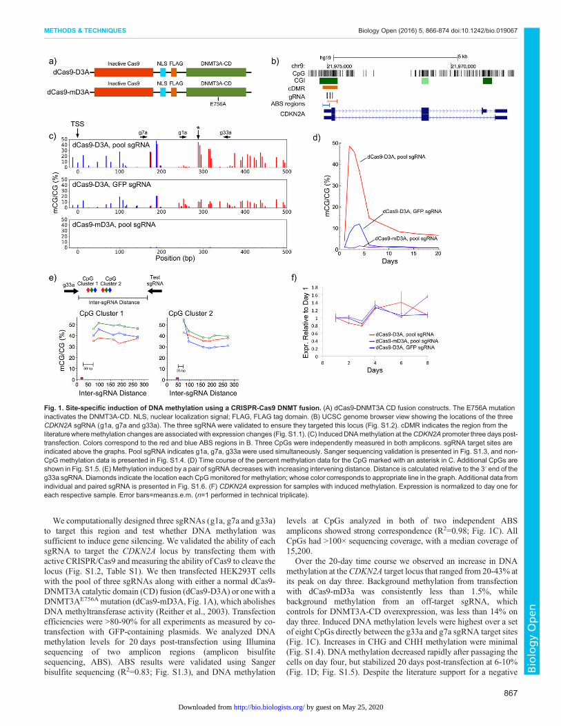
We computationally designed three sgRNAs (g1a, g7a and g33a)to target this region and test whether DNA methylation wassufficient to induce gene silencing. We validated the ability of eachsgRNA to target the CDKN2A locus by transfecting them withactive CRISPR/Cas9 and measuring the ability of Cas9 to cleave thelocus (Fig. S1.2, Table S1). We then transfected HEK293T cellswith the pool of three sgRNAs along with either a normal dCas9-DNMT3A catalytic domain (CD) fusion (dCas9-D3A) or onewith aDNMT3AE756A mutation (dCas9-mD3A, Fig. 1A), which abolishesDNA methyltransferase activity (Reither et al., 2003). Transfectionefficiencies were >80-90% for all experiments as measured by co-transfection with GFP-containing plasmids. We analyzed DNAmethylation levels for 20 days post-transfection using Illuminasequencing of two amplicon regions (amplicon bisulfitesequencing, ABS). ABS results were validated using Sangerbisulfite sequencing (R2=0.83; Fig. S1.3), and DNA methylation
levels at CpGs analyzed in both of two independent ABSamplicons showed strong correspondence (R2=0.98; Fig. 1C). AllCpGs had >100× sequencing coverage, with a median coverage of15,200.
Over the 20-day time course we observed an increase in DNAmethylation at theCDKN2A target locus that ranged from 20-43% atits peak on day three. Background methylation from transfectionwith dCas9-mD3a was consistently less than 1.5%, whilebackground methylation from an off-target sgRNA, whichcontrols for DNMT3A-CD overexpression, was less than 14% onday three. Induced DNA methylation levels were highest over a setof eight CpGs directly between the g33a and g7a sgRNA target sites(Fig. 1C). Increases in CHG and CHH methylation were minimal(Fig. S1.4). DNA methylation decreased rapidly after passaging thecells on day four, but stabilized 20 days post-transfection at 6-10%(Fig. 1D; Fig. S1.5). Despite the literature support for a negative
Fig. 1. Site-specific induction of DNA methylation using a CRISPR-Cas9 DNMT fusion. (A) dCas9-DNMT3A CD fusion constructs. The E756A mutationinactivates the DNMT3A-CD. NLS, nuclear localization signal; FLAG, FLAG tag domain. (B) UCSC genome browser view showing the locations of the threeCDKN2A sgRNA (g1a, g7a and g33a). The three sgRNA were validated to ensure they targeted this locus (Fig. S1.2). cDMR indicates the region from theliteraturewheremethylation changes are associated with expression changes (Fig. S1.1). (C) Induced DNAmethylation at theCDKN2A promoter three days post-transfection. Colors correspond to the red and blue ABS regions in B. Three CpGs were independently measured in both amplicons. sgRNA target sites areindicated above the graphs. Pool sgRNA indicates g1a, g7a, g33a were used simultaneously. Sanger sequencing validation is presented in Fig. S1.3, and non-CpG methylation data is presented in Fig. S1.4. (D) Time course of the percent methylation data for the CpG marked with an asterisk in C. Additional CpGs areshown in Fig. S1.5. (E) Methylation induced by a pair of sgRNA decreases with increasing intervening distance. Distance is calculated relative to the 3′ end of theg33a sgRNA. Diamonds indicate the location each CpGmonitored for methylation; whose color corresponds to appropriate line in the graph. Additional data fromindividual and paired sgRNA is presented in Fig. S1.6. (F) CDKN2A expression for samples with induced methylation. Expression is normalized to day one foreach respective sample. Error bars=mean±s.e.m. (n=1 performed in technical triplicate).
867
METHODS & TECHNIQUES Biology Open (2016) 5, 866-874 doi:10.1242/bio.019067
BiologyOpen
by guest on May 25, 2020http://bio.biologists.org/Downloaded from
correlation between expression and DNAmethylation in this region,we did not observe a measurable effect on CDKN2A geneexpression by RT-qPCR (Fig. 1F). This suggests that a limitedincrease in methylation in the region 100-400 bp downstream of theCDKN2A TSS is insufficient to trigger gene silencing.Spatially, the induced DNA methylation spiked near the sgRNA
target sites and dropped quickly toward background levels atsurrounding CpG sites. Analysis of DNA methylation induced bysingle sgRNAs indicates that methylation occurs primarily within50 bp of the sgRNA binding site (Fig. S1.6). Higher DNAmethylation levels were often observed 3′ of the sgRNA bindingsite (Fig. S1.6). Our initial data from the three pooled sgRNAssuggested that CpG methylation was higher between pairs ofsgRNAs. To investigate this effect, we transfected pairs of sgRNAwith varying intervening distances and monitored methylation ofsix clustered CpGs between the sgRNA pairs using ABS (Fig. 1E).DNA methylation of the three CpGs (cluster 1) within 20 bp of thefixed sgRNA (g33a) did not change with addition of a secondsgRNA 77 bp or further away. However, the methylation levelincreased from 30-40% to 42-53% when sgRNAs were pairedwithin 80 bp and both sgRNAs were within 50 bp of CpG cluster 2(Fig. 1E). This suggests the DNMT3A-CD activity at the targetlocus is additive.
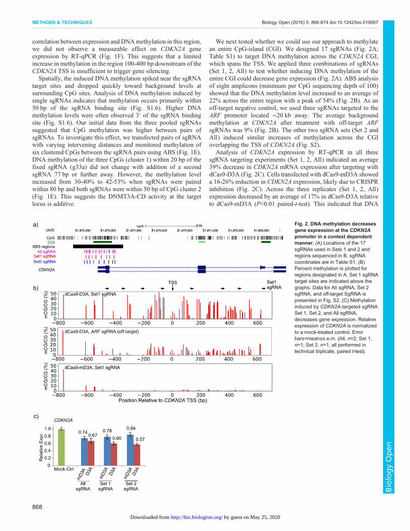
We next tested whether we could use our approach to methylatean entire CpG-island (CGI). We designed 17 sgRNAs (Fig. 2A;Table S1) to target DNA methylation across the CDKN2A CGI,which spans the TSS. We applied three combinations of sgRNAs(Set 1, 2, All) to test whether inducing DNA methylation of theentire CGI could decrease gene expression (Fig. 2A). ABS analysisof eight amplicons (minimum per CpG sequencing depth of 100)showed that the DNA methylation level increased to an average of22% across the entire region with a peak of 54% (Fig. 2B). As anoff-target negative control, we used three sgRNAs targeted to theARF promoter located ∼20 kb away. The average backgroundmethylation at CDKN2A after treatment with off-target ARFsgRNAs was 9% (Fig. 2B). The other two sgRNA sets (Set 2 andAll) induced similar increases of methylation across the CGIoverlapping the TSS of CDKN2A (Fig. S2).
Analysis of CDKN2A expression by RT-qPCR in all threesgRNA targeting experiments (Set 1, 2, All) indicated an average39% decrease in CDKN2A mRNA expression after targeting withdCas9-D3A (Fig. 2C). Cells transfected with dCas9-mD3A showeda 16-26% reduction in CDKN2A expression, likely due to CRISPRinhibition (Fig. 2C). Across the three replicates (Set 1, 2, All)expression decreased by an average of 17% in dCas9-D3A relativeto dCas9-mD3A (P<0.01 paired-t-test). This indicated that DNA
Fig. 2. DNA methylation decreasesgene expression at the CDKN2Apromoter in a context dependentmanner. (A) Locations of the 17sgRNAs used in Sets 1 and 2 andregions sequenced in B. sgRNAcoordinates are in Table S1. (B)Percent methylation is plotted forregions designated in A. Set 1 sgRNAtarget sites are indicated above thegraphs. Data for All sgRNA, Set 2sgRNA, and off-target SgRNA ispresented in Fig. S2. (C) Methylationinduced by CDKN2A-targeted sgRNASet 1, Set 2, and All sgRNA,decreases gene expression. Relativeexpression of CDKN2A is normalizedto a mock-treated control. Errorbars=mean±s.e.m. (All, n=2; Set 1,n=1; Set 2, n=1; all performed intechnical triplicate, paired t-test).
868
METHODS & TECHNIQUES Biology Open (2016) 5, 866-874 doi:10.1242/bio.019067
BiologyOpen
by guest on May 25, 2020http://bio.biologists.org/Downloaded from
methylation directly decreased CDKN2A expression, but targetingof the entire CGI was required to trigger this effect. Our results areconsistent with other studies that find a similar reduction in geneexpression after inducing methylation at the CDKN2A promoterusing ZFP- and TALE-based systems (Bernstein et al., 2015; Cuiet al., 2015).To verify the effect of our system with a separate locus, we
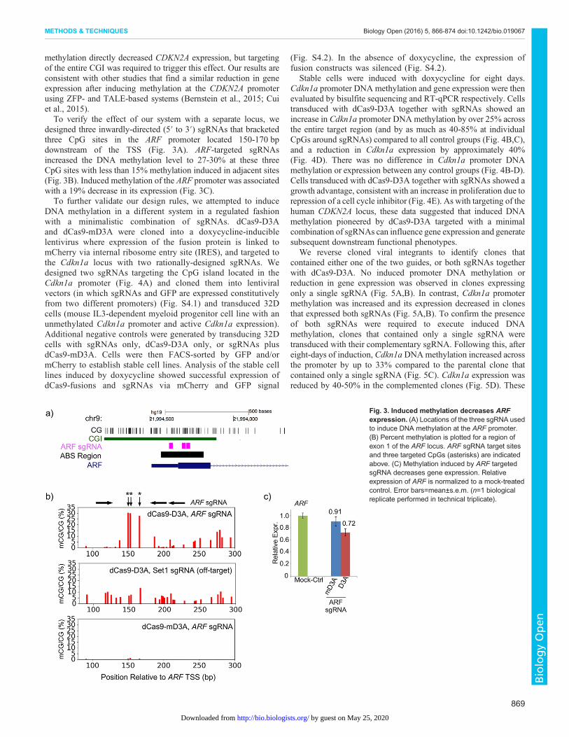
designed three inwardly-directed (5′ to 3′) sgRNAs that bracketedthree CpG sites in the ARF promoter located 150-170 bpdownstream of the TSS (Fig. 3A). ARF-targeted sgRNAsincreased the DNA methylation level to 27-30% at these threeCpG sites with less than 15% methylation induced in adjacent sites(Fig. 3B). Induced methylation of the ARF promoter was associatedwith a 19% decrease in its expression (Fig. 3C).To further validate our design rules, we attempted to induce
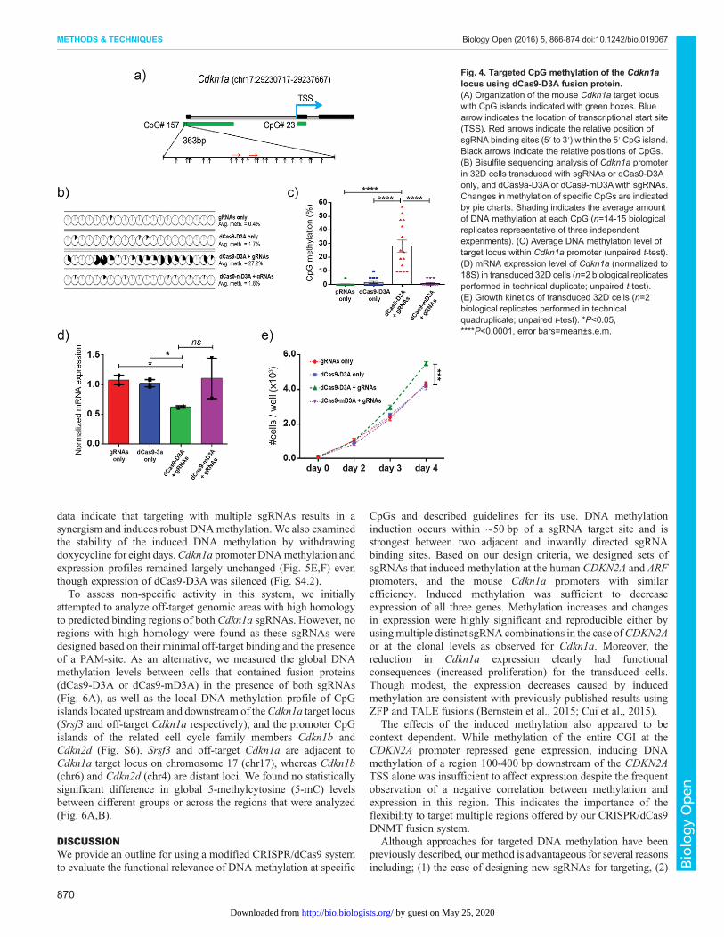
DNA methylation in a different system in a regulated fashionwith a minimalistic combination of sgRNAs. dCas9-D3Aand dCas9-mD3A were cloned into a doxycycline-induciblelentivirus where expression of the fusion protein is linked tomCherry via internal ribosome entry site (IRES), and targeted tothe Cdkn1a locus with two rationally-designed sgRNAs. Wedesigned two sgRNAs targeting the CpG island located in theCdkn1a promoter (Fig. 4A) and cloned them into lentiviralvectors (in which sgRNAs and GFP are expressed constitutivelyfrom two different promoters) (Fig. S4.1) and transduced 32Dcells (mouse IL3-dependent myeloid progenitor cell line with anunmethylated Cdkn1a promoter and active Cdkn1a expression).Additional negative controls were generated by transducing 32Dcells with sgRNAs only, dCas9-D3A only, or sgRNAs plusdCas9-mD3A. Cells were then FACS-sorted by GFP and/ormCherry to establish stable cell lines. Analysis of the stable celllines induced by doxycycline showed successful expression ofdCas9-fusions and sgRNAs via mCherry and GFP signal
(Fig. S4.2). In the absence of doxycycline, the expression offusion constructs was silenced (Fig. S4.2).
Stable cells were induced with doxycycline for eight days.Cdkn1a promoter DNA methylation and gene expression were thenevaluated by bisulfite sequencing and RT-qPCR respectively. Cellstransduced with dCas9-D3A together with sgRNAs showed anincrease in Cdkn1a promoter DNA methylation by over 25% acrossthe entire target region (and by as much as 40-85% at individualCpGs around sgRNAs) compared to all control groups (Fig. 4B,C),and a reduction in Cdkn1a expression by approximately 40%(Fig. 4D). There was no difference in Cdkn1a promoter DNAmethylation or expression between any control groups (Fig. 4B-D).Cells transduced with dCas9-D3A together with sgRNAs showed agrowth advantage, consistent with an increase in proliferation due torepression of a cell cycle inhibitor (Fig. 4E). As with targeting of thehuman CDKN2A locus, these data suggested that induced DNAmethylation pioneered by dCas9-D3A targeted with a minimalcombination of sgRNAs can influence gene expression and generatesubsequent downstream functional phenotypes.
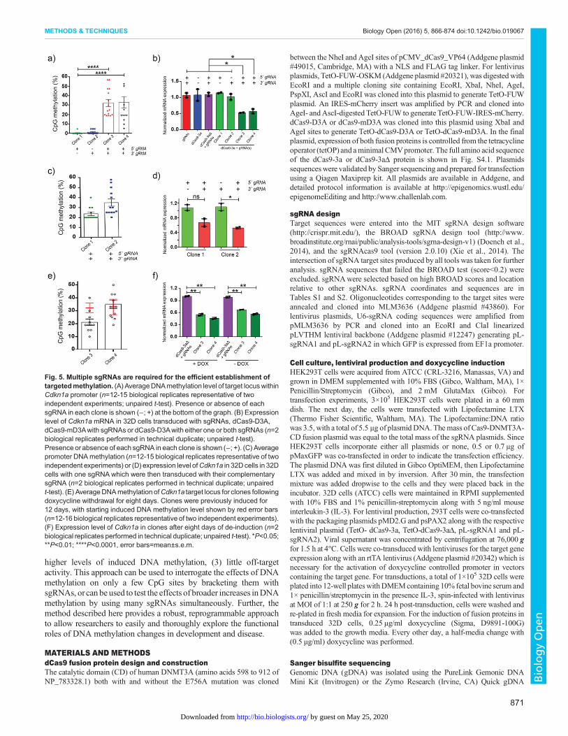
We reverse cloned viral integrants to identify clones thatcontained either one of the two guides, or both sgRNAs togetherwith dCas9-D3A. No induced promoter DNA methylation orreduction in gene expression was observed in clones expressingonly a single sgRNA (Fig. 5A,B). In contrast, Cdkn1a promotermethylation was increased and its expression decreased in clonesthat expressed both sgRNAs (Fig. 5A,B). To confirm the presenceof both sgRNAs were required to execute induced DNAmethylation, clones that contained only a single sgRNA weretransduced with their complementary sgRNA. Following this, aftereight-days of induction, Cdkn1aDNAmethylation increased acrossthe promoter by up to 33% compared to the parental clone thatcontained only a single sgRNA (Fig. 5C). Cdkn1a expression wasreduced by 40-50% in the complemented clones (Fig. 5D). These
Fig. 3. Induced methylation decreases ARFexpression. (A) Locations of the three sgRNA usedto induce DNA methylation at the ARF promoter.(B) Percent methylation is plotted for a region ofexon 1 of the ARF locus. ARF sgRNA target sitesand three targeted CpGs (asterisks) are indicatedabove. (C) Methylation induced by ARF targetedsgRNA decreases gene expression. Relativeexpression of ARF is normalized to a mock-treatedcontrol. Error bars=mean±s.e.m. (n=1 biologicalreplicate performed in technical triplicate).
869
METHODS & TECHNIQUES Biology Open (2016) 5, 866-874 doi:10.1242/bio.019067
BiologyOpen
by guest on May 25, 2020http://bio.biologists.org/Downloaded from
data indicate that targeting with multiple sgRNAs results in asynergism and induces robust DNAmethylation. We also examinedthe stability of the induced DNA methylation by withdrawingdoxycycline for eight days.Cdkn1a promoter DNAmethylation andexpression profiles remained largely unchanged (Fig. 5E,F) eventhough expression of dCas9-D3A was silenced (Fig. S4.2).To assess non-specific activity in this system, we initially
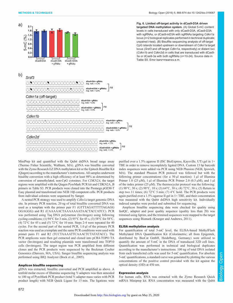
attempted to analyze off-target genomic areas with high homologyto predicted binding regions of both Cdkn1a sgRNAs. However, noregions with high homology were found as these sgRNAs weredesigned based on their minimal off-target binding and the presenceof a PAM-site. As an alternative, we measured the global DNAmethylation levels between cells that contained fusion proteins(dCas9-D3A or dCas9-mD3A) in the presence of both sgRNAs(Fig. 6A), as well as the local DNA methylation profile of CpGislands located upstream and downstream of theCdkn1a target locus(Srsf3 and off-target Cdkn1a respectively), and the promoter CpGislands of the related cell cycle family members Cdkn1b andCdkn2d (Fig. S6). Srsf3 and off-target Cdkn1a are adjacent toCdkn1a target locus on chromosome 17 (chr17), whereas Cdkn1b(chr6) and Cdkn2d (chr4) are distant loci. We found no statisticallysignificant difference in global 5-methylcytosine (5-mC) levelsbetween different groups or across the regions that were analyzed(Fig. 6A,B).
DISCUSSIONWe provide an outline for using a modified CRISPR/dCas9 systemto evaluate the functional relevance of DNA methylation at specific
CpGs and described guidelines for its use. DNA methylationinduction occurs within ∼50 bp of a sgRNA target site and isstrongest between two adjacent and inwardly directed sgRNAbinding sites. Based on our design criteria, we designed sets ofsgRNAs that induced methylation at the human CDKN2A and ARFpromoters, and the mouse Cdkn1a promoters with similarefficiency. Induced methylation was sufficient to decreaseexpression of all three genes. Methylation increases and changesin expression were highly significant and reproducible either byusingmultiple distinct sgRNA combinations in the case ofCDKN2Aor at the clonal levels as observed for Cdkn1a. Moreover, thereduction in Cdkn1a expression clearly had functionalconsequences (increased proliferation) for the transduced cells.Though modest, the expression decreases caused by inducedmethylation are consistent with previously published results usingZFP and TALE fusions (Bernstein et al., 2015; Cui et al., 2015).
The effects of the induced methylation also appeared to becontext dependent. While methylation of the entire CGI at theCDKN2A promoter repressed gene expression, inducing DNAmethylation of a region 100-400 bp downstream of the CDKN2ATSS alone was insufficient to affect expression despite the frequentobservation of a negative correlation between methylation andexpression in this region. This indicates the importance of theflexibility to target multiple regions offered by our CRISPR/dCas9DNMT fusion system.
Although approaches for targeted DNA methylation have beenpreviously described, our method is advantageous for several reasonsincluding; (1) the ease of designing new sgRNAs for targeting, (2)
Fig. 4. Targeted CpG methylation of the Cdkn1alocus using dCas9-D3A fusion protein.(A) Organization of the mouse Cdkn1a target locuswith CpG islands indicated with green boxes. Bluearrow indicates the location of transcriptional start site(TSS). Red arrows indicate the relative position ofsgRNA binding sites (5′ to 3′) within the 5′ CpG island.Black arrows indicate the relative positions of CpGs.(B) Bisulfite sequencing analysis of Cdkn1a promoterin 32D cells transduced with sgRNAs or dCas9-D3Aonly, and dCas9a-D3A or dCas9-mD3Awith sgRNAs.Changes in methylation of specific CpGs are indicatedby pie charts. Shading indicates the average amountof DNA methylation at each CpG (n=14-15 biologicalreplicates representative of three independentexperiments). (C) Average DNA methylation level oftarget locus within Cdkn1a promoter (unpaired t-test).(D) mRNA expression level of Cdkn1a (normalized to18S) in transduced 32D cells (n=2 biological replicatesperformed in technical duplicate; unpaired t-test).(E) Growth kinetics of transduced 32D cells (n=2biological replicates performed in technicalquadruplicate; unpaired t-test). *P<0.05,****P<0.0001, error bars=mean±s.e.m.
870
METHODS & TECHNIQUES Biology Open (2016) 5, 866-874 doi:10.1242/bio.019067
BiologyOpen
by guest on May 25, 2020http://bio.biologists.org/Downloaded from
higher levels of induced DNA methylation, (3) little off-targetactivity. This approach can be used to interrogate the effects of DNAmethylation on only a few CpG sites by bracketing them withsgRNAs, or can be used to test the effects of broader increases inDNAmethylation by using many sgRNAs simultaneously. Further, themethod described here provides a robust, reprogrammable approachto allow researchers to easily and thoroughly explore the functionalroles of DNA methylation changes in development and disease.
MATERIALS AND METHODSdCas9 fusion protein design and constructionThe catalytic domain (CD) of human DNMT3A (amino acids 598 to 912 ofNP_783328.1) both with and without the E756A mutation was cloned
between the NheI and AgeI sites of pCMV_dCas9_VP64 (Addgene plasmid#49015, Cambridge, MA) with a NLS and FLAG tag linker. For lentivirusplasmids, TetO-FUW-OSKM (Addgene plasmid #20321), was digested withEcoRI and a multiple cloning site containing EcoRI, XbaI, NheI, AgeI,PspXI, AscI and EcoRI was cloned into this plasmid to generate TetO-FUWplasmid. An IRES-mCherry insert was amplified by PCR and cloned intoAgeI- andAscI-digested TetO-FUW to generate TetO-FUW-IRES-mCherry.dCas9-D3A or dCas9-mD3A was cloned into this plasmid using XbaI andAgeI sites to generate TetO-dCas9-D3A or TetO-dCas9-mD3A. In the finalplasmid, expression of both fusion proteins is controlled from the tetracyclineoperator (tetOP) and aminimal CMVpromoter. The full amino acid sequenceof the dCas9-3a or dCas9-3aΔ protein is shown in Fig. S4.1. Plasmidssequences were validated by Sanger sequencing and prepared for transfectionusing a Qiagen Maxiprep kit. All plasmids are available in Addgene, anddetailed protocol information is available at http://epigenomics.wustl.edu/epigenomeEditing and http://www.challenlab.com.
sgRNA designTarget sequences were entered into the MIT sgRNA design software(http://crispr.mit.edu/), the BROAD sgRNA design tool (http://www.broadinstitute.org/rnai/public/analysis-tools/sgrna-design-v1) (Doench et al.,2014), and the sgRNAcas9 tool (version 2.0.10) (Xie et al., 2014). Theintersection of sgRNA target sites produced by all tools was taken for furtheranalysis. sgRNA sequences that failed the BROAD test (score<0.2) wereexcluded. sgRNAwere selected based on high BROAD scores and locationrelative to other sgRNAs. sgRNA coordinates and sequences are inTables S1 and S2. Oligonucleotides corresponding to the target sites wereannealed and cloned into MLM3636 (Addgene plasmid #43860). Forlentivirus plasmids, U6-sgRNA coding sequences were amplified frompMLM3636 by PCR and cloned into an EcoRI and ClaI linearizedpLVTHM lentiviral backbone (Addgene plasmid #12247) generating pL-sgRNA1 and pL-sgRNA2 in which GFP is expressed from EF1a promoter.
Cell culture, lentiviral production and doxycycline inductionHEK293T cells were acquired from ATCC (CRL-3216, Manassas, VA) andgrown in DMEM supplemented with 10% FBS (Gibco, Waltham, MA), 1×Penicillin/Streptomycin (Gibco), and 2 mM GlutaMax (Gibco). Fortransfection experiments, 3×10⁵ HEK293T cells were plated in a 60 mmdish. The next day, the cells were transfected with Lipofectamine LTX(Thermo Fisher Scientific, Waltham, MA). The Lipofectamine:DNA ratiowas 3.5, with a total of 5.5 μg of plasmid DNA. Themass of Cas9-DNMT3A-CD fusion plasmid was equal to the total mass of the sgRNA plasmids. SinceHEK293T cells incorporate either all plasmids or none, 0.5 or 0.7 μg ofpMaxGFP was co-transfected in order to indicate the transfection efficiency.The plasmid DNAwas first diluted in Gibco OptiMEM, then LipofectamineLTX was added and mixed in by inversion. After 30 min, the transfectionmixture was added dropwise to the cells and they were placed back in theincubator. 32D cells (ATCC) cells were maintained in RPMI supplementedwith 10% FBS and 1% penicillin-streptomycin along with 5 ng/ml mouseinterleukin-3 (IL-3). For lentiviral production, 293T cells were co-transfectedwith the packaging plasmids pMD2.G and psPAX2 along with the respectivelentiviral plasmid (TetO- dCas9-3a, TetO-dCas9-3aΔ, pL-sgRNA1 and pL-sgRNA2). Viral supernatant was concentrated by centrifugation at 76,000 gfor 1.5 h at 4°C. Cells were co-transduced with lentiviruses for the target geneexpression along with an rtTA lentivirus (Addgene plasmid #20342) which isnecessary for the activation of doxycycline controlled promoter in vectorscontaining the target gene. For transductions, a total of 1×10⁵ 32D cells wereplated into 12-well plateswithDMEMcontaining 10% fetal bovine serum and1× penicillin/streptomycin in the presence IL-3, spin-infected with lentivirusat MOI of 1:1 at 250 g for 2 h. 24 h post-transduction, cells were washed andre-plated in fresh media for expansion. For the induction of fusion proteins intransduced 32D cells, 0.25 µg/ml doxycycline (Sigma, D9891-100G)was added to the growth media. Every other day, a half-media change with(0.5 µg/ml) doxycycline was performed.
Sanger bisulfite sequencingGenomic DNA (gDNA) was isolated using the PureLink Gemonic DNAMini Kit (Invitrogen) or the Zymo Research (Irvine, CA) Quick gDNA
Fig. 5. Multiple sgRNAs are required for the efficient establishment oftargetedmethylation. (A) AverageDNAmethylation level of target locuswithinCdkn1a promoter (n=12-15 biological replicates representative of twoindependent experiments; unpaired t-test). Presence or absence of eachsgRNA in each clone is shown (−; +) at the bottom of the graph. (B) Expressionlevel of Cdkn1a mRNA in 32D cells transduced with sgRNAs, dCas9-D3A,dCas9-mD3Awith sgRNAs or dCas9-D3Awith either one or both sgRNAs (n=2biological replicates performed in technical duplicate; unpaired t-test).Presence orabsence of each sgRNA in each clone is shown (−; +). (C) Averagepromoter DNAmethylation (n=12-15 biological replicates representative of twoindependent experiments) or (D) expression level ofCdkn1a in 32Dcells in 32Dcells with one sgRNA which were then transduced with their complementarysgRNA (n=2 biological replicates performed in technical duplicate; unpairedt-test). (E) AverageDNAmethylation ofCdkn1a target locus for clones followingdoxycycline withdrawal for eight days. Clones were previously induced for12 days, with starting induced DNA methylation level shown by red error bars(n=12-16 biological replicates representative of two independent experiments).(F) Expression level of Cdkn1a in clones after eight days of de-induction (n=2biological replicates performed in technical duplicate; unpaired t-test). *P<0.05;**P<0.01; ****P<0.0001, error bars=mean±s.e.m.
871
METHODS & TECHNIQUES Biology Open (2016) 5, 866-874 doi:10.1242/bio.019067
BiologyOpen
by guest on May 25, 2020http://bio.biologists.org/Downloaded from
MiniPrep kit and quantified with the Qubit dsDNA broad range assay(Thermo Fisher Scientific, Waltham, MA). gDNA was bisulfite convertedwith the ZymoResearch EZDNAmethylation kit or the Epitech Bisulfite Kit(Qiagen) according to the manufacturer’s instructions. All samples underwentbisulfite conversion with a high efficiency of at least 98% as determined byconversion of unmethylated, non-CpG cytosines. For CDKN2A, the targetregions were amplified with the Qiagen PyroMark PCR kit and CDKN2A_Bprimers in Table S3. PCR products were cloned into the Promega pGEM-TEasy plasmid and transformed into NEB 10β competent cells. PCR productsfrom individual colonies were sequenced by Sanger.
A nested PCR strategy was used to amplify Cdkn1a target genomic DNAsite. In primary PCR reaction, 20 ng of total bisulfite converted DNA wasused as a template with the primer pair F1 (GTTTAGATTTTTAGAGGGGAGGG) and R1 (CAAAAACTAAAAAAATAACTACCATCC). PCRwas performed using Taq DNA polymerase (Invitrogen) using followingcycling conditions; (1) 94°C for 3 min, (2) 95°C for 45 s, (3) 58°C for 58°C,(4) 72°C for 45 s and (5) 72°C for 10 min. Steps 2-4 were repeated for 40cycles. For the second part of the nested PCR, 1.0 µl of the primary PCRreaction was used as a template and the same PCR conditions were used withprimer pairs F1 and R2 (TCCTAAAATTCAAACTCTATATACC). ThePCR amplicons were then gel extracted and cloned into pCR4-TOPO TAvector (Invitrogen) and resulting plasmids were transformed into TOP10cells (Invitrogen). The target region was PCR amplified from differentclones and the PCR products were sequenced by the Beckman CoulterGenomics (Danvers, MA, USA). Sanger bisulfite sequencing analysis wasperformed using BIQ Analyzer (Bock et al., 2005).
Amplicon bisulfite sequencinggDNA was extracted, bisulfite converted and PCR amplified as above. Atenfold molar excess of Illumina sequencing Y-adapters was then annealedto 100 ng of PyroMark PCR product (total Y-adapter mass varies with PCRproduct length) with NEB Quick Ligase for 15 min. The ligations were
purified over a 1.5% agarose II (ISC BioExpress, Kaysville, UT) gel in 1×TBE in order to remove incompletely ligated DNA. Custom 13 bp barcodeindex sequences were added via PCR using NEB Phusion (NEB, Ipswich,MA). The standard Phusion PCR protocol was followed but with thefollowing primer concentrations (for a 50 μl reaction): 1 μl of IlluminaPrimer 1.0 (25 μM), 1 μl of Illumina PCR Primer 2.0 (0.5 μM), and 1 μlof the index primer (25 μM). The thermocycler protocol was the following:(1) 98°C, 30 s; (2) 98°C, 10 s; (3) 64°C, 30 s; (4) 72°C, 30 s; (5) Return tostep two 11 times; (6) 72°C 5 min; (7) 4°C hold. The PCR products wereagain purified over a 1.5% agarose II gel in 1× TBE, and their concentrationwas measured with the Qubit dsDNA high sensitivity kit. Individuallyindexed samples were pooled and submitted for sequencing.
Amplicon bisulfite sequencing data were checked for quality usingfastQC, adaptor and poor quality sequence (quality less than 20) wastrimmed using fqtrim, and the trimmed sequences were mapped to the targetsequences using Bismark (Krueger and Andrews, 2011).
ELISA methylation analysisFor quantification of total 5-mC level, the ELISA-based MethylFlashMethylated DNA Quantification Kit (Colorimetric; all from Epigentek,distributed by BioCat GmbH, Heidelberg, Germany) were utilized toquantify the amount of 5-mC in the DNA of transduced 32D cell lines.Quantification was performed in technical and biological duplicatesaccording to the manufacturer’s instructions. 100 ng of total DNA isolatedfrom each transfected cells was used for 5-mC quantification. For absolute5-mC quantifications, a standard curvewas generated by plotting the variousconcentrations of the positive control provided with the kit against theoptical density (OD) at 450 nm.
Expression analysisFor human cells, RNA was extracted with the Zymo Research QuickmRNA Miniprep kit. RNA concentration was measured with the Qubit
Fig. 6. Limited off-target activity in dCas9-D3A driventargeted DNA methylation system. (A) Global 5-mC contentlevels in cells transduced with only dCas9-D3A, dCas9-D3Awith sgRNAs, or dCas9-mD3A with sgRNAs targeting Cdkn1alocus (n=2 biological replicates performed in technical duplicate;unpaired t-test). (B) Bisulfite sequencing analysis of off-targetCpG islands located upstream or downstream of Cdkn1a targetlocus (Srsf3 and off-target Cdkn1a, respectively) or distant loci(Cdkn1b and Cdkn2d) in cells that are transduced with dCas9-3a or dCas9-3a with both sgRNAs (n=10-24). Source data inTable S5. Error bars=mean±s.e.m.
872
METHODS & TECHNIQUES Biology Open (2016) 5, 866-874 doi:10.1242/bio.019067
BiologyOpen
by guest on May 25, 2020http://bio.biologists.org/Downloaded from
RNA BR kit. RNA integrity was determined by visualizing rRNA bandsusing agarose gel electrophoresis. Reverse transcription was performedusing the Bio-Rad iScript Reverse Transcriptase kit. Quantitative reverse-transcription polymerase chain reaction (RT-qPCR) was performed withthe Bio-Rad iTaq Universal with SYBR Green reagent on an AppliedBiosystems Viia7 instrument. The thermocycler protocol was thefollowing: (1) 95°C, 20 s; (2) 95°C, 3 s; (3) 60°C, 20 s; for 40 cycles.qPCR primers are listed in Table S4. A melt curve was performed toindicate there was not off-target amplification. Data was analyzed asdescribed by Hellemans et al. (2007) using the geometric mean of ACTB,GAPDH, and RPL0 as an internal control. The All sgRNA samplerepresents data from two independent transfection experiments. The datafor all remaining samples derives from technical replication using thesame RNA sample. P-values were calculated with paired sample t-tests onthe normalized levels of gene expression.
For mouse cells, total RNA was isolated from 5×10³ FACS-sortedcells using NucleoSpin RNA XS (Macherey-Nagel) and reversetranscribed with the SuperScript VILO kit (Life Technologies). cDNAinput was standardized and real-time polymerase chain reaction (PCR)was performed with TaqMan master Mix (Applied Biosystems), 18s-rRNA probe (VIC-MGB; Applied Biosystems), and Cdkn1a geneprobe (FAM-MGB; Mm04205640_g1, Applied Biosystems) on aStepOnePlus Real-Time PCR System (Life Technologies). Sampleswere normalized to expression of 18S and fold change determined bythe ΔΔCt method.
Western blot32D cells transduced with dCas9-D3A or dCas9-mD3A with or withoutsgRNAs were grown in the presence of doxycycline for 12 days, collectedandwashedwith ice cold PBS twice. Cells were then lysed in complete RIPAbuffer containing protease inhibitors (Santa Cruz Biotechnology). 20 µg ofprotein lysates were separated on 10% SDS-PAGE gels and transferred tonitrocellulose membranes (Millipore). Membranes were subsequentlyprobed to detect fusion proteins using primary antibodies recognizingCas9 (Active Motif ) or β-actin (Santa Cruz) and detection was performedusing horseradish-peroxidase-conjugated secondary mouse antibody (SantaCruz) and chemiluminescence (Millipore).
AcknowledgementsThe authors thank the Genome Engineering and iPSC Center at WashingtonUniversity School of Medicine for production and validation of sgRNAs. We thankJessica Hoisington-Lopez for sequencing assistance, and we thank C. Scholsbergfor critical reading of this manuscript. We thank the Alvin J. Siteman Cancer Centerat Washington University School of Medicine and Barnes-Jewish Hospital for theuse of the Siteman Flow Cytometry Core, which provided cell sorting and analysisservices. The Siteman Cancer Center is supported in part by NCI Cancer CenterSupport Grant #P30 CA91842.
Competing interestsThe authors declare no competing or financial interests.
Author contributionsJ.I.M and H.C were equally responsible for cell culture, methylation and expressionexperiments, data analysis, and writing the manuscript. L.E.R. performedmethylation analyses and assisted with writing the manuscript. T.F. performedmethylation analyses. G.F. and R.R. performed cloning of target loci for sequencinganalysis. A.K. helped with cell culture and off-target analysis. A.M. designed thecloning of dCas9-D3A and dCas9-mD3A fusion proteins. J.R.E and G.A.Csupervised the work, edited and approved the manuscript.
FundingWe acknowledge the following funding sources: National Institutes of Health [NIDDKR01DK102428], the Edward Mallinckrodt Jr. Foundation, the American Society ofHematology, Alex’s Lemonade Stand Foundation for Childhood Cancer, theChildren’s Discovery Institute, the Sidney Kimmel Foundation for Cancer Researchand the V Foundation for Cancer Research (to G.A.C.), the Siteman Cancer Center,U.S. Department of Defense Congressionally Directed Medical Research Programfor Breast Cancer [W81XWH-11-1-0401], and the National Institute of GeneralMedicine Sciences [NIGMS 5R01GM108811, NLM R21LM011199] (to J.R.E.), andNational Institutes of Health T32 CMB Training Grant [2T32GM007067-37] for pre-doctoral support to J.I.M.
Supplementary informationSupplementary information available online athttp://bio.biologists.org/lookup/suppl/doi:10.1242/bio.019067/-/DC1
ReferencesBell, J. T., Pai, A. A., Pickrell, J. K., Gaffney, D. J., Pique-Regi, R., Degner, J. F.,
Gilad, Y. and Pritchard, J. K. (2011). DNA methylation patterns associate withgenetic and gene expression variation in HapMap cell lines.Genome Biol. 12, R10.
Bernstein, D. L., Le Lay, J. E., Ruano, E. G. and Kaestner, K. H. (2015). TALE-mediated epigenetic suppression of CDKN2A increases replication in humanfibroblasts. J. Clin. Invest. 125, 1998-2006.
Bestor, T. H., Edwards, J. R. and Boulard, M. (2015). Notes on the role of dynamicDNA methylation in mammalian development. Proc. Natl. Acad. Sci. USA 112,6796-6799.
Bock, C., Reither, S., Mikeska, T., Paulsen, M., Walter, J. and Lengauer, T.(2005). BiQ analyzer: visualization and quality control for DNA methylation datafrom bisulfite sequencing. Bioinformatics 21, 4067-4068.
Bock,C.,Beerman, I., Lien,W.-H., Smith,Z.D.,Gu,H.,Boyle,P.,Gnirke,A., Fuchs,E., Rossi, D. J. andMeissner, A. (2012). DNAmethylation dynamics during in vivodifferentiation of blood and skin stem cells.Mol. Cell 47, 633-647.
Busslinger, M., Hurst, J. and Flavell, R. A. (1983). DNA methylation and theregulation of globin gene expression. Cell 34, 197-206.
Ciriello, G., Miller, M. L., Aksoy, B. A., Senbabaoglu, Y., Schultz, N. and Sander,C. (2013). Emerging landscape of oncogenic signatures across human cancers.Nat. Genet. 45, 1127-1133.
Cui, C., Gan, Y., Gu, L., Wilson, J., Liu, Z., Zhang, B. and Deng, D. (2015).P16-specific DNA methylation by engineered zinc finger methyltransferaseinactivates gene transcription and promotes cancer metastasis. Genome Biol.16, 252.
Doench, J. G., Hartenian, E., Graham, D. B., Tothova, Z., Hegde, M., Smith, I.,Sullender, M., Ebert, B. L., Xavier, R. J. and Root, D. E. (2014). Rational designof highly active sgRNAs for CRISPR-Cas9-mediated gene inactivation. Nat.Biotechnol. 32, 1262-1267.
Egger, G., Liang, G., Aparicio, A. and Jones, P. A. (2004). Epigenetics in humandisease and prospects for epigenetic therapy. Nature 429, 457-463.
Hellemans, J., Mortier, G., De Paepe, A., Speleman, F. and Vandesompele, J.(2007). qBase relative quantification framework and software for managementand automated analysis of real-time quantitative PCR data.Genome Biol. 8, R19.
Hinshelwood, R. A., Melki, J. R., Huschtscha, L. I., Paul, C., Song, J. Z.,Stirzaker, C., Reddel, R. R. and Clark, S. J. (2009). Aberrant de novomethylation of the p16INK4A CpG island is initiated post gene silencing inassociation with chromatin remodelling and mimics nucleosome positioning.Hum. Mol. Genet. 18, 3098-3109.
Jinek, M., Chylinski, K., Fonfara, I., Hauer, M., Doudna, J. A. andCharpentier, E.(2012). A programmable dual-RNA–guided DNA endonuclease in adaptivebacterial immunity. Science 337, 816-821.
Krueger, F. and Andrews, S. R. (2011). Bismark: a flexible aligner and methylationcaller for Bisulfite-Seq applications. Bioinformatics 27, 1571-1572.
Liggett, W. H. and Sidransky, D. (1998). Role of the p16 tumor suppressor gene incancer. J. Clin. Oncol. 16, 1197-1206.
Lou, S., Lee, H.-M., Qin, H., Li, J.-W., Gao, Z., Liu, X., Chan, L. L., Kl Lam, V., So,W.-Y., Wang, Y. et al. (2014). Whole-genome bisulfite sequencing of multipleindividuals reveals complementary roles of promoter and gene body methylationin transcriptional regulation. Genome Biol. 15, 6.
Lund, K., Cole, J. J., VanderKraats, N. D., McBryan, T., Pchelintsev, N. A., Clark,W., Copland, M., Edwards, J. R. and Adams, P. D. (2014). DNMT inhibitorsreverse a specific signature of aberrant promoter DNAmethylation and associatedgene silencing in AML. Genome Biol. 15, 406.
Maeder, M. L., Linder, S. J., Cascio, V. M., Fu, Y., Ho, Q. H. and Joung, J. K.(2013). CRISPR RNA-guided activation of endogenous human genes. Nat.Methods 10, 977-979.
Mali, P., Aach, J., Stranges, P. B., Esvelt, K. M., Moosburner, M., Kosuri, S.,Yang, L. and Church, G. M. (2013). CAS9 transcriptional activators for targetspecificity screening and paired nickases for cooperative genome engineering.Nat. Biotechnol. 31, 833-838.
Perez-Pinera, P., Kocak, D. D., Vockley, C. M., Adler, A. F., Kabadi, A. M.,Polstein, L. R., Thakore, P. I., Glass, K. A., Ousterout, D. G., Leong, K.W. et al.(2013a). RNA-guided gene activation by CRISPR-Cas9-based transcriptionfactors. Nat. Methods 10, 973-976.
Perez-Pinera, P., Ousterout, D. G., Brunger, J. M., Farin, A. M., Glass, K. A.,Guilak, F., Crawford, G. E., Hartemink, A. J. and Gersbach, C. A. (2013b).Synergistic and tunable human gene activation by combinations of synthetictranscription factors. Nat. Methods 10, 239-242.
Reither, S., Li, F., Gowher, H. and Jeltsch, A. (2003). Catalytic mechanism ofDNA-(cytosine-C5)-methyltransferases revisited: covalent intermediate formationis not essential for methyl group transfer by the murine Dnmt3a enzyme. J. Mol.Biol. 329, 675-684.
Shima, K., Nosho, K., Baba, Y., Cantor, M., Meyerhardt, J. A., Giovannucci,E. L., Fuchs, C. S. and Ogino, S. (2011). Prognostic significance of CDKN2A
873
METHODS & TECHNIQUES Biology Open (2016) 5, 866-874 doi:10.1242/bio.019067
BiologyOpen
by guest on May 25, 2020http://bio.biologists.org/Downloaded from
(p16) promoter methylation and loss of expression in 902 colorectal cancers:cohort study and literature review. Int. J. Cancer 128, 1080-1094.
Siddique, A. N., Nunna, S., Rajavelu, A., Zhang, Y., Jurkowska, R. Z., Reinhardt,R., Rots, M. G., Ragozin, S., Jurkowski, T. P. and Jeltsch, A. (2013). Targetedmethylation and gene silencing of VEGF-A in human cells by using a designedDnmt3a–Dnmt3L single-chain fusion protein with increased DNA methylationactivity. J. Mol. Biol. 425, 479-491.
VanderKraats, N. D., Hiken, J. F., Decker, K. F. and Edwards, J. R.(2013). Discovering high-resolution patterns of differential DNA methylationthat correlate with gene expression changes. Nucleic Acids Res. 41,6816-6827.
Xie, S., Shen, B., Zhang, C., Huang, X. and Zhang, Y. (2014). sgRNAcas9: asoftware package for designing CRISPR sgRNA and evaluating potential off-target cleavage sites. PLoS ONE 9, e100448.
874
METHODS & TECHNIQUES Biology Open (2016) 5, 866-874 doi:10.1242/bio.019067
BiologyOpen
by guest on May 25, 2020http://bio.biologists.org/Downloaded from


![Generation of Targeted Knockout Mutants in Arabidopsis ... · Keywords: CRISPR/Cas9, Genome editing, Arabidopsis thaliana, Plants, Knockout [Background] The CRISPR/Cas9 system (Cas9)](https://img.pdfslide.net/doc/110x75/5fcbdfb69ddbe939ee10f004/generation-of-targeted-knockout-mutants-in-arabidopsis-keywords-crisprcas9.jpg)