Embed Size (px)
Citation preview
Research ArticleCharacterization of Shiga Toxigenic Escherichia coli O157 andNon-O157 Isolates from Ruminant Feces in Malaysia
Asanthi Perera,1 Charles M. Clarke,1 Gary A. Dykes,1 and Narelle Fegan2
1School of Science, Monash University, Jalan Lagoon Selatan, 46150 Bandar Sunway, Selangor, Malaysia2CSIRO, Food and Nutrition Flagship, 671 Sneydes Road, Werribee, VIC 3030, Australia
Correspondence should be addressed to Narelle Fegan; [email protected]
Received 11 November 2014; Revised 15 February 2015; Accepted 23 February 2015
Academic Editor: Miguel Prieto
Copyright © 2015 Asanthi Perera et al. This is an open access article distributed under the Creative Commons Attribution License,which permits unrestricted use, distribution, and reproduction in any medium, provided the original work is properly cited.
Shiga toxigenic Escherichia coli (STEC) O157 and several other serogroups of non-O157 STEC are causative agents of severe diseasein humans world-wide. The present study was conducted to characterize STEC O157 and non-O157 serogroups O26, O103, O111,O121, O45, and O145 in ruminants in Malaysia. A total of 136 ruminant feces samples were collected from 6 different farms inPeninsular Malaysia. Immunomagnetic beads were used to isolate E. coli O157 and non-O157 serogroups, while PCR was usedfor the detection and subtyping of STEC isolates. STEC O157:H7 was isolated from 6 (4%) feces samples and all isolates obtainedcarried 𝑠𝑡𝑥2c, eaeA-𝛾1, and ehxA. Non-O157 STEC was isolated from 2 (1.5%) feces samples with one isolate carrying 𝑠𝑡𝑥1a, 𝑠𝑡𝑥2a,𝑠𝑡𝑥2c, and ehxA and the other carrying 𝑠𝑡𝑥1a alone. The presence of STEC O157 and non-O157 in a small percentage of ruminantsin this study together with their virulence characteristics suggests that they may have limited impact on public health.
1. Introduction
Shiga toxin producing E. coli (STEC), a serologically diversegroup of zoonotic pathogens, have emerged as one of themost virulent groups of bacteria associated with cases of foodborne disease in humans [1]. STEC can cause a spectrumof diseases ranging from mild diarrhea to severe bloodydiarrhea, called hemorrhagic colitis (HC), and even life-threatening sequelae such as hemolytic uremic syndrome(HUS). Patients with HUS were often diagnosed as hav-ing thrombotic thrombocytopenic purpura (TTP), althoughthrombotic microangiopathy is now considered amore accu-rate description of the condition associated with HUS causedby STEC [2]. Production of Shiga toxin (Stx) is consideredas the major virulence factor of STEC [1] which contributesto the development of HUS in humans [2]. Stx productionalone is not sufficient for STEC to cause disease. Accessoryvirulence factors include a 34 kb chromosomal pathogenicityisland called the “locus for enterocyte effacement” (LEE) car-rying several virulence associated genes, such as the attachingand effacing (eaeA) gene, and a large plasmid (60MDa) withan ehxA gene encoding an enterohemolysin. EaeA encodesan outer-membrane protein called intimin which enables
the intimate adherence of STEC to the intestinal epitheliumof the host [3]. The enterohemolysin protein is implicated inextracting iron from the blood released into the intestine [4].
The prototype STEC serotype is E. coli O157:H7 and itsability to cause HC and HUS in many regions and countriesis well established. The pathogenic potential and publichealth significance of several non-O157 STEC serogroups,particularly O26, O103, O111, O121, O145, and O45 referred toas the “big 6” non-O157 STEC serogroups [5], have also beendescribed in recent years due to their association with clinicalHC and HUS in humans. In some geographical areas, such asin Europe, the disease caused by non-O157 strains is signifi-cantly more common than that caused by O157:H7 [6, 7].
Ruminants are considered an important source of bothE. coli O157 and non-O157 with cattle being identified as theprimary reservoir. Intestinal carriage of E. coliO157 and non-O157 in ruminants results in their fecal shedding and releaseinto the environment. As a result, infections of E. coli O157and non-O157 can be transmitted to humans via the con-sumption of food and water contaminated by animal feces.
Data on E. coli O157 and non-O157 serotypes in rumi-nants is limited in countries of the tropical regions includingMalaysia. In addition, the data reported so far on E. coliO157
Hindawi Publishing CorporationBioMed Research InternationalVolume 2015, Article ID 382403, 8 pageshttp://dx.doi.org/10.1155/2015/382403
2 BioMed Research International
Table 1: Distribution of ruminant feces samples collected from farms A–F.
Farm Location Ruminant feces samples Total samplesCattle Buffalo Goat Sheep
A Serdang 25 — — — 25B Kluang 9 20 7 8 44C Sentul 9 — — — 9D Gemas 24 — — — 24E Puchong 13 — 5 — 18F Lumut 16 — — — 16
and non-O157 in ruminants from tropical countries otherthan Malaysia demonstrates substantial variation in theirprevalence and virulence properties. In West Bengal, India,a total of fourteen STEC O157 isolates were obtained fromtwo (2.04%) slaughtered cattle feces samples and six (7.59%)diarrhoeic calf feces samples [8]. The majority of STEC O157isolates (85.71%) obtained from this study carried stx
2alone.
STECO157 was obtained from 0.6% of cattle feces samples inBrazil [9], where the majority of isolates carried ehxA eitherwith both stx
1and stx
2or with stx
2alone. The prevalence of
E. coli O157 was found to be 1.25% in cattle farms in centralMexico [10]. Non-O157 STECwas found in 18% of cattle fecessamples in Calcutta, India [11], in which stx
1predominated.
In another study in Brazil, non-O157 STEC was isolated from5.81% of calf feces samples [12] where stx
1was the dominant
stx genotype observed.Only three studies which isolated STEC O157 from
beef samples have to our knowledge been conducted inMalaysia [13–15]. Apart from a single study which reportedsporadic cases of STECO157 infection among 14% of patientspresented with bloody diarrhea at a local hospital in KualaLumpur, Malaysia [16], there are no other published reportsof sporadic cases or outbreaks of STEC O157 and non-O157 in the country. Although studies have demonstratedthe presence of STEC O157 in foods of animal origin, thepresence and characterization of STEC O157 or non-O157 inruminant feces fromMalaysia has not yet been determined.
The aim of the present study was to examine ruminantfeces samples for the presence of STEC O157 and the “big 6”non-O157 STEC serogroups in Malaysia. The isolated strainsof E. coli O157 and non-O157 were further characterized todetermine their genetic diversity and presence of virulencefactors to indicate the risk potential of these strains to publichealth.
2. Materials and Methods
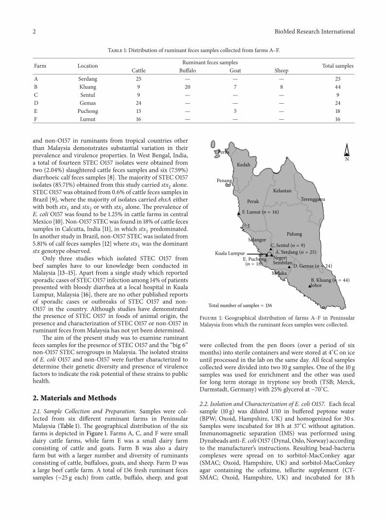
2.1. Sample Collection and Preparation. Samples were col-lected from six different ruminant farms in PeninsularMalaysia (Table 1). The geographical distribution of the sixfarms is depicted in Figure 1. Farms A, C, and F were smalldairy cattle farms, while farm E was a small dairy farmconsisting of cattle and goats. Farm B was also a dairyfarm but with a larger number and diversity of ruminantsconsisting of cattle, buffaloes, goats, and sheep. Farm D wasa large beef cattle farm. A total of 136 fresh ruminant fecessamples (∼25 g each) from cattle, buffalo, sheep, and goat
Perlis
Kedah
Penang
Perak
KelantanTerengganu
PahangSelangor
Kuala LumpurNegeriSembilan
Melaka
Johor
F. Lumut (n = 16)
C. Sentul (n = 9)A. Serdang (n = 25)
D. Gemas (n = 24)
E. Puchong(n = 18)
B. Kluang (n = 44)
N
Total number of samples = 136
Figure 1: Geographical distribution of farms A–F in PeninsularMalaysia from which the ruminant feces samples were collected.
were collected from the pen floors (over a period of sixmonths) into sterile containers and were stored at 4∘C on iceuntil processed in the lab on the same day. All fecal samplescollected were divided into two 10 g samples. One of the 10 gsamples was used for enrichment and the other was usedfor long term storage in tryptone soy broth (TSB; Merck,Darmstadt, Germany) with 25% glycerol at −70∘C.
2.2. Isolation and Characterization of E. coli O157. Each fecalsample (10 g) was diluted 1/10 in buffered peptone water(BPW; Oxoid, Hampshire, UK) and homogenized for 30 s.Samples were incubated for 18 h at 37∘C without agitation.Immunomagnetic separation (IMS) was performed usingDynabeads anti-E. coliO157 (Dynal, Oslo,Norway) accordingto the manufacturer’s instructions. Resulting bead-bacteriacomplexes were spread on to sorbitol-MacConkey agar(SMAC; Oxoid, Hampshire, UK) and sorbitol-MacConkeyagar containing the cefixime, tellurite supplement (CT-SMAC; Oxoid, Hampshire, UK) and incubated for 18 h
BioMed Research International 3
at 37∘C. A total of 10 presumptive E. coli O157 colonies persample were serotyped using an E. coli O157 Latex Test Kit(Oxoid, Hampshire, UK). All isolates agglutinating with theO157 antiserum were further characterized by polymerasechain reaction (PCR) to detect the presence of rfbE, stx
1,
stx2, eaeA, ehxA, and fliC genes using primers and reaction
conditions as previously described [17].Characterization of lineage-specific polymorphisms-6
(LSPA-6) of E. coli O157 isolates was performed using targetamplification and capillary electrophoresis as described pre-viously [18, 19]. AnAppliedBiosystems 3130GeneticAnalyzer(Applied Biosystems, California, USA) with a DS-33 matrixand GeneScan 600 LIZsize standard was used for capillaryelectrophoresis, while a Peak Scanner software (Version 1.0;Applied Biosystems, California, USA) was used to interpretamplicon sizes. LSPA-6 alleles were defined according to [18].Isolates with LSPA-6 genotype 111111 or 211111 were classifiedas lineage I (LI) or lineage I/II (LI/II), respectively, while allother allele combinations were grouped as lineage II (LII)[18, 20].
Analysis of Shiga toxin encoding bacteriophage insertionsites (SBI) of E. coli O157 isolates was determined as previ-ously described [21].
2.3. Detection, Isolation, and Characterization of Non-O157 E.coli. Samples (10 g) which were initially stored at −70∘C inTSB with 25% glycerol were diluted 1/10 in BPW, homog-enized for 30 s, and incubated for 18 h at 37∘C withoutagitation. DNA was extracted from 1mL of the enrichedsample using the Nucleospin Soli DNA extraction kit(Macherey Nagel, Duren, Germany) following the manufac-turer’s instructions. A multiplex PCR was used to screenenrichments for the presence of STEC virulence genes stx
1,
stx2, eaeA, and ehxA using primers and reaction conditions
as described by A. W. Paton and J. C. Paton [22] with severalmodifications. A reaction volume of 25 𝜇L was used with2 𝜇L of DNA template and final concentration of 0.25𝜇Mof each primer, 5x Green GoTaq Flexi Buffer (Promega,Madison, USA), 200𝜇M of dNTP, 2mM of MgCl
2, and 1
unit of GoTaq DNA polymerase (Promega, Madison, USA).The PCR products were separated by electrophoresis on a2% agarose gel, stained with ethidium bromide (0.5𝜇g/mL)and visualized under UV light. Enriched samples positivefor stx and eaeA by PCR were streaked on chromocult-TBX agar (Merck, Darmstadt, Germany) and coliformenagar enhanced selectivity (Merck, Darmstadt, Germany) andincubated overnight at 37∘C. Following incubation, up to 50E. coli colonies per sample were chosen based on colonymorphology and screened individually by multiplex PCR forthe presence of stx
1, stx2, eaeA, and ehxA as described above.
Colonies that were positive for stx and eaeA were then testedfor the “big 6” E. coli non-O157 serogroups by PCR usingprimers and conditions described previously [17, 23].
The enriched samples were also tested for the presenceof genes specific to the “big 6” E. coli non-O157 sero-groups. Samples that tested positive by PCR for any of thetarget serogroups were subjected to IMS for O26, O111,O103, and O145 using Dynabeads (Dynal, Oslo, Norway)following the manufacturer’s instructions. The bead-bacteria
complexes formed during IMS of O26 were plated ontorhamnose MacConkey agar, while those of O111, O103, andO145 were plated onto chromocult-TBX agar and coliformenagar-enhanced selectivity and incubated overnight at 37∘C.Following incubation, 10 presumptive colonies (per sample)based on colony morphology were subjected to serogroupspecific PCR and those confirmed as a specific serogroupwere tested by PCR for the presence of STEC virulence genes.Isolation of serogroups O45 and O121 was performed onenriched fecal samples positive for STEC virulence markerswhich were directly plated onto chromocult-TBX agar asdescribed above.
2.4. Biochemical Confirmation of E. coli Isolates. All theisolates were biochemically identified as E. coli by citrateutilization and indole production tests [24].
2.5. Bacterial Strains. Thebacterial strains used as controls inthis study are listed in Table 2.
2.6. Pulsed-Field Gel Electrophoresis (PFGE). PFGE usingXbaI was performed on all E. coliO157 and non-O157 isolatesin a CHEF Mapper (Bio-Rad, California, USA) accordingto the standardized PulseNet protocol [25]. Banding pat-terns were analysed using BioNumerics software, version6.5 (Applied Maths BVBA, Sint-Martens-Latem, Belgium)following the PulseNet protocol.
2.7. Subtyping of stx and Intimin (eaeA) Genes of E. coli O157and Non-O157. The subtypes of stx and eaeA in isolatescarrying thesemarkers were determined following previouslypublished methods [26, 27].
2.8. Detection of Shiga Toxin Expression. Stx expression bythe STEC strains was determined according to the methodadapted from Shringi et al. [28] using an ELISA kit (PremierEHEC, Meridian Bioscience, Ohio, USA). Mitomycin C(Sigma Aldrich, Missouri, USA) was used at a final concen-tration of 0.5 𝜇g/mL to induce Stx production. After induc-tion, the cells were lysed using Polymixin B (Sigma Aldrich,Missouri, USA) at a final concentration of 0.5mg/mL andincubated at 37∘C for 1 h with rotary shaking (250 rpm).Polymixin B treated cultures were diluted 1 : 100 in sterile LBbroth immediately followed by 1 : 2 dilution in sample diluentof the ELISA kit. Absorbance readings were obtained atwavelengths 450 nm and 630 nm using a Victor X microtiterplate reader (Perkin Elmer, GlenWaverley, Australia) and theresults were displayed as the mean value of two independentbiological replicates.
3. Results
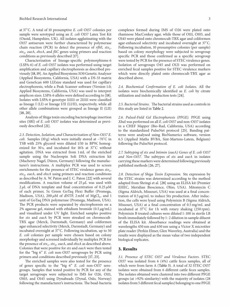
3.1. Presence of STEC O157 and Virulence Factors. STECO157 was isolated from 6 (4%) cattle feces samples, all ofwhich were from farm A (Table 3). A total of 32 STEC O157isolates were obtained from 6 different cattle feces samples.The isolates obtained were clustered into two different PFGEgroups (at >92% similarity) with the majority of isolates (28isolates from5different fecal samples) belonging to onePFGE
4 BioMed Research International
Table 2: Bacterial strains used in the study.
Strain ID Serogroup Source Country Virulence traitsSakai O157 Radish sprouts Japan 𝑠𝑡𝑥
1, 𝑠𝑡𝑥2, eaeA, ehxA
ATCC 43895 O157 Ground beef USA 𝑠𝑡𝑥1, 𝑠𝑡𝑥2, eaeA, ehxA
EC543a O157 Cattle feces Australia 𝑠𝑡𝑥1, 𝑠𝑡𝑥2, eaeA, ehxA
EC6a O157 Cattle feces Australia 𝑠𝑡𝑥2, eaeA, ehxA
1 UPMa O157 Bovine milk Malaysia 𝑠𝑡𝑥1, 𝑠𝑡𝑥2, eaeA, ehxA
2 UPMa O157 Bovine milk Malaysia 𝑠𝑡𝑥1, 𝑠𝑡𝑥2, eaeA, ehxA
3 UPMa O157 Beef Malaysia 𝑠𝑡𝑥1, 𝑠𝑡𝑥2, eaeA, ehxA
4 UPMa O157 Beef Malaysia 𝑠𝑡𝑥1, 𝑠𝑡𝑥2, eaeA, ehxA
MG1655 (E. coli K-12) OR:H48:K-b Laboratory strain USA NoneEC3008ac O26 Cattle feces Australia eaeAEC3009ac O45 Cattle feces Australia NoneEC2998ac O103 Cattle feces Australia NoneEC3113ac O111 Cattle feces Australia NoneEC3111ac O121 Cattle feces Australia NoneaProvided by Professor Son Radu at Universiti Putra Malaysia.bOR = O antigen rough strain which does not produce a typeable O antigen.c𝐸. coli non-O157 strains used as controls in the study, provided by Lesley Duffy at CSIRO, Brisbane, Australia.
Table 3: STEC O157 and non-O157 and their virulence profiles.
STEC serogroup Number of STEC+samples (%) Source Number of
isolatesVirulencefactors Lineage SBI profile
O157:H7a6 (4%) Cattle feces 28b 𝑠𝑡𝑥2c, eaeA-𝛾1,
ehxA II SY2c
4b 𝑠𝑡𝑥2c, eaeA-𝛾1ehxA II SY2c
Non-O157a(unknown) 2 (1.5%) Cattle feces 1 𝑠𝑡𝑥1a, 𝑠𝑡𝑥2a,
𝑠𝑡𝑥2c, ehxA— —
1 𝑠𝑡𝑥1
— —aIsolates of STEC O157:H7 and non-O157 were only present in samples obtained from farm A. All samples from farms B–F were negative for STEC O157:H7and non-O157 isolates.bOn farm A, 28 of the STEC O157 isolates belonged to one PFGE group (at >92% similarity) and the remaining 4 isolates belonged to another PFGE group.—: not applicable.
group and the remaining isolates (4 isolates froma single fecalsample) belonging to the other. All 32 STEC O157 isolateswere positive for the virulence factors stx
2, eaeA, and ehxA
and also for fliC specific for the H7 antigen indicating theybelong to the O157:H7 genotype. All samples from farms B–Fwere negative for the presence STEC O157.
LSPA-6 target amplification indicated that all the STECO157:H7 isolates collected from cattle feces samples in farmA belong to lineage II (Table 3). According to the SBIgenotyping code, genotype SY2c was observed in all STECO157:H7 isolates collected from cattle feces samples in farmA indicating the association of 𝑠𝑡𝑥
2c with prophage insertionin the sbcB locus (Table 3).
In addition, all STEC O157:H7 isolates obtained fromUPM carried the virulencemarkers stx
1, stx2, eaeA, and ehxA
and belonged to a single PFGE group (at >92% similarity).They were of lineage I and contained the SBI genotypeWY12indicating the association of stx
1and 𝑠𝑡𝑥
2a with prophageinsertion in the yehV and wrbA loci, respectively.
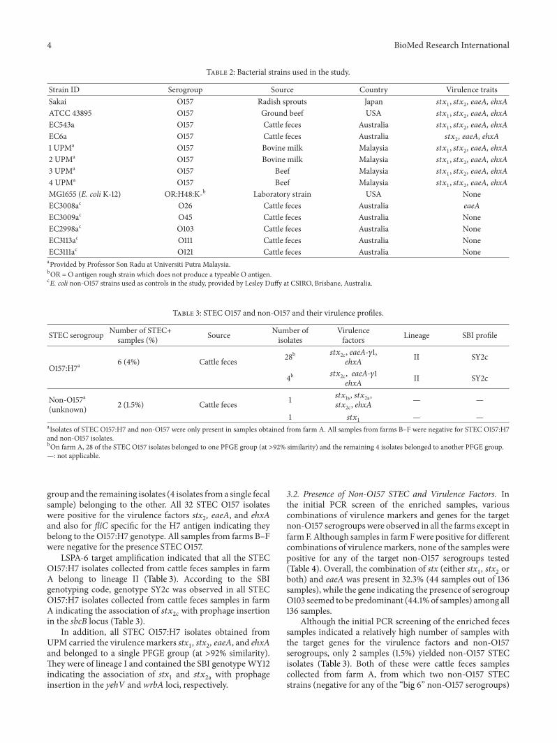
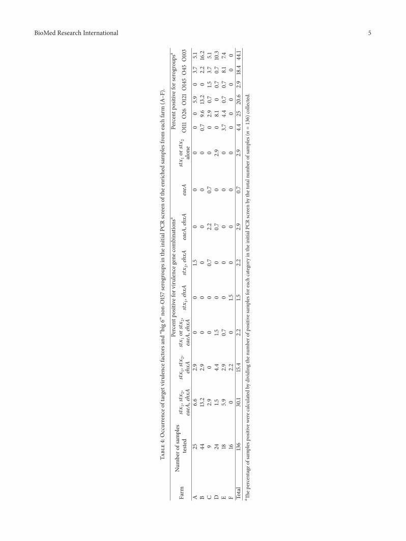
3.2. Presence of Non-O157 STEC and Virulence Factors. Inthe initial PCR screen of the enriched samples, variouscombinations of virulence markers and genes for the targetnon-O157 serogroups were observed in all the farms except infarm F. Although samples in farm Fwere positive for differentcombinations of virulence markers, none of the samples werepositive for any of the target non-O157 serogroups tested(Table 4). Overall, the combination of stx (either stx
1, stx2or
both) and eaeA was present in 32.3% (44 samples out of 136samples), while the gene indicating the presence of serogroupO103 seemed to be predominant (44.1%of samples) among all136 samples.
Although the initial PCR screening of the enriched fecessamples indicated a relatively high number of samples withthe target genes for the virulence factors and non-O157serogroups, only 2 samples (1.5%) yielded non-O157 STECisolates (Table 3). Both of these were cattle feces samplescollected from farm A, from which two non-O157 STECstrains (negative for any of the “big 6” non-O157 serogroups)
BioMed Research International 5
Table4:Occurrenceo
ftargetviru
lencefactorsand“big6”
non-O157serogrou
psin
theinitia
lPCR
screen
ofthee
nrichedsamples
from
each
farm
(A–F
).
Farm
Num
bero
fsam
ples
teste
d
Percentp
ositive
forv
irulenceg
enec
ombinatio
nsa
Percentp
ositive
forserogroup
sa𝑠𝑡𝑥1,𝑠𝑡𝑥2,
eaeA
,ehxA𝑠𝑡𝑥1,𝑠𝑡𝑥2,
ehxA
𝑠𝑡𝑥1or𝑠𝑡𝑥2,
eaeA
,ehxA𝑠𝑡𝑥1,ehxA𝑠𝑡𝑥2,ehxA
eaeA
,ehxA
eaeA
𝑠𝑡𝑥1or𝑠𝑡𝑥2
alon
eO111O26
O121O145O45
O103
A25
6.6
2.9
00
1.50
00
00
5.9
03.7
5.1
B44
13.2
2.9
00
00
00
0.7
9.613.2
02.2
16.2
C9
2.9
00
00.7
2.2
0.7
00
2.9
0.7
1.53.7
5.1
D24
1.54.4
1.50
00.7
02.9
08.1
00.7
0.7
10.3
E18
5.9
2.9
0.7
00
00
03.7
4.4
0.7
0.7
8.1
7.4F
160
2.2
01.5
00
00
00
00
00
Total
136
30.1
15.4
2.2
1.52.2
2.9
0.7
2.9
4.4
2520.6
2.9
18.4
44.1
a Thep
ercentageo
fsam
ples
positivew
erec
alculated
bydividing
then
umbero
fpositive
samples
fore
achcategory
intheinitia
lPCR
screen
bythetotalnu
mbero
fsam
ples
(𝑛=136)collected.
6 BioMed Research International
Table 5: Isolation and virulence profiles of E. coli O157 and “big 6” E. coli non-O157 serogroups lacking stx.
Serogroup Farm Source Number of +samples
Number ofisolates tested Virulence factors Intimin
subtypeO103 A Cattle feces 3 3 None —
O157 BCattle feces 1 2 eaeA NTa
Sheep feces 2 3 eaeA (1 isolate)none (2 isolates) NT
Buffalo feces 3 5 eaeA NT
O26 B Buffalo feces 2 5eaeA, ehxA (2
isolates) none (3isolates)
eaeA-𝛽1
O103 B Buffalo feces 1 7 None —O26 C Cattle feces 2 11 None —aNT = non-typable.—: not applicable.
were isolated which belonged to two unique PFGE groups.One of the two non-O157 STEC isolates was positive for stx
1,
stx2, and ehxA while the other isolate was positive for stx
1
alone.
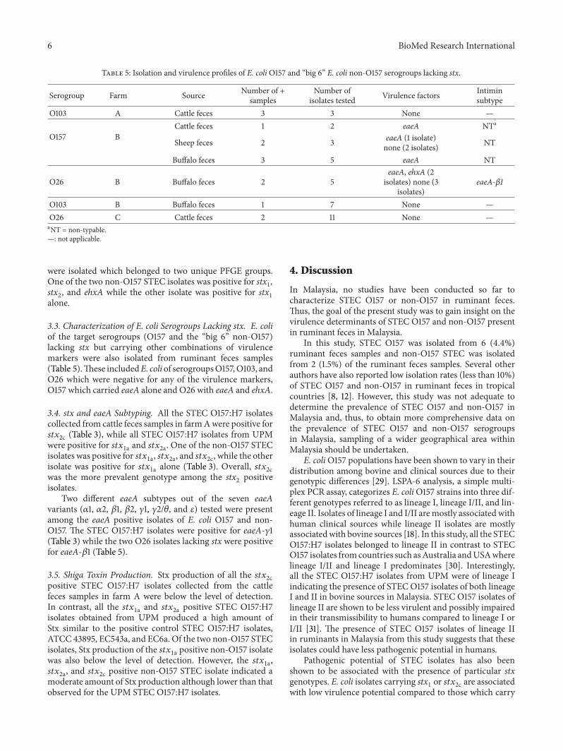
3.3. Characterization of E. coli Serogroups Lacking stx. E. coliof the target serogroups (O157 and the “big 6” non-O157)lacking stx but carrying other combinations of virulencemarkers were also isolated from ruminant feces samples(Table 5).These includedE. coli of serogroupsO157,O103, andO26 which were negative for any of the virulence markers,O157 which carried eaeA alone and O26 with eaeA and ehxA.
3.4. stx and eaeA Subtyping. All the STEC O157:H7 isolatescollected from cattle feces samples in farmAwere positive for𝑠𝑡𝑥2c (Table 3), while all STEC O157:H7 isolates from UPM
were positive for 𝑠𝑡𝑥1a and 𝑠𝑡𝑥2a. One of the non-O157 STEC
isolates was positive for 𝑠𝑡𝑥1a, 𝑠𝑡𝑥2a, and 𝑠𝑡𝑥2c, while the other
isolate was positive for 𝑠𝑡𝑥1a alone (Table 3). Overall, 𝑠𝑡𝑥
2cwas the more prevalent genotype among the stx
2positive
isolates.Two different eaeA subtypes out of the seven eaeA
variants (𝛼1, 𝛼2, 𝛽1, 𝛽2, 𝛾1, 𝛾2/𝜃, and 𝜀) tested were presentamong the eaeA positive isolates of E. coli O157 and non-O157. The STEC O157:H7 isolates were positive for eaeA-𝛾1(Table 3) while the two O26 isolates lacking stx were positivefor eaeA-𝛽1 (Table 5).
3.5. Shiga Toxin Production. Stx production of all the 𝑠𝑡𝑥2c
positive STEC O157:H7 isolates collected from the cattlefeces samples in farm A were below the level of detection.In contrast, all the 𝑠𝑡𝑥
1a and 𝑠𝑡𝑥2a positive STEC O157:H7
isolates obtained from UPM produced a high amount ofStx similar to the positive control STEC O157:H7 isolates,ATCC 43895, EC543a, and EC6a. Of the two non-O157 STECisolates, Stx production of the 𝑠𝑡𝑥
1a positive non-O157 isolatewas also below the level of detection. However, the 𝑠𝑡𝑥
1a,𝑠𝑡𝑥2a, and 𝑠𝑡𝑥2c positive non-O157 STEC isolate indicated a
moderate amount of Stx production although lower than thatobserved for the UPM STEC O157:H7 isolates.
4. Discussion
In Malaysia, no studies have been conducted so far tocharacterize STEC O157 or non-O157 in ruminant feces.Thus, the goal of the present study was to gain insight on thevirulence determinants of STEC O157 and non-O157 presentin ruminant feces in Malaysia.
In this study, STEC O157 was isolated from 6 (4.4%)ruminant feces samples and non-O157 STEC was isolatedfrom 2 (1.5%) of the ruminant feces samples. Several otherauthors have also reported low isolation rates (less than 10%)of STEC O157 and non-O157 in ruminant feces in tropicalcountries [8, 12]. However, this study was not adequate todetermine the prevalence of STEC O157 and non-O157 inMalaysia and, thus, to obtain more comprehensive data onthe prevalence of STEC O157 and non-O157 serogroupsin Malaysia, sampling of a wider geographical area withinMalaysia should be undertaken.
E. coliO157 populations have been shown to vary in theirdistribution among bovine and clinical sources due to theirgenotypic differences [29]. LSPA-6 analysis, a simple multi-plex PCR assay, categorizes E. coliO157 strains into three dif-ferent genotypes referred to as lineage I, lineage I/II, and lin-eage II. Isolates of lineage I and I/II aremostly associated withhuman clinical sources while lineage II isolates are mostlyassociatedwith bovine sources [18]. In this study, all the STECO157:H7 isolates belonged to lineage II in contrast to STECO157 isolates fromcountries such asAustralia andUSAwherelineage I/II and lineage I predominates [30]. Interestingly,all the STEC O157:H7 isolates from UPM were of lineage Iindicating the presence of STECO157 isolates of both lineageI and II in bovine sources in Malaysia. STEC O157 isolates oflineage II are shown to be less virulent and possibly impairedin their transmissibility to humans compared to lineage I orI/II [31]. The presence of STEC O157 isolates of lineage IIin ruminants in Malaysia from this study suggests that theseisolates could have less pathogenic potential in humans.
Pathogenic potential of STEC isolates has also beenshown to be associated with the presence of particular stxgenotypes. E. coli isolates carrying stx
1or 𝑠𝑡𝑥
2c are associatedwith low virulence potential compared to those which carry
BioMed Research International 7
stx2(𝑠𝑡𝑥2a) [32]. In this study, all the STEC O157:H7 isolates
obtained carried 𝑠𝑡𝑥2c indicating low virulence potential in
humans compared to the STEC O157:H7 isolates from UPMwith 𝑠𝑡𝑥
1a and 𝑠𝑡𝑥2a. One of the two non-O157 STEC isolatesof unknown serogroupwith 𝑠𝑡𝑥
1a, 𝑠𝑡𝑥2a, and 𝑠𝑡𝑥2c indicated ahigh pathogenic potential compared to the other isolate with𝑠𝑡𝑥1a alone.Not all E. coli isolates carrying stx produce Stx [33]. This
was true for all 𝑠𝑡𝑥2c positive STEC O157:H7 isolates and one
of the non-O157 isolates positive for 𝑠𝑡𝑥1a obtained in this
study. In contrast, theUPMSTECO157 isolates produced Stx.Although the exact reasons for the discrepancy observed inStx production of stx positive E. coli isolates from this studyis not fully understood, previous studies have also identifiedE. coli isolates positive for stx but negative for Stx production[33, 34]. In fact, the study by Koitabashi et al. [34] suggestedthat stx
2positive E. coli O157 strains that produce little or no
Stx2 may be widely distributed in the Asian environment.Particular stx genotypes of STEC O157 have been shown
to be associated both with particular SBI genotypes and withtheir relative frequency of isolation from clinical and bovinesources [21]. Clinical isolates are generally characterizedby the carrying of stx
2and stx
2-associated bacteriophage
sequences adjacent to either wrbA or argW (SBI genotypes:WY12, AY2, ASY2, ASY22c), while bovine isolates are char-acterized by carrying of 𝑠𝑡𝑥
2c and 𝑠𝑡𝑥2c-associated bacte-
riophage sequences adjacent to sbcB (SBI genotypes: SY2c,SY12c, and ASY12c). In agreement with these observations,the STEC O157 isolates obtained from cattle feces from thisstudy carried 𝑠𝑡𝑥
2c with an occupied sbcB locus (SY2c).However, the STEC O157 from UPM which were collectedfrom bovine sources carried stx
2and an occupiedwrbA locus
indicating characteristics of clinical isolates.All the STECO157:H7 isolates in this study and the STEC
O157:H7 isolates from UPM carried eaeA-𝛾1 as reported foreaeA positive E. coli O157 in previous studies [27, 35]. Noneof the eaeA positive non-STECO157 could be subtyped usingthe primers for eaeA subtypes 𝛼1, 𝛼2, 𝛽1, 𝛽2, 𝛾1, 𝛾2/𝜃, and𝜀. It is possible that these isolates belonged to other intiminsubtypes such as 𝛿/𝜅, 𝜁, 𝜂, 𝜄, 𝜆, 𝜇, and ] which were not testedfor in this study. The two eaeA positive E. coli O26 isolatescarried eaeA-𝛽1 similar to several other E. coli O26 isolatespreviously associated with human STEC strains that causeHUS [27].
5. Conclusions
Despite the use of specific and sensitive methods of enrich-ment and IMS followed in this study to isolate STEC O157and non-O157, it appears that the presence of both STECO157and non-O157 in ruminant feces was low (4% and 1.5%, resp.).The 𝑠𝑡𝑥
2c carrying STEC O157:H7 isolates of lineage II fromthis study suggests that these bacteria potentially represent aless pathogenic clone of STECO157 inMalaysia.This togetherwith the presence of STEC O157 and non-O157 in a smallpercentage of ruminants in this study could contribute to thereasons for the lack of reported sporadic cases and outbreakscaused by STEC O157 in Malaysia. Similar to STEC O157, thelow percentage of non-O157 STEC isolates observed together
with their low pathogenic potential indicated by the lackof eaeA and moderate to no Stx production suggests a lowprobability of causing disease in humans.
Conflict of Interests
The authors declare that there is no conflict of interestsregarding the publication of this paper.
Acknowledgments
The authors thank Monash University, Malaysia, and Com-monwealth Scientific and Industrial Research Organization(CSIRO) Animal, Food and Health Science, Australia, forfunding this study. The authors also thank Lesley Duffyfor the provision of E. coli non-O157 isolates, Glen Mellor,Edward Fox, and Sean Moore for expert technical assistanceand Professor Son Radu of Universiti Putra Malaysia for theprovision of STEC O157 isolates.
References
[1] C. L. Gyles, “Shiga toxin-producing Escherichia coli: anoverview,” Journal of Animal Science, vol. 85, no. 13, pp. E45–E62, 2007.
[2] P. I. Tarr, C. A. Gordon, and W. L. Chandler, “Shiga-toxin-producing Escherichia coli and haemolytic uraemic syndrome,”The Lancet, vol. 365, no. 9464, pp. 1073–1086, 2005.
[3] J. P. Nataro and J. B. Kaper, “Diarrheagenic Escherichia coli,”Clinical Microbiology Reviews, vol. 11, no. 1, pp. 142–201, 1998.
[4] D. Law and J. Kelly, “Use of heme and hemoglobin byEscherichia coli O157 and other Shiga-like-toxin-producing E.coli serogroups,” Infection and Immunity, vol. 63, no. 2, pp. 700–702, 1995.
[5] J. M. Bosilevac and M. Koohmaraie, “Predicting the presenceof non-O157 shiga toxin-producing Escherichia coli in groundbeef by using molecular tests for shiga toxins, intimin, and Oserogroups,” Applied and Environmental Microbiology, vol. 78,no. 19, pp. 7152–7155, 2012.
[6] M. Bielaszewska, J. Janda, K. Blahova, L. Sramkova, J. Havlik,and V. Potuznik, “Verocytotoxin-producing Escherichia coli inchildren with hemolytic uremic syndrome and diarrhea inthe Czech Republic,” in Proceedings of the 2nd InternationalSymposium and Workshop on Verocytotoxin (Shiga-like Toxin)-Producing Escherichia Coli Infections, M. A. Kamali and A. G.Goglio, Eds., pp. 37–40, Bergamo, Italy, 1994.
[7] A. Caprioli and A. E. Tozzi, “Epidemiology of Shiga toxin-producing Escherichia coli infections in continental Europe,” inEscherichia coli O157:H7 andOther ShigaToxin-Producing E. coliStrains, J. B. Kaper andA.D.O’Brien, Eds., pp. 38–48, AmericanSociety for Microbiology, Washington, DC, USA, 1998.
[8] S. K. Manna, M. P. Brahmane, C. Manna, K. Batabyal, and R.Das, “Occurrence, virulence characteristics and antimicrobialresistance of Escherichia coli O157 in slaughtered cattle anddiarrhoeic calves in West Bengal, India,” Letters in AppliedMicrobiology, vol. 43, no. 4, pp. 405–409, 2006.
[9] K. Irino, M. A. M. F. Kato, T. M. I. Vaz et al., “Serotypes andvirulence markers of Shiga toxin-producing Escherichia coli(STEC) isolated from dairy cattle in Sao Paulo State, Brazil,”Veterinary Microbiology, vol. 105, no. 1, pp. 29–36, 2005.
8 BioMed Research International
[10] T. R. Callaway, R. C. Anderson, G. Tellez et al., “Prevalenceof Escherichia coli O157 in cattle and swine in central Mexico,”Journal of Food Protection, vol. 67, no. 10, pp. 2274–2276, 2004.
[11] A. Khan, S. Yamasaki, T. Sato et al., “Prevalence and geneticprofiling of virulence determinants of non-O157 Shiga toxin-producing Escherichia coli isolated from cattle, beef, andhumans, Calcutta, India,” Emerging Infectious Diseases, vol. 8,no. 1, pp. 54–62, 2002.
[12] L. Leomil, L. Aidar-Ugrinovich, B. E. C. Guth et al., “Frequencyof Shiga toxin-producing Escherichia coli (STEC) isolatesamong diarrheic and non-diarrheic calves in Brazil,”VeterinaryMicrobiology, vol. 97, no. 1-2, pp. 103–109, 2003.
[13] K. Apun, P. P. Chang, E. H. U. Sim, and V. Micky, “Clonaldiversity of Escherichia coli isolates from marketed beef in EastMalaysia,” World Journal of Microbiology & Biotechnology, vol.22, no. 7, pp. 661–667, 2006.
[14] S. Radu, S. A. Mutalib, G. Rusul et al., “Detection of Escherichiacoli O157:H7 in the beef marketed in Malaysia,” Applied andEnvironmental Microbiology, vol. 64, no. 3, pp. 1153–1156, 1998.
[15] P. Sukhumungoon, Y. Nakaguchi, N. Ingviya et al., “Investiga-tion of stx2+, eae+ Escherichia coli O157:H7 in beef importedfrom Malaysia to Thailand,” International Food Research Jour-nal, vol. 18, no. 1, pp. 381–386, 2011.
[16] R. Son, A. Ansary, G. Rusul, and M. I. A. Karim, “Isolationof verotoxin-producing Escherichia coli associated with diar-rhoea inMalaysia containing plasmids showing homology withbiotinylated Shiga-like toxin DNA gene probes,”World Journalof Microbiology and Biotechnology, vol. 12, no. 3, pp. 243–246,1996.
[17] J. Bai, X. Shi, and T. G. Nagaraja, “A multiplex PCR procedurefor the detection of six major virulence genes in Escherichia coliO157:H7,” Journal of Microbiological Methods, vol. 82, no. 1, pp.85–89, 2010.
[18] Z. Yang, J. Kovar, J. Kim et al., “Identification of commonsubpopulations of non-sorbitol-fermenting, 𝛽-glucuronidase-negative Escherichia coli O157:H7 from bovine productionenvironments and human clinical samples,” Applied and Envi-ronmental Microbiology, vol. 70, no. 11, pp. 6846–6854, 2004.
[19] J. Whitworth, Y. Zhang, J. Bono, E. Pleydell, N. French, and T.Besser, “Diverse genetic markers concordantly identify bovineorigin Escherichia coli O157 genotypes underrepresented inhuman disease,” Applied and Environmental Microbiology, vol.76, no. 1, pp. 361–365, 2010.
[20] Y. Zhang, C. Laing,M. Steele et al., “Genome evolution inmajorEscherichia coliO157:H7 lineages,”BMCGenomics, vol. 8, article121, 2007.
[21] S. Shringi, C. Schmidt, K. Katherine, K. A. Brayton, D. D.Hancock, and T. E. Besser, “Carriage of stx2a differentiatesclinical and bovine-biased strains of Escherichia coliO157,” PLoSONE, vol. 7, no. 12, Article ID e51572, 2012.
[22] A. W. Paton and J. C. Paton, “Detection and characterization ofshiga toxigenic Escherichia coli by using multiplex PCR assaysfor stx1, stx2, eaeA, enterohemorrhagic E. coli hlyA, rfb(O111),and rfb(O157),” Journal of Clinical Microbiology, vol. 36, no. 2,pp. 598–602, 1998.
[23] Z. Paddock, X. Shi, J. Bai, and T. G. Nagaraja, “Applicability of amultiplex PCR to detect O26, O45, O103, O111, O121, O145, andO157 serogroups of Escherichia coli in cattle feces,” VeterinaryMicrobiology, vol. 156, no. 3-4, pp. 381–388, 2012.
[24] J. Aslanzadeh, “Biochemical profile-based microbial identifica-tion systems,” in Advanced Techniques in Diagnostic Microbiol-ogy, Y. W. Tang and C. W. Stratton, Eds., pp. 84–116, Springer,New York, NY, USA, 2006.
[25] Environmental Science and Research Ltd, “Standard operatingprocedure for PulseNet PFGE of Escherichia coli O157:H7,Escherichia coli non-O157 (STEC), Salmonella serotypes,Shigella sonnei and Shigella flexneri,” 2013, http://www.pulsene-tinternational.org/assets/PulseNet/uploads/pfge/PNL05 Ec-Sal-ShigPFGEprotocol.pdf.
[26] F. Scheutz, L. D. Teel, L. Beutin et al., “Multicenter evaluation ofa sequence-based protocol for subtyping Shiga toxins and stan-dardizing Stx nomenclature,” Journal of Clinical Microbiology,vol. 50, no. 9, pp. 2951–2963, 2012.
[27] M. Blanco, J. E. Blanco, A. Mora et al., “Serotypes, virulencegenes, and intimin types of Shiga toxin (verotoxin)-producingEscherichia coli isolates from cattle in Spain and identificationof a new intimin variant gene (eae-xi),” Journal of ClinicalMicrobiology, vol. 42, no. 2, pp. 645–651, 2004.
[28] S. Shringi, A.Garcıa, K.K. Lahmers et al., “Differential virulenceof clinical and bovine-biased enterohemorrhagic Escherichiacoli O157:H7 genotypes in piglet and dutch belted rabbitmodels,” Infection and Immunity, vol. 80, no. 1, pp. 369–380,2012.
[29] J. Kim, J. Nietfeldt, J. Ju et al., “Ancestral divergence, genomediversification, and phylogeographic variation in subpopula-tions of sorbitol-negative, 𝛽-glucuronidase-negative enterohe-morrhagic Escherichia coli O157,” Journal of Bacteriology, vol.183, no. 23, pp. 6885–6897, 2001.
[30] G. E. Mellor, T. E. Besser, M. A. Davis et al., “Multilocus geno-type analysis ofEscherichia coliO157 Isolates fromAustralia andthe United States provides evidence of geographic divergence,”Applied and Environmental Microbiology, vol. 79, no. 16, pp.5050–5058, 2013.
[31] S. R. Leopold, V. Magrini, N. J. Holt et al., “A precisereconstruction of the emergence and constrained radiationsof Escherichia coli O157 portrayed by backbone concatenomicanalysis,” Proceedings of the National Academy of Sciences of theUnited States of America, vol. 106, no. 21, pp. 8713–8718, 2009.
[32] K. Kawano, M. Okada, T. Haga, K. Maeda, and Y. Goto, “Rela-tionship between pathogenicity for humans and stx genotype inShiga toxin-producingEscherichia coli serotypeO157,”EuropeanJournal of Clinical Microbiology and Infectious Diseases, vol. 27,no. 3, pp. 227–232, 2008.
[33] T. Koitabashi, V. Vuddhakul, S. Radu et al., “Genetic char-acterization of Escherichia coli O157:H7/- strains carrying the𝑠𝑡𝑥2gene but not producing shiga toxin 2,” Microbiology and
Immunology, vol. 50, no. 2, pp. 135–148, 2006.[34] T. Koitabashi, S. Cui, M. Kamruzzaman, and M. Nishibuchi,
“Isolation and characterization of the Shiga toxin gene (stx)-bearing Escherichia coli O157 and non-O157 from retail meatsin Shandong Province, China, and characterization of the O157-derived stx2 phages,” Journal of FoodProtection, vol. 71, no. 4, pp.706–713, 2008.
[35] V. Ramachandran, K. Brett,M.A.Hornitzky et al., “Distributionof intimin subtypes among Escherichia coli isolates from rumi-nant and human sources,” Journal of Clinical Microbiology, vol.41, no. 11, pp. 5022–5032, 2003.

Submit your manuscripts athttp://www.hindawi.com
Hindawi Publishing Corporationhttp://www.hindawi.com Volume 2014
Anatomy Research International
PeptidesInternational Journal of
Hindawi Publishing Corporationhttp://www.hindawi.com Volume 2014
Hindawi Publishing Corporation http://www.hindawi.com
International Journal of
Volume 2014
Zoology
Hindawi Publishing Corporationhttp://www.hindawi.com Volume 2014
Molecular Biology International
GenomicsInternational Journal of
Hindawi Publishing Corporationhttp://www.hindawi.com Volume 2014
The Scientific World JournalHindawi Publishing Corporation http://www.hindawi.com Volume 2014
Hindawi Publishing Corporationhttp://www.hindawi.com Volume 2014
BioinformaticsAdvances in
Marine BiologyJournal of
Hindawi Publishing Corporationhttp://www.hindawi.com Volume 2014
Hindawi Publishing Corporationhttp://www.hindawi.com Volume 2014
Signal TransductionJournal of
Hindawi Publishing Corporationhttp://www.hindawi.com Volume 2014
BioMed Research International
Evolutionary BiologyInternational Journal of
Hindawi Publishing Corporationhttp://www.hindawi.com Volume 2014
Hindawi Publishing Corporationhttp://www.hindawi.com Volume 2014
Biochemistry Research International
ArchaeaHindawi Publishing Corporationhttp://www.hindawi.com Volume 2014
Hindawi Publishing Corporationhttp://www.hindawi.com Volume 2014
Genetics Research International
Hindawi Publishing Corporationhttp://www.hindawi.com Volume 2014
Advances in
Virolog y
Hindawi Publishing Corporationhttp://www.hindawi.com
Nucleic AcidsJournal of
Volume 2014
Stem CellsInternational
Hindawi Publishing Corporationhttp://www.hindawi.com Volume 2014
Hindawi Publishing Corporationhttp://www.hindawi.com Volume 2014
Enzyme Research
Hindawi Publishing Corporationhttp://www.hindawi.com Volume 2014
International Journal of
Microbiology