Embed Size (px)
Citation preview
Research ArticleIn Silico Investigation of Potential Pyruvate Kinase M2Regulators from Traditional Chinese Medicine against Cancers
Kuan-Chung Chen,1 Kuen-Bao Chen,2,3,4 Hsin-Yi Chen,3 and Calvin Yu-Chian Chen2,3,5,6
1 School of Pharmacy, China Medical University, Taichung 40402, Taiwan2 School of Medicine, College of Medicine, China Medical University, Taichung 40402, Taiwan3Department of Biomedical Informatics, Asia University, Taichung 41354, Taiwan4Department of Anesthesiology, China Medical University Hospital, Taichung 40447, Taiwan5Human Genetic Center, Department of Medical Research, China Medical University Hospital, Taichung, Taiwan6Research Center for Chinese Medicine & Acupuncture, China Medical University, Taichung 40402, Taiwan
Correspondence should be addressed to Calvin Yu-Chian Chen; [email protected]
Received 22 February 2014; Revised 5 March 2014; Accepted 5 March 2014; Published 25 June 2014
Academic Editor: Chung Y. Hsu
Copyright © 2014 Kuan-Chung Chen et al. This is an open access article distributed under the Creative Commons AttributionLicense, which permits unrestricted use, distribution, and reproduction in any medium, provided the original work is properlycited.
A recent research in cancer research demonstrates that tumor-specific pyruvate kinase M2 (PKM2) plays an important role inchromosome segregation and mitosis progression of tumor cells. To improve the drug development of TCM compounds, we aimto identify potent TCM compounds as lead compounds of PKM2 regulators. PONDR-Fit protocol was utilized to predict thedisordered disposition in the binding domain of PKM2 protein before virtual screening as the disordered structure in the proteinmay cause the side effect and downregulation of the possibility of ligand to bind with target protein. MD simulation was performedto validate the stability of interactions between PKM2 proteins and each ligand after virtual screening. The top TCM compounds,saussureamine C and precatorine, extracted from Lycium chinenseMill. and Abrus precatorius L., respectively, have higher bindingaffinities with target protein in docking simulation than control. They have stable H-bonds with residues A:Lys311 and some otherresidues in both chains of PKM2 protein. Hence, we propose the TCM compounds, saussureamine C and precatorine, as potentialcandidates as lead compounds for further study in drug development process with the PKM2 protein against cancer.
1. Introduction
Recently, more and more pathogenesis of diseases has beenidentified [1, 2] to reveal potential target proteins for drugdesign [3–5]. A recent research in cancer research demon-strates that tumor-specific pyruvate kinase M2 (PKM2) playsan important role in chromosome segregation and mitosisprogression of tumor cells [6, 7]. PKM2 proteins can betreated as drug target proteins against cancers [8, 9].
The computer-aided drug design hadwildly been used forvirtual drug screening in the drug design [10, 11]. In previousstudy, many compounds from traditional Chinese medicine
(TCM) have been identified as potential lead compoundsin computer-aided drug design for the treatment of cancers[12–14], metabolic syndrome [15], diabetes [16], stroke [17,18], inflammation [19], and some other diseases [20]. Toimprove the drug development of TCM compounds, weemployed TCM compounds from TCM Database@Taiwan[21] to virtual screening of the potent lead compounds ofPKM2 regulators. As the disordered structure in the proteinmay cause the side effect and downregulation of the possi-bility of ligand to bind with target protein [22], PONDR-Fitprotocol was performed to predict the disordered dispositionin binding domain of PKM2 protein before virtual screening.
Hindawi Publishing CorporationBioMed Research InternationalVolume 2014, Article ID 189495, 14 pageshttp://dx.doi.org/10.1155/2014/189495
2 BioMed Research International
1.0
0.5
0.0
Diso
rder
disp
ositi
on
0 50 100 150 200 250 300 350 400 450 500 550
Residue index
P-Fit
Figure 1: Disordered disposition predicted by PONDR-Fit.
RMSD: 0.4873
CrystalRedock
Chain AChain BBinding site
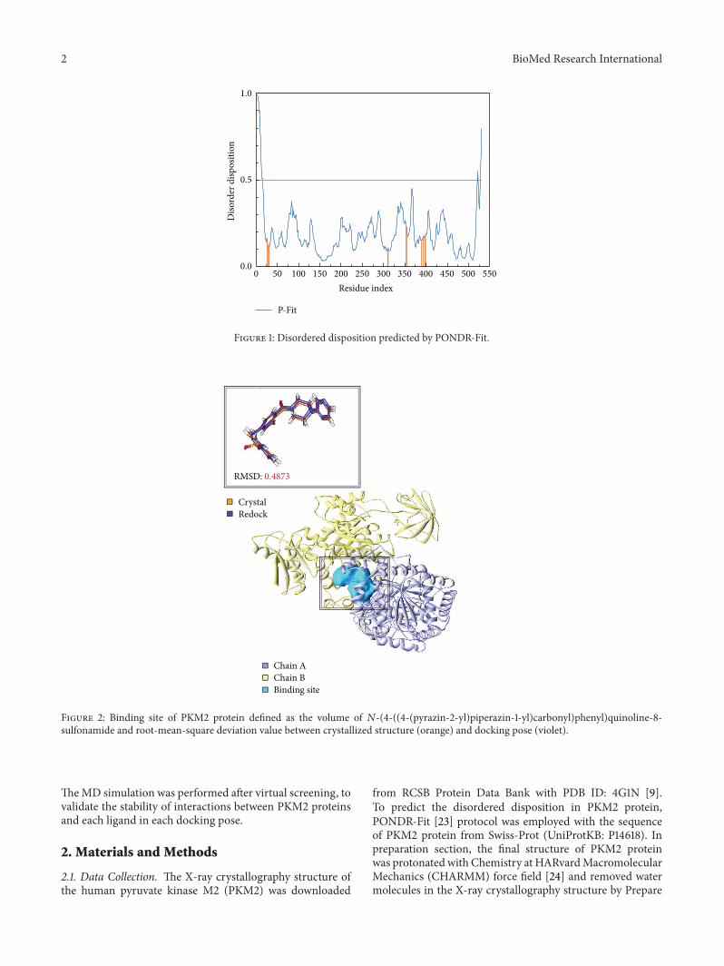
Figure 2: Binding site of PKM2 protein defined as the volume of 𝑁-(4-((4-(pyrazin-2-yl)piperazin-1-yl)carbonyl)phenyl)quinoline-8-sulfonamide and root-mean-square deviation value between crystallized structure (orange) and docking pose (violet).
TheMD simulation was performed after virtual screening, tovalidate the stability of interactions between PKM2 proteinsand each ligand in each docking pose.
2. Materials and Methods
2.1. Data Collection. The X-ray crystallography structure ofthe human pyruvate kinase M2 (PKM2) was downloaded
from RCSB Protein Data Bank with PDB ID: 4G1N [9].To predict the disordered disposition in PKM2 protein,PONDR-Fit [23] protocol was employed with the sequenceof PKM2 protein from Swiss-Prot (UniProtKB: P14618). Inpreparation section, the final structure of PKM2 proteinwas protonated with Chemistry at HARvardMacromolecularMechanics (CHARMM) force field [24] and removed watermolecules in the X-ray crystallography structure by Prepare
BioMed Research International 3
O
O
+H2N O
NH2
O
O−
(a)
N+
O
O
OH
OOH
O−
(b)
N SO
OHN
ONNN
N
(c)
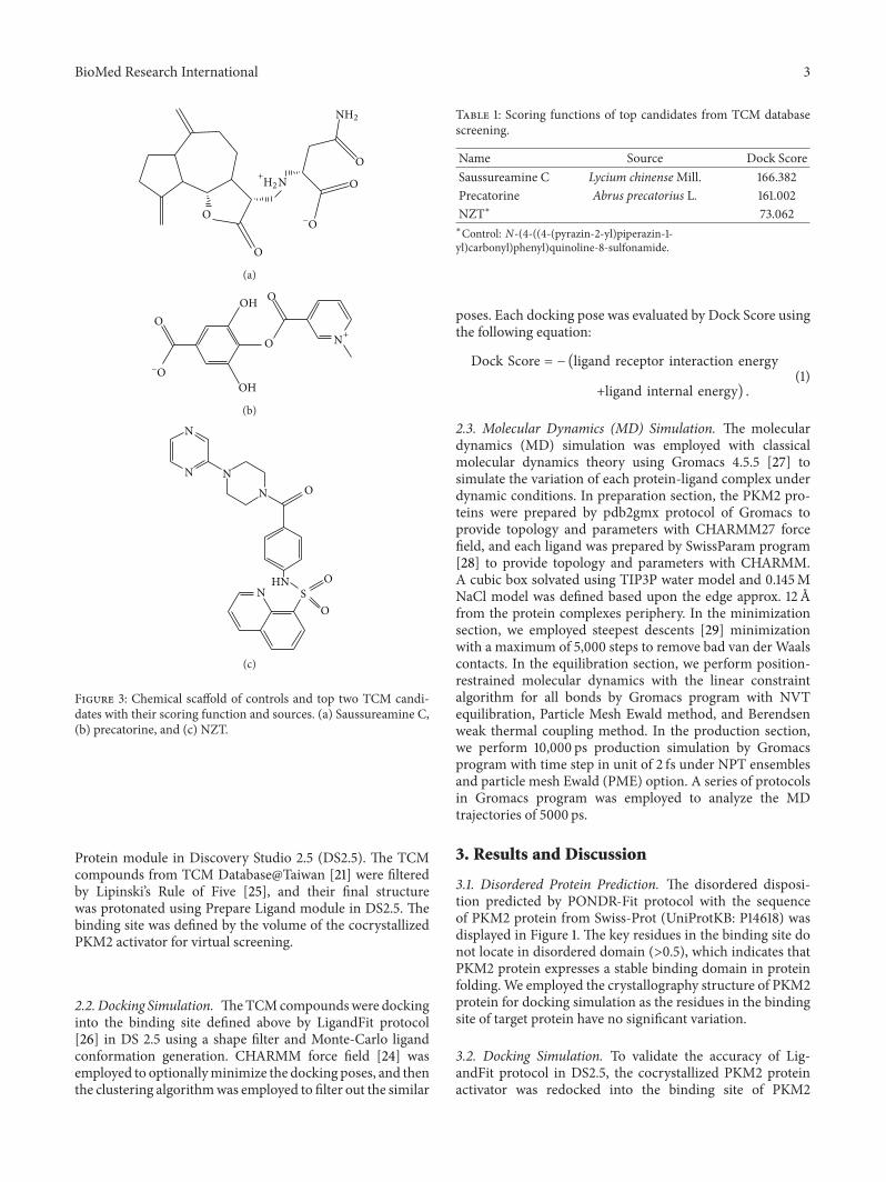
Figure 3: Chemical scaffold of controls and top two TCM candi-dates with their scoring function and sources. (a) Saussureamine C,(b) precatorine, and (c) NZT.
Protein module in Discovery Studio 2.5 (DS2.5). The TCMcompounds from TCM Database@Taiwan [21] were filteredby Lipinski’s Rule of Five [25], and their final structurewas protonated using Prepare Ligand module in DS2.5. Thebinding site was defined by the volume of the cocrystallizedPKM2 activator for virtual screening.
2.2. Docking Simulation. TheTCMcompoundswere dockinginto the binding site defined above by LigandFit protocol[26] in DS 2.5 using a shape filter and Monte-Carlo ligandconformation generation. CHARMM force field [24] wasemployed to optionallyminimize the docking poses, and thenthe clustering algorithmwas employed to filter out the similar
Table 1: Scoring functions of top candidates from TCM databasescreening.
Name Source Dock ScoreSaussureamine C Lycium chinenseMill. 166.382Precatorine Abrus precatorius L. 161.002NZT∗ 73.062∗Control: N-(4-((4-(pyrazin-2-yl)piperazin-1-yl)carbonyl)phenyl)quinoline-8-sulfonamide.
poses. Each docking pose was evaluated by Dock Score usingthe following equation:
Dock Score = − (ligand receptor interaction energy
+ligand internal energy) .(1)
2.3. Molecular Dynamics (MD) Simulation. The moleculardynamics (MD) simulation was employed with classicalmolecular dynamics theory using Gromacs 4.5.5 [27] tosimulate the variation of each protein-ligand complex underdynamic conditions. In preparation section, the PKM2 pro-teins were prepared by pdb2gmx protocol of Gromacs toprovide topology and parameters with CHARMM27 forcefield, and each ligand was prepared by SwissParam program[28] to provide topology and parameters with CHARMM.A cubic box solvated using TIP3P water model and 0.145MNaCl model was defined based upon the edge approx. 12 Afrom the protein complexes periphery. In the minimizationsection, we employed steepest descents [29] minimizationwith a maximum of 5,000 steps to remove bad van der Waalscontacts. In the equilibration section, we perform position-restrained molecular dynamics with the linear constraintalgorithm for all bonds by Gromacs program with NVTequilibration, Particle Mesh Ewald method, and Berendsenweak thermal coupling method. In the production section,we perform 10,000 ps production simulation by Gromacsprogram with time step in unit of 2 fs under NPT ensemblesand particle mesh Ewald (PME) option. A series of protocolsin Gromacs program was employed to analyze the MDtrajectories of 5000 ps.
3. Results and Discussion
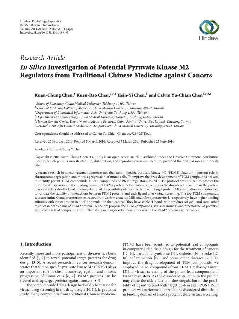
3.1. Disordered Protein Prediction. The disordered disposi-tion predicted by PONDR-Fit protocol with the sequenceof PKM2 protein from Swiss-Prot (UniProtKB: P14618) wasdisplayed in Figure 1. The key residues in the binding site donot locate in disordered domain (>0.5), which indicates thatPKM2 protein expresses a stable binding domain in proteinfolding. We employed the crystallography structure of PKM2protein for docking simulation as the residues in the bindingsite of target protein have no significant variation.
3.2. Docking Simulation. To validate the accuracy of Lig-andFit protocol in DS2.5, the cocrystallized PKM2 proteinactivator was redocked into the binding site of PKM2
4 BioMed Research International
A:ASN350
A:LEU353
A:ASP354
A:MET30
B:ASN350
B:LYS311
B:ASP354
B:GLY355
A:LEU27
B:ASN318
B:LEU394
B:GLU397
B:GLN393
B:TYR390
Pi
+
B:PHE26
A:LYS311 A:PHE
26
B:LEU353
(a)
B:ASP354
A:MET30
B:ASN350
A:ASP354
B:ILE389
B:ASN318
A:ASN350
B:LEU394
B:GLY355
B:GLN393
A:PHE26
B:LYS311
B:PHE26
B:LEU353
A:LYS311
(b)
Figure 4: Continued.
BioMed Research International 5
A:GLU397
A:GLY355
B:LEU27 A:ASP
354
A:GLY315 B:MET
30 B:LEU353
B:LEU394
B:HIS391
B:ILE389
B:ASP354
A:GLN393
A:TYR390
B:PHESigma
i
26A:LEU394
A:LEU353
B:TYR390
A:ALA388
B:GLN393
A:PHE26 A:ILE
389
A:MET30
B:LYS311
B:LEU353
B:LEU394
B:TYR390
(c)
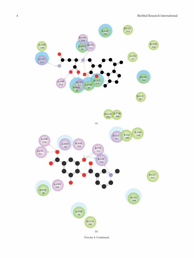
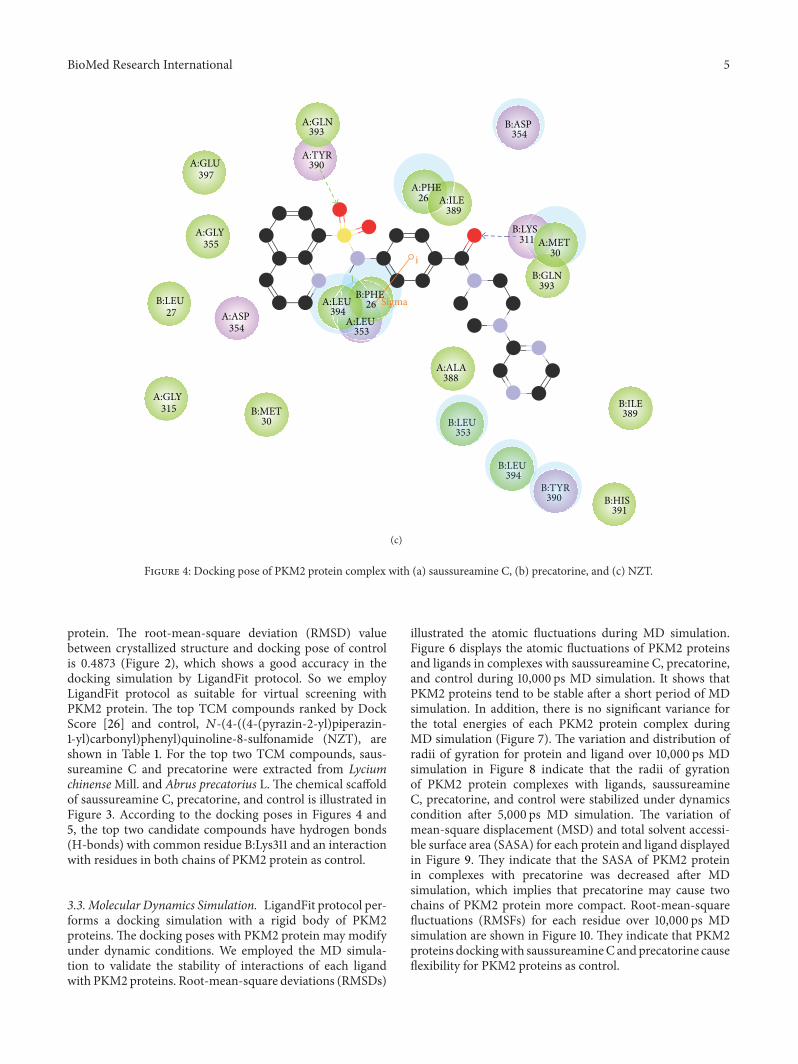
Figure 4: Docking pose of PKM2 protein complex with (a) saussureamine C, (b) precatorine, and (c) NZT.
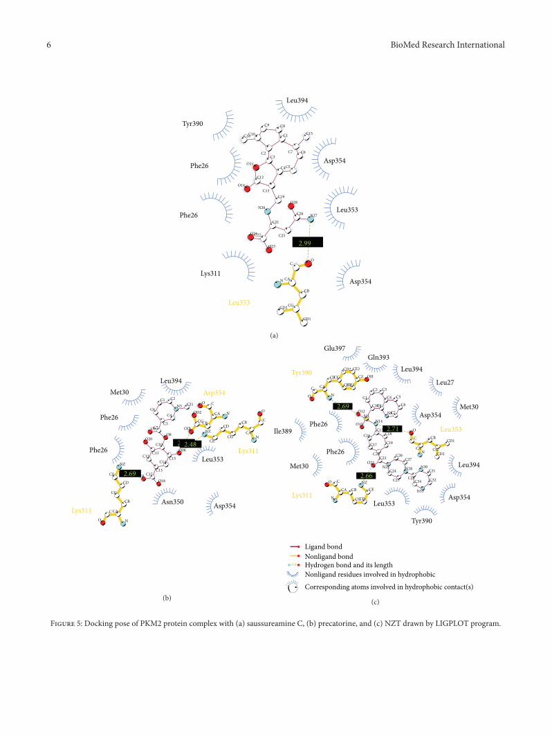
protein. The root-mean-square deviation (RMSD) valuebetween crystallized structure and docking pose of controlis 0.4873 (Figure 2), which shows a good accuracy in thedocking simulation by LigandFit protocol. So we employLigandFit protocol as suitable for virtual screening withPKM2 protein. The top TCM compounds ranked by DockScore [26] and control, 𝑁-(4-((4-(pyrazin-2-yl)piperazin-1-yl)carbonyl)phenyl)quinoline-8-sulfonamide (NZT), areshown in Table 1. For the top two TCM compounds, saus-sureamine C and precatorine were extracted from LyciumchinenseMill. and Abrus precatorius L. The chemical scaffoldof saussureamine C, precatorine, and control is illustrated inFigure 3. According to the docking poses in Figures 4 and5, the top two candidate compounds have hydrogen bonds(H-bonds) with common residue B:Lys311 and an interactionwith residues in both chains of PKM2 protein as control.
3.3. Molecular Dynamics Simulation. LigandFit protocol per-forms a docking simulation with a rigid body of PKM2proteins. The docking poses with PKM2 protein may modifyunder dynamic conditions. We employed the MD simula-tion to validate the stability of interactions of each ligandwith PKM2 proteins. Root-mean-square deviations (RMSDs)
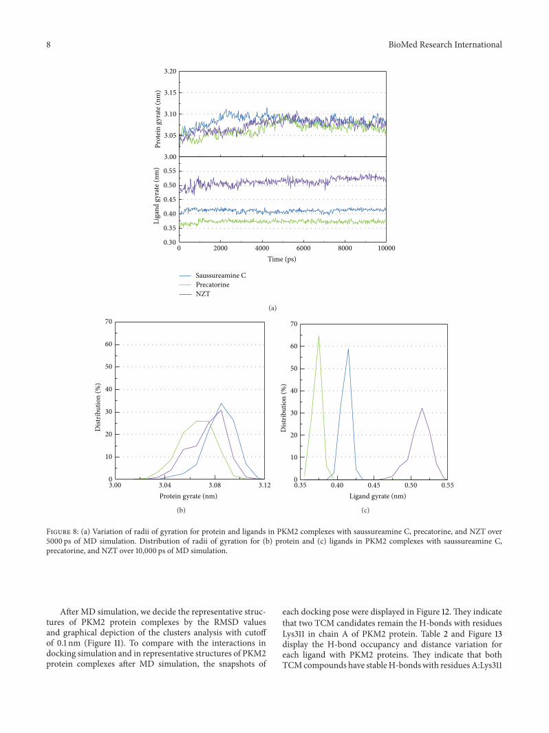
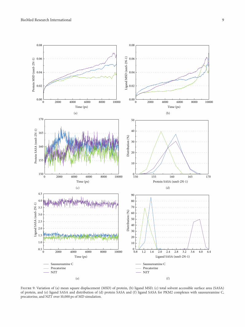
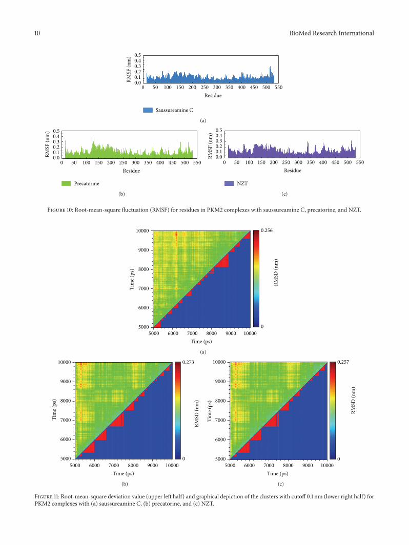
illustrated the atomic fluctuations during MD simulation.Figure 6 displays the atomic fluctuations of PKM2 proteinsand ligands in complexes with saussureamine C, precatorine,and control during 10,000 ps MD simulation. It shows thatPKM2 proteins tend to be stable after a short period of MDsimulation. In addition, there is no significant variance forthe total energies of each PKM2 protein complex duringMD simulation (Figure 7). The variation and distribution ofradii of gyration for protein and ligand over 10,000 ps MDsimulation in Figure 8 indicate that the radii of gyrationof PKM2 protein complexes with ligands, saussureamineC, precatorine, and control were stabilized under dynamicscondition after 5,000 ps MD simulation. The variation ofmean-square displacement (MSD) and total solvent accessi-ble surface area (SASA) for each protein and ligand displayedin Figure 9. They indicate that the SASA of PKM2 proteinin complexes with precatorine was decreased after MDsimulation, which implies that precatorine may cause twochains of PKM2 protein more compact. Root-mean-squarefluctuations (RMSFs) for each residue over 10,000 ps MDsimulation are shown in Figure 10. They indicate that PKM2proteins dockingwith saussureamineC andprecatorine causeflexibility for PKM2 proteins as control.
6 BioMed Research International
2.99
C1
C2C3
C4C5
C6C7
C8C9
C10
O11
C12
C13O14
C15C16
C19
N20
C21
C22
O23
O24 C25
C26 N27
O28
N CA
CB
CG
CD1
CD2
CO
Leu353
Phe26
Phe26Leu353
Asp354
Lys311
Leu394
Asp354
Tyr390
(a)
2.722.48
2.69
C1 C2
N3
C4
C5
C6
C7
O8O9
C10
C11C12
C13
C14C15
O16
C17O18
O19
O20
C21
NCA
CBCG
OD1
OD2
CO
Asp354
NCA
CB
CG
CD
CE
NZ
C
O
Lys311
Met30
N
CA
CB
CG
CD
CE
NZ
C
O
Lys311
Phe26
Phe26
Asn350
Leu353
Leu394
Asp354
(b)
2.66
2.69
2.71
C1
C2 C3
C4 C5
C6
C7N8
C9C10
S11O12
O13N14
C15C16
C17
C18
C19
C20C21
O22N23
C24
C25
C26C27
N28
C29
N30C31
C32
N33
C34
NCA
CB
CG
CD1
CD2
C
O Leu353
N
CA
CBCG
CD1CE1
CD2 CE2
CZ OH
C
O
Tyr390 Leu394
N
CA CB
CGCD
CE
NZCO
Lys311
Phe26
Ile389
Tyr390
Gln393
Phe26
Leu353
Met30
Asp354
Leu394Met30
Asp354
Glu397
Leu27
Ligand bondNonligand bondHydrogen bond and its length Nonligand residues involved in hydrophobic
Corresponding atoms involved in hydrophobic contact(s)
3.0
(c)
Figure 5: Docking pose of PKM2 protein complex with (a) saussureamine C, (b) precatorine, and (c) NZT drawn by LIGPLOT program.

BioMed Research International 7
0 2000 4000 6000 8000 10000
Time (ps)Li
gand
RMSD
_2Li
gand
RMSD
_1C
ompl
exRM
SD
0.50.6
0.41.30.20.10.0
0.3
0.2
0.1
0.00.3
0.2
0.1
0.0
Saussureamine CPrecatorineNZT
Figure 6: Root-mean-square deviations in units of nm for protein and ligand for PKM2protein complexes with saussureamineC, precatorine,and NZT. Ligand RMSD 1 and Ligand RMSD 2 are calculated with least squares fit by protein and ligand, respectively.
−2406000
−2408000
−2410000
−2412000
−2414000
−2416000
−2418000
−2420000
−2422000
−2424000
Tota
l ene
rgy
(KJ/m
ol)
0 2000 4000 6000 8000 10000
Time (ps)
Saussureamine CPrecatorineNZT
(a)
12
10
8
6
4
2
0−2410000 −2415000 −2420000 −2425000 −2430000
Dist
ribut
ion
(%)
Total energy (KJ/mol)
(b)
Figure 7: (a) Distribution and (b) variation of total energy for PKM2 protein complexes with saussureamine C, precatorine, and NZT over5000 ps of MD simulation.
8 BioMed Research International
0 2000 4000 6000 8000 10000
Time (ps)
3.20
3.15
3.10
3.05
3.00
0.55
0.50
0.45
0.40
0.35
0.30
Liga
nd g
yrat
e (nm
)Pr
otei
n gy
rate
(nm
)
Saussureamine CPrecatorineNZT
(a)
70
60
50
40
30
20
10
0
Dist
ribut
ion
(%)
3.00 3.04 3.08 3.12
Protein gyrate (nm)
(b)
0.35 0.40 0.45 0.50 0.55
Ligand gyrate (nm)
70
60
50
40
30
20
10
0
Dist
ribut
ion
(%)
(c)
Figure 8: (a) Variation of radii of gyration for protein and ligands in PKM2 complexes with saussureamine C, precatorine, and NZT over5000 ps of MD simulation. Distribution of radii of gyration for (b) protein and (c) ligands in PKM2 complexes with saussureamine C,precatorine, and NZT over 10,000 ps of MD simulation.
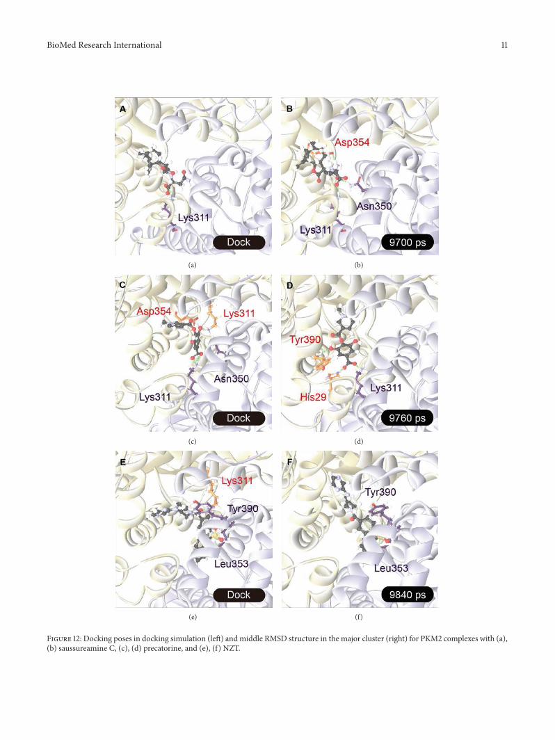
After MD simulation, we decide the representative struc-tures of PKM2 protein complexes by the RMSD valuesand graphical depiction of the clusters analysis with cutoffof 0.1 nm (Figure 11). To compare with the interactions indocking simulation and in representative structures of PKM2protein complexes after MD simulation, the snapshots of
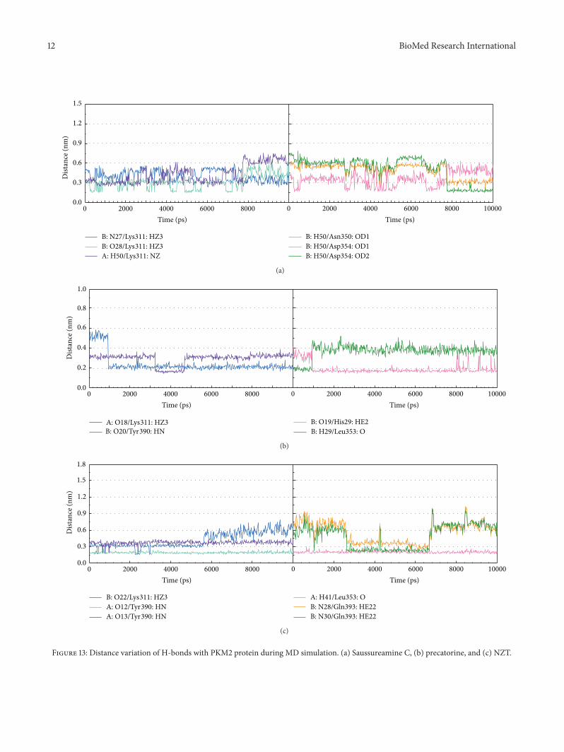
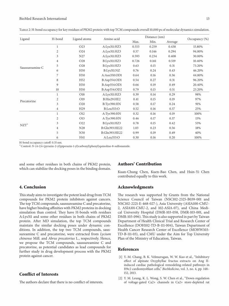
each docking pose were displayed in Figure 12. They indicatethat two TCM candidates remain the H-bonds with residuesLys311 in chain A of PKM2 protein. Table 2 and Figure 13display the H-bond occupancy and distance variation foreach ligand with PKM2 proteins. They indicate that bothTCMcompounds have stableH-bondswith residuesA:Lys311
BioMed Research International 9
0 2000 4000 6000 8000 10000
Time (ps)
0.08
0.06
0.04
0.02
0.00
Prot
ein
MSD
(nm
S-2
N-1
)
(a)
0 2000 4000 6000 8000 10000
Time (ps)
Liga
nd M
SD (n
mS-2
N-1
)
0.08
0.06
0.04
0.02
0.00
(b)
0 2000 4000 6000 8000 10000
Time (ps)
Prot
ein
SASA
(nm
S-2
N-1
)
170
165
160
155
150
(c)
Protein SASA (nmS-2N-1)170165160155150
50
40
30
20
10
0
Dist
ribut
ion
(%)
(d)
Saussureamine CPrecatorineNZT
0 2000 4000 6000 8000 10000
Time (ps)
Liga
nd S
ASA
(nm
S-2
N-1
)
4.5
4.0
3.5
3.0
2.5
2.0
1.5
1.0
0.5
(e)
Saussureamine CPrecatorineNZT
Ligand SASA (nmS-2N-1)0.8 1.2 1.6 2.0 2.4 2.8 3.2 3.6 4.0 4.4
20
10
40
30
60
50
80
90
70
0
Dist
ribut
ion
(%)
(f)
Figure 9: Variation of (a) mean square displacement (MSD) of protein, (b) ligand MSD, (c) total solvent accessible surface area (SASA)of protein, and (e) ligand SASA and distribution of (d) protein SASA and (f) ligand SASA for PKM2 complexes with saussureamine C,precatorine, and NZT over 10,000 ps of MD simulation.
10 BioMed Research International
0.5
0.4
0.3
0.2
0.1
0.0
RMSF
(nm
)
Saussureamine C
0 50 100 150 200 250 300 350 400 450 500 550
Residue
(a)
RMSF
(nm
)
Precatorine
0 50 100 150 200 250 300 350 400 450 500 550
Residue
0.4
0.5
0.3
0.2
0.1
0.0
(b)
0 50 100 150 200 250 300 350 400 450 500 550
Residue
0.4
0.5
0.3
0.2
0.1
0.0RMSF
(nm
)
NZT
(c)
Figure 10: Root-mean-square fluctuation (RMSF) for residues in PKM2 complexes with saussureamine C, precatorine, and NZT.
Time (ps)
Tim
e (ps
)
5000 6000 7000 8000 9000 100005000
6000
7000
8000
9000
10000 0.256
0
RMSD
(nm
)
(a)
Time (ps)
Tim
e (ps
)
5000 6000 7000 8000 9000 100005000
6000
7000
8000
9000
10000
0
0.273
RMSD
(nm
)
(b)
Time (ps)
Tim
e (ps
)
5000 6000 7000 8000 9000 100005000
6000
7000
8000
9000
10000 0.257
0
RMSD
(nm
)
(c)
Figure 11: Root-mean-square deviation value (upper left half) and graphical depiction of the clusters with cutoff 0.1 nm (lower right half) forPKM2 complexes with (a) saussureamine C, (b) precatorine, and (c) NZT.
BioMed Research International 11
(a) (b)
(c) (d)
(e) (f)
Figure 12: Docking poses in docking simulation (left) andmiddle RMSD structure in the major cluster (right) for PKM2 complexes with (a),(b) saussureamine C, (c), (d) precatorine, and (e), (f) NZT.
12 BioMed Research International
B: N27/Lys311: HZ3 B: H50/Asn350: OD1B: H50/Asp354: OD1B: H50/Asp354: OD2
B: O28/Lys311: HZ3A: H50/Lys311: NZ
Dist
ance
(nm
)
1.5
1.2
0.9
0.6
0.3
0.0
0 2000 4000 6000 8000 100000 2000 4000 6000 8000
Time (ps) Time (ps)
(a)
B: O19/His29: HE2B: H29/Leu353: O
A: O18/Lys311: HZ3B: O20/Tyr390: HN
Dist
ance
(nm
)
0.8
1.0
0.6
0.4
0.2
0.00 2000 4000 6000 8000 100000 2000 4000 6000 8000
Time (ps) Time (ps)
(b)
B: O22/Lys311: HZ3A: O12/Tyr390: HNA: O13/Tyr390: HN
A: H41/Leu353: OB: N28/Gln393: HE22B: N30/Gln393: HE22
Dist
ance
(nm
)
1.5
1.8
1.2
0.9
0.6
0.3
0.0
0 2000 4000 6000 8000 100000 2000 4000 6000 8000
Time (ps) Time (ps)
(c)
Figure 13: Distance variation of H-bonds with PKM2 protein during MD simulation. (a) Saussureamine C, (b) precatorine, and (c) NZT.
BioMed Research International 13
Table 2: H-bond occupancy for key residues of PKM2 protein with top TCMcompounds overall 10,000 ps ofmolecular dynamics simulation.
Ligand H-bond Ligand atoms Amino acid Distance (nm) Occupancy (%)Max. Min. Average
Saussureamine C
1 O23 A:Lys311:HZ3 0.553 0.259 0.438 15.80%2 O24 A:Lys311:HZ3 0.57 0.146 0.294 94.80%3 N27 A:Lys311:HZ3 0.593 0.234 0.408 30.00%4 O28 B:Lys311:HZ3 0.726 0.161 0.519 10.40%5 O28 B:Lys311:HZ3 0.63 0.15 0.31 73.20%6 H50 B:Lys311:NZ 0.76 0.24 0.43 46.20%7 H50 A:Asn350:OD1 0.64 0.16 0.36 44.80%8 H52 B:Asp354:OD1 0.54 0.27 0.31 96.20%9 H50 B:Asp354:OD1 0.66 0.19 0.49 20.40%10 H50 B:Asp354:OD2 0.79 0.15 0.51 23.20%
Precatorine
1 O18 A:Lys311:HZ3 0.39 0.14 0.29 98%2 O19 B:His29:HE2 0.41 0.15 0.19 97%3 O20 B:Tyr390:HN 0.58 0.17 0.24 91%4 H29 B:Leu353:O 0.52 0.16 0.37 25%
NZT∗
1 O12 A:Tyr390:HN 0.32 0.16 0.19 100%2 O13 A:Tyr390:HN 0.46 0.17 0.37 15%3 O22 B:Lys311:HZ3 0.78 0.15 0.42 53%4 N28 B:Gln393:HE22 1.03 0.23 0.56 18%5 N30 B:Gln393:HE22 0.99 0.19 0.49 40%6 H41 A:Leu353:O 0.30 0.16 0.20 100%
H-bond occupancy cutoff: 0.35 nm.∗Control: N-(4-((4-(pyrazin-2-yl)piperazin-1-yl)carbonyl)phenyl)quinoline-8-sulfonamide.
and some other residues in both chains of PKM2 protein,which can stabilize the docking poses in the binding domain.
4. Conclusion
This study aims to investigate the potent lead drug fromTCMcompounds for PKM2 protein inhibitors against cancers.The top TCM compounds, saussureamine C and precatorine,have higher binding affinities with PKM2 proteins in dockingsimulation than control. They have H-bonds with residuesA:Lys311 and some other residues in both chains of PKM2protein. After MD simulation, the top TCM compoundsmaintain the similar docking poses under dynamic con-ditions. In addition, the top two TCM compounds, saus-sureamine C and precatorine, were extracted from Lyciumchinense Mill. and Abrus precatorius L., respectively. Hence,we propose the TCM compounds, saussureamine C andprecatorine, as potential candidates as lead compounds forfurther study in drug development process with the PKM2protein against cancer.
Conflict of Interests
The authors declare that there is no conflict of interests.
Authors’ Contribution
Kuan-Chung Chen, Kuen-Bao Chen, and Hsin-Yi Chencontributed equally to this work.
Acknowledgments
The research was supported by Grants from the NationalScience Council of Taiwan (NSC102-2325-B039-001 andNSC102-2221-E-468-027-), Asia University (ASIA100-CMU-2, ASIA101-CMU-2, and 102-ASIA-07), and China Medi-cal University Hospital (DMR-103-058, DMR-103-001, andDMR-103-096).This study is also supported in part byTaiwanDepartment of Health Clinical Trial and Research Center ofExcellence (DOH102-TD-B-111-004), Taiwan Department ofHealth Cancer Research Center of Excellence (MOHW103-TD-B-111-03), and CMU under the Aim for Top UniversityPlan of the Ministry of Education, Taiwan.
References
[1] Y.-M. Chang, B. K. Velmurugan, W. W. Kuo et al., “Inhibitoryeffect of alpinate Oxyphyllae fructus extracts on Ang II-induced cardiac pathological remodeling-related pathways inH9c2 cardiomyoblast cells,” BioMedicine, vol. 3, no. 4, pp. 148–152, 2013.
[2] Y. M. Leung, K. L. Wong, S. W. Chen et al., “Down-regulationof voltage-gated Ca2+ channels in Ca2+ store-depleted rat
14 BioMed Research International
insulinoma RINm5F cells,” BioMedicine, vol. 3, no. 3, pp. 130–139, 2013.
[3] M. A. Leissring, E. Malito, S. Hedouin et al., “Designedinhibitors of insulin-degrading enzyme regulate the catabolismand activity of insulin,” PLoS ONE, vol. 5, no. 5, Article IDe10504, 2010.
[4] M.-C. Lin, S.-Y. Tsai, F.-Y. Wang et al., “Leptin induces cellinvasion and the upregulation of matrilysin in human coloncancer cells,” BioMedicine, vol. 3, no. 4, pp. 174–180, 2013.
[5] V. Janssens and J. Goris, “Protein phosphatase 2A: a highlyregulated family of serine/threonine phosphatases implicated incell growth and signalling,” Biochemical Journal, vol. 353, no. 3,pp. 417–439, 2001.
[6] Y. Jiang, X. Li, W. Yang et al., “PKM2 regulates chromosomesegregation and mitosis progression of tumor cells,” MolecularCell, vol. 53, no. 1, pp. 75–87, 2014.
[7] H. R. Christofk, M. G. Vander Heiden, M. H. Harris et al., “TheM2 splice isoform of pyruvate kinase is important for cancermetabolism and tumour growth,”Nature, vol. 452, no. 7184, pp.230–233, 2008.
[8] D. Anastasiou, Y. Yu, W. J. Israelsen et al., “Pyruvate kinase M2activators promote tetramer formation and suppress tumorige-nesis,”Nature Chemical Biology, vol. 8, no. 10, pp. 839–847, 2012.
[9] C. Kung, J. Hixon, S. Choe et al., “Small molecule activation ofpkm2 in cancer cells induces serine auxotrophy,” Chemistry andBiology, vol. 19, no. 9, pp. 1187–1198, 2012.
[10] C. Y.-C. Chen, “A novel integrated framework and improvedmethodology of computer-aided drug design,” Current Topicsin Medicinal Chemistry, vol. 13, no. 9, pp. 965–988, 2013.
[11] H. J. Huang, H.W. Yu, C. Y. Chen et al., “Current developmentsof computer-aided drug design,” Journal of the Taiwan Instituteof Chemical Engineers, vol. 41, no. 6, pp. 623–635, 2010.
[12] C. Y. Chen and C. Y. C. Chen, “Insights into designing thedual-targeted HER2/HSP90 inhibitors,” Journal of MolecularGraphics and Modelling, vol. 29, no. 1, pp. 21–31, 2010.
[13] S. C. Yang, S. S. Chang, H. Y. Chen, and C. Y. C. Chen,“Identification of potent EGFR inhibitors from TCMDatabase@Taiwan,” PLoS Computational Biology, vol. 7,no. 10, Article ID e1002189, 2011.
[14] Y. A. Tsou, K. C. Chen, H. C. Lin, S. S. Chang, and C. Y. C.Chen, “Uroporphyrinogen decarboxylase as a potential targetfor specific components of traditional chinese medicine: avirtual screening and Molecular Dynamics Study,” PLoS ONE,vol. 7, no. 11, Article ID e50087, 2012.
[15] K. C. Chen, S. S. Chang, H. J. Huang, T. L. Lin, Y. J. Wu,and C. Y. C. Chen, “Three-in-one agonists for PPAR-a, PPAR-𝛾, and PPAR-d from traditional Chinese medicine,” Journal ofBiomolecular Structure and Dynamics, vol. 30, no. 6, pp. 662–683, 2012.
[16] K. C. Chen, S. S. Chang, F. J. Tsai, and C. Y. Chen, “Hanethnicity-specific type 2 diabetic treatment from traditionalChinese medicine?” Journal of Biomolecular Structure &Dynamics, vol. 31, no. 11, pp. 1219–1235, 2013.
[17] K. C. Chen and C. Yu-Chian Chen, “Stroke prevention bytraditional Chinese medicine? A genetic algorithm, supportvectormachine andmolecular dynamics approach,” SoftMatter,vol. 7, no. 8, pp. 4001–4008, 2011.
[18] K. C. Chen, K. W. Chang, H. Y. Chen, and C. Y. C. Chen,“Traditional Chinese medicine, a solution for reducing dualstroke risk factors at once?” Molecular BioSystems, vol. 7, no. 9,pp. 2711–2719, 2011.
[19] K. C. Chen,M. F. Sun, S. C. Yang et al., “Investigation into potentinflammation inhibitors from traditional Chinese medicine,”Chemical Biology & Drug Design, vol. 78, no. 4, pp. 679–688,2011.
[20] W. I. Tou, S. S. Chang, C. C. Lee, and C. Y. C. Chen, “Drugdesign for neuropathic pain regulation from traditional Chinesemedicine,” Scientific reports, vol. 3, p. 844, 2013.
[21] C. Y. C. Chen, “TCM Database@Taiwan: the world's largesttraditional Chinese medicine database for drug screening inSilico,” PLoS ONE, vol. 6, no. 1, Article ID e15939, 2011.
[22] C. Y. C. Chen and W. I. Tou, “How to design a drug for thedisordered proteins?” Drug Discovery Today, vol. 18, no. 19-20,pp. 910–915, 2013.
[23] B. Xue, R. L. Dunbrack, R. W. Williams, A. K. Dunker, and V.N. Uversky, “PONDR-FIT: a meta-predictor of intrinsically dis-ordered amino acids,” Biochimica et Biophysica Acta—Proteinsand Proteomics, vol. 1804, no. 4, pp. 996–1010, 2010.
[24] B. R. Brooks, R. E. Bruccoleri, B. D.Olafson et al., “CHARMM: aprogram formacromolecular energyminimization and dynam-ics calculations,” Journal of Computational Chemistry, vol. 4, pp.187–217, 1983.
[25] C. A. Lipinski, F. Lombardo, B. W. Dominy, and P. J. Feeney,“Experimental and computational approaches to estimate sol-ubility and permeability in drug discovery and developmentsettings,” Advanced Drug Delivery Reviews, vol. 46, no. 1–3, pp.3–26, 2001.
[26] C. M. Venkatachalam, X. Jiang, T. Oldfield, and M. Wald-man, “LigandFit: a novel method for the shape-directed rapiddocking of ligands to protein active sites,” Journal of MolecularGraphics and Modelling, vol. 21, no. 4, pp. 289–307, 2003.
[27] B. Hess, C. Kutzner, D. van der Spoel, and E. Lindahl,“GRGMACS 4: algorithms for highly efficient, load-balanced,and scalable molecular simulation,” Journal of Chemical Theoryand Computation, vol. 4, no. 3, pp. 435–447, 2008.
[28] V. Zoete, M. A. Cuendet, A. Grosdidier, and O. Michielin,“SwissParam: a fast force field generation tool for small organicmolecules,” Journal of Computational Chemistry, vol. 32, no. 11,pp. 2359–2368, 2011.
[29] R. Fletcher,Optimization, Academic Press, NewYork, NY, USA,1969.

Submit your manuscripts athttp://www.hindawi.com
Hindawi Publishing Corporationhttp://www.hindawi.com Volume 2014
Anatomy Research International
PeptidesInternational Journal of
Hindawi Publishing Corporationhttp://www.hindawi.com Volume 2014
Hindawi Publishing Corporation http://www.hindawi.com
International Journal of
Volume 2014
Zoology
Hindawi Publishing Corporationhttp://www.hindawi.com Volume 2014
Molecular Biology International
GenomicsInternational Journal of
Hindawi Publishing Corporationhttp://www.hindawi.com Volume 2014
The Scientific World JournalHindawi Publishing Corporation http://www.hindawi.com Volume 2014
Hindawi Publishing Corporationhttp://www.hindawi.com Volume 2014
BioinformaticsAdvances in
Marine BiologyJournal of
Hindawi Publishing Corporationhttp://www.hindawi.com Volume 2014
Hindawi Publishing Corporationhttp://www.hindawi.com Volume 2014
Signal TransductionJournal of
Hindawi Publishing Corporationhttp://www.hindawi.com Volume 2014
BioMed Research International
Evolutionary BiologyInternational Journal of
Hindawi Publishing Corporationhttp://www.hindawi.com Volume 2014
Hindawi Publishing Corporationhttp://www.hindawi.com Volume 2014
Biochemistry Research International
ArchaeaHindawi Publishing Corporationhttp://www.hindawi.com Volume 2014
Hindawi Publishing Corporationhttp://www.hindawi.com Volume 2014
Genetics Research International
Hindawi Publishing Corporationhttp://www.hindawi.com Volume 2014
Advances in
Virolog y
Hindawi Publishing Corporationhttp://www.hindawi.com
Nucleic AcidsJournal of
Volume 2014
Stem CellsInternational
Hindawi Publishing Corporationhttp://www.hindawi.com Volume 2014
Hindawi Publishing Corporationhttp://www.hindawi.com Volume 2014
Enzyme Research
Hindawi Publishing Corporationhttp://www.hindawi.com Volume 2014
International Journal of
Microbiology