Embed Size (px)
Citation preview
719
Journal of Scientific and Innovative Research 2013; 2 (4): 719-735
Available online at: www.jsirjournal.com
Research Article
ISSN 2320-4818
JSIR 2013; 2(4): 719-735
© 2013, All rights reserved
Received: 24-08-2013
Accepted: 30-09-2013
Dr. Kalyani Bai Kunte* Department of Zoology, Sri
Venkateswara University, Tirupati,
Andhra Pradesh, India
Prof. Yellamma Kuna
Chairperson, Board of Studies
(BOS), Department of Zoology, Sri
Venkateswara University, Tirupati,
Andhra Pradesh, India
Correspondence: Dr. Kalyani Bai Kunte
Department of Zoology, Sri
Venkateswara University, Tirupati,
Andhra Pradesh, India- 517502
Tel: +91-9951294934
E-mail:
Neuroprotective effect of Bacopa monniera on memory
deficits and ATPase system in Alzheimer’s disease (AD)
induced mice
Kalyani Bai Kunte, Yellamma Kuna
Abstract
Theme of the present study is to evaluate neuroprotective properties of Bacopa monniera
(Brahmi) extract (BME) on memory deficits and biochemical changes in ATPases system of
AD induced mice. Mus musculus, of one month old weighing 20 ± 2 grams, were used as
experimental model and were maintained in Animal House according to the ethical guidelines
for animal protection and welfare, bearing the Resolution No. 02/(i)/a/CPCSEA/ IAEC/ SVU/
KY-KK/ Dt. 21-03-2011. The mice were divided in to four groups as follows: Group I: Control
mice; Group II: mice treated with BME; Group III (AD induced): mice treated with D-Gal and
NaNO2; Group IV: AD induced mice simultaneously treated with BME. Changes in
Morphometric and Behavioural aspects of four groups were analyzed by using Morris Water
Maze technique. Various constituents of ATPase system were determined in selected regions of
mice brain through standard biochemical assay methods. Results revealed that BME showed
positive effects on body weight, learning skills, memory and concentration, whereas D-Gal and
NaNO2 caused learning and memory deficits in mice which could be ameliorated by
simultaneous administration of BME. Similar, protective effects of BME were noticed on the
ATPase system which could revert all the constituents of ATPase system to normal levels in AD
induced mice. From these observations, it was concluded that BME had potential compounds
which can prevent the learning and memory deficits effectively; and to maintain ion gradients
across biological membranes, thus confer significant neuroprotection against AD by stabilizing
the structural and functional integrity of the membrane.
Keywords: Albino mice, Bacopa monniera (Brahmi), Alzheimer’s disease (AD),
Morphometric and Behavioral aspects, ATPase system
Introduction
Brain aging is a risk factor of neurodegenerative diseases such as Alzheimer’s disease
(AD), the most common cause of dementia which accounts 70% of dementia causes in
the most industrialized countries and is characterized by cell atrophy and extensive
neuronal loss. It is a complex and heterogeneous disorder particularly prevalent in
those over the 60 years of age. The incidence of AD rises from 2.8 per 1000 person
years in the 65-69 year age group to 56.1 per 1000 person years in the older age group
beyond 90 years.1 According to the World Health Organisation (WHO), it is estimated
that there are currently about 18 million people worldwide with Alzheimer’s disease.
This figure is expected to nearly double by 2025 to 34 million. Much of this increase
will be in the developing countries, and will be due to the ageing population.
Journal of Scientific and Innovative Research
720
In Alzheimer’s disease, there is a progressive degeneration
of basal forebrain cholinergic neurons innervating the
hippocampus and the cortex. Although other
neurotransmitters decline during Alzheimer’s-associated
neurodegeneration, the degree of brain Acetylcholine
(ACh) reduction directly correlates with deterioration of
cognition and of daily activity in AD patients.2 Since
deficits in cholinergic function contribute to the pathology
of Alzheimer’s disease, attempts to delay the progression
of the illness and improve patients’ daily activities are
based on pharmacological strategies to increase ACh levels
by means of anti-cholinesterasic agents.3 But, some
anticholinesterasic drugs have serious side effects on
patients because they not only act specifically on the
acetylcholinesterases, but also affect other ion channels
such as potassium channels.4 Disturbances in the ionic
equilibrium of the cells as a result of inactivation of
ATPases are believed to be the major factors in the
pathogenesis of various neurological disorders.5 Neuronal
membrane damage was evident from the decreased
activities of membrane bound enzymes such as Na+/K
+,
Mg2+
and Ca2+
-ATPases. Treatment strategies have been
investigated to cure AD, and the developed anti-
Alzheimer’s drugs showed positive results, but with
relevant side effects. Therefore it is worthwhile to choose
the application of alternative traditional medical system for
treatment of Alzheimer’s disease. Many natural herbal
medicines for treatments of Alzheimer's disease have been
touted to extend desirable and promising positive effects
beyond that of modern allopathy drugs.
Bacopa monniera (Brahmi) is a well known plant with
wide medicinal properties that is being used for treatment
of memory-related disorders.6 In Ayurveda, Bacopa
monniera has been classified under medicinal plants for
rejuvenating intellect and memory. The medicinal efficacy
of Bacopa monniera is extensively reported in Indian
Traditional literature such as Athar-Ved, Carak Samhita,
Susrutu Samhita7 for treatment of epilepsy, insomnia,
8
anxiety and as a mild sedative and memory enhancer.8
Besides, Bacopa monniera displays antistress9 and
anxiolytic10
activities too in animals. It has also been
shown to exert antioxidant effects through the chelating of
metal ions, breaking oxidative chain reaction8 improving
activities of antioxidative defense enzymes11
and
scavenging the free radicals.12
It also exhibits antistress
activity in rats, repairing the damaged neurons by
enhanced kinase activity, neuronal synthesis coupled with
restoration of synaptic activity and nerve impulse
transmission.7 In view of the above mentioned multiple
beneficial qualities of bacopa, an attempt has been made in
the present study to explore the protective effects of
Bacopa monniera extract on membrane bound enzymes
viz. Na+/K
+, Mg
2+ and Ca
2+ -ATPases in the brain of
normal and AD induces mice with particular reference to
Morphometric and Behavioural aspects.
Materials and Methods
Chemicals: All chemicals used in the present study were
Analar Grade (AR) and were obtained from Sigma (St.
Louis, MO, USA), Fisher (Pittsburg, PA, USA), Merck
(Mumbai, India), Ranbaxy (New Delhi, India), Qualigens
(Mumbai, India) Scientific Companies. For the present
investigation, Barnstead Thermoline water purification
plant was used for Nano pure water; Hahnvapor Rotary
Evaporator HS-2005V for plant exraction; KR 2000T
centrifuge for centrifugation of homogenates; Hitachi UV-
2800 spectrophotometer, RF 1501 Schimadzu Fluorimeter
and other standard equipments for
biochemical/physiological analyses.
Maintenance of Animals
Male albino mice, Mus musculus, of one month old
weighing 20 ± 2 grams, obtained from Sri Venkateswara
enterprises, Bangalore was selected as the experimental
model. The mice were maintained in the laboratory
conditions according to the instructions of Behringer ,1973
and as per the approval of the Institutional Animal Ethical
Committee (Resolution No. 02/(i)/a/CPCSEA/ IAEC/
SVU/ KY-KK/ Dt. 21-03-2011).
Collection and preparation of Bacopa monniera extract
Bacopa monniera plant was collected from Talacona forest
area which is around 50 Km from Tirupati. The whole
plant was dried in shade, powdered and used for extraction
by using methanol as solvent. Powdered plant material was
soaked in 95% methanol for 2 days at room temperature
and the solvent was filtered. This was repeated 3 to 4 times
until the extract gave no colouration. The extract was
distilled and concentrated under reduced pressure in the
Hahnvapor Rotary Evaporator HS-2005V. The resulting
methanol crude extract was air-dried and used in the
present study.
Induction of Alzheimer’s disease in mice
Until now, a combination of the chemicals, D-Galactose
and Sodium nitrite together was considered to be quite
successful in inducing Alzheimer’s disease in mice.13,14
Hence, in the present study, AD in mice was induced by an
intraperitoneal (i.p.) injection of D-Galactose (120mg/kg
Journal of Scientific and Innovative Research
721
body weight) and sodium nitrite (90mg/kg body weight) by
dissolving in distilled water.
Experiment protocol
Grouping of animals: After the mice were acclimated to
the laboratory conditions for 10 days, they were randomly
divided in to four main groups. Each main group was again
divided in to 12 sub groups of six each and was housed in
separate cages. All the animals in each Group were
administered with the following compounds as given
below. All doses were given once in a day in the morning
hours between 8 to 9 AM, keeping in view the altered
activity of mice during the nights.
Table 1: Grouping of animals
Group I Control (C)
Group II Mice treated with BME (100 mg/kg body weight
for 180 days)
Group
II(AD
induced):
Mice treated with D-Galactose ( 120 mg/kg body
weight ) + Sodium nitrite (90 mg/kg body weight
) for 60 days
Group IV AD induced mice simultaneously treated with
BME from 10th
day up to 180th
days, at Doses
mentioned above.
Isolation of tissues: Mice in all groups were sacrificed by
cervical dislocation at the selected time periods viz., 15th,
30th, 45th, 60th, 75th, 90th, 105th, 120th, 135th, 150th,
165th and 180th day. Selected regions of mice brain such
as Olfactory Lobe(OL), Cerebral Cortex(CC),
Hippocampus(Hc), Cerebellum(Cb), Ponsmedulla(Pm) and
Spinal cord(Spc) were isolated and immediately
homogenized in suitable media for assay of Cholinergic
constituents.
Parameters studied
Morphometric aspects: The basic Morphometric aspects
such as size and total body weight of control and
experimental groups have been recorded for every 15 days
up to 180th day. The data thus obtained was analyzed and
used to correlate with the behavioural aspects and the
ATPase system.
Behavioural aspects: Morris Water Maze test: Learning
and memory abilities were determined through Morris
water maze technique.15
A great deal of knowledge has
been obtained on the neurochemical, neuroanatomical and
neurophysiological basis for the behavior associated with
this paradigm. The apparatus consisted of a circular tank,
100 cm in diameter and 50 cm in depth. The tank was
filled with water (21-260C) up to a height of 30 cm and the
transparent escape platform made of plexiglass (10 cm in
diameter and 29 cm in height) was hidden at 1.5 cm below
the surface of water in a fixed location. The water was
made opaque with powdered non-fat milk or non-toxic
white colored dye. The platform was not visible from just
above the water level and transfer trials have indicated that
escape on to the platform was not achieved by visual or
other proximal cues.16
The time spent by the animal to
reach the hidden platform was used as the index of
memory. Before starting the experiment the mice were
acclimatize to the maze environment. The water maze test
was conducted for all groups of mice on selected days
viz., 15th , 30th , 45th, 60th, 75th, 90th, 105th, 120th,
135th, 150th, 165th and 180th for all six animals in a
group separately. For each trial, the time required (in
seconds) for individual mouse to find the hidden platform
was recorded and the mean data from the tests were used
for statistical analysis.
Biochemical analysis: Tissue samples from different
regions of control and AD induced mice were analyzed for
Na+/K
+ -ATPase and Mg
2+ -ATPase activity levels
according to the method of Tirri et al., 1973.17
Ca2+
-
ATPase was assayed according to the method of Fritz and
Hamrick (1966)18
as supported by Desaiah and Ho (1979)
and Inorganic phosphates was estimated by the method of
Fiske and Subba Row (1925).19
Statistical Analysis
Values of the measured parameters were expressed as
Mean ± SEM. Repeated Measures of ANOVA was used to
test the significance of difference among four different
groups followed by Dunnet’s Multiple Range Test
(DMRT). Statistical analysis was performed by using
Statistical Program of Social Sciences (SPSS) for windows
(Version 19; SPSS Inc., Chicago, 1L, USA). The results
were presented with the F-value and p-value. In all cases
F-value was found to be significant with p-value less than
0.01**. This indicates that the effects of factors are
significant.
Results
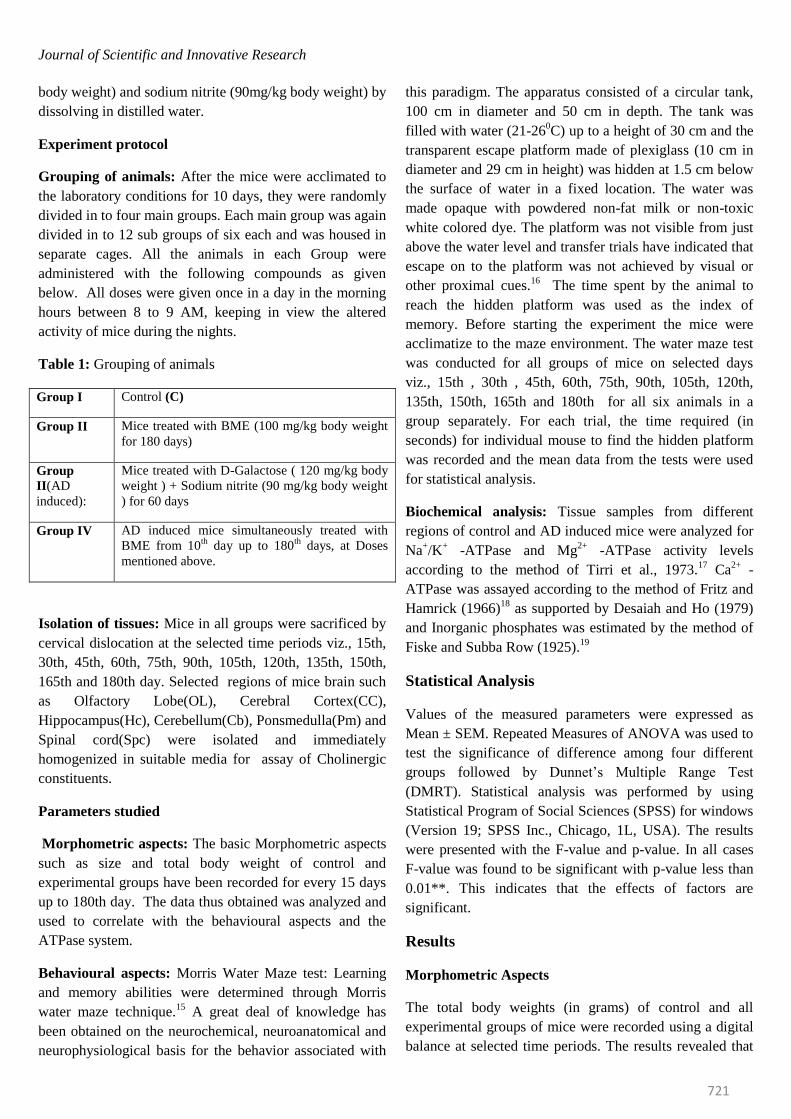
Morphometric Aspects
The total body weights (in grams) of control and all
experimental groups of mice were recorded using a digital
balance at selected time periods. The results revealed that
Journal of Scientific and Innovative Research
722
the control mice showed a gradual increase in their body
weights from 15th day (21 grams) to 180th day (43 grams).
When compared to the control ones, BME treated mice
gained more weight at all time periods from 15th day (23
grams) to 180th day (57.17 grams) whereas the D-
Galactose and NaNO2 treated mice gained less weight
throughout the period of experiment (18 grams to 31
grams). Observations on Group IV (D-Galactose and NaNO2,
simultaneously treated with BME) revealed that the body
weights were lesser than the control mice (19 grams).
From 165th day onwards the mice gained more weight (42
grams) against control ones indicating that BME could
effectively revert the AD induced changes gradually
(Table 1; Figure 1).
Figure 1: Graphical representation of differences in the body weights of Control and Experimental groups of mice treated
by BME, D-Galactose & NaNO2 and D-Galactose & NaNO2 + BME at selected time intervals.
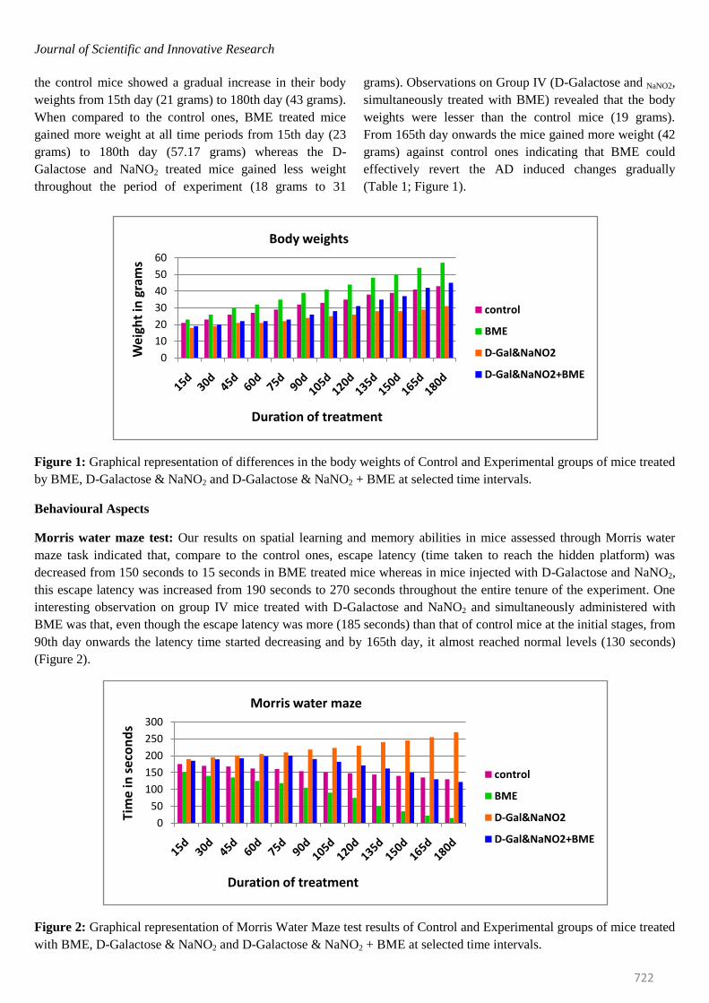
Behavioural Aspects
Morris water maze test: Our results on spatial learning and memory abilities in mice assessed through Morris water
maze task indicated that, compare to the control ones, escape latency (time taken to reach the hidden platform) was
decreased from 150 seconds to 15 seconds in BME treated mice whereas in mice injected with D-Galactose and NaNO2,
this escape latency was increased from 190 seconds to 270 seconds throughout the entire tenure of the experiment. One
interesting observation on group IV mice treated with D-Galactose and NaNO2 and simultaneously administered with
BME was that, even though the escape latency was more (185 seconds) than that of control mice at the initial stages, from
90th day onwards the latency time started decreasing and by 165th day, it almost reached normal levels (130 seconds)
(Figure 2).
Figure 2: Graphical representation of Morris Water Maze test results of Control and Experimental groups of mice treated
with BME, D-Galactose & NaNO2 and D-Galactose & NaNO2 + BME at selected time intervals.
0
10
20
30
40
50
60
Wei
ght
in g
ram
s
Duration of treatment
Body weights
control
BME
D-Gal&NaNO2
D-Gal&NaNO2+BME
0
50
100
150
200
250
300
Tim
e in
se
con
ds
Duration of treatment
Morris water maze
control
BME
D-Gal&NaNO2
D-Gal&NaNO2+BME
Journal of Scientific and Innovative Research
723
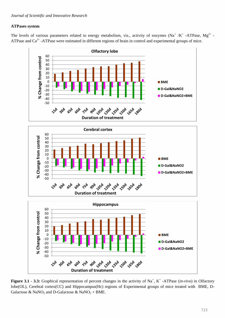
ATPases system
The levels of various parameters related to energy metabolism, viz., activity of enzymes (Na+ /K
+ -ATPase, Mg
2+ -
ATPase and Ca2+
-ATPase were estimated in different regions of brain in control and experimental groups of mice.
Figure 3.1 - 3.3: Graphical representation of percent changes in the activity of Na+, K
+ -ATPase (in-vivo) in Olfactory
lobe(OL), Cerebral cortex(CC) and Hippocampus(Hc) regions of Experimental groups of mice treated with BME, D-
Galactose & NaNO2 and D-Galactose & NaNO2 + BME.
-50-40-30-20-10
0102030405060
% C
han
ge f
rom
co
ntr
ol
Duration of treatment
Olfactory lobe
BME
D-Gal&NaNO2
D-Gal&NaNO2+BME
-50-40-30-20-10
0102030405060
% C
han
ge f
rom
co
ntr
ol
Duration of treatment
Cerebral cortex
BME
D-Gal&NaNO2
D-Gal&NaNO2+BME
-50-40-30-20-10
0102030405060
% C
han
ge f
rom
co
ntr
ol
Duration of treatment
Hippocampus
BME
D-Gal&NaNO2
D-Gal&NaNO2+BME
Journal of Scientific and Innovative Research
724
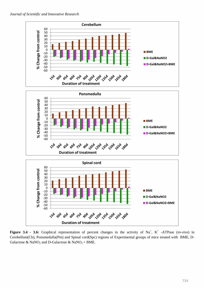
Figure 3.4 - 3.6: Graphical representation of percent changes in the activity of Na+, K
+ -ATPase (in-vivo) in
Cerebellum(Cb), Ponsmedulla(Pm) and Spinal cord(Spc) regions of Experimental groups of mice treated with BME, D-
Galactose & NaNO2 and D-Galactose & NaNO2 + BME.
-60-50-40-30-20-10
0102030405060
% C
han
ge f
rom
co
ntr
ol
Duration of treatment
Cerebellum
BME
D-Gal&NaNO2
D-Gal&NaNO2+BME
-60-50-40-30-20-10
0102030405060
% C
han
ge f
rom
co
ntr
ol
Duration of treatment
Ponsmedulla
BME
D-Gal&NaNO2
D-Gal&NaNO2+BME
-60-50-40-30-20-10
0102030405060
% C
han
ge f
rom
co
ntr
ol
Duration of treatment
Spinal cord
BME
D-Gal&NaNO2
D-Gal&NaNO2+BME
Journal of Scientific and Innovative Research
725
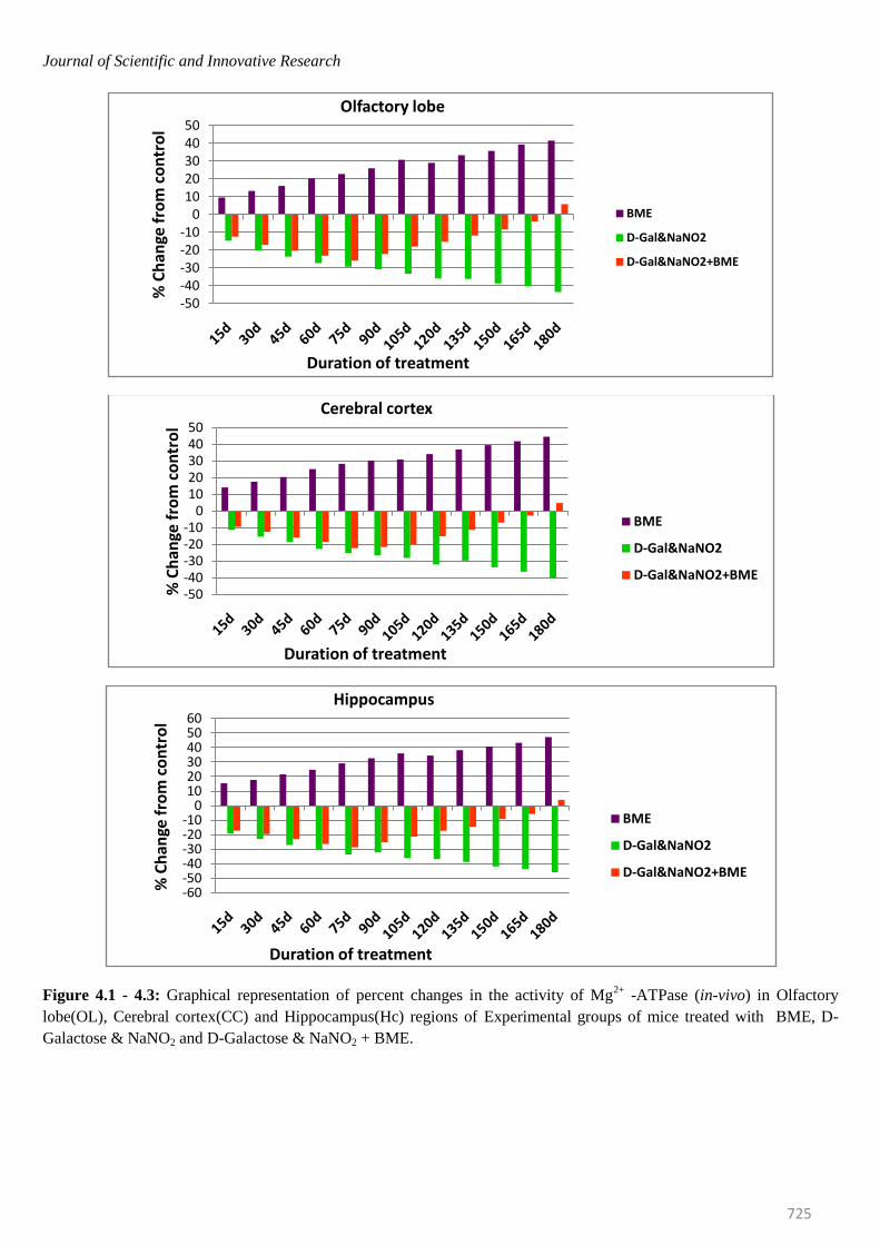
Figure 4.1 - 4.3: Graphical representation of percent changes in the activity of Mg2+
-ATPase (in-vivo) in Olfactory
lobe(OL), Cerebral cortex(CC) and Hippocampus(Hc) regions of Experimental groups of mice treated with BME, D-
Galactose & NaNO2 and D-Galactose & NaNO2 + BME.
-50-40-30-20-10
01020304050
% C
han
ge f
rom
co
ntr
ol
Duration of treatment
Olfactory lobe
BME
D-Gal&NaNO2
D-Gal&NaNO2+BME
-50-40-30-20-10
01020304050
% C
han
ge f
rom
co
ntr
ol
Duration of treatment
Cerebral cortex
BME
D-Gal&NaNO2
D-Gal&NaNO2+BME
-60-50-40-30-20-10
0102030405060
% C
han
ge f
rom
co
ntr
ol
Duration of treatment
Hippocampus
BME
D-Gal&NaNO2
D-Gal&NaNO2+BME
Journal of Scientific and Innovative Research
726
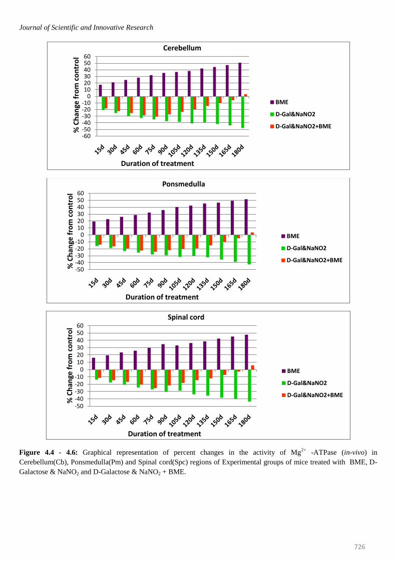
Figure 4.4 - 4.6: Graphical representation of percent changes in the activity of Mg2+
-ATPase (in-vivo) in
Cerebellum(Cb), Ponsmedulla(Pm) and Spinal cord(Spc) regions of Experimental groups of mice treated with BME, D-
Galactose & NaNO2 and D-Galactose & NaNO2 + BME.
-60-50-40-30-20-10
0102030405060
% C
han
ge f
rom
co
ntr
ol
Duration of treatment
Cerebellum
BME
D-Gal&NaNO2
D-Gal&NaNO2+BME
-50-40-30-20-10
0102030405060
% C
han
ge f
rom
co
ntr
ol
Duration of treatment
Ponsmedulla
BME
D-Gal&NaNO2
D-Gal&NaNO2+BME
-50-40-30-20-10
0102030405060
% C
han
ge f
rom
co
ntr
ol
Duration of treatment
Spinal cord
BME
D-Gal&NaNO2
D-Gal&NaNO2+BME
Journal of Scientific and Innovative Research
727
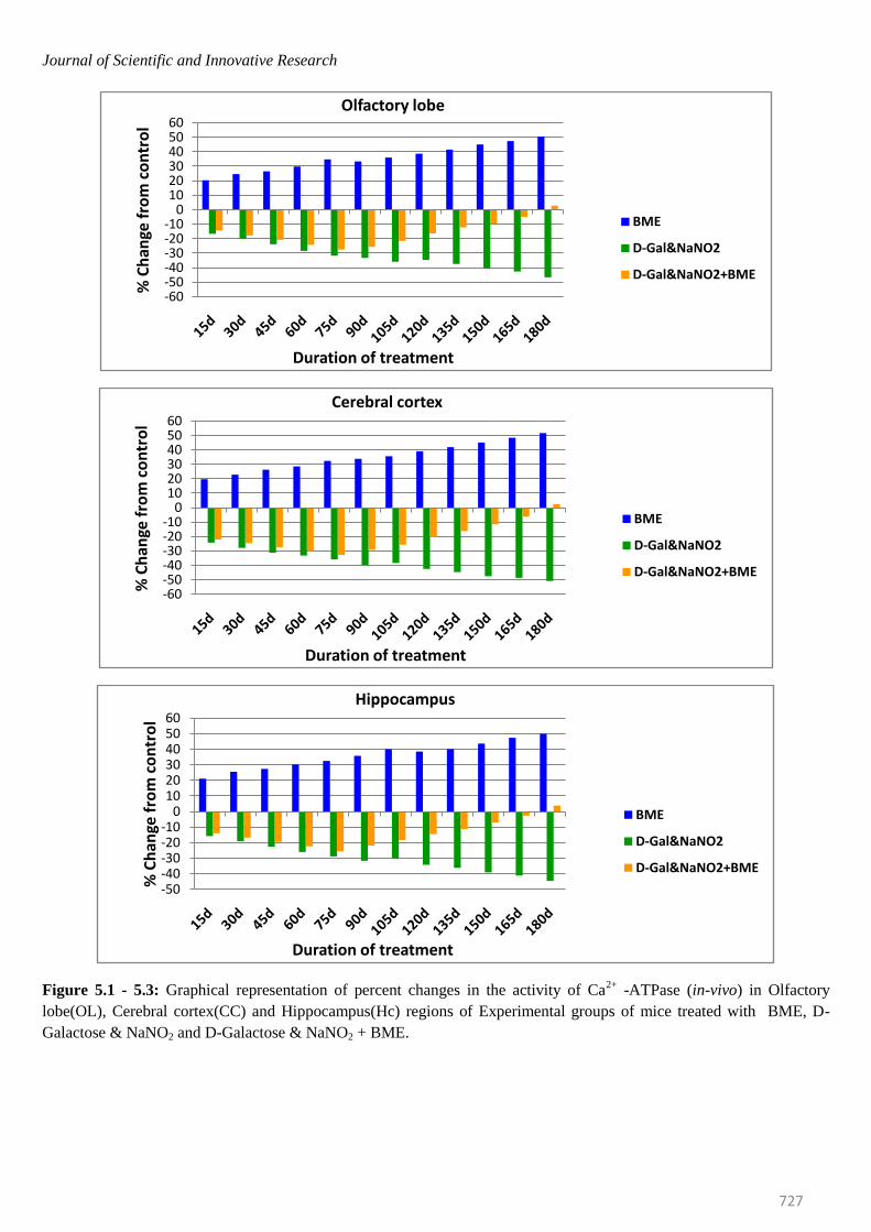
Figure 5.1 - 5.3: Graphical representation of percent changes in the activity of Ca2+
-ATPase (in-vivo) in Olfactory
lobe(OL), Cerebral cortex(CC) and Hippocampus(Hc) regions of Experimental groups of mice treated with BME, D-
Galactose & NaNO2 and D-Galactose & NaNO2 + BME.
-60-50-40-30-20-10
0102030405060
% C
han
ge f
rom
co
ntr
ol
Duration of treatment
Olfactory lobe
BME
D-Gal&NaNO2
D-Gal&NaNO2+BME
-60-50-40-30-20-10
0102030405060
% C
han
ge f
rom
co
ntr
ol
Duration of treatment
Cerebral cortex
BME
D-Gal&NaNO2
D-Gal&NaNO2+BME
-50-40-30-20-10
0102030405060
% C
han
ge f
rom
co
ntr
ol
Duration of treatment
Hippocampus
BME
D-Gal&NaNO2
D-Gal&NaNO2+BME
Journal of Scientific and Innovative Research
728
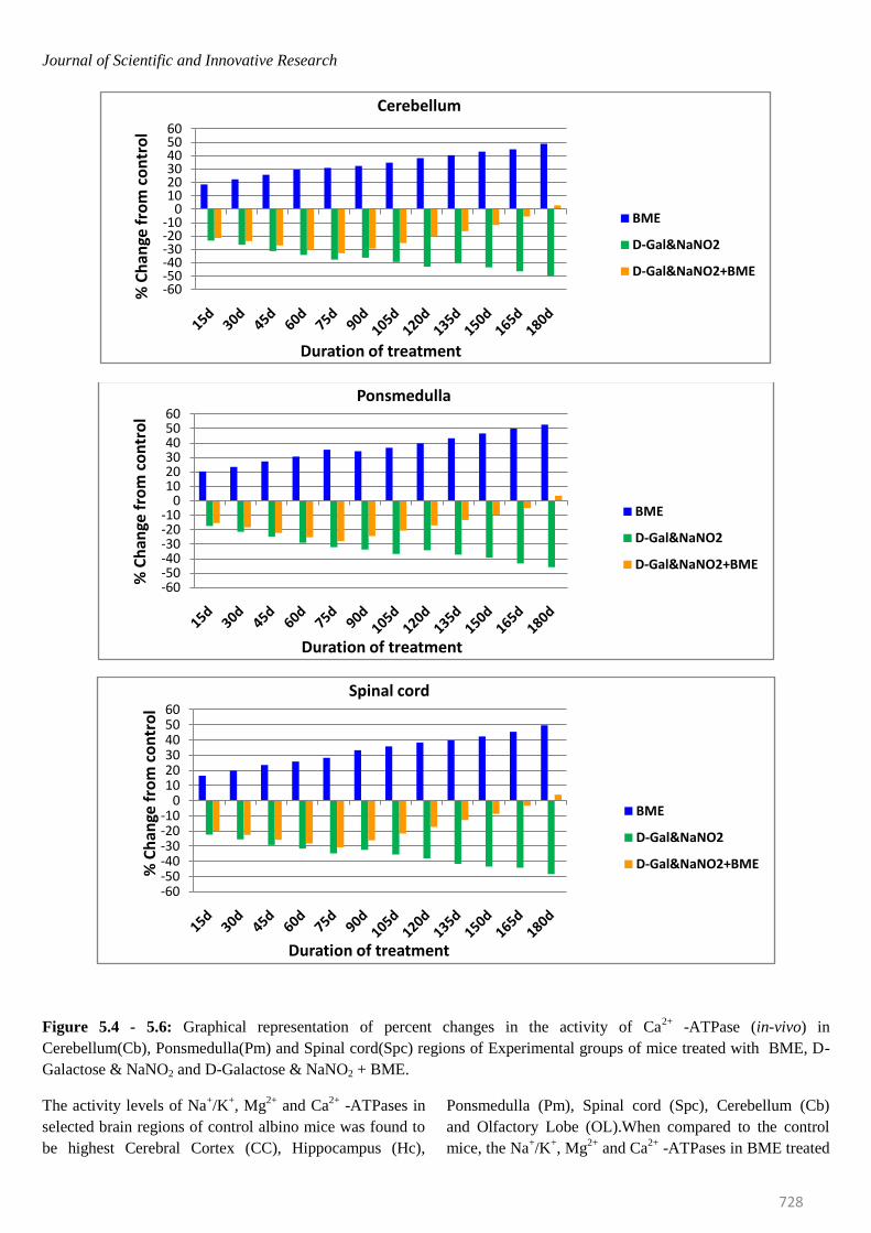
Figure 5.4 - 5.6: Graphical representation of percent changes in the activity of Ca2+
-ATPase (in-vivo) in
Cerebellum(Cb), Ponsmedulla(Pm) and Spinal cord(Spc) regions of Experimental groups of mice treated with BME, D-
Galactose & NaNO2 and D-Galactose & NaNO2 + BME.
The activity levels of Na+/K
+, Mg
2+ and Ca
2+ -ATPases in
selected brain regions of control albino mice was found to
be highest Cerebral Cortex (CC), Hippocampus (Hc),
Ponsmedulla (Pm), Spinal cord (Spc), Cerebellum (Cb)
and Olfactory Lobe (OL).When compared to the control
mice, the Na+/K
+, Mg
2+ and Ca
2+ -ATPases in BME treated
-60-50-40-30-20-10
0102030405060
% C
han
ge f
rom
co
ntr
ol
Duration of treatment
Cerebellum
BME
D-Gal&NaNO2
D-Gal&NaNO2+BME
-60-50-40-30-20-10
0102030405060
% C
han
ge f
rom
co
ntr
ol
Duration of treatment
Ponsmedulla
BME
D-Gal&NaNO2
D-Gal&NaNO2+BME
-60-50-40-30-20-10
0102030405060
% C
han
ge f
rom
co
ntr
ol
Duration of treatment
Spinal cord
BME
D-Gal&NaNO2
D-Gal&NaNO2+BME
Journal of Scientific and Innovative Research
729
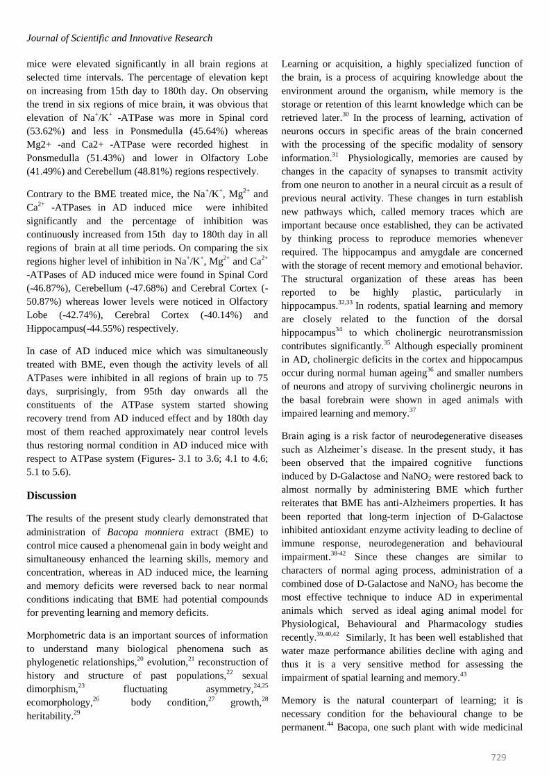
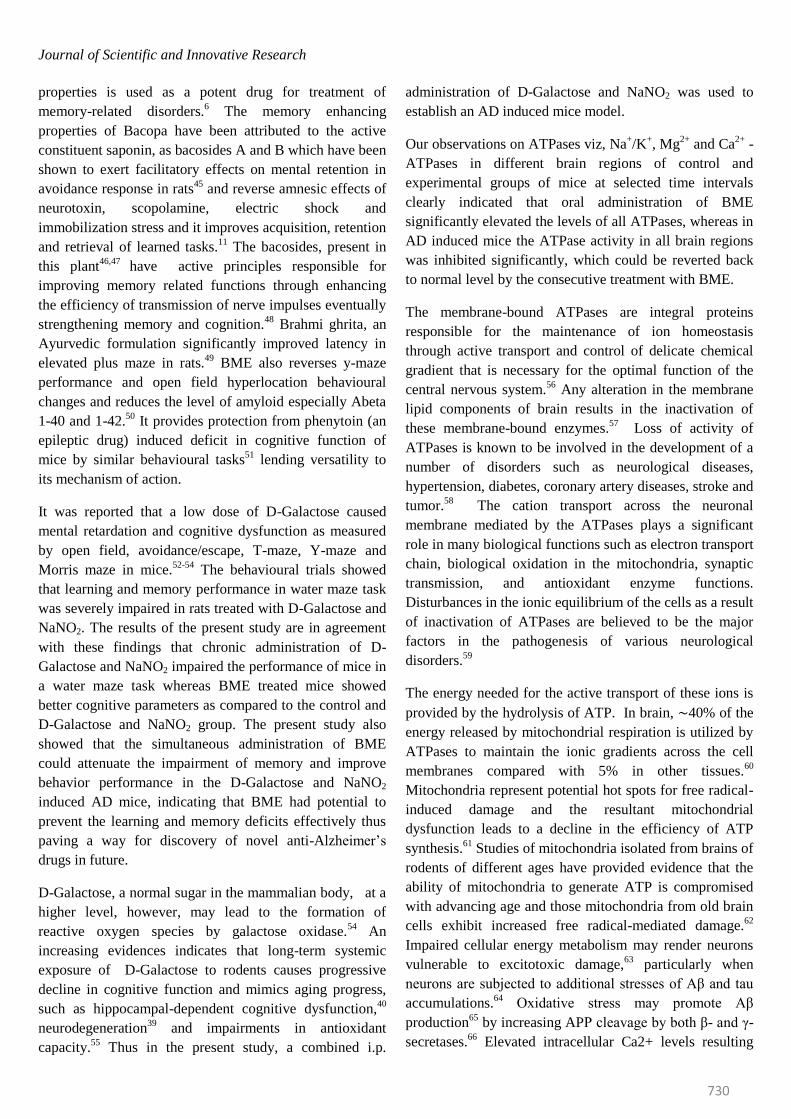
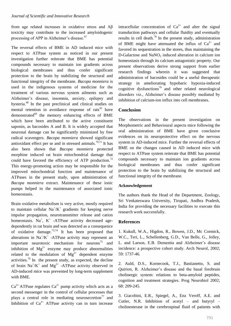
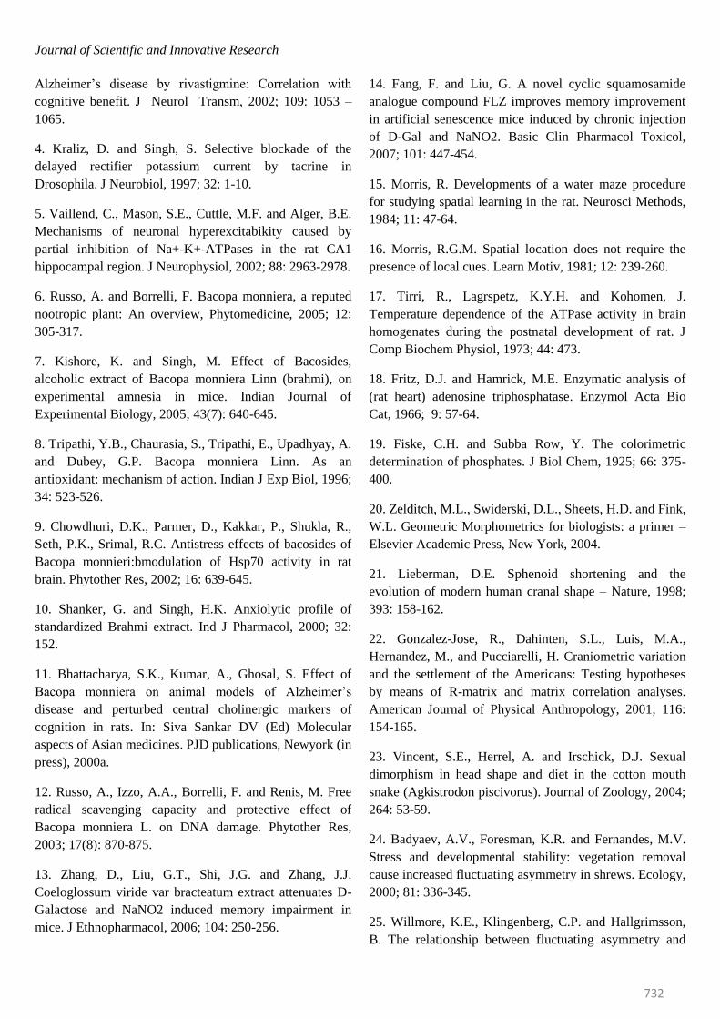
mice were elevated significantly in all brain regions at
selected time intervals. The percentage of elevation kept
on increasing from 15th day to 180th day. On observing
the trend in six regions of mice brain, it was obvious that
elevation of Na+/K
+ -ATPase was more in Spinal cord
(53.62%) and less in Ponsmedulla (45.64%) whereas
Mg2+ -and Ca2+ -ATPase were recorded highest in
Ponsmedulla (51.43%) and lower in Olfactory Lobe
(41.49%) and Cerebellum (48.81%) regions respectively.
Contrary to the BME treated mice, the Na+/K
+, Mg
2+ and
Ca2+
-ATPases in AD induced mice were inhibited
significantly and the percentage of inhibition was
continuously increased from 15th day to 180th day in all
regions of brain at all time periods. On comparing the six
regions higher level of inhibition in Na+/K
+, Mg
2+ and Ca
2+
-ATPases of AD induced mice were found in Spinal Cord
(-46.87%), Cerebellum (-47.68%) and Cerebral Cortex (-
50.87%) whereas lower levels were noticed in Olfactory
Lobe (-42.74%), Cerebral Cortex (-40.14%) and
Hippocampus(-44.55%) respectively.
In case of AD induced mice which was simultaneously
treated with BME, even though the activity levels of all
ATPases were inhibited in all regions of brain up to 75
days, surprisingly, from 95th day onwards all the
constituents of the ATPase system started showing
recovery trend from AD induced effect and by 180th day
most of them reached approximately near control levels
thus restoring normal condition in AD induced mice with
respect to ATPase system (Figures- 3.1 to 3.6; 4.1 to 4.6;
5.1 to 5.6).
Discussion
The results of the present study clearly demonstrated that
administration of Bacopa monniera extract (BME) to
control mice caused a phenomenal gain in body weight and
simultaneousy enhanced the learning skills, memory and
concentration, whereas in AD induced mice, the learning
and memory deficits were reversed back to near normal
conditions indicating that BME had potential compounds
for preventing learning and memory deficits.
Morphometric data is an important sources of information
to understand many biological phenomena such as
phylogenetic relationships,20
evolution,21
reconstruction of
history and structure of past populations,22
sexual
dimorphism,23
fluctuating asymmetry,24,25
ecomorphology,26
body condition,27
growth,28
heritability.29
Learning or acquisition, a highly specialized function of
the brain, is a process of acquiring knowledge about the
environment around the organism, while memory is the
storage or retention of this learnt knowledge which can be
retrieved later.30
In the process of learning, activation of
neurons occurs in specific areas of the brain concerned
with the processing of the specific modality of sensory
information.31
Physiologically, memories are caused by
changes in the capacity of synapses to transmit activity
from one neuron to another in a neural circuit as a result of
previous neural activity. These changes in turn establish
new pathways which, called memory traces which are
important because once established, they can be activated
by thinking process to reproduce memories whenever
required. The hippocampus and amygdale are concerned
with the storage of recent memory and emotional behavior.
The structural organization of these areas has been
reported to be highly plastic, particularly in
hippocampus.32,33
In rodents, spatial learning and memory
are closely related to the function of the dorsal
hippocampus34
to which cholinergic neurotransmission
contributes significantly.35
Although especially prominent
in AD, cholinergic deficits in the cortex and hippocampus
occur during normal human ageing36
and smaller numbers
of neurons and atropy of surviving cholinergic neurons in
the basal forebrain were shown in aged animals with
impaired learning and memory.37
Brain aging is a risk factor of neurodegenerative diseases
such as Alzheimer’s disease. In the present study, it has
been observed that the impaired cognitive functions
induced by D-Galactose and NaNO2 were restored back to
almost normally by administering BME which further
reiterates that BME has anti-Alzheimers properties. It has
been reported that long-term injection of D-Galactose
inhibited antioxidant enzyme activity leading to decline of
immune response, neurodegeneration and behavioural
impairment.38-42
Since these changes are similar to
characters of normal aging process, administration of a
combined dose of D-Galactose and NaNO2 has become the
most effective technique to induce AD in experimental
animals which served as ideal aging animal model for
Physiological, Behavioural and Pharmacology studies
recently.39,40,42
Similarly, It has been well established that
water maze performance abilities decline with aging and
thus it is a very sensitive method for assessing the
impairment of spatial learning and memory.43
Memory is the natural counterpart of learning; it is
necessary condition for the behavioural change to be
permanent.44
Bacopa, one such plant with wide medicinal
Journal of Scientific and Innovative Research
730
properties is used as a potent drug for treatment of
memory-related disorders.6 The memory enhancing
properties of Bacopa have been attributed to the active
constituent saponin, as bacosides A and B which have been
shown to exert facilitatory effects on mental retention in
avoidance response in rats45
and reverse amnesic effects of
neurotoxin, scopolamine, electric shock and
immobilization stress and it improves acquisition, retention
and retrieval of learned tasks.11
The bacosides, present in
this plant46,47
have active principles responsible for
improving memory related functions through enhancing
the efficiency of transmission of nerve impulses eventually
strengthening memory and cognition.48
Brahmi ghrita, an
Ayurvedic formulation significantly improved latency in
elevated plus maze in rats.49
BME also reverses y-maze
performance and open field hyperlocation behavioural
changes and reduces the level of amyloid especially Abeta
1-40 and 1-42.50
It provides protection from phenytoin (an
epileptic drug) induced deficit in cognitive function of
mice by similar behavioural tasks51
lending versatility to
its mechanism of action.
It was reported that a low dose of D-Galactose caused
mental retardation and cognitive dysfunction as measured
by open field, avoidance/escape, T-maze, Y-maze and
Morris maze in mice.52-54
The behavioural trials showed
that learning and memory performance in water maze task
was severely impaired in rats treated with D-Galactose and
NaNO2. The results of the present study are in agreement
with these findings that chronic administration of D-
Galactose and NaNO2 impaired the performance of mice in
a water maze task whereas BME treated mice showed
better cognitive parameters as compared to the control and
D-Galactose and NaNO2 group. The present study also
showed that the simultaneous administration of BME
could attenuate the impairment of memory and improve
behavior performance in the D-Galactose and NaNO2
induced AD mice, indicating that BME had potential to
prevent the learning and memory deficits effectively thus
paving a way for discovery of novel anti-Alzheimer’s
drugs in future.
D-Galactose, a normal sugar in the mammalian body, at a
higher level, however, may lead to the formation of
reactive oxygen species by galactose oxidase.54
An
increasing evidences indicates that long-term systemic
exposure of D-Galactose to rodents causes progressive
decline in cognitive function and mimics aging progress,
such as hippocampal-dependent cognitive dysfunction,40
neurodegeneration39
and impairments in antioxidant
capacity.55
Thus in the present study, a combined i.p.
administration of D-Galactose and NaNO2 was used to
establish an AD induced mice model.
Our observations on ATPases viz, Na+/K
+, Mg
2+ and Ca
2+ -
ATPases in different brain regions of control and
experimental groups of mice at selected time intervals
clearly indicated that oral administration of BME
significantly elevated the levels of all ATPases, whereas in
AD induced mice the ATPase activity in all brain regions
was inhibited significantly, which could be reverted back
to normal level by the consecutive treatment with BME.
The membrane-bound ATPases are integral proteins
responsible for the maintenance of ion homeostasis
through active transport and control of delicate chemical
gradient that is necessary for the optimal function of the
central nervous system.56
Any alteration in the membrane
lipid components of brain results in the inactivation of
these membrane-bound enzymes.57
Loss of activity of
ATPases is known to be involved in the development of a
number of disorders such as neurological diseases,
hypertension, diabetes, coronary artery diseases, stroke and
tumor.58
The cation transport across the neuronal
membrane mediated by the ATPases plays a significant
role in many biological functions such as electron transport
chain, biological oxidation in the mitochondria, synaptic
transmission, and antioxidant enzyme functions.
Disturbances in the ionic equilibrium of the cells as a result
of inactivation of ATPases are believed to be the major
factors in the pathogenesis of various neurological
disorders.59
The energy needed for the active transport of these ions is
provided by the hydrolysis of ATP. In brain, ∼40% of the
energy released by mitochondrial respiration is utilized by
ATPases to maintain the ionic gradients across the cell
membranes compared with 5% in other tissues.60
Mitochondria represent potential hot spots for free radical-
induced damage and the resultant mitochondrial
dysfunction leads to a decline in the efficiency of ATP
synthesis.61
Studies of mitochondria isolated from brains of
rodents of different ages have provided evidence that the
ability of mitochondria to generate ATP is compromised
with advancing age and those mitochondria from old brain
cells exhibit increased free radical-mediated damage.62
Impaired cellular energy metabolism may render neurons
vulnerable to excitotoxic damage,63
particularly when
neurons are subjected to additional stresses of Aβ and tau
accumulations.64
Oxidative stress may promote Aβ
production65
by increasing APP cleavage by both β- and γ-
secretases.66
Elevated intracellular Ca2+ levels resulting
Journal of Scientific and Innovative Research
731
from age related increases in oxidative stress and Aβ
toxicity may contribute to the increased amyloidogenic
processing of APP in Alzheimer’s disease.67
The reversal effects of BME in AD induced mice with
respect to ATPase system as noticed in our present
investigation further reiterate that BME has potential
compounds necessary to maintain ion gradients across
biological membranes and thus confer significant
protection to the brain by stabilizing the structural and
functional integrity of the membrane. Bacopa monniera is
used in the indigenous systems of medicine for the
treatment of various nervous system ailments such as
Alzheimer’s disease, insomnia, anxiety, epilepsy and
hysteria.68
In the past preclinical and clinical studies on
mental retention in avoidance response of rats45
have
demonstrated69
the memory enhancing effects of BME
which have been attributed to the active constituent
saponin, as bacosides A and B. It is widely accepted that
neuronal damage can be significantly minimized by free
radical scavengers. Bacopa monniera showed significant
antioxidant effect per se and in stressed animals.70,71
It has
also been shown that Bacopa monniera protected
morphine induced rat brain mitochondrial damage that
could have favored the efficiency of ATP production.72
This energy-promoting action may be responsible for the
improved mitochondrial function and maintenance of
ATPases in the present study, upon administration of
Bacopa monniera extract. Maintenance of these ionic
pumps helped in the maintenance of associated ionic
homeostasis.
Brain oxidative metabolism is very active, mostly required
to maintain cellular Na+/K
+ gradients for keeping nerve
impulse propagation, neurotransmitter release and cation
homeostasis. Na+, K
+ -ATPase activity decreased age-
dependently in rat brain and was detected as a consequence
of oxidative damage.73,74
It has been proposed that
alterations in Na+/K
+ -ATPase activity may represent an
important neurotoxic mechanism for neurons75
and
inhibition of Mg2+
enzyme may produce abnormalities
related to the modulation of Mg2+
dependent enzyme
activities.76
In the present study, as expected, the decline
of brain Na+/K
+ and Mg
2+ -ATPase activity observed in
AD-induced mice was prevented by long-term supplement
with BME.
Ca2+-
ATPase regulates Ca2+
pump activity which acts as a
second messenger in the control of cellular processes that
plays a central role in mediating neurosecretion77
and
Inhibition of Ca2+
ATPase activity can in turn increase
intracellular concentration of Ca2+
and alter the signal
transduction pathways and cellular fluidity and eventually
results in cell death.78
In the present study, administration
of BME might have attenuated the influx of Ca2+
and
favored its sequestration in the stores, thus maintaining the
D-Galactose and NaNO2 induced alteration in calcium ion
homeostasis through its calcium antagonistic property. Our
present observations derive strong support from earlier
research findings wherein it was suggested that
administration of bacosides could be a useful therapeutic
strategy in ameliorating hypobaric hypoxia-induced
cognitive dysfunctions79
and other related neurological
disorders viz., Alzheimer’s disease possibly mediated by
inhibition of calcium-ion influx into cell membranes.
Conclusion
The observations in the present investigation on
Morphometric and Behavioural aspects mice following the
oral administration of BME have given conclusive
evidences on its neuroprotective effect on the nervous
system in AD-induced mice. Further the reversal effects of
BME on the changes caused in AD induced mice with
respect to ATPase system reiterate that BME has potential
compounds necessary to maintain ion gradients across
biological membranes and thus confer significant
protection to the brain by stabilizing the structural and
functional integrity of the membrane.
Acknowledgement
The authors thank the Head of the Department, Zoology,
Sri Venkateswara University, Tirupati, Andhra Pradesh,
India for providing the necessary facilities to execute this
research work successfully.
References
1. Kukull, W.A., Higdon, R., Bowen, J.D., Mc Cormick,
W.C., Teri, L., Schellenberg, G.D., Van Belle, G., Jolley,
L. and Larson, E.B. Dementia and Alzheimer’s disease
incidence: a prospective cohort study. Arch Neurol, 2002;
59: 1737-46.
2. Auld, D.S., Kornecook, T.J., Bastianetto, S. and
Quirion, R. Alzheimer’s disease and the basal forebrain
cholinergic system: relations to beta-amyloid peptides,
cognition and treatment strategies. Prog Neurobiol 2002;
68: 209-245.
3. Giacobini, E.R., Spiegel, A., Enz Veroff, A.E. and
Cutler, N.R. Inhibition of acetyl – and butyryl –
cholinesterase in the cerebrospinal fluid of patients with
Journal of Scientific and Innovative Research
732
Alzheimer’s disease by rivastigmine: Correlation with
cognitive benefit. J Neurol Transm, 2002; 109: 1053 –
1065.
4. Kraliz, D. and Singh, S. Selective blockade of the
delayed rectifier potassium current by tacrine in
Drosophila. J Neurobiol, 1997; 32: 1-10.
5. Vaillend, C., Mason, S.E., Cuttle, M.F. and Alger, B.E.
Mechanisms of neuronal hyperexcitabikity caused by
partial inhibition of Na+-K+-ATPases in the rat CA1
hippocampal region. J Neurophysiol, 2002; 88: 2963-2978.
6. Russo, A. and Borrelli, F. Bacopa monniera, a reputed
nootropic plant: An overview, Phytomedicine, 2005; 12:
305-317.
7. Kishore, K. and Singh, M. Effect of Bacosides,
alcoholic extract of Bacopa monniera Linn (brahmi), on
experimental amnesia in mice. Indian Journal of
Experimental Biology, 2005; 43(7): 640-645.
8. Tripathi, Y.B., Chaurasia, S., Tripathi, E., Upadhyay, A.
and Dubey, G.P. Bacopa monniera Linn. As an
antioxidant: mechanism of action. Indian J Exp Biol, 1996;
34: 523-526.
9. Chowdhuri, D.K., Parmer, D., Kakkar, P., Shukla, R.,
Seth, P.K., Srimal, R.C. Antistress effects of bacosides of
Bacopa monnieri:bmodulation of Hsp70 activity in rat
brain. Phytother Res, 2002; 16: 639-645.
10. Shanker, G. and Singh, H.K. Anxiolytic profile of
standardized Brahmi extract. Ind J Pharmacol, 2000; 32:
152.
11. Bhattacharya, S.K., Kumar, A., Ghosal, S. Effect of
Bacopa monniera on animal models of Alzheimer’s
disease and perturbed central cholinergic markers of
cognition in rats. In: Siva Sankar DV (Ed) Molecular
aspects of Asian medicines. PJD publications, Newyork (in
press), 2000a.
12. Russo, A., Izzo, A.A., Borrelli, F. and Renis, M. Free
radical scavenging capacity and protective effect of
Bacopa monniera L. on DNA damage. Phytother Res,
2003; 17(8): 870-875.
13. Zhang, D., Liu, G.T., Shi, J.G. and Zhang, J.J.
Coeloglossum viride var bracteatum extract attenuates D-
Galactose and NaNO2 induced memory impairment in
mice. J Ethnopharmacol, 2006; 104: 250-256.
14. Fang, F. and Liu, G. A novel cyclic squamosamide
analogue compound FLZ improves memory improvement
in artificial senescence mice induced by chronic injection
of D-Gal and NaNO2. Basic Clin Pharmacol Toxicol,
2007; 101: 447-454.
15. Morris, R. Developments of a water maze procedure
for studying spatial learning in the rat. Neurosci Methods,
1984; 11: 47-64.
16. Morris, R.G.M. Spatial location does not require the
presence of local cues. Learn Motiv, 1981; 12: 239-260.
17. Tirri, R., Lagrspetz, K.Y.H. and Kohomen, J.
Temperature dependence of the ATPase activity in brain
homogenates during the postnatal development of rat. J
Comp Biochem Physiol, 1973; 44: 473.
18. Fritz, D.J. and Hamrick, M.E. Enzymatic analysis of
(rat heart) adenosine triphosphatase. Enzymol Acta Bio
Cat, 1966; 9: 57-64.
19. Fiske, C.H. and Subba Row, Y. The colorimetric
determination of phosphates. J Biol Chem, 1925; 66: 375-
400.
20. Zelditch, M.L., Swiderski, D.L., Sheets, H.D. and Fink,
W.L. Geometric Morphometrics for biologists: a primer –
Elsevier Academic Press, New York, 2004.
21. Lieberman, D.E. Sphenoid shortening and the
evolution of modern human cranal shape – Nature, 1998;
393: 158-162.
22. Gonzalez-Jose, R., Dahinten, S.L., Luis, M.A.,
Hernandez, M., and Pucciarelli, H. Craniometric variation
and the settlement of the Americans: Testing hypotheses
by means of R-matrix and matrix correlation analyses.
American Journal of Physical Anthropology, 2001; 116:
154-165.
23. Vincent, S.E., Herrel, A. and Irschick, D.J. Sexual
dimorphism in head shape and diet in the cotton mouth
snake (Agkistrodon piscivorus). Journal of Zoology, 2004;
264: 53-59.
24. Badyaev, A.V., Foresman, K.R. and Fernandes, M.V.
Stress and developmental stability: vegetation removal
cause increased fluctuating asymmetry in shrews. Ecology,
2000; 81: 336-345.
25. Willmore, K.E., Klingenberg, C.P. and Hallgrimsson,
B. The relationship between fluctuating asymmetry and
Journal of Scientific and Innovative Research
733
environmental variance in rhesus macaque skulls.
Evolution, 2005; 59: 898-909.
26. Klingenberg, C.P. and Ekau, W. Acombined
morphometric and phylogenetic analysis of an
ecomorphological trend: pelagization in Antarctic fishes
(Perciformes: nototheniidae). Biological Journal of the
Linnean Society, 1996; 59: 143-177.
27. Green, A.J. Mass/length residuals: measures of body
condition or generators of spurious results? Ecology, 2001;
82: 1473-1483.
28. Ackermann, R.R. Ontogenetic integration of the
homoinoid face. Journal of Human Evolution, 2005; 48:
175-197.
29. Krunk, L.E.B., Clutton-Brock, T.H., Slate, J.,
Pemberton, J.M., Brother stone, S. and Guinness, F.E.
Heritability of fitness in a wild mammal population.
Proceedings of the National Academy of Sciences of the
United Statesof America, 2000; 97: 698-703.
30. Squire, L.R. and Schlafer, W.T. Handbook of
Biological Psychiatry, Vanpraag HM, Lader MH,
Rafaelsen OJ, Sachar EJ (Eds). Raven Press: Newyork,
USA, 1981; 249.
31. Rolls, E.T. Memory systems in the brain. Annu Rev
Psychol, 2000; 51: 599-530.
32. Richter-Levin, G. and Akirav, I. Amygdala-
hippocampus dynamic interaction in relation to memory.
Mol Neurobiol, 2000; 22(1-3): 11-20.
33. Antoniadis, E.A. and Mc Donald, R.J. Amygdala
hippocampus and discriminative fear conditioning to
context. Behav Brain Res, 2003; 108: 1-19.
34. Moser, E., Moser, M.B. and Andersen, P. Spatial;
earning impairment parallels the magnitude of dorsal
hippocampal lesions, but is hardly present following
ventral lesions. J Neurosci, 1993; 13: 3916-3925.
35. Bartus, R.T. On neurodegenerative diseases models
and treatment strategies: lessons learned and lessons
forgotten a generation following the cholinergic
hypothesis. Exp Neurol, 2000; 163: 495-529.
36. Colom, L.V. Septal networks: Relevance to theta
rhythm, epilepsy and Alzheimer’s disease. J Neurochem,
2006; 96: 609-623.
37. Muir, J.L. Acetylcholine, aging and Alzheimer’s
disease. Pharmacol Biochem Behav, 1997; 56(4): 687-696.
38. Zhang, C., Wang, S.Z., Zuo, P.P. and Cui, X.
Protective effect of tetramethylpyrazine on learning and
memory function in D-Galactose lesioned mice. J Chin
Med Sci, 2004; 19: 180-4.
39. Zhang, Q., Li, X.K., Cui, X. and Zuo, P.P. D-Galactose
injured neurogenesis in the hippocampus of adult mice.
Neurol Res, 2005; 27: 552-556.
40. Cui, X., Zuo, P., Zhang, Q., Li, X., Hu, Y., Long, J. et
al., Chronic systemic D-Galactose exposure indices
memory loss, neurodegeneration and oxidative damage in
mice: protective effects of R-alpha-Lipoic acid. J Neurosci
Res, 2006; 83(8): 1584-90.
41. Hua, X.D., Lei, M., Zhang, Y.J., Ding, J., Han, Q.Y.
and Hu, G. Long-term D-Galactose injection combines
with ovirectomy serves as a new rodent model for
Alzheimer’s disease. Life Sci, 2007; 80: 1897-905.
42. Lu, J., Zheng, Y., Wu, D., Luo, L., Sun, D. and Shan,
Q. Urosolic acid ameliorates cognition deficits and
attenuates oxidative damage in the brain of senescent mice
induced by D-Galactose. Biochemical Pharmacology,
2007; 74: 1078-1090.
43. Brandeis, R., Brandys, Y., Yehuda, S. The use of
Morris water maze in the study of memory and learning.
The international Journal of Neuroscience, 1989; 48 (1-2):
29-69.
44. Vervilet, B. Acta Psychologica, 2008; 127(3): 601-613.
45. Singh, H.K., Rastogi, R.P., Sriman, R.C., Dhawan,
B.N. Effect of Bacoside A and B on avoidance response in
rats. Phytother, 1988; Res 2: 70-75.
46. Rastogi, S., Pal, R. and Kulshreshtha, D.K. Bacoside
A3-a triterpinoid saponin from Bacopa monniera,
Phytochemistry, 1994; 36(1): 133-137.
47. Sivaramakrishna et al., Triterprnoid glycosides from
Bacopa monniera. Phytochemistry, 2005; 66: 2719-2728.
48. Anon, Bacopa monnieri Monograph. Altern Med Rev,
2004; 9: 79-85.
49. Achilya, C., Barabde, U., Wadodkar, S. and Dorle, A.
Effect of Brahmi Ghrita, a polyherbal formulation on
learning and memory paradigms in experimental animals.
Indian J Pharmacol, 2004; 36(3): 159-162.
Journal of Scientific and Innovative Research
734
50. Holcomb, L.A., Dhanasekaran, A.R., Hitt, K.A.,
Young, M. Riggs and Manyam, B.V. Bacopa monniera
extract reduces amyloid levels in PSAPP mice. Journal of
Alzheimer’s disease, 2006; 9(3): 243-251.
51. Vohora, D., Pal, S.N., Pillai, K.K. Protection from
phenytoin induced cognitive deficit by Bacopa monniera, a
reputed Indian nootropic plant. J ethnopharmacol, 2000;
71: 383-90.
52. Shen, Y.X., Xu, S.Y., Wei, W., Sun, X.X., Yang, J.,
Liu, L.H. and Dong, C. Melatonin reduces memory
changes and neural oxidative damage in mice treated with
D-galactose. J Pineal Res, 2002; 32: 173-178.
53. Xu, X.H. and Zhang Z.G. Effect of Puerarin on
learning-memory behaviout and synaptic structure of
hippocampus in the aging mice induced by D-Galactose.
Acta Pharm Sin, 2002; 37: 1-4.
54. Ho, S.C., Liu, J.H. and Wu, R.Y. Establishment of the
mimetic aging effect in mice caused by D-Galactose.
Biogerontology, 2003; 4: 15-18.
55. Lu, J., Zheng, Y.L., Luo, L., Wu, D.M., Sun, D.X. and
Feng, Y.J. Quercetin reverses D-Galactose induced
neurotoxicity in mouse brain. Behavioural Brain Research,
2006; 171(2): 251-260.
56. Dzhafaroz, A.I., Magomedov, N.M., Babaev, Kh.F.,
Akhmedova, G.Sh., and Bekhbudova, Z.A. Lipid
peroxidation and ATPase activity in synaptosomal and
mitochondrial fractions of the brain in Hypoxia. Vopr Med
Kim, 1989; 35: 51-56.
57. Barriviera, M.L. and Hasson-Voloch, A. Lipids
associated with the (Na+-K+) ATPase of normal and
denervated electric organs of Electrophorus electricus (L.).
Z Naturforsch[c], 1996; 51: 883-892.
58. Dhanya, C.R., Indu, Ar., Deepadevi, K.V. and Kurup,
P.A. Inhibition of membrane Na+-K+-ATPase of brain,
liver and RBC in rats administered di (2-ethyl hexyl)
phthalate (DEHP), a plasticizer used in polyvinyl chloride
(PVC) blood storage bags. Indian J Exp Biol, 2003; 41:
814-820.
59. Vaillend, C., Mason, S.E., Cuttle, M.F. and Alger, B.E.
Mechanisms of neuronal hyperexcitabikity caused by
partial inhibition of Na+-K+-ATPases in the rat CA1
hippocampal region. J Neurophysiol, 2002; 88: 2963-2978.
60. Rani, P.J.A. and Panneerselvam, C. The role of L-
Carnitine in the activities of membrane-bound enzymes in
the brain of aged rats. J Anti-aging Med, 2001; 4: 147-155.
61. Nicholls, D.G. Mitochondrial function and dysfunction
in the cell: Its relevance to aging and aging related
diseases. Int J Biochem Cell Biol, 2002; 34: 1372-1381.
62. Toescu, E.C., Myronove, N. and Verkhratsky, A. Age-
related structural and functional changes of brain
mitochondria. Cell Calcium, 2000; 28: 329-338.
63. Beal, M.F. Does impairment of energy metabolism
result in excitotoxic neuronal death in neurodegenerative
illness? Ann Neurol, 1992; 31: 119-130.
64. Mattson, M.P. Pathways towards and away from
Alzheimer’s disease. Nature, 2004; 430(7000): 631-639.
65. Li, F., Clingasan, N.Y., Yu, F., Mauck, W.M., Toidze,
M., Almeida, C.G., Takahashi, R.H., Carlson, G.A., Beal,
M.F., Lin, M.T. and Gouras, G.K. Increased plaque burden
in brains of APP mutant Mn SOD hetero zygous knockout
mice. J Neurochem, 2004; 89: 1308-1312.
66. Jo, D.G., Arumugam, T.V., Woo, H.N., Park, J.S.,
Tang, S.C., Mughal, M., Hyun, D.H., Park, J.H., Choi,
Y.H., Gwon, A.R., Camandola, S., Cheng, A., Cai, H.,
Song, W., Markesbery, W.R. and Mattson, M.P. Evidence
that gamma-secretase mediates oxidative stress-induced
beta-secretase expression in Alzheimer’s disease.
Neurobiol Aging, 2010; 31: 917-925.
67. Liang, B., Duan, B.Y., Zhou, X.P., Gong, J.X. and
Luo, Z.G. Calpain activation promotes BACE1 expression,
amyloid precursor protein processing and amyloid plaque
formation in a transgenic mouse model of Alzheimer’s
disease. J Biol Chem, 2010; 285: 27737-27744.
68. Nadkarni, K.M. Indian Materia Medica. Bombay:
Popular prakashan pvt. Ltd., 1976; pp 624-625.
69. Roodenrys, S., Booth, D., Bulzoni, S., Phipps, A.,
Micallef, C. and Smoker, J. Chronic effects of Brahmi
(BM) on human memory. Neuropsychopharmacology,
2002; 27: 279-81.
70. Sairam, K., Rao, C.V., Babu, M.D. and Goel, R.K.
Prophylactic and curative effects of Bacopa monniera in
gastric ulcer models. Phytomedicine, 2001; 8: 423-430.
71. Bhattacharya, S.K., Bhattacharya, A., Kumar, A. and
Ghosal, S. Antioxidant activity of Bacopa monniera in rat
Journal of Scientific and Innovative Research
735
frontal cortex, striatum and hippocampus. Phytother Res,
2000; 14: 174-179.
72. Sumathy, T., Govindasamy, S., Balakrishna, K. and
Veluchamy, G. Protective role of Bacopa monniera on
Morphine-induces brain mitochondrial enzyme activity in
rats. Fitoterapia, 2002; 73: 381-385.
73. Gorini, A., Canosi, U. and Devecchi, E. ATPase a
enzyme activities during ageing in different types of
somatic and synaptic plasma membranes from rat frontal
cerebral cortex. Prog Neuro-Psychopharmacol Biol
Psychiatry, 2002; 26: 81-90.
74. Chakraborty, H., Sen, P. and Sur, A. Age related
oxidative inactivation of Na+-K+-ATPase in rat brain
crude synaptosomes. Exp Gerontol, 2003; 38: 705-10.
75. Lees, G.J. Contributory mechanisms in the causation of
neurodegenerative disorders. Neuroscience, 1993; 54: 287-
322.
76. Tsakiris, S., Marinou, K. and Schulpis, K.H. The
invitro effects of D-Galactose and its derivatives on rat
brain Mg2+ -ATPase activity. Pharmacol Toxicol, 2002;
91: 254-257.
77. Pelletier, M.R., Wadia, J.S., Millis, L.R. and Carlen,
P.L. Seizure induced cell death produced by repeated
titanic stimulation invitro: Possible role of endoplsmic
reticulam calcium stores. J Neurophysiol, 1999; 81: 3054-
3064.
78. Aubier, M. and Viires, N. Calcium ATPase and
respiratory muscle function. Eur Respir J, 1998; 11: 758-
766.
79. Channa, S., Dar, A., Yaqoob, M., Anjum, S., Sultani,
Z. and Rahman, A. Bronchovasodilatory activity of
fractions of pure constituents isolated from Bacopa
monniera. J Ethnopharmacol, 2003; 86: 27-35.