Embed Size (px)
Citation preview
Fisher et al. BMC Microbiology 2012, 12:117http://www.biomedcentral.com/1471-2180/12/117
RESEARCH ARTICLE Open Access
The Madagascar hissing cockroach as a novelsurrogate host for Burkholderia pseudomallei,B. mallei and B. thailandensisNathan A Fisher1, Wilson J Ribot2, Willard Applefeld2,3 and David DeShazer2*
Abstract
Background: Burkholderia pseudomallei and Burkholderia mallei are gram-negative pathogens responsible for thediseases melioidosis and glanders, respectively. Both species cause disease in humans and animals and have beendesignated as category B select agents by the Centers for Disease Control and Prevention (CDC). Burkholderiathailandensis is a closely related bacterium that is generally considered avirulent for humans. While it can causedisease in rodents, the B. thailandensis 50% lethal dose (LD50) is typically≥ 104-fold higher than the B. pseudomalleiand B. mallei LD50 in mammalian models of infection. Here we describe an alternative to mammalian hosts in thestudy of virulence and host-pathogen interactions of these Burkholderia species.
Results: Madagascar hissing cockroaches (MH cockroaches) possess a number of qualities that make them desirablefor use as a surrogate host, including ease of breeding, ease of handling, a competent innate immune system, andthe ability to survive at 37°C. MH cockroaches were highly susceptible to infection with B. pseudomallei, B. malleiand B. thailandensis and the LD50 was <10 colony-forming units (cfu) for all three species. In comparison, the LD50
for Escherichia coli in MH cockroaches was >105 cfu. B. pseudomallei, B. mallei, and B. thailandensis cluster 1 type VIsecretion system (T6SS-1) mutants were all attenuated in MH cockroaches, which is consistent with previousvirulence studies conducted in rodents. B. pseudomallei mutants deficient in the other five T6SS gene clusters,T6SS-2 through T6SS-6, were virulent in both MH cockroaches and hamsters. Hemocytes obtained from MHcockroaches infected with B. pseudomallei harbored numerous intracellular bacteria, suggesting that this facultativeintracellular pathogen can survive and replicate inside of MH cockroach phagocytic cells. The hemolymph extractedfrom these MH cockroaches also contained multinuclear giant cells (MNGCs) with intracellular B. pseudomallei,which indicates that infected hemocytes can fuse while flowing through the insect’s open circulatorysystem in vivo.
Conclusions: The results demonstrate that MH cockroaches are an attractive alternative to mammals to studyhost-pathogen interactions and may allow the identification of new Burkholderia virulence determinants. Theimportance of T6SS-1 as a virulence factor in MH cockroaches and rodents suggests that the primary role of thissecretion system is to target evasion of the innate immune system.
Keywords: Pathogenesis, Melioidosis, Glanders, Virulence, Surrogate host, Type VI secretion system
* Correspondence: [email protected] Division, United States Army Medical Research Institute ofInfectious Diseases, 1425 Porter St., Fort Detrick, Frederick, MD (301)21702-5011, USAFull list of author information is available at the end of the article
© 2012 Fisher et al.; licensee BioMed Central Ltd. This is an Open Access article distributed under the terms of the CreativeCommons Attribution License (http://creativecommons.org/licenses/by/2.0), which permits unrestricted use, distribution, andreproduction in any medium, provided the original work is properly cited.
Fisher et al. BMC Microbiology 2012, 12:117 Page 2 of 10http://www.biomedcentral.com/1471-2180/12/117
BackgroundBurkholderia mallei is an obligate parasite of horses,mules and donkeys and no other natural reservoir is known[1]. The organism is a nonmotile gram-negative bacillusthat is closely related to Burkholderia pseudomallei andBurkholderia thailandensis. B. pseudomallei is a pathogenicmicrobe that causes the glanders-like disease melioidosis[2] and B. thailandensis is a weakly pathogenic soil sapro-phyte [3]. While a handful of Burkholderia virulence deter-minants have been identified using rodent models ofinfection [4], research on the molecular mechanism(s) ofpathogenesis is still a fertile area. B. mallei, B. pseudomallei,and B. thailandensis are able to survive and replicate insidephagocytic cells in a process that involves escape from theendocytic vacuole, replication in the cytosol, intra- andintercellular spread by actin polymerization, and fusionwith uninfected cells to form multinucleated giant cells(MNGCs) [4]. Gram-negative pathogens often use secretionsystems to deliver virulence factors to the cytosol of hostcells, where they modulate cell physiology to favor the mi-crobe. The exploitation of host phagocytic cells by B.pseudomallei involves two type III secretion systems(T3SS-1 & T3SS-3) [5–7], a type V secretion system(BimA) [8], and the cluster 1 type VI secretion system(T6SS-1) [9]. T6SS-1, occasionally referred to as tss-5[10], is also important for host cell interactions andvirulence in B. mallei and B. thailandensis [11,12].Small mammal models of infection have long been
employed to characterize virulence factors of bacterialpathogens, but over the last decade there has been anincrease in the use of surrogate hosts to study the patho-genic mechanisms of bacteria [13,14]. Several surrogatehosts have been used as alternatives to mammals tostudy virulence factors and host-pathogen interactionswith B. pseudomallei, B. mallei, and B. thailandensis, in-cluding Galleria mellonella larvae (wax worms) [15,16],Dictyostelium discoideum (phagocytic amoeba) [17],Caenorhabditis elegans (soil nematode) [18–20], andSolanum lycopersicum (tomato plantlets) [21]. These al-ternative hosts have allowed the identification of newBurkholderia virulence determinants and have con-firmed the importance of virulence factors previouslycharacterized using rodent models of infection.Insects are popular alternatives to mammalian hosts
in large-scale screening studies, owing largely to thehigh degree of similarity between the innate immunesystems of insects and mammals [22]. In both, the rec-ognition of pathogen-associated molecular patterns(PAMPs) by Toll receptors (insects) and Toll-like recep-tors (mammals) results in the production of antimicro-bial peptides [23]. Furthermore, insect hemocytes andmammalian neutrophils can both engulf and kill mostinvading microorganisms [24]. Insects are also affordedprotection from microorganisms through the coagulation
and melanization of hemolymph, but they do not have anadaptive immune system.In addition to biological similarities, several logistical
issues contribute to the recent adoption of insects as al-ternative hosts for bacterial pathogens. Insects can bereadily obtained, housed, and cared for at considerablecost savings compared to mammals. Moreover, the useof insects is not governed by animal use regulations orcommittees and even very large-scale experimentsusing insects are considered ethically acceptable. As apossible insect alternative to mammalian models of in-fection, we tested several B. pseudomallei, B. mallei,and B. thailandensis strains against juvenile Madagas-car hissing cockroaches (MH cockroaches) obtainedfrom a commercial vendor (Carolina Biological SupplyCompany). MH cockroaches are readily available, easilycultured, and reproduce rapidly. They are larger thanwax moth larvae, slow moving compared to other spe-cies of cockroaches, and have a substantive carapace.These characteristics make them easier to manipulateand inoculate with known numbers of bacteria com-pared with other species of insects commonly used forsimilar studies. MH cockroaches thrive at 37°C, a char-acteristic that is essential for the analysis of mammalianpathogens.In this study, we found the MH cockroach to be a suit-
able surrogate host for B. pseudomallei, B. mallei, and B.thailandensis. Burkholderia type VI secretion systemmutants were attenuated in MH cockroaches, which isconsistent with what is seen in rodent models of infec-tion [9,25]. B. pseudomallei multiplied inside MH cock-roach hemocytes and may be the primary mechanism bywhich this pathogen avoids elimination by the MH cock-roach innate immune system. The results suggest thatMH cockroaches are a good alternative to mammals forthe study of Burkholderia species and possibly othermammalian pathogens.
Results and discussionB. pseudomallei is virulent in the MH cockroach andT6SS-1 mutants exhibit attenuated virulenceIn an attempt to determine if the MH cockroach mightserve as a surrogate host for B. pseudomallei, we chal-lenged juvenile MH cockroaches (Figure 1) with K96243and T6SS mutant derivatives. T6SS-1 is a critical virulencedeterminant for B. pseudomallei in the hamster model ofinfection [9], while T6SS-2, T6SS-3, T6SS-4, T6SS-5, andT6SS-6 are dispensable for virulence in hamsters. Groupsof eight MH cockroaches were challenged by the intra-abdominal route with 101-105 bacteria and deaths wererecorded for 5 days at 37°C (Figure 2).Figure 2A shows that only one MH cockroach sur-
vived for 5 days after challenge with 101 B. pseudomalleiK96243 (Bp), demonstrating that the 50% lethal dose
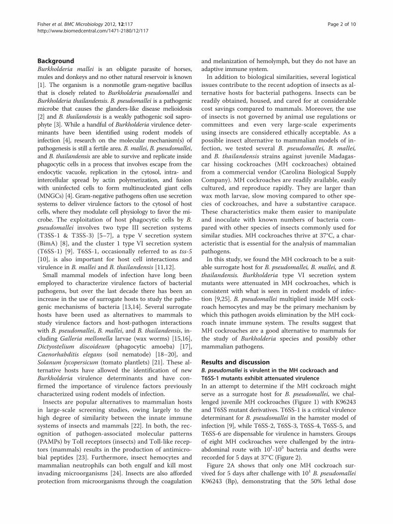
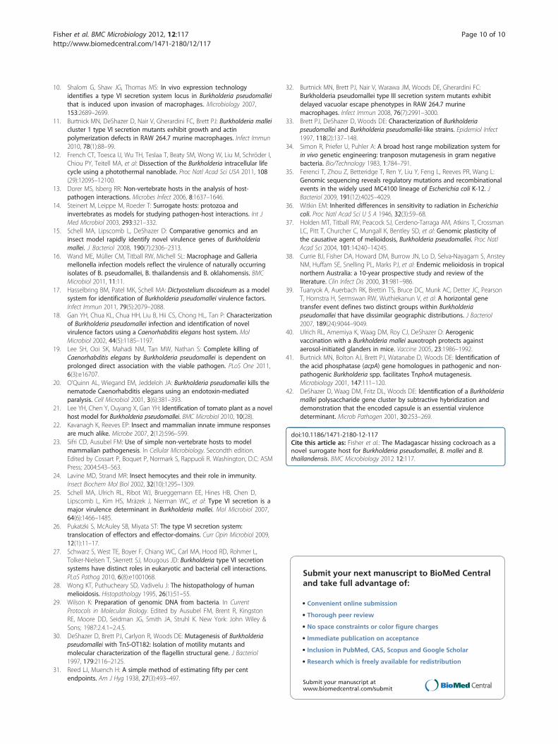
Figure 1 A representative juvenile Madagascar hissingcockroach used as a surrogate host for B. pseudomallei, B.mallei, and B. thailandensis infection studies. The black arrowsshow the locations where bacteria were inoculated into the dorsalabdominal section of the MH cockroach, between the third and thefifth terga from the posterior.
Liv
e ro
ache
sL
ive
roac
hes
Liv
e ro
ache
s
A
B
C
Figure 2 B. pseudomallei is virulent for the MH cockroach and T6SS-1challenged by the intra-abdominal route of infection and MH cockroach de103 cfu. (D) 104 cfu. (E) 105 cfu. Bp, K96243; Bp Δhcp1, DDS1498A; Bp ΔvgrG
Fisher et al. BMC Microbiology 2012, 12:117 Page 3 of 10http://www.biomedcentral.com/1471-2180/12/117
(LD50) is <10 bacteria. Similarly, the LD50 for K96243 inthe hamster model of infection was <10 bacteria [9]. B.pseudomallei Δhcp1 is a derivative of K96243 that lacksthe essential tail tube component of the T6SS-1 struc-tural apparatus (Hcp1) and is highly attenuated in thehamster [9,26]. B. pseudomallei Δhcp1 was also attenu-ated in the MH cockroach (Figure 2A-E) and the LD50
was ~ 2 x 10² bacteria on day 5, which was >20 timeshigher than the K96243 LD50 (Table 1). In addition, adose response was readily apparent with this strain. Asthe challenge dose increased from 101 to 105 bacteria,the number and rate of MH cockroach deaths increasedaccordingly (Figure 2A-E). It took a challenge dose of104 Δhcp1 to kill all eight MH cockroaches, whereas theminimum lethal dose for K96243 was only 102 bacteria(Figure 2). The results demonstrate that B. pseudomalleiis highly virulent in MH cockroaches and that T6SS-1 isa critical virulence factor in this insect host. Further-more, there is a clear correlation between the virulence
Liv
e ro
ache
sL
ive
roac
hes
D
E
mutants are attenuated. Groups of eight MH cockroaches wereaths were monitored for 5 days at 37°C. (A) 101 cfu. (B) 102 cfu. (C)1-5’, DDS1503-1A; Bp ΔvgrG1-3’, DDS1503-2A.
Table 1 Relative virulence of bacterial strains in Syrianhamsters and Madagascar hissing cockroaches
Bacterial strain SyrianhamsterLD50
a
Madagascarhissing
cockroach LD50
E. coli
MC4100 NDb > 105
B/r ND >105
B. pseudomallei
K96243 <10 <10
DDS1498A (Δhcp1) >1000 207
DDS0518A (Δhcp2) <10 <10
DDS2098A (Δhcp3) <10 <10
DDS0171A (Δhcp4) <10 <10
DDS0099A (Δhcp5) <10 <10
DDL3105A (Δhcp6) <10 <10
DDS1503-1A (ΔvgrG1-5’) 102 <10
DDS1503-2A (ΔvgrG1-3’) >450 <10
1026b <10 <10
MSHR305 ND <10
B. mallei
SR1 <10 <10
DDA0742 (Δhcp1) >103 >103
B. thailandensis
DW503 ND <10
DDII0868 (Δhcp1) ND >103
a LD50, 50% lethal dose [9,25,33]; b ND, not determined.
Fisher et al. BMC Microbiology 2012, 12:117 Page 4 of 10http://www.biomedcentral.com/1471-2180/12/117
capacity of B. pseudomallei in the MH cockroach andthe hamster (Table 1).B. pseudomallei ΔvgrG1-5’ and ΔvgrG1-3’ are K96243
derivatives that have deletions within the gene encodingthe tail spike protein (VgrG1) of the T6SS-1 structuralapparatus [9,26]. These mutants were more virulent thanB. pseudomallei Δhcp1 in the hamster model of infection[9], but were less virulent than K96243 (Table 1). Therewas no difference in the LD50 of ΔvgrG1-5’, ΔvgrG1-3’,and K96243 in MH cockroaches (Table 1), however,there was a slight but not statistically significant differ-ence in the time to death with these strains. In general,it took longer for MH cockroaches infected withΔvgrG1-5’ and ΔvgrG1-3’ to die relative to K96243(Figure 2A-C). Thus, these strains appear to have anintermediate virulence phenotype in both MH cock-roaches and in hamsters (Table 1 and Figure 2).We next examined the relative virulence of the B. pseu-
domallei Δhcp2, Δhcp3, Δhcp4, Δhcp5, and Δhcp6mutantsin MH cockroaches [9]. These mutants are each deficientin one of the other five T6SSs present in B. pseudomalleiand all are virulent in the hamster (Table 1). Figure 3
shows that these strains are also virulent in the MH cock-roach and all exhibit a clear dose response. The majorityof MH cockroaches infected with a challenge dose of 101
bacteria were dead by day 3 (Figure 3A), but most weredead by day 1 with a challenge dose of 105 bacteria(Figure 3E). Interestingly, the LD50 results with thesestrains are remarkably similar in both MH cockroachesand hamsters (Table 1).The virulence of two additional isolates of B. pseudo-
mallei and two isolates of Escherichia coli were also testedin the MH cockroach. The LD50s of B. pseudomallei1026b and MSHR305 were <10 bacteria and the LD50sfor E. coli MC4100 and B/r were >105 bacteria, the high-est dose tested (Table 1). The results suggest that viru-lence for the MH cockroach is common among B.pseudomallei isolates and that not all gram-negative bac-teria are pathogenic for this surrogate host (Table 1).Taken together, the results demonstrate that B.
pseudomallei is highly virulent in the MH cockroachand indicate that this insect might serve as a surrogatehost for high throughput virulence screening assays. Inaddition, the MH cockroach challenge results are con-sistent with what is seen in the hamster model of infec-tion and suggest that the primary function of the T6SS-1is to evade the innate immune system.
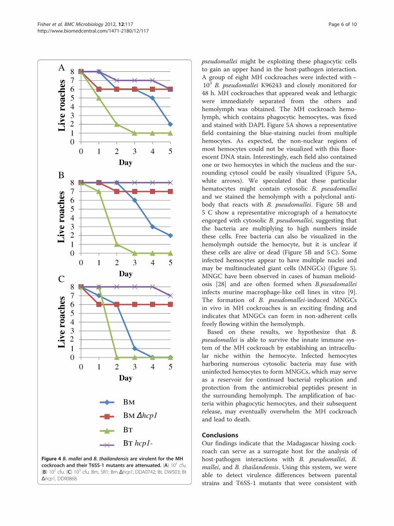
The MH cockroach can serve as a surrogate host forB. mallei and B. thailandensisWe also evaluated the virulence of B. mallei and B.thailandensis in the MH cockroach. The LD50s for B.mallei SR1 (Bm) and B. thailandensis DW503 (Bt) were< 10 bacteria (Table 1) and the number and rate ofdeaths increased as the challenge dose increased from101 to 103 bacteria (Figure 4). Interestingly, B. malleikilled the MH cockroaches at a slower rate than B.thailandensis (and B. pseudomallei). It took only 2 daysfor B. thailandensis to kill 75% of the MH cockroacheswith a dose of 101 bacteria, whereas it took B. mallei5 days (Figure 4A). Similar trends were apparent withchallenge doses of 102 and 103 bacteria (Figure 4Band C). B. mallei does not kill rodents as quickly asB. pseudomallei and it is more fastidious than B.pseudomallei and B. thailandensis, so it may not be toosurprising that it took longer to kill MH cockroaches [4].These experiments demonstrate that B. mallei and B.thailandensis are both virulent in the MH cockroach andsuggest that the MH cockroach might serve as a surrogatehost for these bacterial species.As mentioned above, B. thailandensis is considered
to be avirulent in humans and exhibits a higher LD50
in mammalian models of infection than B. mallei andB. pseudomallei. Mammals, unlike MH cockroaches,possess both an innate and an acquired immune sys-tem. The fact that B. thailandensis is highly virulent
Liv
e ro
ache
sL
ive
roac
hes
Liv
e ro
ache
s
Liv
e ro
ache
sL
ive
roac
hes
Figure 3 B. pseudomallei T6SS-2, T6SS-3, T6SS-4, T6SS-5, and T6SS-6 mutants are virulent in the MH cockroach. (A) 101 cfu. (B) 102 cfu.(C) 103 cfu. (D) 104 cfu. (E) 105 cfu. Bp, K96243; Bp Δhcp2, DDS0518A; Bp Δhcp3, DDS2098A; Bp Δhcp4, DDS0171A; Bp Δhcp5, DDS0099A; BpΔhcp6, DDL3105A.
Fisher et al. BMC Microbiology 2012, 12:117 Page 5 of 10http://www.biomedcentral.com/1471-2180/12/117
in the MH cockroach may suggest that the acquiredimmune system plays an important role in defenceagainst B. thailandensis. B. mallei and B. pseudomallei,on the other hand, may have developed mechanisms tosubvert the acquired immune response in mammalianspecies.T6SS-1 is a critical virulence determinant for B. mallei
in the hamster model of infection [25] and for B.thailandensis in the C57BL/6 mouse model of infection[27]. We challenged MH cockroaches with B. mallei andB. thailandensis hcp1 mutants and found that they werehighly attenuated in this surrogate host (Table 1 andFigure 4). The LD50s for B. mallei Δhcp1 and B.thailandensis hcp1- were> 103 bacteria on day 5, whichwas at least 100 times higher than their respective par-ental strains (Table 1 and Figure 4). The B. mallei resultswere indistinguishable from what was previously described
for SR1 and Δhcp1 using the hamster model of infection[25]. While the B. thailandensis strains used in this studyhave not been tested in hamsters, a B. thailandensis T6SS-1 mutant was recently shown to be avirulent in C57BL/6mice by the aerosol route of infection [27]. Interestingly,MyD88−/− mice were susceptible to the B. thailandensisT6SS-1 mutant, which suggests that T6SS-1 plays a rolein evading the innate immune response [27]. The factthat B. thailandensis hcp1- was attenuated in an insecthost, which lacks an adaptive immune response, furthersupports the notion that the function of the T6SS-1 isto evade the eukaryotic innate immune system.
B. pseudomallei replicates inside MH cockroachhemocytesHemocytes are a key component of the MH cockroachinnate immune system and we next examined if B.
Liv
e ro
ache
sL
ive
roac
hes
Liv
e ro
ache
s
Figure 4 B. mallei and B. thailandensis are virulent for the MHcockroach and their T6SS-1 mutants are attenuated. (A) 101 cfu.(B) 102 cfu. (C) 103 cfu. Bm, SR1; Bm Δhcp1, DDA0742; Bt, DW503; BtΔhcp1, DDII0868.
Fisher et al. BMC Microbiology 2012, 12:117 Page 6 of 10http://www.biomedcentral.com/1471-2180/12/117
pseudomallei might be exploiting these phagocytic cellsto gain an upper hand in the host-pathogen interaction.A group of eight MH cockroaches were infected with ~103 B. pseudomallei K96243 and closely monitored for48 h. MH cockroaches that appeared weak and lethargicwere immediately separated from the others andhemolymph was obtained. The MH cockroach hemo-lymph, which contains phagocytic hemocytes, was fixedand stained with DAPI. Figure 5A shows a representativefield containing the blue-staining nuclei from multiplehemocytes. As expected, the non-nuclear regions ofmost hemocytes could not be visualized with this fluor-escent DNA stain. Interestingly, each field also containedone or two hemocytes in which the nucleus and the sur-rounding cytosol could be easily visualized (Figure 5A,white arrows). We speculated that these particularhematocytes might contain cytosolic B. pseudomalleiand we stained the hemolymph with a polyclonal anti-body that reacts with B. pseudomallei. Figure 5B and5 C show a representative micrograph of a hematocyteengorged with cytosolic B. pseudomallei, suggesting thatthe bacteria are multiplying to high numbers insidethese cells. Free bacteria can also be visualized in thehemolymph outside the hemocyte, but it is unclear ifthese cells are alive or dead (Figure 5B and 5C). Someinfected hemocytes appear to have multiple nuclei andmay be multinucleated giant cells (MNGCs) (Figure 5).MNGC have been observed in cases of human melioid-osis [28] and are often formed when B.pseudomalleiinfects murine macrophage-like cell lines in vitro [9].The formation of B. pseudomallei-induced MNGCsin vivo in MH cockroaches is an exciting finding andindicates that MNGCs can form in non-adherent cellsfreely flowing within the hemolymph.Based on these results, we hypothesize that B.
pseudomallei is able to survive the innate immune sys-tem of the MH cockroach by establishing an intracellu-lar niche within the hemocyte. Infected hemocytesharboring numerous cytosolic bacteria may fuse withuninfected hemocytes to form MNGCs, which may serveas a reservoir for continued bacterial replication andprotection from the antimicrobial peptides present inthe surrounding hemolymph. The amplification of bac-teria within phagocytic hemocytes, and their subsequentrelease, may eventually overwhelm the MH cockroachand lead to death.
ConclusionsOur findings indicate that the Madagascar hissing cock-roach can serve as a surrogate host for the analysis ofhost-pathogen interactions with B. pseudomallei, B.mallei, and B. thailandensis. Using this system, we wereable to detect virulence differences between parentalstrains and T6SS-1 mutants that were consistent with
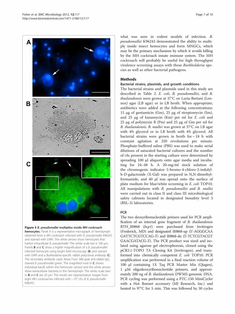
Figure 5 B. pseudomallei multiplies inside MH cockroachhemocytes. Panel A is a representative micrograph of hemolymphobtained from a MH cockroach infected with B. pseudomallei K96243and stained with DAPI. The white arrows show hemocytes thatharbor intracellular B. pseudomallei. The white scale bar is 100 μm.Panels B and C show a higher magnification of a B. pseudomallei-infected hemocyte using bright field microscopy (B) and stainedwith DAPI and a Burkholderia-specific rabbit polyclonal antibody (C).The secondary antibody used, Alexa Fluor 588 goat anti-rabbit IgG,stained B. pseudomallei green. The magnified inset in C showsindividual bacilli within the hemocyte cytosol and the white arrowsshow extracellular bacteria in the hemolymph. The white scale barsin B and C are 20 μm. The results are representative images fromeight MH cockroaches infected with ~ 103 cfu of B. pseudomalleiK96243.
Fisher et al. BMC Microbiology 2012, 12:117 Page 7 of 10http://www.biomedcentral.com/1471-2180/12/117
what was seen in rodent models of infection. B.pseudomallei K96243 demonstrated the ability to multi-ply inside insect hemocytes and form MNGCs, whichmay be the primary mechanism by which it avoids killingby the MH cockroach innate immune system. The MHcockroach will probably be useful for high throughputvirulence screening assays with these Burkholderia spe-cies as well as other bacterial pathogens.
MethodsBacterial strains, plasmids, and growth conditionsThe bacterial strains and plasmids used in this study aredescribed in Table 2. E. coli, B. pseudomallei, and B.thailandensis were grown at 37°C on Luria-Bertani (Len-nox) agar (LB agar) or in LB broth. When appropriate,antibiotics were added at the following concentrations:15 μg of gentamicin (Gm), 25 μg of streptomycin (Sm),and 25 μg of kanamycin (Km) per ml for E. coli and25 μg of polymyxin B (Pm) and 25 μg of Gm per ml forB. thailandensis. B. mallei was grown at 37°C on LB agarwith 4% glycerol or in LB broth with 4% glycerol. Allbacterial strains were grown in broth for ~ 18 h withconstant agitation at 250 revolutions per minute.Phosphate-buffered saline (PBS) was used to make serialdilutions of saturated bacterial cultures and the numberof cfu present in the starting culture were determined byspreading 100 μl aliquots onto agar media and incuba-ting for 24–48 h. A 20-mg/ml stock solution ofthe chromogenic indicator 5-bromo-4-chloro-3-indolyl-b-D-galactoside (X-Gal) was prepared in N,N-dimethyl-formamide, and 40 μl was spread onto the surface ofplate medium for blue/white screening in E. coli TOP10.All manipulations with B. pseudomallei and B. malleiwere carried out in class II and class III microbiologicalsafety cabinets located in designated biosafety level 3(BSL-3) laboratories.
PCRThe two deoxyribonucleotide primers used for PCR ampli-fication of an internal gene fragment of B. thailandensisBTH_II0868 (hcp1) were purchased from Invitrogen(Frederick, MD) and designated II0868-up (5’-AGGGCAAGATTCTCGTCCAG-3’) and II0868-dn (5’-TCTCGTACGTGAACGATACG-3’). The PCR product was sized and iso-lated using agarose gel electrophoresis, cloned using thepCR2.1-TOPO TA Cloning Kit (Invitrogen), and trans-formed into chemically competent E. coli TOP10. PCRamplification was performed in a final reaction volume of100 μl containing 1X Taq PCR Master Mix (Qiagen),1 μM oligodeoxyribonucleotide primers, and approxi-mately 200 ng of B. thailandensis DW503 genomic DNA.PCR cycling was performed using a PTC-150 MiniCyclerwith a Hot Bonnet accessory (MJ Research, Inc.) andheated to 97°C for 5 min. This was followed by 30 cycles
Table 2 Strains and plasmids used in this study
Strain or plasmid Relevant characteristicsa Source or reference
E. coli
TOP10 General cloning and blue/white screening Invitrogen
S17-1 Mobilizing strain with transfer genes of RP4 integratedon chromosome; Smr, Pms
[34]
MC4100 K-12 laboratory strain [35]
B/r B laboratory strain [36]
B. pseudomallei
K96243 Isolated in Thailand from a diabetic patient with a clinicalhistory of short incubation, septicemic infection, and rapid progression to death
[37]
DDS1498A K96243 derivative harboring a 162-bp in-frame deletion mutation in hcp1 (Δhcp1) [9]
DDS0518A K96243 derivative harboring a 303-bp in-frame deletion mutation in hcp2 (Δhcp2) [9]
DDS2098A K96243 derivative harboring a 186-bp in-frame deletion mutation in hcp3 (Δhcp3) [9]
DDS0171A K96243 derivative harboring a 321-bp in-frame deletion mutation in hcp4 (Δhcp4) [9]
DDS0099A K96243 derivative harboring a 192-bp in-frame deletion mutation in hcp5 (Δhcp5) [9]
DDL3105A K96243 derivative harboring a 216-bp in-frame deletion mutation in hcp6 (Δhcp6) [9]
DDS1503-1A K96243 derivative harboring a deletion of the 743-bp StuI fragment at the 5’ end ofvgrG1 (ΔvgrG1-5’)
[9]
DDS1503-2A K96243 derivative harboring a deletion of the 894-bp PstI fragment at the 3’ end ofvgrG1 (ΔvgrG1-3’)
[9]
1026b Isolated in Thailand from a human case of septicemic melioidosis with skin, soft tissue,and spleen involvement
[30]
MSHR305 Isolated from the brain of a fatal human melioidosis encephalomyelitis case in Australia [38,39]
B. mallei
SR1 ATCC 23344 sucrose-resistant derivative [40]
DDA0742 SR1 derivative harboring a deletion of the 156 bp NarI–SfuI fragment internalto hcp1; Δhcp1
[25]
B. thailandensis
DW503 E264 derivative; Δ(amrR-oprA) (Gms) rpsL (Smr) [41]
DDII0868 DW503::pGSV3-0868; Gmr; hcp1- This study
Plasmids
pCR2.1-TOPO 3,931-bp TA vector; pMB1 oriR; Kmr Invitrogen
pCR2.1-0868 pCR2.1-TOPO containing 342-bp PCR product generated with II0868-upand II0868-dn
This study
pGSV3 Mobilizabile Gmr suicide vector [42]
pGSV3-0868 pGSV3 derivative containing EcoRI insert from pCR2.1-0868 This studya r, resistant; s, susceptible.
Fisher et al. BMC Microbiology 2012, 12:117 Page 8 of 10http://www.biomedcentral.com/1471-2180/12/117
of a three-temperature cycling protocol (97°C for 30 s,55°C for 30 s, and 72°C for 1 min) and one cycle at 72°Cfor 10 min.
DNA manipulation and plasmid conjugationRestriction enzymes, Antarctic phosphatase, and T4DNA ligase were purchased from Roche Molecular Bio-chemicals and were used according to the manufac-turer’s instructions. DNA fragments used in cloningprocedures were excised from agarose gels and purifiedwith a GeneClean III kit (Q � BIOgene). Bacterial gen-omic DNA was prepared by a previously described
protocol [29]. Plasmids were purified from overnightcultures by using Wizard Plus SV Minipreps (Promega).Plasmid pGSV3-0868 (Table 2) was electroporated intoE. coli S17-1 (12.25 kV/cm) and conjugated with B.thailandensis for 8 h, as described elsewhere [30]. Pmwas used to counterselect E. coli S17-1 (pGSV3-0868).
MH cockroach housing and manipulationMadagascar hissing cockroaches, Gromphadorhinalaevigata, were purchased from Carolina BiologicalSupply Company (Burlington, NC) as 1–2 inchnymphs (Figure 1) and were housed in the dark at
Fisher et al. BMC Microbiology 2012, 12:117 Page 9 of 10http://www.biomedcentral.com/1471-2180/12/117
room temperature in a 7.5" w x 11.75 l x 5" h mousecage with a filtered top (Allentown Caging EquipmentCo., Inc., Allentown, NJ). The bottom of the cage waslined with cocoa mulch and a thin layer of petroleumjelly was spread around the top portion of the cage toprevent MH cockroaches from climbing up the sides.Dog food was spread on the bottom of the cage forfood and the top of a petri dish was inverted and filledwith water for drinking. On occasion, sliced applewedges were placed in the cage as an additional sourceof food.For bacterial infection experiments, 1.5-2 inch ju-
venile MH cockroaches were used (Figure 1). We alsotested larger MH cockroaches (> 3 inches) and theydisplayed the same susceptibility as the juveniles (datanot shown). Bacteria were inoculated into the dorsalabdominal section of MH cockroaches, between thethird and the fifth terga (from the posterior), using a1 ml syringe fitted with a 3/8 inch, 26-gauge needle(see Figure 1). The syringe was loaded into a TridakSTEPPER series repetitive pipette (Tridak LLC, Tor-rington, CT) and a 25 μl aliquot was injected intoMH cockroaches. A group of eight infected MHcockroaches were placed in a 16-ounce plastic con-tainer with a few pieces of dog food and 1–2 ml ofwater. The containers were placed in a 37°C incuba-tor and deaths were recorded for 5 days. Food andwater levels were checked daily and replenished ifneeded. The LD50s were calculated 5 days postinfec-tion according to the Reed-Muench method [31].
Extraction and staining of hemolymph from infected MHcockroachesEight MH cockroaches were infected with ~ 103 B. pseu-domallei K96243 and monitored daily as describedabove. Hemolymph was extracted from MH cockroachesthat were lethargic and on the verge of death. Holdingthe MH cockroach with its ventral side up, one hindleg was folded up towards the head to expose themembrane at the base of the leg. The membrane waspunctured with a 26-gauge needle and hemolymphwas immediately collected using a P200 GilsonPIPETMAN. We used a pipette tip cut with scissorsapproximately a 1/2 inch from the end to aid in up-take of the viscous hemolyph. The amber-coloredhemolymph was transferred to a glass slide, allowedto air dry, and then fixed with methanol. The sampleswere initially stained with 4′, 6-diamidino-2-phenylindole(DAPI) and viewed on a Nikon Eclipse TE2000-S invertedmicroscope equipped with a Spot-RT digital camera(Image Systems, Columbia, MD). Subsequently, the sam-ples were incubated for 1 h with a 1:1000 dilution of rabbitpolyclonal Burkholderia antiserum [32] and then reactedfor 1 h with a 1:500 dilution of an Alexa Fluor 588 goat
anti-rabbit IgG secondary antibody (Molecular Probes)and visualized by fluorescence microscopy.
AbbreviationsMH: Cockroach; Madagascar: Hissing cockroach; T6SS-1: Ccluster 1 type VIsecretion system; LD50: 50% lethal dose; MNGC: Multinucleated giant cell;Cfu: Colony forming units; DAPI: 4′, 6-diamidino-2-phenylindole.
Authors’ contributionsNAF conceived use of the MH cockroach as a surrogate host, contributed tothe experimental design, and helped draft the manuscript. WJR was involvedwith the extraction, staining, and fluorescence microscopy of MH cockroachhemolymph. WA participated in the study design and conductedexperiments. DD designed and conducted the experiments and drafted themanuscript. All authors read and approved the final manuscript.
Authors’ informationOpinions, interpretations, conclusions, and recommendations are those ofthe author and are not necessarily endorsed by the U.S. Army or Departmentof Homeland Security.
AcknowledgementsThis project received support from DTRA/JSTO-CBD proposal number CBS.MEDBIO.02.10.RD.034 (to D.D.).
Author details1Center for Genomic Sciences, United States Army Medical Research Instituteof Infectious Diseases, Fort Detrick, Frederick, MD 21702, USA. 2BacteriologyDivision, United States Army Medical Research Institute of InfectiousDiseases, 1425 Porter St., Fort Detrick, Frederick, MD (301)21702-5011, USA. 3University of Maryland School of Medicine, Baltimore21201 (WA), MD, USA.
Received: 18 April 2012 Accepted: 7 June 2012Published: 22 June 2012
References1. Waag DM, DeShazer D: Glanders: new insights into an old disease. In
Biological Weapons Defense: Infectious Diseases and Counterbioterrorism.Edited by Lindler LE, Lebeda FJ, Korch GW. Totowa, New Jersey: HumanaPress Inc; 2004:209–237.
2. Vietri NJ, DeShazer D: Melioidosis. In Medical Aspects of Biological Warfare.Edited by Dembek ZF. Washington, DC: Department of the Army, Office ofThe Surgeon General, Borden Institute; 2007:147–166.
3. Brett PJ, DeShazer D, Woods DE: Burkholderia thailandensis sp. nov.,description of a Burkholderia pseudomallei-like species. Int J Syst Bacteriol1998, 48:317–320.
4. Galyov EE, Brett PJ, DeShazer D: Molecular insights into Burkholderiapseudomallei and Burkholderia mallei pathogenesis. Annu Rev Microbiol2010, 64:495–517.
5. D'Cruze T, Gong L, Treerat P, Ramm G, Boyce JD, Prescott M, Adler B,Devenish RJ: Role for the Burkholderia pseudomallei type threesecretion system cluster 1 bpscN gene in virulence. Infect Immun 2011,79(9):3659–3664.
6. Stevens MP, Haque A, Atkins T, Hill J, Wood MW, Easton A, Nelson M,Underwood-Fowler C, Titball RW, Bancroft GJ, et al: Attenuated virulenceand protective efficacy of a Burkholderia pseudomallei bsa type IIIsecretion mutant in murine models of melioidosis. Microbiology 2004,150(Pt 8):2669–2676.
7. Warawa J, Woods DE: Type III secretion system cluster 3 is required formaximal virulence of Burkholderia pseudomallei in a hamster infectionmodel. FEMS Microbiol Lett 2005, 242:101–108.
8. Stevens MP, Stevens JM, Jeng RL, Taylor LA, Wood MW, Hawes P,Monaghan P, Welch MD, Galyov EE: Identification of a bacterial factorrequired for actin-based motility of Burkholderia pseudomallei. MolMicrobiol 2005, 56:40–53.
9. Burtnick MN, Brett PJ, Harding SV, Ngugi SA, Ribot WJ, Chantratita N,Scorpio A, Milne TS, Dean RE, Fritz DL, et al: The cluster 1 type VI secretionsystem is a major virulence determinant in Burkholderia pseudomallei.Infect Immun 2011, 79(4):1512–1525.
Fisher et al. BMC Microbiology 2012, 12:117 Page 10 of 10http://www.biomedcentral.com/1471-2180/12/117
10. Shalom G, Shaw JG, Thomas MS: In vivo expression technologyidentifies a type VI secretion system locus in Burkholderia pseudomalleithat is induced upon invasion of macrophages. Microbiology 2007,153:2689–2699.
11. Burtnick MN, DeShazer D, Nair V, Gherardini FC, Brett PJ: Burkholderia malleicluster 1 type VI secretion mutants exhibit growth and actinpolymerization defects in RAW 264.7 murine macrophages. Infect Immun2010, 78(1):88–99.
12. French CT, Toesca IJ, Wu TH, Teslaa T, Beaty SM, Wong W, Liu M, Schröder I,Chiou PY, Teitell MA, et al: Dissection of the Burkholderia intracellular lifecycle using a photothermal nanoblade. Proc Natl Acad Sci USA 2011, 108(29):12095–12100.
13. Dorer MS, Isberg RR: Non-vertebrate hosts in the analysis of host-pathogen interactions. Microbes Infect 2006, 8:1637–1646.
14. Steinert M, Leippe M, Roeder T: Surrogate hosts: protozoa andinvertebrates as models for studying pathogen-host interactions. Int JMed Microbiol 2003, 293:321–332.
15. Schell MA, Lipscomb L, DeShazer D: Comparative genomics and aninsect model rapidly identify novel virulence genes of Burkholderiamallei. J Bacteriol 2008, 190(7):2306–2313.
16. Wand ME, Müller CM, Titball RW, Michell SL: Macrophage and Galleriamellonella infection models reflect the virulence of naturally occurringisolates of B. pseudomallei, B. thailandensis and B. oklahomensis. BMCMicrobiol 2011, 11:11.
17. Hasselbring BM, Patel MK, Schell MA: Dictyostelium discoideum as a modelsystem for identification of Burkholderia pseudomallei virulence factors.Infect Immun 2011, 79(5):2079–2088.
18. Gan YH, Chua KL, Chua HH, Liu B, Hii CS, Chong HL, Tan P: Characterizationof Burkholderia pseudomallei infection and identification of novelvirulence factors using a Caenorhabditis elegans host system. MolMicrobiol 2002, 44(5):1185–1197.
19. Lee SH, Ooi SK, Mahadi NM, Tan MW, Nathan S: Complete killing ofCaenorhabditis elegans by Burkholderia pseudomallei is dependent onprolonged direct association with the viable pathogen. PLoS One 2011,6(3):e16707.
20. O'Quinn AL, Wiegand EM, Jeddeloh JA: Burkholderia pseudomallei kills thenematode Caenorhabditis elegans using an endotoxin-mediatedparalysis. Cell Microbiol 2001, 3(6):381–393.
21. Lee YH, Chen Y, Ouyang X, Gan YH: Identification of tomato plant as a novelhost model for Burkholderia pseudomallei. BMC Microbiol 2010, 10(28).
22. Kavanagh K, Reeves EP: Insect and mammalian innate immune responsesare much alike. Microbe 2007, 2(12):596–599.
23. Sifri CD, Ausubel FM: Use of simple non-vertebrate hosts to modelmammalian pathogenesis. In Cellular Microbiology. Secondth edition.Edited by Cossart P, Boquet P, Normark S, Rappuoli R. Washington, D.C: ASMPress; 2004:543–563.
24. Lavine MD, Strand MR: Insect hemocytes and their role in immunity.Insect Biochem Mol Biol 2002, 32(10):1295–1309.
25. Schell MA, Ulrich RL, Ribot WJ, Brueggemann EE, Hines HB, Chen D,Lipscomb L, Kim HS, Mrázek J, Nierman WC, et al: Type VI secretion is amajor virulence determinant in Burkholderia mallei. Mol Microbiol 2007,64(6):1466–1485.
26. Pukatzki S, McAuley SB, Miyata ST: The type VI secretion system:translocation of effectors and effector-domains. Curr Opin Microbiol 2009,12(1):11–17.
27. Schwarz S, West TE, Boyer F, Chiang WC, Carl MA, Hood RD, Rohmer L,Tolker-Nielsen T, Skerrett SJ, Mougous JD: Burkholderia type VI secretionsystems have distinct roles in eukaryotic and bacterial cell interactions.PLoS Pathog 2010, 6(8):e1001068.
28. Wong KT, Puthucheary SD, Vadivelu J: The histopathology of humanmelioidosis. Histopathology 1995, 26(1):51–55.
29. Wilson K: Preparation of genomic DNA from bacteria. In CurrentProtocols in Molecular Biology. Edited by Ausubel FM, Brent R, KingstonRE, Moore DD, Seidman JG, Smith JA, Struhl K. New York: John Wiley &Sons; 1987:2.4.1–2.4.5.
30. DeShazer D, Brett PJ, Carlyon R, Woods DE: Mutagenesis of Burkholderiapseudomallei with Tn5-OT182: Isolation of motility mutants andmolecular characterization of the flagellin structural gene. J Bacteriol1997, 179:2116–2125.
31. Reed LJ, Muench H: A simple method of estimating fifty per centendpoints. Am J Hyg 1938, 27(3):493–497.
32. Burtnick MN, Brett PJ, Nair V, Warawa JM, Woods DE, Gherardini FC:Burkholderia pseudomallei type III secretion system mutants exhibitdelayed vacuolar escape phenotypes in RAW 264.7 murinemacrophages. Infect Immun 2008, 76(7):2991–3000.
33. Brett PJ, DeShazer D, Woods DE: Characterization of Burkholderiapseudomallei and Burkholderia pseudomallei-like strains. Epidemiol Infect1997, 118(2):137–148.
34. Simon R, Priefer U, Puhler A: A broad host range mobilization system forin vivo genetic engineering: tranposon mutagenesis in gram negativebacteria. Bio/Technology 1983, 1:784–791.
35. Ferenci T, Zhou Z, Betteridge T, Ren Y, Liu Y, Feng L, Reeves PR, Wang L:Genomic sequencing reveals regulatory mutations and recombinationalevents in the widely used MC4100 lineage of Escherichia coli K-12. JBacteriol 2009, 191(12):4025–4029.
36. Witkin EM: Inherited differences in sensitivity to radiation in Escherichiacoli. Proc Natl Acad Sci U S A 1946, 32(3):59–68.
37. Holden MT, Titball RW, Peacock SJ, Cerdeno-Tarraga AM, Atkins T, CrossmanLC, Pitt T, Churcher C, Mungall K, Bentley SD, et al: Genomic plasticity ofthe causative agent of melioidosis, Burkholderia pseudomallei. Proc NatlAcad Sci 2004, 101:14240–14245.
38. Currie BJ, Fisher DA, Howard DM, Burrow JN, Lo D, Selva-Nayagam S, AnsteyNM, Huffam SE, Snelling PL, Marks PJ, et al: Endemic melioidosis in tropicalnorthern Australia: a 10-year prospective study and review of theliterature. Clin Infect Dis 2000, 31:981–986.
39. Tuanyok A, Auerbach RK, Brettin TS, Bruce DC, Munk AC, Detter JC, PearsonT, Hornstra H, Sermswan RW, Wuthiekanun V, et al: A horizontal genetransfer event defines two distinct groups within Burkholderiapseudomallei that have dissimilar geographic distributions. J Bacteriol2007, 189(24):9044–9049.
40. Ulrich RL, Amemiya K, Waag DM, Roy CJ, DeShazer D: Aerogenicvaccination with a Burkholderia mallei auxotroph protects againstaerosol-initiated glanders in mice. Vaccine 2005, 23:1986–1992.
41. Burtnick MN, Bolton AJ, Brett PJ, Watanabe D, Woods DE: Identification ofthe acid phosphatase (acpA) gene homologues in pathogenic and non-pathogenic Burkholderia spp. facilitates TnphoA mutagenesis.Microbiology 2001, 147:111–120.
42. DeShazer D, Waag DM, Fritz DL, Woods DE: Identification of a Burkholderiamallei polysaccharide gene cluster by subtractive hybridization anddemonstration that the encoded capsule is an essential virulencedeterminant. Microb Pathogen 2001, 30:253–269.
doi:10.1186/1471-2180-12-117Cite this article as: Fisher et al.: The Madagascar hissing cockroach as anovel surrogate host for Burkholderia pseudomallei, B. mallei and B.thailandensis. BMC Microbiology 2012 12:117.
Submit your next manuscript to BioMed Centraland take full advantage of:
• Convenient online submission
• Thorough peer review
• No space constraints or color figure charges
• Immediate publication on acceptance
• Inclusion in PubMed, CAS, Scopus and Google Scholar
• Research which is freely available for redistribution
Submit your manuscript at www.biomedcentral.com/submit