Embed Size (px)
Citation preview

Research ArticlePrevalence of Tinea Capitis among School Children in NokCommunity of Kaduna State, Nigeria
Josephine Dogo,1 Seniyat Larai Afegbua,1 and Edward Christopher Dung2
1Department of Microbiology, Ahmadu Bello University, Zaria, Kaduna State, Nigeria2Department of Dermatophilosis, National Veterinary Research Institute, Vom, Plateau State, Nigeria
Correspondence should be addressed to Seniyat Larai Afegbua; [email protected]
Received 31 October 2015; Revised 28 March 2016; Accepted 17 April 2016
Academic Editor: Nongnuch Vanittanakom
Copyright © 2016 Josephine Dogo et al.This is an open access article distributed under the Creative CommonsAttribution License,which permits unrestricted use, distribution, and reproduction in any medium, provided the original work is properly cited.
In recent years, the prevalence of tinea capitis, an infection of the scalp by dermatophytes, has increased in children worldwide.This cross-sectional study was carried out to determine the prevalence and risk factor of tinea capitis among school children inNok community of Kaduna State, Nigeria. A total of 100 children were screened and 45% were diagnosed to have tinea capitis afterfungal culture and microscopy. The prevalence of tinea capitis among girls was higher (51.4%) than that among boys (41.5%) butnot significantly different (𝑝 = 0.402). The prevalence with respect to age was lower for the age group 5–10 years (42.6%) than thatof 11–15 years (50%) but was not significantly different (𝑝 = 0.524). Trichophyton rubrum (28.8%) andMicrosporum canis (22.7%)were the most prevalent dermatophytes isolated and the least were Trichophyton verrucosum (4.5%) and Trichophyton tonsurans(4.5%). There were 73.3% single infection while 26.7% had 2–4 dermatophytes of the genera Microsporum and Trichophyton. Thepredisposing factors with statistically significant association with tinea capitis were number of children in the family (𝑝 = 0.02)and sharing of the same bed (𝑝 = 0.002). This indicates the high tendencies of spread of tinea capitis through human-to-humanmode of transmission and possible animal contact. Community health education on the cause, mode of transmission, prevention,and prompt treatment of tinea capitis is recommended.
1. Introduction
Dermatophytosis refers to mycotic infections caused by agroup of fungi that possess the propensity to colonize andinfect superficial layers of the skin, hair, or nails [1]. Thisgroup of fungi that invade keratinized tissue associated withthe stratum corneum of the skin, hair, and nails on the livinghost are called dermatophytes [1]. Dermatophytosis is causedby a group of fungi with predilection to colonize and infectstratum corneumof the skin, hair, and nails on the living host.Such fungi referred to as dermatophytes include members ofthe genera Epidermophyton, Microsporum, and Trichophyton[1]. Dermatophytes are grouped as either anthropophilic(e.g., Trichophyton violaceum, Trichophyton rubrum, andMicrosporum audouinii), geophilic (e.g., Microsporum gyp-seum andMicrosporumnanum), or zoophilic (e.g.,Trichophy-ton mentagrophytes, Trichophyton verrucosum, andMicrospo-rum canis) based on their ecological niches; however, some
dermatophytes are not strictly restricted to a particularecological niche [1, 2].
Dermatophytosis has a worldwide distribution but isendemic to tropical regions as the growth of dermatophytesis facilitated by the warm andmoist conditions [1, 3, 4]. Apartfrom climate, the variability in distribution of dermatophytesworldwide is attributed to other factors such as populationmigration patterns, lifestyle, primary host range, secondaryhost immunity, presence of immunodeficiency diseases, andpatients attitude to prompt treatment following clinical pre-sentation and standard of living [1, 5].
Depending on the body site affected, dermatophytosismay result in different clinical syndromes such as tinea capitis(hair shaft and scalp), tinea corporis (body), tinea cruris(groin), tinea pedis (foot), and tinea unguium (hand). Tineacapitis is an infection of the scalp and hair shaft caused byseveral species of Trichophyton and Microsporum [1, 6–8].It is the most common dermatophytosis in children aged
Hindawi Publishing CorporationJournal of PathogensVolume 2016, Article ID 9601717, 6 pageshttp://dx.doi.org/10.1155/2016/9601717
2 Journal of Pathogens
between six months and prepubertal age [8–11]. Tinea capitisis reported more in males than in females within prepubertalage [7, 12].
Tinea capitis is quite common inAfrica and its prevalenceamong children is 14–86% [1]. Studies carried out in differentparts of Nigeria have proven that causative agents of tineacapitis vary fromone location to another.Nweze [13] reportedTrichophyton schoenleinii as the predominant cause of tineacapitis in Borno. In a similar study carried out on 2150 itiner-ant Quranic scholars in Kano State,Trichophyton rubrumwasthe most prevalent followed by Microsporum audouinii [14].Ayanlowo et al. [8] reported Trichophyton mentagrophytes asthe most prevalent causative agent in a community in thesouthwestern part ofNigeria.Nweze andOkafor [15] reportedTrichophyton tonsurans as the most prevalent aetiologicalagent of tinea capitis in Anambra State, Nigeria.
Studies are required to obtain information on its epi-demiology and familial, social, and geographical factors[16, 17]. Although there are studies on prevalence of tineacapitis in different parts of Nigeria, there are no reports onepidemiologic studies on tinea capitis among school childrenin Kaduna State. Nok community, a rural settlement in Jabalocal government area of Kaduna State, was selected for thisstudy.This study was designed to determine the prevalence oftinea capitis and identify factors associated with the infectionamong primary school children in Nok community, KadunaState, Nigeria.
2. Methods
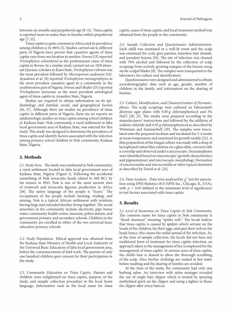
2.1. StudyArea. Thestudywas conducted inNok community,a rural settlement located in Jaba local government area ofKaduna State, Nigeria (Figure 1). Following the accidentalunearthing of Nok terracotta heads (dated to 500 BC) bytin miners in 1943, Nok is one of the most ancient sitesof ironwork and terracotta figurine production in Africa[18]. The native language of the people is “hyam.” Theoccupations of the people include farming, trading, andmining. Nok is a typical African settlement with residentshaving large and extended families living together.The socialamenities in the community include electricity, pipe bornewater, community health centre, museum, police station, andgovernment primary and secondary schools. Children in thecommunity are enrolled in either of the two universal basiceducation primary schools.
2.2. Study Population. Ethical approval was obtained fromthe Kaduna State Ministry of Health and Local Authority ofthe Universal Basic Education of Jaba local government area,before the commencement of field work. The parents of onlyone hundred children gave consent for their participation inthe study.
2.3. Community Education on Tinea Capitis. Parents andchildren were enlightened on tinea capitis, purpose of thestudy, and sample collection procedure in the local hyamlanguage. Information such as the local name for tinea
capitis, cause of tinea capitis, and local treatmentmethod wasobtained from the people in the community.
2.4. Sample Collection and Questionnaire Administration.Each child was examined in a well-lit room and the scalpwas examined for scaly grey patches, lusterless hair strands,and purulent lesions [19]. The site of infection was cleanedwith 70% alcohol and followed by the collection of scalpscrapings from actively growing margins of the lesions usingsterile scalpel blades [8].The samples were transported to thelaboratory for culture and identification.
Questionnaireswere designed and administered to obtainsociodemographic data such as age, gender, number ofchildren in the family, and information on the sharing offomites.
2.5. Culture, Identification, and Characterization of Dermato-phytes. The scalp scrapings were cultured on Sabouraud’sdextrose agar plates with 0.05 g chloramphenicol and 5%NaCl [20, 21]. The media were prepared according to themanufacturers’ instructions and followed by the addition ofsodium chloride and 0.05 g chloramphenicol as described byWeitzman and Summerbell [20]. The samples were inocu-lated onto the prepared medium and incubated for 3-4 weeksat room temperature and examined for growthweekly [22]. Athin preparation of the fungal culturewasmadewith a drop oflactophenol cotton blue solution on a glass slide, coveredwitha coverslip and observed under amicroscope.Dermatophyteswere identified based onmacroscopic (growth characteristicsand pigmentation) and microscopic morphology (formationof macroconidia andmicroconidia or other typical elements)as described by David et al. [21].
2.6. Data Analysis. Data were analysed by 𝜒2 test for associa-tion using SPSS Statistics 18.0 (SPSS Inc., Chicago, IL, USA),with 𝑝 < 0.05 defined as the minimum level of significanceto test factors associated with tinea capitis.
3. Results
3.1. Level of Awareness on Tinea Capitis in Nok Community.The common name for tinea capitis in Nok community is“shaak shantaan” meaning “spider web.” The locals believethat tinea capitis is caused by spiders which urinate on theheads of the children, lay their eggs, and spin their web on thehead; hence, this causes the radial spread of the infection. As,at the time of sample collection, the locals did not have anytraditional form of treatment for tinea capitis infection, anapproach taken in themanagement of lice is employed for themanagement of tinea capitis. In serious cases of tinea capitis,the child’s hair is shaved to allow the thorough scrubbingof the scalp. Also, his/her clothings are soaked in hot waterbefore washing and the sharing of fomites are avoided.
At the time of the study, the community had only onebarbing salon. An interview with salon manager revealedthe use of single hair clipper which is treated by sprayingmethylated spirit on the clipper and using a lighter to flamethe clipper after every haircut.
Journal of Pathogens 3
Figure 1: Maps showing Nok village in Kaduna State, Nigeria (taken from Google Maps).
3.2. Sociodemographic Characteristics of Study Subjects. Atotal of 100 scalp scrapings were collected and analysed fordermatophytes. The characteristics of the study participantsare shown in Table 1. The age range of the study participantswas 5–15 years. Of the 100 participants, 65% (65/100) weremale and 35% (35/100) were female, giving a gender ratio of1.86 : 1 (males : females). 68% and 32%of the participantswereof the age range 5–10 and 11–15 years, respectively. 74% of thestudy participants had a family size of 1–5 members and 26%of the participants had a family size of 6–10 members. Theaverage number of children per family was 4.3.
3.3. Prevalence of Tinea Capitis. After the incubation periodand examining the microscopic and macroscopic features offungal growth, there were 45 cases of dermatophytosis ofwhich 33 cases and 12 cases had single and mixed dermato-phytes, respectively. The overall prevalence of tinea capitis inthe study population is 45%. Tinea capitis wasmore prevalentamong females (51.4%; 18/35) than in males (41.5%; 27/65).However, the gender ratio in terms of tinea capitis was 1 : 1.3(male : female) and did not differ significantly (𝑝 = 0.402).The prevalence of tinea capitis with respect to age was lowerfor the age group 5–10 years (42.6%; 29/68) than that of 11–15 years (50%; 16/32) but was not significantly different (𝑝 =0.524).There was an association between tinea capitis and thenumber of children in a family (𝑝 = 0.02) and sharing of bed(𝑝 = 0.002) (Table 1).There was no association between tineacapitis and variables such as sharing of combs (𝑝 = 0.188),sharing of caps/scarves (𝑝 = 0.107), sharing of towels, hair cutat a barbing salon (𝑝 = 0.28), keeping and playing with pets(𝑝 = 1.00), and carrying objects on a bare head (𝑝 = 1.00).
3.4. Dermatophytes Frequency. Of the 45 cases, a total of66 dermatophytes were isolated belonging to the generaMicrosporum and Trichophyton (Table 2). The most fre-quently isolated dermatophyte was Trichophyton rubrum(28.8%; 19/66), followed byMicrosporum canis (22.7%; 15/66),and the least were Trichophyton verrucosum (4.5%; 3/66)and Trichophyton tonsurans (4.5%; 3/66). Although 33 cases(73.3%) had single infections with dermatophytes, mixedinfections with 2–4 dermatophytes of the genera Microspo-rum and Trichophyton were observed in 12 cases (26.7%).This is presented in relation to age group and gender inTable 3. 75% (9/12) of the mixed infections were with
Microsporum and Trichophyton species, 16.7% (2/12) werewith Microsporum species alone, and 8.3% (1/12) was withTrichophyton species alone.
4. Discussion
The high prevalence of tinea capitis (45%) is close to theprevalence of 43.53% obtained in a study by Akinboroet al. [19] in Osogbo, southwestern Nigeria. However, theprevalence of this study was higher than those obtained inother studies: 9.5% for study carried out on 2150 Quranicscholars in Kano State [14], 15.4% for a study on 604 childrenin a rural settlement in southwestern Nigeria [8], and 9.4%for a study in Anambra State involving 47723 children [23].Moto et al. [24] recently reported a higher prevalence of 81.2%(122/150) among school children in Mathare, an informalsettlement in Nairobi, Kenya. A number of factors may beattributed to the varying prevalence and frequency of tineacapitis in different parts of Nigeria as well as other countries[22]. These include population growth, close contact amonginfected children at home and school, and poor personalhygiene [24].
With respect to gender, prevalence was higher amongfemales than males, but the association was not significant(𝑝 = 0.402). This is supported by the findings of some pre-vious studies that reported higher infections among femalesin Nigeria [25] and Egypt [26].
Furthermore, it was observed that the prevalence of tineacapitis in prepubertal females (56.7%; 17/30) was higher thanthat of the males (35.3%; 12/34) while the prevalence of tineacapitis among the pubertal age range of 11–15 years was higherin males (48.4%; 15/31) compared to females (20%; 1/5). Thisis contrary to the findings of Sidat et al. [12] and Balci etal. [7]. This is contrary to some findings and the suggestionthat scalp infection in females is less due to steroid-mediatedinhibition of dermatophyte growth by progesterone and othersimilar compounds while males may be predisposed to scalpinfection due to prepubertal factors such as level of fungistaticfatty acids [23].
Various conflicting views exist regarding the sexual pre-dominance of tinea capitis which may be attributed to hairdressing and styling practices such as tight hair braiding,shaving of the scalp, plaiting, and the use of hair oils whichmay promote disease transmission. However, the precise roleof such practices remains a subject of study [3, 27, 28].
4 Journal of Pathogens
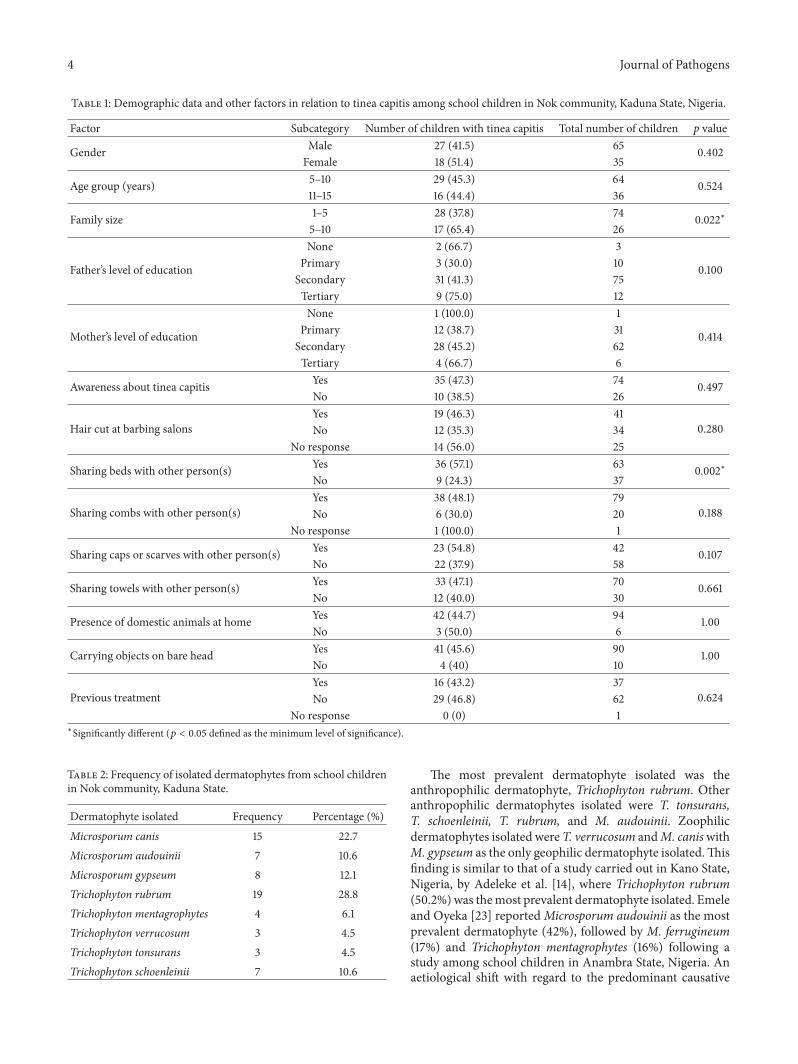
Table 1: Demographic data and other factors in relation to tinea capitis among school children in Nok community, Kaduna State, Nigeria.
Factor Subcategory Number of children with tinea capitis Total number of children p value
Gender Male 27 (41.5) 65 0.402Female 18 (51.4) 35
Age group (years) 5–10 29 (45.3) 64 0.52411–15 16 (44.4) 36
Family size 1–5 28 (37.8) 74 0.022∗5–10 17 (65.4) 26
Father’s level of education
None 2 (66.7) 3
0.100Primary 3 (30.0) 10Secondary 31 (41.3) 75Tertiary 9 (75.0) 12
Mother’s level of education
None 1 (100.0) 1
0.414Primary 12 (38.7) 31Secondary 28 (45.2) 62Tertiary 4 (66.7) 6
Awareness about tinea capitis Yes 35 (47.3) 74 0.497No 10 (38.5) 26
Hair cut at barbing salonsYes 19 (46.3) 41
0.280No 12 (35.3) 34No response 14 (56.0) 25
Sharing beds with other person(s) Yes 36 (57.1) 63 0.002∗No 9 (24.3) 37
Sharing combs with other person(s)Yes 38 (48.1) 79
0.188No 6 (30.0) 20No response 1 (100.0) 1
Sharing caps or scarves with other person(s) Yes 23 (54.8) 42 0.107No 22 (37.9) 58
Sharing towels with other person(s) Yes 33 (47.1) 70 0.661No 12 (40.0) 30
Presence of domestic animals at home Yes 42 (44.7) 94 1.00No 3 (50.0) 6
Carrying objects on bare head Yes 41 (45.6) 90 1.00No 4 (40) 10
Previous treatmentYes 16 (43.2) 37
0.624No 29 (46.8) 62No response 0 (0) 1
∗Significantly different (𝑝 < 0.05 defined as the minimum level of significance).
Table 2: Frequency of isolated dermatophytes from school childrenin Nok community, Kaduna State.
Dermatophyte isolated Frequency Percentage (%)Microsporum canis 15 22.7Microsporum audouinii 7 10.6Microsporum gypseum 8 12.1Trichophyton rubrum 19 28.8Trichophyton mentagrophytes 4 6.1Trichophyton verrucosum 3 4.5Trichophyton tonsurans 3 4.5Trichophyton schoenleinii 7 10.6
The most prevalent dermatophyte isolated was theanthropophilic dermatophyte, Trichophyton rubrum. Otheranthropophilic dermatophytes isolated were T. tonsurans,T. schoenleinii, T. rubrum, and M. audouinii. Zoophilicdermatophytes isolated were T. verrucosum andM. caniswithM. gypseum as the only geophilic dermatophyte isolated.Thisfinding is similar to that of a study carried out in Kano State,Nigeria, by Adeleke et al. [14], where Trichophyton rubrum(50.2%)was themost prevalent dermatophyte isolated. Emeleand Oyeka [23] reportedMicrosporum audouinii as the mostprevalent dermatophyte (42%), followed by M. ferrugineum(17%) and Trichophyton mentagrophytes (16%) following astudy among school children in Anambra State, Nigeria. Anaetiological shift with regard to the predominant causative
Journal of Pathogens 5
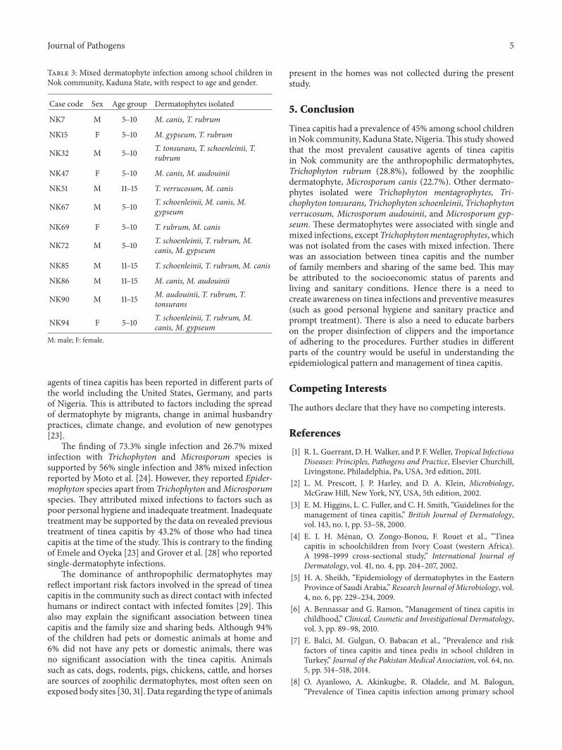
Table 3: Mixed dermatophyte infection among school children inNok community, Kaduna State, with respect to age and gender.
Case code Sex Age group Dermatophytes isolated
NK7 M 5–10 M. canis, T. rubrum
NK15 F 5–10 M. gypseum, T. rubrum
NK32 M 5–10 T. tonsurans, T. schoenleinii, T.rubrum
NK47 F 5–10 M. canis, M. audouinii
NK51 M 11–15 T. verrucosum, M. canis
NK67 M 5–10 T. schoenleinii, M. canis, M.gypseum
NK69 F 5–10 T. rubrum, M. canis
NK72 M 5–10 T. schoenleinii, T. rubrum, M.canis, M. gypseum
NK85 M 11–15 T. schoenleinii, T. rubrum, M. canis
NK86 M 11–15 M. canis, M. audouinii
NK90 M 11–15 M. audouinii, T. rubrum, T.tonsurans
NK94 F 5–10 T. schoenleinii, T. rubrum, M.canis, M. gypseum
M: male; F: female.
agents of tinea capitis has been reported in different parts ofthe world including the United States, Germany, and partsof Nigeria. This is attributed to factors including the spreadof dermatophyte by migrants, change in animal husbandrypractices, climate change, and evolution of new genotypes[23].
The finding of 73.3% single infection and 26.7% mixedinfection with Trichophyton and Microsporum species issupported by 56% single infection and 38% mixed infectionreported by Moto et al. [24]. However, they reported Epider-mophyton species apart from Trichophyton andMicrosporumspecies. They attributed mixed infections to factors such aspoor personal hygiene and inadequate treatment. Inadequatetreatment may be supported by the data on revealed previoustreatment of tinea capitis by 43.2% of those who had tineacapitis at the time of the study. This is contrary to the findingof Emele and Oyeka [23] and Grover et al. [28] who reportedsingle-dermatophyte infections.
The dominance of anthropophilic dermatophytes mayreflect important risk factors involved in the spread of tineacapitis in the community such as direct contact with infectedhumans or indirect contact with infected fomites [29]. Thisalso may explain the significant association between tineacapitis and the family size and sharing beds. Although 94%of the children had pets or domestic animals at home and6% did not have any pets or domestic animals, there wasno significant association with the tinea capitis. Animalssuch as cats, dogs, rodents, pigs, chickens, cattle, and horsesare sources of zoophilic dermatophytes, most often seen onexposed body sites [30, 31]. Data regarding the type of animals
present in the homes was not collected during the presentstudy.
5. Conclusion
Tinea capitis had a prevalence of 45% among school childreninNok community, Kaduna State, Nigeria.This study showedthat the most prevalent causative agents of tinea capitisin Nok community are the anthropophilic dermatophytes,Trichophyton rubrum (28.8%), followed by the zoophilicdermatophyte, Microsporum canis (22.7%). Other dermato-phytes isolated were Trichophyton mentagrophytes, Tri-chophyton tonsurans, Trichophyton schoenleinii, Trichophytonverrucosum, Microsporum audouinii, and Microsporum gyp-seum. These dermatophytes were associated with single andmixed infections, exceptTrichophytonmentagrophytes, whichwas not isolated from the cases with mixed infection. Therewas an association between tinea capitis and the numberof family members and sharing of the same bed. This maybe attributed to the socioeconomic status of parents andliving and sanitary conditions. Hence there is a need tocreate awareness on tinea infections and preventive measures(such as good personal hygiene and sanitary practice andprompt treatment). There is also a need to educate barberson the proper disinfection of clippers and the importanceof adhering to the procedures. Further studies in differentparts of the country would be useful in understanding theepidemiological pattern and management of tinea capitis.
Competing Interests
The authors declare that they have no competing interests.
References
[1] R. L. Guerrant, D. H.Walker, and P. F.Weller, Tropical InfectiousDiseases: Principles, Pathogens and Practice, Elsevier Churchill,Livingstone, Philadelphia, Pa, USA, 3rd edition, 2011.
[2] L. M. Prescott, J. P. Harley, and D. A. Klein, Microbiology,McGraw Hill, New York, NY, USA, 5th edition, 2002.
[3] E. M. Higgins, L. C. Fuller, and C. H. Smith, “Guidelines for themanagement of tinea capitis,” British Journal of Dermatology,vol. 143, no. 1, pp. 53–58, 2000.
[4] E. I. H. Menan, O. Zongo-Bonou, F. Rouet et al., “Tineacapitis in schoolchildren from Ivory Coast (western Africa).A 1998–1999 cross-sectional study,” International Journal ofDermatology, vol. 41, no. 4, pp. 204–207, 2002.
[5] H. A. Sheikh, “Epidemiology of dermatophytes in the EasternProvince of Saudi Arabia,” Research Journal ofMicrobiology, vol.4, no. 6, pp. 229–234, 2009.
[6] A. Bennassar and G. Ramon, “Management of tinea capitis inchildhood,” Clinical, Cosmetic and Investigational Dermatology,vol. 3, pp. 89–98, 2010.
[7] E. Balci, M. Gulgun, O. Babacan et al., “Prevalence and riskfactors of tinea capitis and tinea pedis in school children inTurkey,” Journal of the Pakistan Medical Association, vol. 64, no.5, pp. 514–518, 2014.
[8] O. Ayanlowo, A. Akinkugbe, R. Oladele, and M. Balogun,“Prevalence of Tinea capitis infection among primary school
6 Journal of Pathogens
children in a rural setting in south-west Nigeria,” Journal ofPublic Health in Africa, vol. 5, no. 1, 2014.
[9] M. Feuilhade and C. Lacroix, “Epidemiology of tinea capitis,”Presse Medicale, vol. 30, no. 1, pp. 499–504, 2001.
[10] V. Mane, A. D. Urheka, S. Mali, N. Patil, S. A. Patil, andK. G. Ajit, “Tinea capitis infection in children along withtertiary care hospitals with reference to in vitro antifungalsusceptibility testing of dermatophytes isolate,” InternationalJournal of Research and Reviews in Pharmacy and AppliedScience, vol. 3, pp. 199–208, 2013.
[11] M. Mayowa, R. Godson, and A. M. K. Sridhar, “Use ofAzadirachta indica derived germicidal in the management oftinea capitis among pupils in selected public primary schoolsin Ibadan, Nigeria,” Peak Journal of Medicinal Plant Research,vol. 3, no. 1, pp. 9–15, 2015.
[12] M. M. Sidat, D. Correia, and T. P. Buene, “Tinea capitis amongchildren at one suburban primary school in the City of Maputo,Mozambique,” Revista da Sociedade Brasileira de MedicinaTropical, vol. 40, no. 4, pp. 473–475, 2007.
[13] E. I. Nweze, “Etiology of dermatophytoses amongst children innortheastern Nigeria,”Medical Mycology, vol. 39, no. 2, pp. 181–184, 2001.
[14] S. Adeleke, B. Usman, and G. Ihesiulor, “Dermatophytosisamong itinerant quranic scholars in Kano (Northwest) Nigeria,”Nigerian Medical Practitioner, vol. 53, no. 3, pp. 33–35, 2008.
[15] E. I. Nweze and J. I. Okafor, “Prevalence of dermatophyticfungal infections in children: a recent study in Anambra State,Nigeria,”Mycopathologia, vol. 160, no. 3, pp. 239–243, 2005.
[16] I. J. Frieden and R. Howard, “Tinea capitis: epidemiology,diagnosis, treatment, and control,” Journal of the AmericanAcademy ofDermatology, vol. 31, no. 3, part 2, pp. S42–S46, 1994.
[17] A. Yazdanfar, “Tinea capitis in primary school children inHamedan (west of Iran),” International Journal of Medicine andMedical Sciences, vol. 2, pp. 29–33, 2010.
[18] Department ofArts ofAfrica,Oceania, andTheAmericas, “NokTerracottas (500 B.C.-200 A.D.),” in Heilbrunn Timeline of ArtHistory,TheMetropolitanMuseum of Art, NewYork, NY, USA,2000, http://www.metmuseum.org/toah/hd/nok/hd nok.htm.
[19] O. Akinboro, A. Olayinka, A. Olasode, and O. Onayemi, “Thepattern, Risk factors and clinico-aetiological correlate of tineacapitis among the children in a tropical community setting ofOsogbo, South-Western Nigeria,” South-Western Nigeria. Afro-Egypt Journal of Endemic Diseases, vol. 1, no. 2, pp. 53–64, 2011.
[20] I. Weitzman and R. C. Summerbell, “The dermatophytes,”Clinical Microbiology Reviews, vol. 8, no. 2, pp. 240–259, 1995.
[21] E. David, S. Davis, H. Alexiou, R. Handke, and R. Bartley,Description of Medical Fungi, The Authors, Adelaide, Australia,2nd edition, 2007.
[22] I. Ahmed, A. Zaffar, and N. Sarwat, “Prevalence of tinea capitisand asymptomatic carriage amongst school going children,”Journal of Pakistan Association of Dermatologists, vol. 16, pp.215–219, 2006.
[23] F. E. Emele and C. A. Oyeka, “Tinea capitis among primaryschool children in Anambra state of Nigeria,” Mycoses, vol. 51,no. 6, pp. 536–541, 2008.
[24] J. N. Moto, J. M. Maingi, and A. K. Nyamache, “Prevalence ofTinea capitis in school going children from Mathare, informalsettlement in Nairobi, Kenya,” BMC Research Notes, vol. 8,article 274, 2015.
[25] J. C. Anosike, I. R. Keke, J. C. Uwaezuoke et al., “Prevalenceand distribution of ringworm infection in primary schools
in parts of Eastern Nigeria,” Journal of Applied Sciences andEnvironmental Management, vol. 9, no. 3, pp. 21–25, 2006.
[26] A. A.Omar, “Ringwormof the scalp in primary-school childrenin Alexandria: infection and carriage,” Eastern MediterraneanHealth Journal, vol. 6, no. 5-6, pp. 961–967, 2000.
[27] S. F. Friedlander,M. Rueda, B. K. Chen, andH.W.Caceros-Rios,“Fungal, protozoal and helminthic infections,” in PaediatricDermatology, L. A. Schachner and R. C. Hansen, Eds., pp. 1093–1140, Mosby, St. Louis, Mo, USA, 3rd edition, 2003.
[28] C. Grover, P. Arora, and V. Manchanda, “Tinea capitis in thepediatric population: a study from North India,” Indian Journalof Dermatology, Venereology and Leprology, vol. 76, no. 5, pp.527–532, 2010.
[29] L. Johnson, “Dermatophytes-the skin eaters,”Mycologist, vol. 17,no. 4, pp. 147–149, 2003.
[30] P. M. Rabinowitz, Z. Gordon, and L. Odofin, “Pet-relatedinfections,” American Family Physician, vol. 76, no. 9, pp. 1314–1322, 2007.
[31] B. D. Michaels and J. Q. Del Rosso, “Tinea capitis in infants:recognition, evaluation, and management suggestions,” TheJournal of Clinical and Aesthetic Dermatology, vol. 5, no. 2, pp.49–59, 2012.

Submit your manuscripts athttp://www.hindawi.com
Stem CellsInternational
Hindawi Publishing Corporationhttp://www.hindawi.com Volume 2014
Hindawi Publishing Corporationhttp://www.hindawi.com Volume 2014
MEDIATORSINFLAMMATION
of
Hindawi Publishing Corporationhttp://www.hindawi.com Volume 2014
Behavioural Neurology
EndocrinologyInternational Journal of
Hindawi Publishing Corporationhttp://www.hindawi.com Volume 2014
Hindawi Publishing Corporationhttp://www.hindawi.com Volume 2014
Disease Markers
Hindawi Publishing Corporationhttp://www.hindawi.com Volume 2014
BioMed Research International
OncologyJournal of
Hindawi Publishing Corporationhttp://www.hindawi.com Volume 2014
Hindawi Publishing Corporationhttp://www.hindawi.com Volume 2014
Oxidative Medicine and Cellular Longevity
Hindawi Publishing Corporationhttp://www.hindawi.com Volume 2014
PPAR Research
The Scientific World JournalHindawi Publishing Corporation http://www.hindawi.com Volume 2014
Immunology ResearchHindawi Publishing Corporationhttp://www.hindawi.com Volume 2014
Journal of
ObesityJournal of
Hindawi Publishing Corporationhttp://www.hindawi.com Volume 2014
Hindawi Publishing Corporationhttp://www.hindawi.com Volume 2014
Computational and Mathematical Methods in Medicine
OphthalmologyJournal of
Hindawi Publishing Corporationhttp://www.hindawi.com Volume 2014
Diabetes ResearchJournal of
Hindawi Publishing Corporationhttp://www.hindawi.com Volume 2014
Hindawi Publishing Corporationhttp://www.hindawi.com Volume 2014
Research and TreatmentAIDS
Hindawi Publishing Corporationhttp://www.hindawi.com Volume 2014
Gastroenterology Research and Practice
Hindawi Publishing Corporationhttp://www.hindawi.com Volume 2014
Parkinson’s Disease
Evidence-Based Complementary and Alternative Medicine
Volume 2014Hindawi Publishing Corporationhttp://www.hindawi.com
![PREVALENCE OF DERMATOPHYTIC INFECTION AND … · body and tend to the following conditions, Tinea capitis, Tinea cruris, Tinea corporis and Tinea pedis [14]. A total of two hundred](https://img.pdfslide.net/doc/110x75/5cd4c0d988c993e4698dd664/prevalence-of-dermatophytic-infection-and-body-and-tend-to-the-following-conditions.jpg)