Embed Size (px)
Citation preview

Sede Amministrativa: Università degli Studi di Padova
Dipartimento di Scienze Cardiologiche, Toraciche e Vascolari
___________________________________________________________________
SCUOLA DI DOTTORATO DI RICERCA IN : Scienze Mediche, Cliniche e Sperimentali
INDIRIZZO: Scienze Cardiovascolari
CICLO 27°
RESEARCH OF PREDICTIVE AND PROGNOSTIC TISSUE AND MOLECULAR MARKERS AND OF NEW
THERAPEUTIC TARGETS IN MALIGNANT PLEURAL MESOTHELIOMA
Direttore della Scuola : Ch.mo Prof. Gaetano Thiene
Coordinatore d’indirizzo: Ch.mo Prof. Gaetano Thiene
Supervisore :Ch.mo Prof. Federico Rea
Cotutor: Ch.ma Prof. Fiorella Calabrese
Dottorando : Giulia Pasello
1
Table of contents
1. Abstract/Riassunto Pag. 3-‐10
2. Background Pag. 11-‐28
2.1. Malignant Pleural Mesothelioma Pag. 11-‐20
2.2. Extrinsec and intrinsec apoptosis: Pag. 20-‐27
TRAIL and p53-‐MDM2 pathway
2.3. Neoangiogenesis and MPM Pag. 27-‐28
3. Aims Pag. 29-‐32
3.1 Preclinical evaluation of the anticancer activity of Pag.30-‐31
the extrinsic apoptosis activator (rhApo2L/TRAIL) in combination with
intrinsic apoptosis triggers acting through p53 activation
3.2 Translational study of the identification of Pag. 31-‐32
pathological and molecular differences in chemonaive tumor samples from
different MPM histologic subtypes (epithelioid versus non-‐epithelioid)
4. Methods Pag. 32-‐42
5. Results Pag. 42-‐51
5.1 Preclinical evaluation of the anticancer activity of Pag. 42-‐47
rhApo2L/TRAIL in combination with chemotherapy (carboplatin plus
pemetrexed) or p53-‐MDM2 inhibitor RG7112
5.2 Translational study of the identification of Pag. 47-‐51
pathological and molecular differences in chemonaive tumor samples from
different MPM histologic subtypes (epithelioid versus non-‐epithelioid)
6. Discussion Pag. 51-‐60
7. Conclusion and summary of topic results Pag. 60

2
8. Appendix: figures and tables Pag. 61-‐77
9. References Pag. 78-‐94
10. European curriculum vitae and summary of Pag. 95-‐104
scientific and research activity
Acknowledgments Pag. 105
3
1. Abstract/Riassunto
1.1 Abstract
BACKGROUND: Malignant pleural mesothelioma (MPM) is an aggressive tumor
with increasing incidence in industrialized countries, because of previous
widespread asbestos exposure and long latency time before symptoms
appearance. Tumor necrosis factor-‐related apoptosis-‐inducing ligand (TRAIL)
belongs to the tumor necrosis factor (TNF) family of death ligands; it was
identified as a promising anticancer agent thanks to its property of killing
cancer cells while sparing normal cells. Conflicting evidences about MPM
resistance rather than sensitivity to TRAIL-‐induced apoptosis were previously
reported. While TRAIL-‐dependent apoptosis is thought to be p53-‐independent,
p53 wild type cancer cells can be sensitized to TRAIL through p53 activation. In
contrast to most solid tumors, MPM cells frequently express wild type p53,
thus p53 reactivation might be considered as an effective strategy to sensitize
MPM cells to TRAIL-‐dependent apoptosis. DNA-‐damaging agents such as
chemotherapy or radiotherapy and other agents targeting negative regulators
of p53, may be considered as useful strategies to reactivate p53. Murine
Double Minute 2 (MDM2) is a transcriptional target of p53: once activated,
MDM2 binds p53 to the amino-‐terminus, targeting it for ubiquitylation and
subsequent proteasomal degradation. Recently, many researchers have
investigated a possible role of MDM2 in promoting tumor neoangiogenesis
(Vascular Endothelial Growth Factor, VEGF; hypoxia inducible factor,
HIF1alpha). Thus MDM2 might be a promising target for anticancer treatment
because of its antiapoptotic and proangiogenetic role. The poor prognosis of
4
affected patients, the lack of effective treatment options, with particular
reference to biologic drugs, the absence of predictive markers for targeted
treatment and the lack of knowledge about the basis of different biological and
clinical behaviour of the two main histologic subtypes, epithelioid versus non-‐
epithelioid (sarcomatoid/biphasic), constitute the rationale for the present
study.
AIMS: The first purpose of the study was to investigate new treatment options
through preclinical evaluation of extrinsic apoptosis triggers (recombinant
human Apo2L/TRAIL) in combination with intrinsic apoptosis inducers acting
through the reactivation of p53, such as DNA-‐damaging agents
(carboplatin/pemetrexed) or p53-‐MDM2 inhibitors (nutlin3-‐RG7112), both in
vitro and in vivo. Moreover, the study aims to investigate new targets (MDM2,
HIF1alpha, VEGF) for treatment in MPM tumor samples, testing possible
different expression levels of such targets in the different histologic subtypes.
Some morphological features, such as inflammation, necrosis and proliferation
were quantified in the different histotypes and correlated with MDM2 and
HIF1alpha. Finally, correlations between molecular data and clinical features
were performed.
METHODS: Anticancer effects of rhApo2L/TRAIL (Amgen, Genentech) plus
chemotherapy (Carboplatin/Pemetrexed) or nutlin3-‐RG7112 (Roche) was
evaluated in different cell lines through annexin V and caspases assay, and in a
Severe Combined ImmunoDeficiency (SCID) mouse model. p53 expression
levels were evaluated through western blot. TRAIL receptors were evaluated
through flow cytometry. Formalin-‐Fixed Paraffin Embedded (FFPE) chemonaive
5
tumor samples from MPM patients were analyzed: MDM2, VEGF and HIF1alpha
mRNA and protein expression levels were investigated through RT-‐qPCR and
immunohistochemistry (IHC) with specific antibodies, respectively. Proliferation
was quantified through staining with Ki67 antibodies. Necrosis and
inflammation were also quantified on histological sections using a grading
score. Normal pleura samples from patients undergoing diagnostic surgery for
non cancer disease were used as negative controls. Clinical data of the patients
under study were collected in a password-‐protected database: age, gender,
ECOG PS (Performance Status), EORTC score, stage, systemic treatments,
surgery, radiotherapy, first progression and last follow-‐up date, status
(alive/dead).
RESULTS: In vitro and in vivo results showed a significant increase of apoptosis
in cell lines and reduction of tumor volume in animal models treated with
rhApo2L/TRAIL plus chemotherapy or nutlin3-‐RG7112 compared with those
receiving single treatments. This synergistic effect was dependent on the ability
of chemotherapy or nutlin3-‐RG7112 to increase the expression of the TRAIL
receptors DR4 and DR5 in a p53 manner. Higher MDM2 and HIF1alpha IHC
expression was significantly associated with sarcomatoid/biphasic histologic
subtype (p=0.010 and p=0.007, respectively) with positive correlation between
MDM2 and HIF1alpha expression levels (correlation coefficient=0.533; p value=
0.00626). Proliferation index was significantly higher in sarcomatoid/biphasic
compared with epithelioid samples (p=0.005) and also significantly higher in
tumor samples with higher MDM2 expression (p=0.008). Clinical and
pathological features or biomarker did not show any correlation with
6
prognosis, except for proliferation index and Progression Free Survival (PFS),
even though the results of this exploratory analysis should be considered with
caution because of the limited number of patients, the heterogeneous
treatment received and the insufficient follow-‐up time in some patients.
CONCLUSION: Our preclinical in vitro and in vivo results confirm that
reactivation of p53 by chemotherapy or p53-‐MDM2 inhibitors effectively
sensitizes to TRAIL-‐dependent apoptosis in malignant pleural mesothelioma.
Our translational study in tumor samples from MPM patients confirmed
different biological and pathological features and molecular targets expression
in the two main histologic subtypes. It is tempting to speculate that MDM2 and
Ki67 might be considered as further useful diagnostic tools to identify poor
prognosis patients. Moreover, MDM2 and HIF1alpha might be considered as
promising targets for anticancer treatment of MPM.
1.2 Riassunto
BACKGROUND: Il mesotelioma pleurico maligno (MPM) è una neoplasia
aggressiva con incidenza in aumento nei paesi industrializzati per la pregressa
esposizione ad amianto e il lungo periodo di latenza tra l’esposizione e la
comparsa dei sintomi. TRAIL (Tumor necrosis factor-‐related apoptosis-‐inducing
ligand) appartiene alla famiglia dei ligandi di morte apoptotica di TNF (tumor
necrosis factor), ed è stato recentemente identificato come promettente
agente antitumorale in considerazione della sua proprietà di uccidere le cellule
tumorali, risparmiando le cellule normali. Evidenze contrastanti riportano la
presenza di resistenza piuttosto che di sensibilità delle cellule di mesotelioma
maligno all’apoptosi mediata da TRAIL. Sebbene l’apoptosi indotta da TRAIL (via
7
estrinseca dell’apoptosi) sembra essere indipendente da p53, alcune cellule
tumorali portatrici di p53 wild-‐type possono essere sensibilizzate alla morte da
TRAIL attraverso l’attivazione di p53 (via intrinseca dell’apoptosi).
Contrariamente alla maggior parte delle neoplasie, le cellule di mesotelioma
pleurico esprimono più frequentemente p53 wild-‐type, e quindi la riattivazione
di p53 potrebbe essere una strategia efficace per sensibilizzare le cellule di
mesotelioma all’apoptosi mediata da TRAIL. Agenti in grado di danneggiare il
DNA (chemioterapia, radioterapia) ed altri agenti in grado di “down-‐regolare”
gli inibitori di p53, possono essere considerati come valide strategie per
riattivare p53. Murine Double Minute 2 (MDM2) è un bersaglio dell’attività
trascrizionale di p53: una volta attivata, MDM2 lega il dominio ammino-‐
terminale di p53 e la conduce al processo di ubiquitilazione e successiva
degradazione proteasomica. Negli anni recenti, molti ricercatori hanno studiato
un possibile ruolo di MDM2 nella attivazione di marcatori di neoangiogenesi
tumorale (Vascular Endothelial Growth Factor, VEGF; hypoxia inducible factor,
HIF1alpha), pertanto MDM2 potrebbe rappresentare un promettente bersaglio
per il trattamento antitumorale in considerazione della sua possibile duplice
attività antiapoptotica e proangiogenetica. La prognosi infausta dei pazienti
affetti, l’assenza di opzioni terapeutiche efficaci, in particolare di farmaci
biologici, l’assenza di marcatori predittivi di risposta ai farmaci a bersaglio
molecolare, e la scarsità di conoscenze sui meccanismi che sottendono al
diverso comportamento biologico e clinico dei due principali sottotipi istologici
(epitelioide versus non-‐epitelioide), costituiscono il razionale del presente
studio.
8
OBIETTIVI: Il primo obiettivo è stato valutare nuove opzioni terapeutiche
attraverso studi preclinici in vitro ed in vivo con associazione di induttori della
via estrinseca dell’apoptosi (rhApo2L/TRAIL) e induttori della via intrinseca
dell’apoptosi che agiscono attraverso riattivazione di p53, come agenti
danneggianti il DNA (carboplatino/pemetrexed) o inibitori del legame p53-‐
MDM2 (nutlin3-‐RG7112). Secondariamente, lo studio si è proposto di ricercare
l’espressione dei nuovi bersagli terapeutici (MDM2, HIF1alpha) nei campioni
tumorali di pazienti affetti da mesotelioma maligno, e di valutarne la diversa
espressione nei diversi sottotipi istologici. Inoltre, il progetto si è focalizzato
sulla valutazione di alcuni parametri morfologici come infiammazione, necrosi
ed indice proliferativo nei campioni tumorali dei diversi istotipi e sulla loro
correlazione con MDM2 e HIF1alpha. Infine, sono state valutate le correlazioni
tra dati molecolari e caratteristiche cliniche dei pazienti in studio.
MATERIALI E METODI: l’attività antitumorale di rhApo2L/TRAIL (Amgen,
Genentech) in associazione a chemioterapia (Carboplatino/Pemetrexed) o
nutlin3-‐RG7112 (Roche) è stata valutata in diverse linee cellulari attraverso il
saggio di Annessina V e delle caspasi, e in un modello di topo Severe Combined
ImmunoDeficiency (SCID). I livelli di espressione di p53 sono stati analizzati
attraverso western blot. I recettori di TRAIL sono stati rilevati attraverso
citofluorimetria. Campioni tumorali fissati in formalina e inclusi in paraffina da
pazienti chemonaive sono stati analizzati con immunoistochimica e valutando
l’espressione di mRNA per MDM2 e HIF1alpha. L’indice proliferativo è stato
quantificato mediante anticorpo monoclonale di Ki67. La presenza di
infiammazione e necrosi è stata valutata su sezioni istologiche. Campioni di
9
pleura normale da pazienti sottoposti a chirurgia toracica per patologia non
oncologica sono stati utilizzati come controlli negativi. I dati clinici dei pazienti
in studio sono stati raccolti un un database protetto da password: età, sesso,
ECOG PS (Performance Status), score prognostico EORTC, stadio, trattamenti
sistemici, chirurgia, radioterapia, prima progressione, data di ultimo follow-‐up
e status (vivo/morto).
RISULTATI: I risultati in vitro ed in vivo mostrano un significativo aumento di
apoptosi in linee cellulari e riduzione di volume tumorale in modelli animali
trattati con rhApo2L/TRAIL in associazione a chemioterapia o nutlin3-‐RG7112,
confrontato ai singoli trattamenti. Tale effetto sinergico è correlato
all’incremento di espressione dei recettori di TRAIL (DR4 e 5) conseguente alla
riattivazione di p53 da chemioterapia o nutlin3-‐RG7112. Abbiamo poi valutato i
livelli di espressione di MDM2 e del suo possibile target HIF1alpha in campioni
tumorali di pazienti affetti da mesotelioma. I livelli di espressione di MDM2 e
HIF1alpha erano significativamente più elevati nel sottotipo istologico
sarcomatoide/bifasico (p=0.010 and p=0.007, respectively), ed è stata
osservata una correlazione positiva tra i livelli di espressione di MDM2 e
HIF1alpha (coefficiente di correlazione =0.533; p = 0.00626). Infine, l’indice
proliferativo (Ki67) si è dimostrato significativamente più elevato nel sottotipo
istologico sarcomatoide/bifasico rispetto a quello epitelioide (p=0.005) e
significativamente più elevato nei campioni con iperespressione di MDM2
(p=0.008). Per quanto riguarda gli obiettivi esploratori del progetto, nessuna
correlazione prognostica è stata osservata per alcun parametro clinico o
patologico o per diversi livelli di espressione dei biomarcatori in studio, mentre
10
è stata osservata una correlazione significativa tra i livelli di Ki67 e la
sopravvivenza libera da progressione. I risultati di tale indagine esploratoria
devono, comunque, essere considerati con cautela per la limitata dimensione
campionaria, l’eterogeneità degli interventi terapeutici e l’insufficiente follow-‐
up di alcuni pazienti.
CONCLUSIONI: I risultati in vitro e in vivo di questo progetto di ricerca
dimostrano che la riattivazione di p53 con chemioterapia o molecole inibitrici
del legame p53-‐MDM2 rappresenta un’efficace strategia per sensibilizzare
all’apoptosi mediata da TRAIL. Lo studio traslazionale ha invece confermato
diverse caratteristiche biologiche e patologiche così come differenti livelli di
espressione di nuovi bersagli terapeutici nei due sottotipi istologici di MPM.
MDM2 e Ki67 possono essere considerati come importanti ausili diagnostici per
una migliore caratterizzazione dell’istotipo e soprattutto per identificare i
tumori a peggiore prognosi. Inoltre, MDM2 e HIF1alpha potrebbero
rappresentare promettenti bersagli per il trattamento del mesotelioma
pleurico maligno.
11
2. Background
2.1 Malignant Pleural Mesothelioma
Malignant Pleural Mesothelioma (MPM) is an aggressive cancer involving
pleural surfaces and, in the advanced stage, lung parenchyma, leading to a
typicale clinical picture characterized by chest pain, dyspnoea and cough.
Although the main risk factor is asbestos exposure, other factors have a
central role in MPM pathogenesis. The biological behaviour of MPM is
determined by molecular alterations, such as oncosuppressor genes loss,
like p16INK4A and p14ARF, while rare mutations or deletions of p53 and pRb,
which are commonly involved in the pathogenesis of most cancer. Such
molecular aberrations seem to be the basis of MPM resistance to systemic
treatments currently adopted in the medical management of this disease.
Surgery is feasible only in selected cases and current gold standard
chemotherapy in unresectable disease is a platinum-‐based doublet with an
antifolate agent, which shows a median overall and progression free
survival of approximately 12 and 6 months respectively, and a response
rate of 20-‐40%[1, 2]
High refractoriety to systemic treatment, rare and short-‐term complete
responses make MPM a therapeutic challenge. Improved knowledge about
molecular pathways lead to several clinical trials investigating biologic
agents in the treatment of MPM, even though they have not found a
precise placement in the therapeutic strategy yet.
2.1.1. Epidemiology
12
The incidence of MPM shows some difference among countries, and
epidemiologic data are somewhere lacking. Whereas in some countries
MPM and cancer registers are available, in other countries few areas are
covered with sufficient information and relative rates are estimated by
mortality data; elsewhere, only surveys of medical doctors and researchers
are available. Such heterogeneous picture determines that not all available
data have the same reliability.
In most European countries, such as France, Germany, Italy and
Scandinavia, the incidence rate is between 11 and 20 cases on 1.000.000
inhabitants[3], and these rates may vary about 70 times according to the
geographical area. In Italy, the National Mesothelioma Register (Registro
Nazionale Mesotelioma, ReNaM) reports an incidence of 2.98 and 0.98
(rough rates, on 100.000 inhabitants, per year) in males and females,
respectively.
Analysis on asbestos exposure and MPM incidence performed in several
industrialized countries, showed a statistically significant correlation
between the two variables[4, 5]. At the national level, the geographic
distribution of MPM reflects the location of industries using asbestos such
as shipyards, building construction, production of asbestos cement and
construction/repair of railway cars; in Italy, mortality data for pleural
cancer in males, between 1988 and 1997, showed wide variations between
one province and another. Higher mortality rates, between 4 and
12/100.000, are shown in ares where the main shipyards and cement
13
factories are located. Among the most affected provinces, Casale
Monferrato, Savona, Genova, La Spezia, Alessandria, Gorizia and Trieste.
In Italy the extraction, import and marketing of asbestos were banned in
1992 but due to the long lag time (even 40 years) from exposure to clinical
evidence of MPM the epidemiologic data foresee a sharp rise of MPM
incidence and mortality in the next fifteen years[6]. European
epidemiological surveys foresee the mortality peak for MPM in males
between 2015 and 2020 [7],[8, 9].
The Veneto region is particularly interested by asbestos exposure because
of its strong industrial development. Incidence is about 2.3/100.000/year,
quite superior to the mean national and European incidence. Since 1987,
about 2000 new cases of malignant pleural mesothelioma have been
registered, with mean incidence over 80 new cases/year. Most of them are
males with a previous occupational exposure, but many females living
close to some industries or with asbestos exposed workers are also
affected. Thus malignant mesothelioma has a strong social impact that
deserves attention by the National Health Service (NHS).
2.1.2 Etiology and pathogenesis
Etiopathogenesis of MPM is attributable in 80% of the cases to asbestos
exposure. Asbestos fibers are classified in two main groups, according to
size and bio-‐persistance: serpentine (chrysotile) and amphibole
(crocidolite, tremolite, anthofillite and amosite). Even if in the different
experimental models all asbestos fibers have shown cancerogenetic
potential[10], subsequent evidence confirm a weak potential of chrysotile
14
fibers[11], and currently the scientific community agree on a higher
cancerogenetic potential of thinner and longer asbestos fibers and of the
subtypes amphibole, compared to shorter and chrisotile fibers.
Asbestos fibers may directly damage the DNA of mesothelial cells, by laying
down and penetrating in the pleura, and leading to a process characterized
by damage, repair and local inflammation, pleural plaques or
mesothelioma. Another mechanism implied in asbestos cancerogenesis is
the reactive oxygen species (ROS) and cytokines production by
macrophages[12], which determine DNA damage and an
immunocompromised state. Moreover, asbestos fibers are able to interfer
with mitotic processes, inducing aneuploidy and chromosomal aberrations,
and to activate some kinases such as MAPK (mitogen-‐activated protein
kinase)and ERK1 and 2 (extracellular signal-‐regulated kinases 1 and 2)[13].
The complex cancerogenetic process leading to plural mesothelioma might
be triggered by other factors such as genetics, ionizing radiations, SV40
virus (Simian Virus 40) [14].
Loss of heterozygosity (LOH) is a frequent process in the pathogenesis of
MPM, and the result is the loss or inactivation of several oncosuppressor
genes.
Even though mutation and deletion of p53 and pRb tumour suppressor
genes occur frequently in a lot of human cancers, they are extremely rare
in malignant mesothelioma[15] Probably, a functional apoptotic defect
causing MPM resistance to chemotherapy and radiotherapy occurs
downstream p53 and pRb. The INK4a/ARF locus within 9p21 chromosome

15
encodes two proteins, p16INK4a and p14ARF , and previous series
demonstrated deletion of this genome region in 70% of MPM cell lines.
p16INK4a inhibits cyclinD-‐dependent kinase (CDK), preventing the
phosphorilation and subsequent inactivation of pRb, whereas p14
promotes the degradation of Murine Double Minute 2 (MDM2) protein,
that is responsible for p53 ubiquitination and inactivation. p53 leads the
cell to apoptosis or inhibits the entrance into the cell cycle, whereas pRb
arrests the cell in G1 phase (see section 2.2.2).
The most frequent chromosomal aberration in MPM is on chromosome 22,
where the oncosuppressor gene NF2 (Neurofibromatosis 2) is located and
the protein merlin is encoded. Mutations of NF2 gene have been observed
in 40% of MPM cases[16-‐18]. In animal models with asbestos exposure,
MPM is most commonly observed after inactivation of one NF2 allele,
compared to wild type; moreover, the other allele is often loss in MPM,
thus confirming its role as a gatekeeper[19, 20]. The loss of the remaining
allele is often associated with the loss of INK4a/ARF locus, which might be
responsible for a ‘permissive’ background.
The BCL2 family of genes which regulate the apoptotic process plays an
important role in the cancerogenesis process; this family includes
proapoptotic genes such as BAX, BAK and BAD, and antiapoptotic genes
such as BCL2, BCLXL, MCL1. BCL2 is rarely expressed in MPM, but high
mRNA levels of BCLXL have been observed in MPM cell lines and in tumor
tissue samples [21-‐23], probably in order to contrast with the proapoptotic
effect of BAX, expressed in MPM cell lines[15].
16
Many growth factors play a role in the pathogenesis of MPM, such as IGF-‐1
(Insulin-‐like growth factor-‐1), HGF (hepatocyte growth factor), bFGF (basic
fibroblast growth factor), EGF (epidermal growth factor), VEGF (vascular
endothelial growth factor), PDGF A e B (platelet-‐derived growth factor A e
B), TNF (tumor necrosis factor). These factors come from the surrounding
lung parenchyma, macrophages or mesothelial cells, after different
stimula, inflammatory cytochines, asbestos, SV40 infection. Growth factors
promote tumor growth, proliferation and invasiveness, but also the
neoangiogenesis process which feeds the growing tumor with oxygen and
metabolites (see section 2.3).
2.1.3 Histopathologic and clinical features and treatment
MPM usually arises between the fifth and seventh decade, more frequently
in males (males:females equal to 3-‐5:1). Affected patients tipically come to
the medical evaluation with pleural effusion (80%), and subsequent
symptoms such as chest pain (60%) and exercise dyspnoea (50-‐70%). Other
symptoms such as weight loss and fatigue might be present, especially in
the advanced stages of the disease, and their presence at the diagnosis
characterize a worse prognosis. MPM might spread to the abdomen
through the diaphragm, and in the 30% of the patients the main
complication is bowel occlusion, while rarely the invasion of liver or other
organs is observed. About 10% of the patients might die because of
myocardial or pericardial involvement. Distant metastases are uncommon,
and generally more frequent in the sarcomatoid subtype. MPM patients
usually die because of respiratory insufficiency o pneumonitis.
17
The 2004 WHO (World Health Organization) classification of pleural tumors
have described three main MPM histologic subtypes: epithelioid (50-‐67%),
sarcomatoid (7-‐21%) and biphasic (24-‐35%), each of them characterized by
different biological and clinical behaviour[24] [25].
Epithelioid mesothelioma, associated to a more favourable prognosis, is
characterized by cells distributed into three different patterns: solid,
glandular and tubulo-‐papillar.
Sarcomatoid mesothelioma is biologically more aggressive, and
characterized by fusiform cells organized in layers or folders (typical) or
sometimes in a disorganized pattern associated with abundant stroma
(desmoplastic).
The immunohistochemistry diagnosis uses panels which combine
mesothelioma and cancer associated markers. The immunohistochemistry
usually consists on subsequent steps, using first two markers for MPM
(calretinin, podoplanin) and two for another cancer (usually lung
adenocarcinoma: TTF1, CEA).
Surgical resection of MPM is feasible in a small percentage of patients,
although selection criteria have not been identified yet in prospective
series [26].
Pleurectomy/Decortication (P/D) leads to a significant but incomplete
resection of the tumoral pleura, leaving the involved lung free to expand.
This procedure has not a curative intent, but it may be considered in
symptomatic patients in order to control pain and restrictive deficit[27].
Radical surgery consists on the complete resection of cancer tissue
18
macroscopically visible, and it may be achieved with extrapleural
pneumonectomy (EPP): en-‐bloc resection of pleura, lung, pericardium,
diapraghm, lymphnodes. In the attempt of reducing the incidence of local
recurrences after extrapleural pneumonectomy, a multimodality approach
with surgery followed by postoperative radiotherapy was explored in the
past years. Extrapleural pneumonectomy allows higher doses of
radiotherapy to the whole hemithorax by avoiding pulmonary toxicity and
the result of this approach is a significant reduction of loco-‐regional
relapses.
The issue of extrathoracic metastasis represents a major challenge in the
management of the disease because of the impact on overall survival.
Once a chemotherapy regimen shows activity in advanced malignant
pleural mesothelioma, a subsequent step was the addition of such
treatment to surgery and radiotherapy to improve the
systemic control of the disease. The success with surgical resection after
neoadjuvant chemotherapy in stage IIIA non-‐small cell lung cancer has
been the impetus for several groups to apply this strategy in malignant
mesothelioma aiming at reducing the incidence of distant relapse. As well
as in non-‐small cell lung cancer, neoadjuvant chemotherapy could
maximize cytoreduction and increase the proportion of patients able to
complete the following treatments. Furthermore, the difficult
administration of both postoperative chemotherapy and radiotherapy in
most patients induced many groups to introduce a trimodality approach
19
based on preoperative chemotherapy, surgery and postoperative
radiotherapy in the attempt of improving compliance[28].
Recently the MARS (Mesothelioma And Radical Surgery) study, the first
study which randomized between extrapleural pneumonectomy and no
extrapleural pneumonectomy, showed no survival advantage and worse
quality of life in those patients who underwent surgery. The trial had
several limitations; patients were treated with different chemotherapy
regimens, and in a relevant percentage of cases a sub-‐optimal
chemotherapy was delivered. The heterogeneity of delivered
chemotherapy could have unbalanced the two study arms. The study
population was small but the conclusion of the trial raised the issue of a
less invasive approach as suitable treatment of malignant pleural
mesothelioma[29].
Chemotherapy is still the treatment option for the majority of MPM
patients not suitable for surgical resection. Currently, the gold standard in
the systemic treatment of MPM patients is a chemotherapy regimen based
on a platinum-‐base doublet plus an antifolate agents such as pemetrexed
or raltitrexed, which showed survival and response improvement
compared with platinum single agent. These combinations showed median
overall and progression free survival of approximately 12 and 6 months
respectively, and a response rate of 20-‐40%[1, 2]. Carboplatin is considered
a valid option in the systemic treatment of advanced pleural
mesothelioma, with better toxicity profile compared to cisplatin [30-‐32].
20
After first-‐line treatment failure and disease progression or relapse, there
is not a systemic regimen showing significant improvement in survival and
quality of life, and few data of second line treatment are reported in small
phase II studies or retrospective case series, thus raising some doubts
about which might be the right drug for the right patient in previously
treated subjects.
The improvement of knowledge about biological behaviour, molecular
pathways and genetic alterations of MPM, lead to the preclinical and
clinical investigation of new targeted agents in this setting. VEGF and other
antiangiogenetic drugs, agents against other growth factors, HDAC and
proteasome inhibitors, PI3K/mTOR inhibitors have been explored, although
these drugs have not found a specific role in the therapeutic
armamentarium of this disease[33].
2.2 Extrinsec and intrinsec apoptosis: TRAIL and p53-‐MDM2 pathway
2.2.1 Tumor necrosis factor-‐related apoptosis-‐inducing ligand (TRAIL) and
MPM.
Tumor necrosis factor-‐related apoptosis-‐inducing ligand (TRAIL) belongs to
the tumor necrosis factor (TNF) family of death ligands inducing the
extrinsic apoptotic pathway. Two surface death receptors (TRAIL-‐R1 or DR4
and TRAIL-‐R2 or DR5), two decoy non-‐functional receptors (TRAIL-‐R3 or
DcR1 and TRAIL-‐R4 or DcR2) and the soluble decoy receptor
osteoprotegerin were described [34-‐39]. After the binding of TRAIL to
DR4/5 and the oligomerization of death receptors, the death-‐inducing
21
signalling complex (DISC) is formed, which includes also the Fas-‐associated
death domain (FADD).
FADD recruits and initiates procaspase 8 to active caspase 8 which in turn
cleaves and activates the effector caspases 3, 6 and 7.
TRAIL has been identified as a promising anticancer agent thanks to its
property of killing cancer cells while sparing normal cells [40, 41], even
though both sensitivity and resistance mechanisms to TRAIL-‐induced cell
death are not completely clarified [42].
TRAIL-‐induced death program is successfully executed in “type I” cells,
while “type II” cells need the activation of the intrinsic apoptotic pathway
[43], through activation of the BH3-‐only protein, Bid, which moves to the
mithocondria where contributes to Bax and Bak activation. The resulting
mithocondrial pore formation leads to cytochrome c release into the
cytosol with the final caspase 9 activation. Moreover, “type II” cancer cells
seem sensitive to the synergistic effect of TRAIL, acting on the extrinsic
pathway, and DNA damaging agents, targeting the intrinsic pathway; the
mechanism of this synergy has been proposed to be via the upregulation of
the TRAIL receptor DR5 [44-‐46].
Cancer cells are frequently resistant to TRAIL-‐dependent apoptosis through
different mechanisms: mutations and disfunction of DR4 and DR5; defects
of Fas-‐associated death domain (FADD) and caspase 8; overexpression of
cellular FADD-‐like interleukin-‐1b-‐converting enzyme inhibitory protein
(cFLIP); overexpression of the antiapoptotic proteins Bcl-‐2, Bcl-‐Xl, IAPs
(Inhibitor of Apoptosis Proteins); loss of proapoptotic proteins Bax, Bak;
22
decreased release of second mithocondria-‐derived activator of caspases
(Smac-‐Diablo); activation of mitogen-‐activated protein kinases (MAPK) or
NFKB [42].
Monoclonal agonist antibodies directed against the DR4 and DR5
(Mapatumumab, Lexatumumab, Apomab, AMG655, LBY135) and
recombinant human Apo2L/TRAIL (rhApo2L/TRAIL, Dulanermin) have been
studied at a preclinical and clinical level both as single agents and in
combination with chemotherapy [47-‐52].
Recombinant human (rh) Apo2L/TRAIL (Dulanermin), a receptor agonist
which binds both DR4 and DR5[53, 54], showed antitumor activity in vitro
and in vivo tumor models of different cancer types both as single agent and
in combination with chemotherapy without any toxicity in normal cells [55-‐
57], and was the first TRAIL agonist investigated in human clinical trials [51,
52, 58].
Conflicting evidences about MPM resistance rather than sensitivity to
TRAIL-‐induced apoptosis were previously reported [49, 59]. A Swiss group
showed apoptotic effects of TRAIL or the monoclonal antibodies
Mapatumumab and Lexatumumab directed against DR4 and DR5 on 13
MPM cell lines; moreover, Mapatumumab (anti DR4) and Lexatumumab
(anti DR5) sensitized MPM cell lines to the cytotoxic effects of Cisplatin,
and cell death occurred through a synergistic cooperation of the two
agents (Mapatumumab or Lexatumumab plus Cisplatin) probably through
Reactive Oxygen Species (ROS) induction[49].
23
Increased apoptotic levels were demonstrated in four cell lines treated
with the association of TRAIL and chemotherapy (Cisplatin, Doxorubicin,
Gemcitabine or Etoposide), probably through p53-‐independent apoptotic
pathway; no DR5 increase was observed at the basis of this
sensitization[60].
Other studies showed a p53-‐dependent induction of DR4 and DR5
expression by Alpha-‐tocopheryl succinate, resulting in TRAIL sensitization
[61]. While TRAIL-‐dependent apoptosis is thought to be p53-‐independent,
p53 wild type cancer cells can be sensitized to TRAIL through p53 activation
[62]. In contrast to most solid tumors, MPM cells frequently express wild
type p53 [63], thus p53 reactivation through different strategies might be
considered as an effective strategy to sensitize MPM cells to TRAIL-‐
dependent apoptosis.
2.2.2 p53-‐MDM2 pathway and MPM.
The tumor suppressor p53 acts as a transcription factor regulating genes
involved in DNA repair, metabolism, cell cycle arrest, apoptosis and
senescence. It was defined as the ‘guardian of genome’ because of its
ability of preserving the genomic integrity of the cell under stressed
conditions; p53 disruption, subsequently, leads to increased cancer risk
and to a worse cancer prognosis and treatment response.
Under unstressed conditions, p53 levels are kept low by a feedback
interaction with the RING domain proteins murine double minute 2
(MDM2) and MDM4 (also known as MdmX). MDM2 is a target of p53’s
transcriptional activity, with E3 ubiquitin ligase activity: once activated,
24
MDM2 binds p53 to the amino-‐terminus, targeting it for ubiquitylation and
subsequent proteasomal degradation which represents its main, and
probably first known, function[64]. MDM2 inhibits p53 through multiple
other inhibitory mechanisms, such as the prevention of the transcriptional
coactivator recruitment, the inhibition of p53-‐DNA interaction and p53
indirect translation[65].
Under stressed condition -‐ such as DNA damage-‐ induced p53 decides cell
fate outcomes among apoptosis, cell cycle arrest and senescence through
specific gene transcription; p53’s promoter selectivity, the levels of the
protein itself, antiapoptotic proteins levels, specific cofactors recruitment
for downstream genes transcription, post-‐translational p53 modifications
seem to condition cell death rather than cell cycle arrest or
senescence[66].
p53 is mutated in about 50% of the human cancers[67], while in tumors
with wild-‐type p53 gene, the protein function may be lost because of
overexpression of p53 regulatory proteins such as MDM2 and MDM4, or
because of CDKN2A –encoding for ARF which binds to and rapidly degrades
MDM2-‐ deletions. MDM2 and MDM4 protein overexpression with or
without increased gene copy number occurs in several cancer types, more
frequently in those tumors with a wild-‐type p53[68].
MDM2 and MDM4 amplification have been shown in about 30% and 17%
of soft tissue sarcomas (STS) respectively, while p53 mutations and
CDKN2A deletions were described in about 20 and 15% of the cases,
respectively[69-‐72]. A recent p53-‐pathway mapping in different STS
25
histologic subtypes, confirmed frequent p53 mutations in
leiomyosarcomas, osteosarcomas and pleomorphic sarcomas, while
frequent MDM2 amplifications in well differentiated liposarcomas and
MDM4 in Ewing’s sarcoma/PNETs; MDM2 and MDM4 coamplification was
a common event in synovial sarcomas, Ewing’s/PNET and osteosarcomas.
Moreover, p53 mutations and MDM2 amplifications appeared as mutually
exclusive events, which acquire particular relevance in patients selection
for p53-‐reactivating treatments[73]. However, gene amplification is not the
only mechanism sustaining MDM2 overexpression[74-‐76].
Transcriptional and post-‐transcriptional mechanisms have been proposed
at the basis of MDM2 and MDM4 overexpression, thanks to the dense
interaction network of these proteins[77]. Among post-‐transcriptional
modifications, post-‐translational modifications acquire particular relevance
in MDM2 regulation, and phosphorylation is one of the most commonly
implied in different conditions. This post-‐translational modification leads to
p53 ubiquitination and degradation and to the MDM2-‐p19ARF interaction
prevention, finally to MDM2 nucleus-‐cytoplasm shuttling.
Heterogeneous data across tumor types have been reported about MDM2
overexpression with or without gene amplification, and about a possibile
prognostic role of such marker[78] but its protoncogenic activity, both p53
dependent and independent, suggests this may be a promising target for
treatment.
The majority of MPM tumor specimens have p53 wild type but present
deletion of the locus INK4A/ARF (70-‐80%) that contains the genes p14/ARF
26
and p16/INK4A[79] p14/ARF is crucial in controlling cell proliferation. It is
activated by oncogenic triggers and acts by binding to MDM2, sequestering
it in the nucleolus and, so, inhibiting its functions as p53 negative regulator
[80]. p14/ARF deletion has a significant role in driving MPM pathogenesis
in vivo [81] Despite this, several evidences demonstrate that the p53
pathway, downstream p14/ARF, is functional and that p53 activation is
able to induce apoptosis in absence of p14/ARF[82]. The introduction of
p14 gene in INK4A/ARF-‐deficient MPM cell lines induced activation of p53
and subsequent cell cycle arrest and apoptosis[83].
Investigations on the MDM2-‐p53 interaction provided a basis for the
design of novel small molecules targeting the MDM2 activity, possibly
reactivating the wild-‐type p53 function. Previous evidence reported the
discovery of a series of 4,5-‐dihydroimidazolines called Nutlins. Compound
1, also known as Nutlin-‐3a, has become a tool of choice to study p53
biology and therapeutic applications. Although these early lead compounds
have shown good cellular activity and provided the mechanistic proof-‐of-‐
concept for inhibiting p53-‐MDM2 interaction for cancer therapy, their
pharmacological properties were suboptimal for clinical development[84].
Optimization efforts led to the discovery of a new member of the Nutlin3
family of MDM2 inhibitors, RG7112 which is currently being evaluated in
human clinical trials.
RG7112 is the first clinical small-‐molecule MDM2 inhibitor designed to
occupy the p53-‐binding pocket of MDM2. In cancer cells expressing wild-‐
type p53, RG7112 stabilizes p53 and activates the p53 pathway, leading to
27
cell cycle arrest, apoptosis, and inhibition or regression of human tumor
xenografts[85].
2.3 Neoangiogenesis and MPM
VEGF (Vascular Endothelial Growth Factor) is an autocrine growth factor
released by MPM cells which binds endothelial cell receptors and induces
new blood vessels formation. VEGF levels are higher in MPM patients than
in healthy individuals or patients with non neoplastic pleural-‐pulmonary
disease or patients with other solid malignancies; high levels of this growth
factor are associated to microvessels density and poor prognosis. These
data suggest that VEGF may be a proper target for MPM treatment[33, 86,
87]. Treatment of MPM cell lines with rhVEGF (recombinant human VEGF)
induces MPM proliferation and this effect is abrogated by using VEGF
blocking antibodies[88, 89] demonstrating that VEGF has a role both in
angiogenesis and cell proliferation. These results offer a rationale for the
use of antiangiogenic therapies in MPM patients. Nevertheless,
antiangiogenic therapy in MPM did not achieve the expected results.
Bevacizumab, a recombinant humanized monoclonal antibody that inhibits
the binding of VEGF to its receptors, was clinically investigated in MPM.
Simultaneous administration of Bevacizumab plus cisplatin/gemcitabine,
Cisplatin/Pemetrexed or Carboplatin/Pemetrexed did not improve the
overall survival of MPM patients[90-‐93]. These results demonstrate that,
although anti-‐VEGF target therapy may be a promising strategy for MPM
treatment, it is important to individuate a new molecular marker to predict
the efficacy of anti-‐angiogenic therapies.
28
Recently, many researchers have investigated a possible role of MDM2 in
promoting tumor neoangiogenesis through the regulation of VEGF
expression and probably other factors involved in this biological process.
Physiologically, MDM2 seems to be entailed in exercise-‐induced muscles
vascularization[94]. In pathological condition, hypoxia might induce
expression of MDM2 which in turns binds and stabilizes Hypoxia-‐Inducible
Factor (HIF)-‐1alpha, a transcription factor responsible for VEGF
transcription[95]. In agreements with this observation, LaRusch and co-‐
workers demonstrated that the inhibition of MDM2-‐HIF-‐1alpha interaction
by Nutlin-‐3a reduces VEGF mRNA expression[96].
An alternative mechanism by which MDM2 regulates VEGF it was proposed
by Zhou and colleagues. They demonstrated that MDM2 binds the 3’UTR of
VEGF and stabilizes VEGF mRNA. In their work they suggested that hypoxia
provokes MDM2 translocation from the nucleus to the cytoplasm where
MDM2 interacts with VEGF mRNA inducing high levels of VEGF in the
cells[97].
The same mechanisms seems to be at the basis of VEGF expression
regulation in breast cancer cell lines, independenty from the p53 status.
Moreover, the administration of MDM2 inhibitors in nude mice injected
with breast cancer cells is able to reduce tumor volume compared with
untreated controls[98].
29
3. Aims
The general purpose of the present study comes from unanswered
scientific questions and unmet medical needs in the knowledge and
medical management of Malignant Pleural Mesothelioma.
Considering the poor prognosis of affected patients, the lack of effective
treatment options, with particular reference to biologic drugs, and absence
of predictive markers for targeted treatment, the first purpose was to
investigate new treatment options through preclinical evaluation of
extrinsic apoptosis triggers (recombinant human Apo2L/TRAIL) in
combination with intrinsic apoptosis inducers acting through the
reactivation of p53.
Moreover, the study aims at investigating new targets for treatment in
MPM cell lines and tumor samples, investigating possible different
expression levels of such targets in the different histologic subtypes.
Finally, considering the lack of knowledge about the genetic and molecular
mechanisms at the basis of different biological behaviour of the two main
mesothelioma histologic subtypes (epithelioid and sarcomatoid/biphasic)
the project tried to put on evidence some difference between different
histologic subtypes of mesothelioma samples in terms of pathological
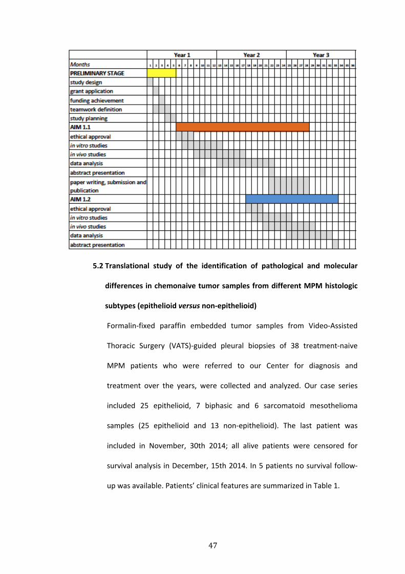
features. We achieved the project’s aims according to the following
timelines (Gannt Chart):
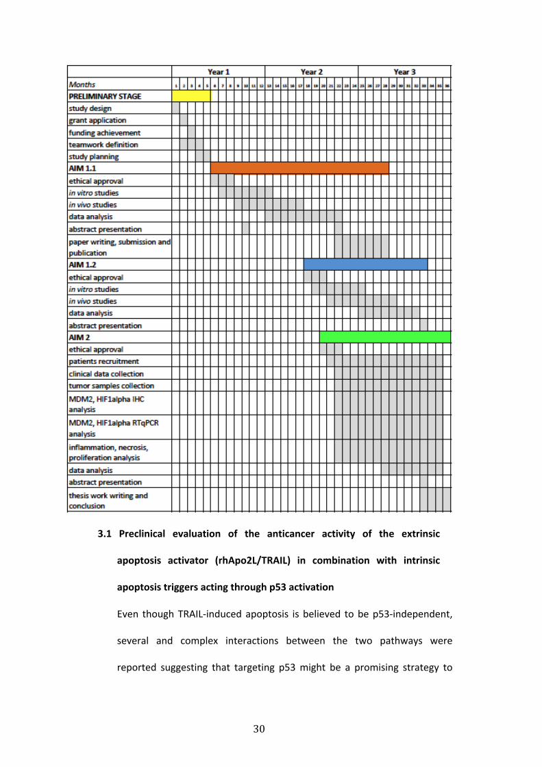
30
3.1 Preclinical evaluation of the anticancer activity of the extrinsic
apoptosis activator (rhApo2L/TRAIL) in combination with intrinsic
apoptosis triggers acting through p53 activation
Even though TRAIL-‐induced apoptosis is believed to be p53-‐independent,
several and complex interactions between the two pathways were
reported suggesting that targeting p53 might be a promising strategy to
31
sensitize tumors with wild-‐type p53 (e.g. MPM) to TRAIL-‐dependent cell
death [15, 62].
The first aim of this study is to investigate the anticancer effects of
rhApo2L/TRAIL (Amgen/Genentech) in combination with p53 reactivating
agents such as chemotherapy and p53-‐MDM2 inhibitors, employing
epithelioid and sarcomatoid MPM cell lines and an in vivo preclinical
model.
3.1.1. First, we investigated the anticancer activity of rhApo2L/TRAIL plus
the current gold standard chemotherapy regimen, a platinum-‐based
doublet associated with the antifolate agent pemetrexed. We furthermore
investigated if the improved cytotoxicity after the combination of
rhApo2L/TRAIL plus chemotherapy was actually p53-‐dependent.
3.1.2. We also explored the association of rhApo2L/TRAIL plus a new
member of the Nutlin3 family of MDM2 inhibitors, RG7112 (Hoffmann-‐La
Roche Inc).
3.2 Translational study of the identification of pathological and molecular
differences in chemonaive tumor samples from different MPM histologic
subtypes (epithelioid versus non-‐epithelioid)
Druggable molecular difference between epithelioid and sarcomatoid
MPM has not been identified so far. Preliminary results suggest that
MDM2 might promote tumor growth through apoptosis inhibition and
neoangiogenesis (VEGF and HIF1alpha) induction. According to our
preliminary data, this protein might be expressed at different levels in the
two MPM histologic subtypes.
32
Through the analysis of chemonaive patients samples the aim of this study
is to describe the different MPM histologic subtypes in terms of:
-‐ possible molecular targets for treatment– MDM2, VEGF, HIF1alpha-‐ at
mRNA and protein level; the correlation of MDM2 and neoangiogenesis
markers expression level have been explored.
-‐ pathological features –necrosis, inflammation and proliferation index-‐
and their correlation with MDM2 expression levels
-‐ as exploratory endpoints we aim at describing a possible prognostic
and/or predictive role of MDM2 and other markers/histological features.
4. Methods
4.1 Preclinical evaluation of the anticancer activity of the extrinsic apoptosis
activator (rhApo2L/TRAIL) in combination with intrinsic apoptosis triggers
acting through p53 activation (chemotherapy; RG7112)
4.1.1.Cell lines and primary cultures
Peripheral Blood Mononuclear Cells (PBMC) were isolated from peripheral
blood of healthy donors using Ficoll-‐Paque PLUS (GE HEALTHCARE, Little
Chalfont, Buckinghamshire, U.K.) according to the manufacturer’s protocol.
We employed three cell lines of epithelioid derivation (ZL55, H28, M14K),
three biphasic cell lines (ZL5, SPC111, MSTO-‐211H) and the sarcomatoid
cell line ZL34. PBMC and MPM cell lines were maintained in Roswell Park
Memorial Institute medium (RPMI) 1640 (Gibco-‐Life Technologies,
Carlsbad, CA, U.S.); Human Foreskin Fibroblasts (HFF) were grown in
Modified Dulbecco’s Eagle Medium (DMEM) (Gibco-‐Life Technologies,
Carlsbad, CA, U.S.); both mediums were supplemented with 2 mM L-‐
33
glutamine, 1 mM sodium pyruvate, 10% FBS and 1% (w/v)
penicillin/streptomycin (Invitrogen-‐Life Technologies, Carlsbad, CA, U.S.).
All cells were cultured at 37°C in a humidified atmosphere containing 5%
CO2. One MPM primary culture (MPM1801) of sarcomatoid mesothelioma
was established from fresh human pleural mesothelioma surgical
specimen. Specimens have been obtained from the Thoracic Surgical Unit
(University of Padua), after patient’s informed consent signature. The
project was submitted for approval to the Ethical Committee of Istituto
Oncologico Veneto and to the Ethical Committee for animal studies of the
University of Padua.
4.1.2 Annexin V staining:
MPM cells were seeded into 12-‐well plates in 1.0 mL/well of complete
RPMI 1640 and treated with Carboplatin/Pemetrexed (27uM and 42 uM
respectively) for 48 hours or Nutlin3a 10uM for 24 hours and/or
rhApo2L/TRAIL (Dulanermin, Amgen Inc, Thousand Oaks, CA, U.S.;
Genentech Inc, South San Francisco, CA, U.S.) 50 ng/mL for 24 hours.
In vitro chemotherapy concentrations were defined according to the dose
inducing the higher cell death in MPM cell lines with the lower cell death in
normal cells (PBMC and fibroblasts).
Thus, we choose concentration of Carboplatin 27uM and Pemetrexed
42uM inducing 10% of apoptosis in ZL55; the same concentration of
Carboplatin induced about 5% of apoptosis in ZL34, while Pemetrexed as
single agent showed no apoptosis induction with any tested concentration
(0-‐100 uM) (data not shown). In vitro rhApo2L/TRAIL concentrations were
34
defined according to previous data showing that these are able to reach
similar blood concentrations [99].
Time and sequence of exposure to chemotherapy and rhApo2L/TRAIL were
established according to previous data with other TRAIL agonists and
considering the pharmacokinetic of the drugs under study (shorter half-‐life
of rhApo2L/TRAIL compared to agonistic antibodies or to chemotherapy)
[49],[53] and the doubling times of cell lines (20.89 hours for ZL55 and
28.12 hours for Zl34, data not shown).
The Annexin V assay was performed using Annexin-‐V-‐Fluos and PI (Roche,
Basel, Switzerland) according to the manufacturers’ instructions. Cells were
collected, centrifuged, and then resuspended in 300 uL of Annexin-‐binding
buffer, followed by incubation with 1 uL of Annexin V-‐Fluos and 1uL of PI
for 10 minutes at room temperature. Cells positive for Annexin V/PI were
detected by flow cytometry using a FACSCalibur apparatus and CellQuest
software (BD Biosciences San Jose, CA, U.S.). Where indicated cells were
pre-‐treated with the ROS scavenger N-‐Acetyl-‐Cysteine (NAC 100 uM)
overnight. Specific Apoptosis was calculated by the following formula:
(percentage of Annexin V positive cells in treated samples-‐ percentage of
Annexin V positive cells in untreated samples) / (100-‐ percentage of
Annexin V positive cells in untreated samples)* 100.
Drug interactions were quantified by determining the combination index
(CI) using the CompuSyn software (ComboSyn, Inc., Paramus, NJ), where
CI< 1, CI=1, and CI > 1 indicated synergistic, additive, and antagonistic
effects, respectively.
35
4.1.3 Caspases Assay:
Caspases Assay was performed using Fluorometric Homogenous Caspase
Assay (Roche, Basel, Switzerland). MPM cells were seeded into 96-‐well
plates in 0.1 mL/well of complete RPMI 1640, treated with
Carboplatin/Pemetrexed (27uM and 42 uM respectively) for 48 hours or
Nutlin3a 10 uM for 24 hours and/or rhApo2L/TRAIL (50 ng/mL) for 24
hours, and then incubated with DEVD-‐Rhodamine 110. Upon cleavage of
the substrate by activated caspases, fluorescence of the released
Rhodamine 110 was measured using Victor microplate reader
(PerkinElmer, Waltham, Massachusetts, U.S.) with an excitation
wavelength of 480 nm and emission wavelength of 520 nm. Specific
caspases activity was calculated by the following formula: (fluorescence
intensity of treated samples-‐ fluorescence intensity of untreated samples) /
(fluorescence intensity of untreated samples).
4.1.4 Western blot:
Tissue specimens were processed by cryogenic grinding with mortar and
pestle to obtain a fine powder. The tissue powders and the cell lines were
lysed in Mammalian Cells Disruption Buffer Paris-‐Kit (Ambion-‐Life
Technologies, Carlsbad, CA, U.S.) supplemented with Phosphatase Inhibitor
Cocktail (Roche, Basel, Switzerland) and Complete Protease Inhibitor
Cocktail (Roche, Basel, Switzerland). Protein concentration was determined
by the Coomassie (Bradford) Protein Assay Kit (Thermo Scientific,
Waltham, Massachusetts, U.S.) using bovine serum albumin as standard,
and equal amounts of proteins were analyzed by SDS-‐PAGE (12%
36
acrylamide/bis-‐acrylamide). Gels were electroblotted onto
polyvinylidenedifluoride membranes (Amersham-‐GE HEALTHCARE, Little
Chalfont, Buckinghamshire, U.K.). In immunoblot analysis, membranes
were blocked for 1 hour with 5% non-‐fat dry milk in Tris Buffered Saline
(TBS) containing 0.1% Tween-‐20, and incubated at 4°C over night with
primary antibody direct against p53 (Santa Cruz Biotechnology), p21 (Cell
Signaling ) and p53 (Abcam) and anti-‐b-‐actin antibody (Sigma) used as
loading control, followed by horseradish peroxidase-‐conjugated secondary
antibodies (Santa Cruz Biotechnology, Dallas, Texas, U.S.). Finally, the
membranes were incubated with chemiluminescence reagents
(Supersignal Pico; Pierce-‐Thermo Scientific, Waltham, Massachusetts, U.S.)
and revealed using Chemidoc XRS System (Biorad, Hercules, CA, U.S.).
4.1.5 Flow cytometry analysis:
Surface expression of TRAIL receptors was evaluated by indirect
immunostaining using the primary antibodies DR4, DR5, DcR1 and DcR2
(Alexis Biochemicals, San Diego, CA, U.S.) followed by Alexa Fluor Goat
anti-‐mouse immunoglobulin G (IgG H+L) (Life Technologies, Life
Technologies, Carlsbad, CA, U.S.). Non-‐specific fluorescence was assessed
using normal mouse IgG followed by secondary antibody. Flow cytometry
analysis was performed using a FACSCalibur apparatus and CellQuest
software (BD Biosciences San Jose, CA, U.S.).Relative expression of TRAIL-‐R
was calculated by the following formula: percentage of positive cells x
mean fluorescence intensity (MFI).
4.1.6 Transfections:
37
The siRNA pool (25nM) for p53 (RIBOXX-‐Life Science, Dresden-‐Radebeul,
Germany) and/or the wild-‐type p53 expression vector (200 ng) were
transiently transfected in MPM cell lines using LIPOFECTAMINE 2000
(Invitrogen-‐Life Technologies, Carlsbad, CA, U.S.), according to the
manufacturers’ instructions. The expression levels of p53 were evaluated
24 hours after transfection by western blot analysis.
4.1.7 In vivo experiments:
4.1.7.1. rhAPO2L/TRAIL plus chemotherapy
In vivo experiments were performed in accordance with the Padua
University Ethic Committee for Animal Testing.
60 SCID male mice at the 6th week were implanted subcutaneously (sc) in
the right flank with 2x10⁶ ZL55 (30 mice) or ZL34 cell lines (30 mice)
suspended in 0.1 ml volume of RPMI. When tumor volume reached 50
mm3, mice were randomized in four groups (N=6 mice/group) and treated
by intraperitoneal (IP) injection: not treated (NT, vehicle 100uL on day 1);
Carboplatin/Pemetrexed (CP, 75 mg/Kg and 100 mg/Kg respectively on day
1); rhApo2L/TRAIL (T, 60 mg/Kg on days 1, 2 and 3); CPT (CP, 75 mg/Kg plus
100 mg/Kg respectively on day 1 plus T, 60 mg/Kg on days 1, 2 and 3).
rhApo2L/TRAIL schedule and dose were established according to previous
studies (data on file, Amgen Inc, Thousand Oaks, CA/Genentech Inc, South
San Francisco, CA, 2009).
Tumor volumes were measured with a caliper every third day; volumes
were calculated using the modified ellipsoidal formula: 1/2(length x
width2). Mice were suppressed at the 21th day or when tumor volume
38
reached 500 mm3. Delta volume was calculated by the following formula:
(tumor volume at the day n-‐ tumor volume at the day 1)/tumor volume at
the day 1*100.
4.1.7.2. rhAPO2L/TRAIL plus RG7112
30 SCID mice at the 6th week were intraperitoneally (IP) injected with
5x10⁶ ZL34 cells previously transduced with lentiviral vector containing a
plasmid encoding for Luciferase. 3 weeks post injection the mice were
randomized in 4 treatment groups and treated with RG7112 (Hoffman-‐La
Roche Inc.) or vehicle (days 1-‐21) by gavage and/or rhAPO2L/TRAIL (days 1-‐
3) by IP. Tumor size was assessed once a week by in vivo bioluminescence
using Xenogen bioluminescence imaging after IP injection of D-‐luciferin in
each mouse. The mice were suppressed at the 22th day. Average Radiance
[p/s/cm²/sr] was proportional to the number of ZL34 cells expressing the
LUC gene and D Average radiance was used as indicator of tumor growth
and calculated by the following formula: (Average Radiance at the day n-‐
Average Radiance at the day 1)/Average radiance at the day 1.
4.2 Translational study of the identification of pathological and molecular
differences in chemonaive tumor samples from different MPM histologic
subtypes (epithelioid versus non-‐epithelioid)
4.2.1 Patients samples and data collection
We retrospectively collected and analyzed epithelioid, biphasic and
sarcomatoid malignant pleural mesothelioma samples from the diagnostic
biopsy. In order to perform RT-‐qPCR and immunohistochemistry the
following tumor samples were required:
39
-‐ 1 hematoxylin and eosin stained slide and paraffin-‐embedded tumor
block or 5-‐10 paraffin embedded tumor sections in IHC slides (for
immunohistochemistry analysis)
-‐ paraffin embedded tumor block or 5 sections (10 u) in eppendorf
RNASE/DNASE free (for RT-‐qPCR).
All the samples (N=38) were analyzed for protein expression levels of
MDM2 and HIF1alpha through IHC and for inflammation, necrosis and Ki67
levels; mRNA expression levels of MDM2 and HIF1alpha levels were
quantified through RT-‐qPCR in 32 tumor samples (20 epithelioid and 12
sarcomatoid/biphasic) where RNA was available. VEGF IHC and RT-‐qPCR
analysis were performed in a training set of 27 tumor samples (17
epithelioid and 10 sarcomatoid/biphasic). As negative controls we used
normal pleural from 4 non cancer patients undergoing thoracic surgery.
Complete clinical information about patients enrolled in the study were
collected: age, gender, ECOG PS, EORTC score, stage, systemic treatments,
surgery, radiotherapy, first progression and last follow-‐up date, status
(alive/dead).
To perform the statistical analyses, all data collected were recorded in a
computer data base (Microsoft Excel) with a protection of password.
We collected the informed consent to the data processing of the data
subjects included in the research if, during the study, it was possible to give
them the information and to get the related consent, and in particular,
when they turned to our Institute also for follow up reasons. The
operations of collection, storage, preservation, and circulation of the
40
biological samples as well as all the data processing operations regarding
health data of the persons involved in the study were uniformed to the
security and organizational measures defined in the document “Guidelines
on data protection in medical and biomedical research”.
Eight slices of 10 μm sections/sample have been collected in 1,5 ml of a
microcentrifuge tube and incubated in xylene at elevated temperatures to
solubilize and remove paraffin from the tissue, then washed in alcohol
solutions to remove the xylene. Total RNA extraction of the deparaffinized
samples have been performed using RecoverAll Total Nucleic Acid Isolation
Kit (Ambion) according to manufacturer’s protocol.
4.2.2 mRNA expression analysis.
Reverse transcription of total mRNA was performed using 500 ng of total
RNA/sample by SuperScript II Reverse Trancriptase (Invitrogen) according
to the manufacturer’s protocol. Quantitative analysis of MDM2, VEGF, and
HIF-‐1 alpha genes have been performed by LightCycler 480 Real time PCR
System and LightCycler 480 SYBR Green I Master Mix (Roche) according to
the manufacturer’s protocol using specific primers for each genes (Sigma).
As internal reference we used B2-‐microglobulin, GAPDH and b-‐actin. The
Real Time reaction was carried out as follows: 95°C for 5 min, 40 cycles of
95°C for 10 s, 60°C for 10 s, and 72°C for 10 s. All reaction have been run in
triplicate and the quantitation of gene expression performed using the
∆∆CT calculation as previously described.
41
4.2.3 Immunohistochemistry and pathologic assessment
Serial sections of 4 micron were immunostained with the monoclonal Ki67,
MDM2, HIF 1alpha and VEGF antibodies. The strong dark stained Ki67 and
MDM2 and HIF1alpha nuclei were counted and expressed as % of total cell
number. The counting of VEGF positive cells was based on the number and
staining intensity of positive cells (from 0 to 300). The presence of necrosis
and inflammation were evaluated on hematoxylin eosin sections and
quantified using a score system from 0 to 3 (0: absent; 1: <10%; 2 from 10
to 20% and 3>20% of the whole tumor section examined).
4.3. Statistics
4.3.1 Preclinical studies
In vitro and in vivo studies results were expressed as mean +/-‐ standard
error and +/-‐ standard deviation, respectively. Mann Whitney and ANOVA
test (followed by post-‐hoc LSD test) were used to compare different
treatment groups in vitro and in vivo, respectively. A p value ≤ 0,05 was
considered as significant.
4.3.2 Translational studies
Low versus high expression levels of each marker and pathological
parameter have been identified over and under the median value, and data
presented as % of patients with high/low expression levels in each
histological subtype. Kruskall-‐Wallis test have been performed to evaluate a
different expression of molecular markers and pathological parameters in
the two main histological subtypes, and to assess different pathological
features in tumor tissues with high/low MDM2 expression. The correlation
42
between MDM2 and HIF1alpha expression levels (and possibly VEGF), and
between RNA and protein expression levels of each marker have been
investigated through the linear correlation analysis of Spearman. Overall and
progression free survival curves have been designed according to Kaplan-‐
Meier method.
The univariate analysis allowed to select the molecular marker and/or
morphological parameter to be analyzed with the clinical features in the
multivariate analysis for the assessment of the impact on PFS and OS (Cox
Regression Proportional Hazards Model).
PFS has been assessed from the date of enrolment to the date of disease
progression to the first-‐line (or relapse) or to the date of death, whichever
occurred first. OS has been assessed from the date of enrolment to the date
of death.
5. Results
5.1 Preclinical evaluation of the anticancer activity of rhApo2L/TRAIL in
combination with chemotherapy (carboplatin plus pemetrexed)[100] or
p53-‐MDM2 inhibitor RG7112
5.1.1 rhApo2L/TRAIL triggers apoptosis in MPM cells but not in normal
cells.
The induction of apoptosis by rhApo2L/TRAIL treatment was tested in
seven MPM cell lines (3 epithelioid: ZL55, H28, M14K; 1 sarcomatoid: ZL34;
3 biphasic: MSTO-‐211, SPC111, ZL5) and one short-‐term primary culture of
sarcomatoid MPM cells established from a patient (MPM1801). Cells were
treated with 50 ng/ml rhApo2L/TRAIL for 24 hours and apoptosis was
43
measured by Annexin V staining and Fluorometric Homogenous Caspase
Assays. The results showed a significant, even if heterogeneous sensitivity
of MPM cells to TRAIL treatment, independent from the histotype.
Interestingly, this effect was specific in MPM cells, as significant death was
not observed in control cells (HFF and PBMC)(Figure 1a and b).
5.1.2 Carboplatin and Pemetrexed or Nutlin3a enhance the pro-‐apoptotic
effects of rhApo2L/TRAIL on MPM cell lines
As Carboplatin and Pemetrexed (CP) are the cornerstone of current MPM
therapies, we next tested whether these drugs might synergize with
rhApo2L/TRAIL (T). Apoptosis was measured by Annexin V staining and
flow cytometry. Cell lines ZL34 and ZL55 were selected as representative of
sarcomatoid and epitheliod MPM, respectively. Results showed a
significant (p < 0.001) synergistic effect of the combination of these drugs
compared with no treatment or with CP or T as single agents (Figure 2 a
and c). These results were also confirmed when apoptosis was assessed
with the caspases assay (Figure 2b). A similar effect was shown in all the
tested cell lines (3 epithelioid, 3 biphasic and 1 sarcomatoid) and in the
sarcomatoid primary culture (Figure 3).
Previous studies [49] suggested that sensitivity to TRAIL might be
dependent on the levels of reactive oxygen species (ROS). However, we did
not observe any difference in specific cell death when both ZL34 and ZL55
cell lines were treated with the ROS scavenger NAC (Figure 4).
We then analyzed in vitro effects of rhAPO2L/TRAIL plus the MDM2-‐p53
inhibitor Nutlin3a by Annexin V and Caspases assay. Apoptosis assay
44
performed in eight MPM cell lines, representing the three different
histotypes (epithelioid, biphasic and sarcomatoid), showed a synergistic
anticancer effect of Nutlin3a plus rhAPO2L/TRAIL. Higher synergistic effect
was shown in sarcomatoid cell (Figure 2d).
5.1.3 p53 activation by Carboplatin and Pemetrexed sensitizes to TRAIL-‐
dependent apoptosis in vitro
We next investigated the mechanisms at the basis of the sensitization to
TRAIL-‐dependent apoptosis induced by CP. Considering that both
Carboplatin and Pemetrexed induced DNA damage resulting in p53
activation and that p53 is not mutated in most MPM cases, we tested the
effect of CP on p53 levels in the ZL55 and ZL34 cell lines. Results indicated
an increase in p53 levels in ZL55 and ZL34 cell lines following CP treatment
(Figure 5a). To test whether this increase in p53 levels accounts for the
ability of CP to sensitize to TRAIL-‐induced apoptosis, we investigated cell
death in ZL55 and ZL34 cell lines treated with CP and/or T after p53 knock–
down by siRNAs. Results demonstrated that the siRNA treatment induced a
significant, specific knock-‐down of p53 expression (Figure 5a and b).
Interestingly, p53 silencing resulted in a significant reduction of cell death
induced by CP and T (Figure 5c); importantly, this effect was reverted by
cotransfection with a vector coding for wild-‐type p53 (Figure 5c). We
observed no or weak p53 expression in five tumor tissues from MPM
patients; in contrast, p53 was readily detected in the ZL34 and ZL55 cell
lines after p53 vector (Figure 5b).
5.1.4 p53 activation increases the expression of TRAIL receptors in vitro

45
To explore the TRAIL-‐sensitizing effect of p53 activation, we next
investigated whether p53-‐inducing treatments enhance the expression of
the TRAIL receptors DR4/DR5, DcR1/DcR2. Results showed that the ZL55
and ZL34 cell lines expressed higher levels of DR4 and DR5 in response to
CP treatment (Figure 6a). Interestingly, knock-‐down of p53 expression
resulted in a significant reduction of CP-‐induced DR4 and DR5 expression in
MPM cells (p< 0.05), while no reduction of DR4 and DR5 expression was
observed in MPM cells cotransfected with the p53 siRNA plus wild-‐type
p53 expression vector (Figure 7). Taken together, these data provide
evidence of a causal link between CP treatment, p53 activation, increased
expression of DR4 and DR5 receptors and sensitivity to TRAIL. The
detection of DcR1 and DcR2 levels in ZL34 and ZL55 cell lines treated or not
with CP seemed not relevant.
When we explored the mechanism at the basis of the synergism between
rhAPO2L/TRAIL and Nutlin3a in sarcomatoid cell lines, we observed the
activation of p53, confirmed also by the activation of the two p53 targets
p21 and inhibition of surviving. Additionally, p53 activation by Nutlin3a
increased the expression of DR4/DR5 TRAIL death receptors (Figure 6b).
5.1.5 Antitumor activity of Apo2L/TRAIL + chemotherapy or RG7112 in
preclinical animal models of MPM
To test the in vivo efficacy of Apo2L/TRAIL as single agent and in
combination with CP, we employed a preclinical model based on the
subcutaneous injection of the ZL55 and ZL34 MPM cell lines in SCID mice.
46
Thirty SCID mice were inoculated with ZL55 and 30 SCID mice were
inoculated with ZL34 cells. Twenty-‐four mice had a measurable tumor and
were randomized in the four treatment groups (N=6/group). Mice
inoculated with ZL55 cells showed a statistically significant reduction of
tumor volume at every time point in the three treatment groups (CP; T;
CPT) compared to not treated (NT) mice; moreover, tumor volume was
significantly reduced in mice treated with CPT compared to CP at the 21th
day (p< 0.05) (Figure 8a).
Mice inoculated with ZL34 cells showed a statistically significant reduction
of tumor volume at every time point in T and CPT treatment groups
compared to not treated; at the day 21 we observed a statistically
significant difference of tumor volume between all three treatment groups
and untreated mice and between CPT and CP groups (Figure 8b).
No significant difference was observed between mice treated with CP
compared to T (Figures 8 a,b).
To test the in vivo efficacy of Apo2L/TRAIL in combination with RG7112, we
employed a preclinical model based on the intraperitoneal injection of
ZL34 MPM cell lines in SCID mice. 30 SCID mice were inoculated with ZL34
cells.
Tumor growth in mice injected with ZL34 cells was significantly reduced in
mice treated with RG7112 plus rhApo2L/TRAIL compared with mice treated
with RG7112 or Apo2L/TRAIL as single agents (Figure 9).
47
5.2 Translational study of the identification of pathological and molecular
differences in chemonaive tumor samples from different MPM histologic
subtypes (epithelioid versus non-‐epithelioid)
Formalin-‐fixed paraffin embedded tumor samples from Video-‐Assisted
Thoracic Surgery (VATS)-‐guided pleural biopsies of 38 treatment-‐naive
MPM patients who were referred to our Center for diagnosis and
treatment over the years, were collected and analyzed. Our case series
included 25 epithelioid, 7 biphasic and 6 sarcomatoid mesothelioma
samples (25 epithelioid and 13 non-‐epithelioid). The last patient was
included in November, 30th 2014; all alive patients were censored for
survival analysis in December, 15th 2014. In 5 patients no survival follow-‐
up was available. Patients’ clinical features are summarized in Table 1.
48
5.2.1 MDM2, HIF1alpha, VEGF expression levels in epithelioid versus non-‐
epithelioid MPM samples
Nuclear expression of MDM2 and HIF1alpha was higher in
sarcomatoid/biphasic tumor samples (77% and 77% of tumor samples) and
lower in epithelioid samples (68% and 64% of tumor samples, respectively);
similar results were observed when we investigated mRNA expression
levels of MDM2 but not of HIF1alpha (Table 2 and 3). No expression of
MDM2 and HIF1alpha was observed in normal pleura samples.
Higher MDM2 and HIF1alpha IHC expression levels were significantly
associated with sarcomatoid/biphasic histologic subtype (p=0.010 and
p=0.007, respectively) (Figure 10 and 11). When we analyzed mRNA
expression levels of MDM2 we observed a correlation trend with histologic
subtype (higher levels in sarcomatoid/biphasic samples), although not
statistically significant (p=0.067); no correlation was observed between
HIF1alpha mRNA expression levels and histologic subtype (p=0.2), and
between RNA and protein expression levels of MDM2(p=0.3) and
HIF1alpha (p=0.9).
Among the 18 tumor samples with high MDM2 expression at the IHC, only
9 showed high levels of mRNA expression at the RT-‐qPCR analysis,
suggesting mechanisms other than gene amplification sustaining protein
overexpression.
Importantly, when we assessed the correlation between nuclear IHC
expression levels of MDM2 and HIF1alpha, we observed a statistically
49
significant positive correlation (correlation coefficient=0.533; p value=
0.00626).
No significant difference between the two main histologic subgroups was
observed when we analyzed VEGF protein and mRNA expression levels in
the training set of 27 tumor samples (Table 4), therefore we considered
this marker not worthy of further investigation through either IHC or RT-‐
qPCR. Moreover, VEGF expression was not completely negative in normal
pleural samples, probably because of a consequence of a pro-‐inflammatory
phenotype of such controls.
5.2.2 Inflammation, necrosis and proliferation index levels in epithelioid
versus non-‐epithelioid MPM samples
When we investigated different pathological features in tumor samples
and compared epithelioid and sarcomatoid/biphasic subtypes, we
observed more frequently low levels of inflammation in sarcomatoid
samples (62% of tumor samples) and low levels of necrosis among
epithelioid tumor samples (72% of tumor samples). Differently,
proliferation index was more frequently low in epithelioid (72%) and high
in sarcomatoid/biphasic (77%) samples (Table 5). No statistically difference
in terms of inflammation (p=0.112) and necrosis (p=0.07) levels was
observed between epithelioid and sarcomatoid/biphasic samples (Figure
12 a,b and d); proliferation index was significantly higher in
sarcomatoid/biphasic compared with epithelioid samples (p=0.005) (Figure
12 c and d).
50
When we divided the whole patients population in two subgroups
according to different MDM2 expression levels (high versus low), we
observed that proliferation index was also significantly higher in tumor
samples with higher MDM2 expression (p=0.008, data not shown).
5.2.3 Exploratory endpoints: prognostic and predictive role of MDM2
expression levels and other markers/histological features
As exploratory analysis, we assessed the impact of some clinical and
pathological features on OS and PFS. Unfortunately, 5 patients were lost to
follow-‐up and no survival data were available, thus reducing the sample
size and impairing this analysis. Moreover, other weak points were that
some sarcomatoid/biphasic mesothelioma patients had no sufficient
follow-‐up time (less than 6 months) and different local or systemic
treatments were considered.
No statistically significant difference in terms of OS (p=0.3) or PFS (p=0.7)
was observed between epithelioid and sarcomatoid/biphasic samples,
even if a trend towards a longer OS and PFS was observed in epithelioid
(median OS 80 weeks; median PFS 48 weeks) compared with
sarcomatoid/biphasic MPM (median OS 56 weeks; median PFS 40 weeks)
(Figure 13). No statistically significant difference in terms of OS (p=0.5) or
PFS (p=0.3) between high and low MDM2-‐expressing samples was
observed. A trend towards a longer OS and PFS was seen in samples with
lower MDM2 expression levels (median OS 96 weeks; median PFS 48
weeks) compared with sarcomatoid/biphasic MPM (median OS 60 weeks;
median PFS 36 weeks).
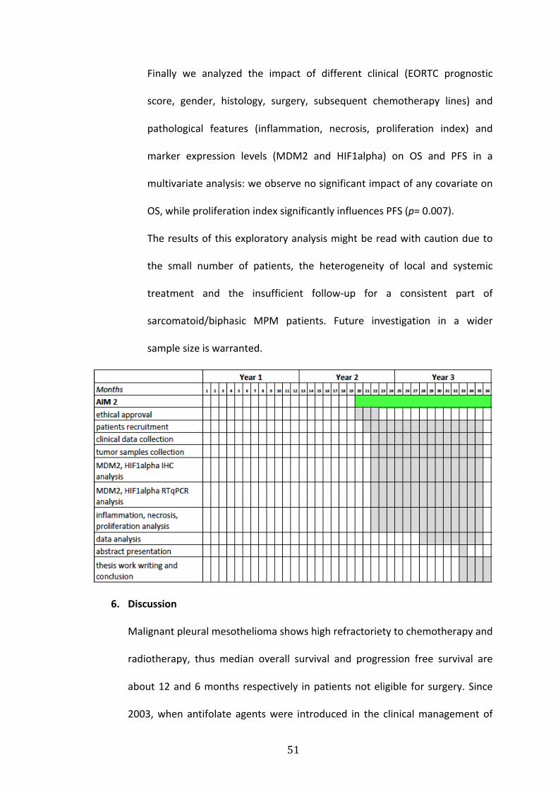
51
Finally we analyzed the impact of different clinical (EORTC prognostic
score, gender, histology, surgery, subsequent chemotherapy lines) and
pathological features (inflammation, necrosis, proliferation index) and
marker expression levels (MDM2 and HIF1alpha) on OS and PFS in a
multivariate analysis: we observe no significant impact of any covariate on
OS, while proliferation index significantly influences PFS (p= 0.007).
The results of this exploratory analysis might be read with caution due to
the small number of patients, the heterogeneity of local and systemic
treatment and the insufficient follow-‐up for a consistent part of
sarcomatoid/biphasic MPM patients. Future investigation in a wider
sample size is warranted.
6. Discussion
Malignant pleural mesothelioma shows high refractoriety to chemotherapy and
radiotherapy, thus median overall survival and progression free survival are
about 12 and 6 months respectively in patients not eligible for surgery. Since
2003, when antifolate agents were introduced in the clinical management of
52
this disease, the gold standard chemotherapy is a Platinum-‐based doublet plus
Pemetrexed or Raltitrexed[1, 2]. Surgery is feasible in highly selected cases, and
patients suitable for trimodal treatment (neoadjuvant chemotherapy, surgery,
postoperative radiotherapy) achieve overall survival longer than 25 months
[28]. After the standard first-‐line Pemetrexed/Platinum combination there is
not a defined regimen for the second line treatment of MPM, and the clinical
benefits are uncertain[101-‐104]. Recent studies tested biologic agents that
target key oncogenic pathways, including phosphatidylinositol3-‐kinase
(PI3K)/mammalian target of Rapamycin (mTOR) pathways, histone
deacetylases (HDAC), Nuclear Factor kB (NFkB) and neoangiogenesis. However,
no one of these therapies proved to significantly impact the natural history of
this neoplasm, thus reinforcing the need for new drugs to improve prognosis of
MPM patients.
TRAIL is a member of the TNF superfamily which has recently emerged as a
potentially interesting anticancer agent because of its ability to kill cancer cell
lines while sparing many normal cells[40, 55, 105]. Several studies showed that
MPM cells are resistant to TRAIL in vitro, although this resistance can be
overcome by combining chemotherapy with alpha-‐tocopherylsuccinate,
anisomycin, HDAC inhibitors, proteasome inhibitors and FLIP siRNA [49, 60, 61,
106-‐109]. Increased apoptotic levels were demonstrated in four cell lines
treated with the association of TRAIL and chemotherapy (Cisplatin,
Doxorubicin, Gemcitabine or Etoposide), probably through p53-‐independent
apoptotic pathway; no DR5 increase was observed at the basis of this
sensitization[60]. Recently, another study showed apoptotic effects of TRAIL or
53
the monoclonal antibodies Mapatumumab and Lexatumumab directed against
DR4 and DR5 on 13 MPM cell lines; this effect was enhanced by Cisplatin
probably trough the induction of ROS [49]. Other studies showed a p53-‐
dependent induction of DR4 and DR5 expression by Alpha-‐tocopheryl
succinate, resulting in TRAIL sensitization [61]. While TRAIL-‐dependent
apoptosis is thought to be p53-‐independent, p53 wild type cancer cells can be
sensitized to TRAIL through p53 activation [62]. In contrast to most solid
tumors, MPM cells frequently express wild type p53 [63].
Recombinant human (rh) Apo2L/TRAIL (Dulanermin), a receptor agonist which
binds both DR4 and DR5[53, 54], showed antitumor activity in in vitro and in
vivo tumor models of different cancer types both as single agent and in
combination with chemotherapy without any toxicity in normal cells [55-‐57],
and was the first TRAIL agonist investigated in human clinical trials [51, 52, 58].
In the present study we investigated proapoptotic effects of rhApo2L/TRAIL
combined to different intrinsic apoptosis triggers: first the DNA-‐damaging
agents Carboplatin and Pemetrexed[100], then the p53-‐MDM2 inhibitor
nutlin3-‐RG7112. To our knowledge, this is the first in vivo study of a TRAIL
agonist in mesothelioma.
We observed heterogeneous sensitivity of seven mesothelioma cell lines and
one primary culture to rhApo2L/TRAIL treatment; higher or lower sensitivity to
the TRAIL agonist did not seem to be dependent on the histologic subtype.
These data seem in line with previous studies of TRAIL agonists in vitro, and
might be explained by the variable expression levels of antiapoptotic proteins
such as Bcl2 and IAPs-‐family proteins, or other proteins involved in the
54
apoptotic pathway[49]. These proteins have been previously suggested as
useful predictive markers of sensitivity to TRAIL-‐dependent apoptosis.
We observed higher apoptotic levels when both epithelioid and sarcomatoid
cell lines were treated with chemotherapy and rhApo2L/TRAIL compared to
Carboplatin/Pemetrexed and rhApo2L/TRAIL alone. This effect seemed to be
dependent upon p53 but not on increased ROS levels. Furthermore we showed
that p53 activation leads to increased expression of the DR4 and DR5
receptors. Previous evidences showed that chemotherapy-‐induced p53
activation lead to antiapoptotic proteins downregulation (such as survivin or
Mcl1) and proapoptotic targets upregulation (such as Bax) [49, 110].
Contrary to previous evidences[63], preliminary data from next-‐generation
sequencing of 123 MPM samples showed that p53 was among the five most
frequently altered genes [111], although the overall frequence of such
mutations may be considered low. Our results in a primary culture of
sarcomatoid MPM (MPM1801) showed no mutation in p53 gene. Thus, based
on our results, it is tempting to speculate that p53 mutational status in MPM
samples may be a predictive marker of sensitivity to the combination of
chemotherapy and rhApo2L/TRAIL. This hypothesis will be thoroughly tested in
further studies in p53 knock-‐out models. In vivo studies showed that the
association of Carboplatin/Pemetrexed with rhApo2L/TRAIL significantly
reduced tumor growth compared to Carboplatin/Pemetrexed in both cell lines
tested. Considering the short-‐term responses achieved with currently used
chemotherapy, especially in the neoadjuvant setting where tumor shrinkage
and distant relapses prevention assume particular relevance, the synergistic
55
effect of chemotherapy combined to rhApo2L/TRAIL might improve patients
prognosis. Moreover, similar antitumor effects of rhApo2L/TRAIL and
chemotherapy in vivo, might suggest a role for such agent at the disease
progression after the first line treatment, where a valid option is still lacking in
patients eligible for further treatments.
Taken together our findings suggest that rhApo2L/TRAIL combined to standard
chemotherapy as first line treatment and as single agent in the second line
setting might prove to be an effective drug in the treatment of MPM,
considering also its low toxicity profile.
On the same way, we adopted the combination of rhApo2L/TRAIL plus nutlin3-‐
RG7112, as alternative strategy to reactivate p53 thus sensitizing to TRAIL-‐
dependent apoptosis in vitro and in vivo. Given the central role of MDM2 in
regulating p53 activity and stability, developing small-‐molecule inhibitors of
MDM2 could offer a novel approach to treating cancers. While nutlin3, the first
developed compound targeting the p53-‐MDM2 complex was optimal for
previous studies of p53 biology, and was effectively adopted in our in vitro
studies, this compound was not optimal in clinical studies, thus recent efforts
lead to a new molecule belonging to the nutlin family of MDM2 inhibitors,
RG7112, which is currently in clinical development[84] (clinicaltrials.gov
NCT00559533; NCT00623870). The high genetic plasticity characteristic for
human tumors, especially at advanced stages, increases the chances for
acquired resistance to most single agent therapies including MDM2
antagonists. Therefore, combination approaches to cancer therapy are
extensively sought.
56
In vitro studies confirmed p53 activation by nutlin3, with subsequent
upregulation of TRAIL receptors and final sensitization to extrinsec apoptosis,
thus confirming previous results by other groups [112]. In vitro studies showed
the higher synergism between the two molecules in sarcomatoid cell lines, thus
raising the question about different MDM2 expression levels in epithelioid
versus non-‐epithelioid (sarcomatoid and biphasic) MPM. This scientific question
was someway reinforced by previous evidence of MDM2 overexpression with
or without gene amplification in soft tissue sarcomas [70-‐73] and by the
evidence of higher effectiveness of RG7112 in killing cancer cells overproducing
MDM2 protein as a result of MDM2 gene amplification [85]. Previous results
from our in vitro studies showed higher MDM2 mRNA levels in sarcomatoid cell
lines, compared with epithelioid (Figure 15). Translational studies performed
on clinical samples from affected patients confirmed these data.
Sarcomatoid/biphasic mesothelioma are characterized by higher aggressive
biological behaviour, higher resistance to systemic treatments, more frequent
distant spread and poor prognosis. Our in vivo results in the sarcomatoid
animal model showed remarkable reduction in tumor growth with the
combination under study (rhApo2L/TRAIL plus nutlin3-‐RG7112). These findings
might open new scenarios to the medical treatment of such histologic MPM
subtype, with a well-‐tolerated alternative to chemotherapy, worthy of clinical
investigation. Moreover a systemic targeted treatment according to a specific
mesothelioma histotype could be planned.
Recently MDM2 disregulation in merlin-‐deficient tumors has been reported.
However to best of our knowledge, different expression level of MDM2 in
57
different mesothelioma histotypes have not been investigated, so far. On the
same way, specific morphological features (inflammation, necrosis,
proliferation index) have not been carefully investigated in epithelioid versus
sarcomatoid/biphasic MPM.
Our results showed that strong MDM2 overexpression-‐ mainly in IHC-‐ was
significantly correlated with sarcomatoid/biphasic histotype even if the mRNA
levels of MDM2 did not reach the statistically significant value. The correlation
test between mRNA and protein analysis was negative. These results might be
explained in different ways. First of all, literature data confirm that MDM2
protein overexpression is not only regulated by gene amplification, but also by
other mechanisms, such as transcriptional and post-‐transcriptional
modifications[77]. Secondarily, IHC detected only strong nuclear expression,
while RT-‐qPCR quantified mRNA derived from the whole tumor sample.
Moreover, there are pools within a cell of active and inactive MDM2 that do
not directly correlate with overall MDM2 expression but which may reflect
different isoforms or modified forms of the protein. Moreover, other factors
could influence intracellular MDM2 expression level such as altered rates of
transcription, mRNA stability, enhanced translation, and diminished destruction
of the protein all will affect intracellular levels of MDM2. The prognostic
significance of MDM2 overexpression are quite controversial in the literature
[78]. We found a trend towards a negative prognostic and predictive
significance of high MDM2 strong nuclear expression levels. In our case series,
this lack of correlation with survival data may be related to several factors such
as the small sample size, especially for sarcomatoid/biphasic samples whose
58
survival data were available in a small number of patients; the insufficient
follow-‐up time of the last patients might have some impact on prognosis. This
underlines the importance to validate our data in a prospective study with a
larger and more homogenous population. Our results reported a statistically
significant correlation of high Ki67 levels with sarcomatoid/biphasic histology
and with high MDM2-‐expressing tumors, and Ki67 was the only covariate
significantly affecting progression free survival to the first line treatment, at the
multivariate analysis.
Thus, from our results, it is tempting to speculate that MDM2 and Ki67 might
be considered as important diagnostic parameters to characterize MPM with
poor prognosis.
Recently, nuclear grading in epithelioid mesothelioma has been shown as a
simple, practical, and cost-‐effective prognostic tool that better stratifies clinical
outcome and time to recurrence than currently available clinicopathologic
factors [113]. Our preliminar data on 25 epithelioid tumor samples where
survival data were available, showed only a trend towards a prognostic value of
nuclear grading, although not statistically significant, probably because of the
small sample size; in fact, we observed a median overall survival of 128, 96 and
60 weeks (p value= 0.5), in those patients with G1 (N=7), G2 (N=9) and G3 (N=9)
nuclear grading, respectively; median progression free survival was 55, 36 and
58 weeks (p=0.2), in patients with G1, G2 and G3 nuclear grading, respectively.
To date, no literature data are available about different levels of inflammation
and necrosis across mesothelioma histotypes; the prognostic significance of
necrosis in mesothelioma was reported by other groups, even though these
59
results were limited to biphasic [87] or epithelioid histotype [114]. Our results
did not show any significant difference, even though a trend towards higher
necrosis features in sarcomatoid/biphasic tumors was detected. This finding
may be due to a different biological behaviour of the two cancer types. The
sarcomatoid histotype would present necrosis following a failure blood supply,
as usually occurs in tumours with great proliferative index.
Overexpression of tumor neoangiogenesis markers has been previously
described as a prognostic factor in MPM patients, with particular reference to
VEGF [86, 87]. MDM2 has been suggested as a possible regulator of
neoangiogenesis, both through direct regulation of VEGF and through
stabilization of HIF1alpha, responsible for VEGF transcription [95-‐98].
Our results did not show significant difference of VEGF expression between
epithelioid and non-‐epithelioid mesothelioma samples both at the protein and
mRNA level. These results, together with recent evidence from negative clinical
studies with Bevacizumab, a monoclonal antibody targeting VEGF, lead us to
leave the investigation of this marker in MPM.
One innovative finding of our study was the significant correlation between
HIF1alpha expression levels and the sarcomatoid/biphasic histotype, as much
as the correlation between expression levels of MDM2 and HIF1alpha.
These results are of particular relevance further supporting the different
biological behaviour of sarcomatoid/biphasic versus epithelioid MPM. As
MDM2, HIF1alpha might be another promising target for antiangiogenetic
treatments in MPM because monoclonal antibodies against VEGF has failed in
the systemic treatment of mesothelioma. Further efforts should be made in
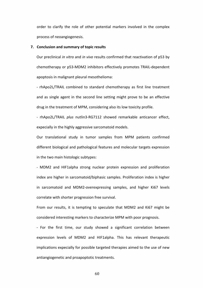
60
order to clarify the role of other potential markers involved in the complex
process of neoangiogenesis.
7. Conclusion and summary of topic results
Our preclinical in vitro and in vivo results confirmed that reactivation of p53 by
chemotherapy or p53-‐MDM2 inhibitors effectively promotes TRAIL-‐dependent
apoptosis in malignant pleural mesothelioma:
-‐ rhApo2L/TRAIL combined to standard chemotherapy as first line treatment
and as single agent in the second line setting might prove to be an effective
drug in the treatment of MPM, considering also its low toxicity profile.
-‐ rhApo2L/TRAIL plus nutlin3-‐RG7112 showed remarkable anticancer effect,
expecially in the highly aggressive sarcomatoid models.
Our translational study in tumor samples from MPM patients confirmed
different biological and pathological features and molecular targets expression
in the two main histologic subtypes:
-‐ MDM2 and HIF1alpha strong nuclear protein expression and proliferation
index are higher in sarcomatoid/biphasic samples. Proliferation index is higher
in sarcomatoid and MDM2-‐overexpressing samples, and higher Ki67 levels
correlate with shorter progression free survival.
From our results, it is tempting to speculate that MDM2 and Ki67 might be
considered interesting markers to characterize MPM with poor prognosis.
-‐ For the first time, our study showed a significant correlation between
expression levels of MDM2 and HIF1alpha. This has relevant therapeutic
implications expecially for possible targeted therapies aimed to the use of new
antiangiogenetic and proapoptotic treatments.
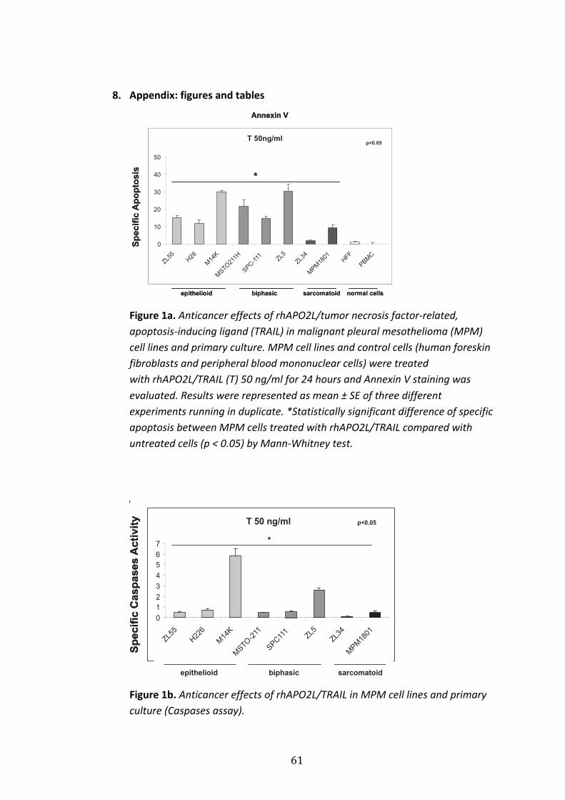
61
8. Appendix: figures and tables
Figure 1a. Anticancer effects of rhAPO2L/tumor necrosis factor-‐related, apoptosis-‐inducing ligand (TRAIL) in malignant pleural mesothelioma (MPM) cell lines and primary culture. MPM cell lines and control cells (human foreskin fibroblasts and peripheral blood mononuclear cells) were treated with rhAPO2L/TRAIL (T) 50 ng/ml for 24 hours and Annexin V staining was evaluated. Results were represented as mean ± SE of three different experiments running in duplicate. *Statistically significant difference of specific apoptosis between MPM cells treated with rhAPO2L/TRAIL compared with untreated cells (p < 0.05) by Mann-‐Whitney test.
Figure 1b. Anticancer effects of rhAPO2L/TRAIL in MPM cell lines and primary culture (Caspases assay).
Sp
ec
ific
Ap
op
tos
isT 50ng/ml
0
10
20
30
40
50
ZL55
H28
M14
K
MSTO
211H
SPC-1
11 ZL5
ZL34
MPM
1801
HFF
PBM
C
Annexin V
epithelioid biphasic sarcomatoid normal cells
*
p<0.05
Sp
ec
ific
Ap
op
tos
isT 50ng/ml
0
10
20
30
40
50
ZL55
H28
M14
K
MSTO
211H
SPC-1
11 ZL5
ZL34
MPM
1801
HFF
PBM
C
Annexin V
epithelioid biphasic sarcomatoid normal cells
**
p<0.05
Figure 1
T 50 ng/ml
0
1
2
3
4
5
6
7
ZL55
H22
6
M14
K
MSTO
-211
SPC11
1ZL5
ZL34
MPM
1801
Sp
ec
ific
Ca
sp
as
es
Ac
tiv
ity T 50 ng/ml
0
1
2
3
4
5
6
7
ZL55
H22
6
M14
K
MSTO
-211
SPC11
1ZL5
ZL34
MPM
1801
T 50 ng/ml
0
1
2
3
4
5
6
7
ZL55
H22
6
M14
K
MSTO
-211
SPC11
1ZL5
ZL34
MPM
1801
Sp
ec
ific
Ca
sp
as
es
Ac
tiv
ity
epithelioid biphasic sarcomatoid
*
p<0.05
A
B
0
0,5
1
1,5
2
2,5
ZL55 ZL34
*
*
p<0.001
Sp
ec
ific
Ca
sp
as
es
Ac
tiv
ity
CP
T
CPT
ZL55 ZL34
Me
an
CI
Synergistic interaction CI<1
0
0,1
0,2
0,3
0,4
0,5
0,6
0,7
0,8
ZL55 ZL34
0
0,1
0,2
0,3
0,4
0,5
0,6
0,7
0,8
ZL55 ZL34
C
Supplementary Figure 1
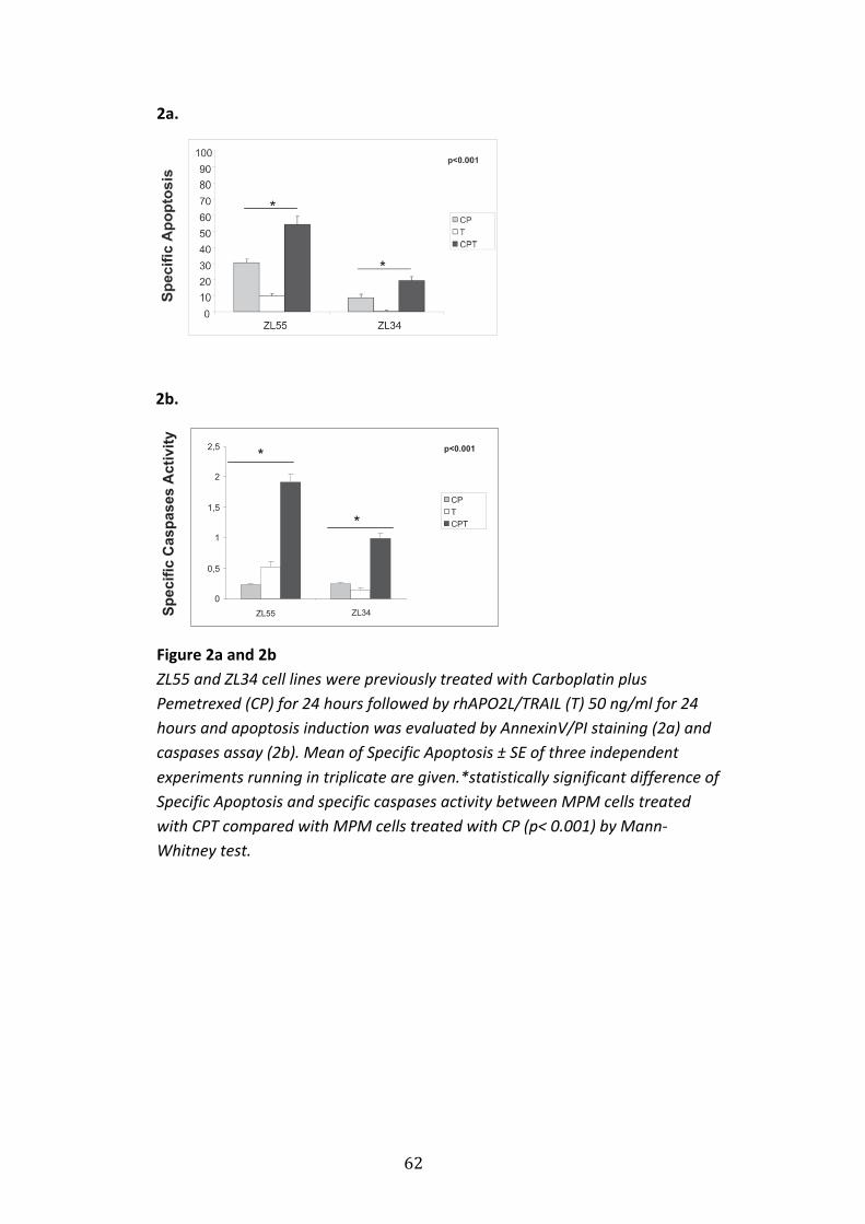
62
2a.
2b.
Figure 2a and 2b ZL55 and ZL34 cell lines were previously treated with Carboplatin plus Pemetrexed (CP) for 24 hours followed by rhAPO2L/TRAIL (T) 50 ng/ml for 24 hours and apoptosis induction was evaluated by AnnexinV/PI staining (2a) and caspases assay (2b). Mean of Specific Apoptosis ± SE of three independent experiments running in triplicate are given.*statistically significant difference of Specific Apoptosis and specific caspases activity between MPM cells treated with CPT compared with MPM cells treated with CP (p< 0.001) by Mann-‐Whitney test.
T 50 ng/ml
0
1
2
3
4
5
6
7
ZL55
H22
6
M14
K
MSTO
-211
SPC11
1ZL5
ZL34
MPM
1801
Sp
ec
ific
Ca
sp
as
es
Ac
tiv
ity T 50 ng/ml
0
1
2
3
4
5
6
7
ZL55
H22
6
M14
K
MSTO
-211
SPC11
1ZL5
ZL34
MPM
1801
T 50 ng/ml
0
1
2
3
4
5
6
7
ZL55
H22
6
M14
K
MSTO
-211
SPC11
1ZL5
ZL34
MPM
1801
Sp
ec
ific
Ca
sp
as
es
Ac
tiv
ity
epithelioid biphasic sarcomatoid
*
p<0.05
A
B
0
0,5
1
1,5
2
2,5
ZL55 ZL34
*
*
p<0.001
Sp
ec
ific
Ca
sp
as
es
Ac
tiv
ity
CP
T
CPT
ZL55 ZL34
Me
an
CI
Synergistic interaction CI<1
0
0,1
0,2
0,3
0,4
0,5
0,6
0,7
0,8
ZL55 ZL34
0
0,1
0,2
0,3
0,4
0,5
0,6
0,7
0,8
ZL55 ZL34
C
Supplementary Figure 1
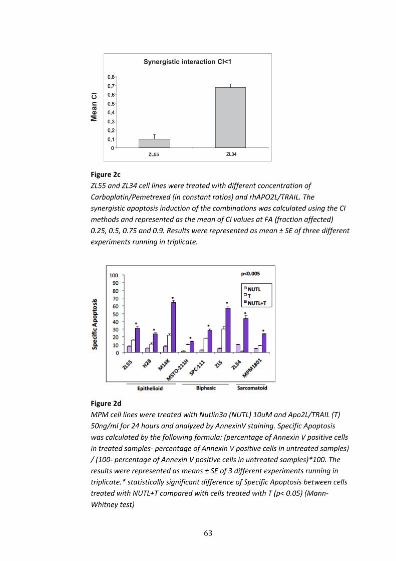
63
Figure 2c ZL55 and ZL34 cell lines were treated with different concentration of Carboplatin/Pemetrexed (in constant ratios) and rhAPO2L/TRAIL. The synergistic apoptosis induction of the combinations was calculated using the CI methods and represented as the mean of CI values at FA (fraction affected) 0.25, 0.5, 0.75 and 0.9. Results were represented as mean ± SE of three different experiments running in triplicate.
Figure 2d MPM cell lines were treated with Nutlin3a (NUTL) 10uM and Apo2L/TRAIL (T) 50ng/ml for 24 hours and analyzed by AnnexinV staining. Specific Apoptosis was calculated by the following formula: (percentage of Annexin V positive cells in treated samples-‐ percentage of Annexin V positive cells in untreated samples) / (100-‐ percentage of Annexin V positive cells in untreated samples)*100. The results were represented as means ± SE of 3 different experiments running in triplicate.* statistically significant difference of Specific Apoptosis between cells treated with NUTL+T compared with cells treated with T (p< 0.05) (Mann-‐Whitney test)
T 50 ng/ml
0
1
2
3
4
5
6
7
ZL55
H22
6
M14
K
MSTO
-211
SPC11
1ZL5
ZL34
MPM
1801
Sp
ec
ific
Ca
sp
as
es
Ac
tiv
ity T 50 ng/ml
0
1
2
3
4
5
6
7
ZL55
H22
6
M14
K
MSTO
-211
SPC11
1ZL5
ZL34
MPM
1801
T 50 ng/ml
0
1
2
3
4
5
6
7
ZL55
H22
6
M14
K
MSTO
-211
SPC11
1ZL5
ZL34
MPM
1801
Sp
ec
ific
Ca
sp
as
es
Ac
tiv
ity
epithelioid biphasic sarcomatoid
*
p<0.05
A
B
0
0,5
1
1,5
2
2,5
ZL55 ZL34
*
*
p<0.001
Sp
ec
ific
Ca
sp
as
es
Ac
tiv
ity
CP
T
CPT
ZL55 ZL34
Mean
CI
Synergistic interaction CI<1
0
0,1
0,2
0,3
0,4
0,5
0,6
0,7
0,8
ZL55 ZL34
0
0,1
0,2
0,3
0,4
0,5
0,6
0,7
0,8
ZL55 ZL34
C
Supplementary Figure 1
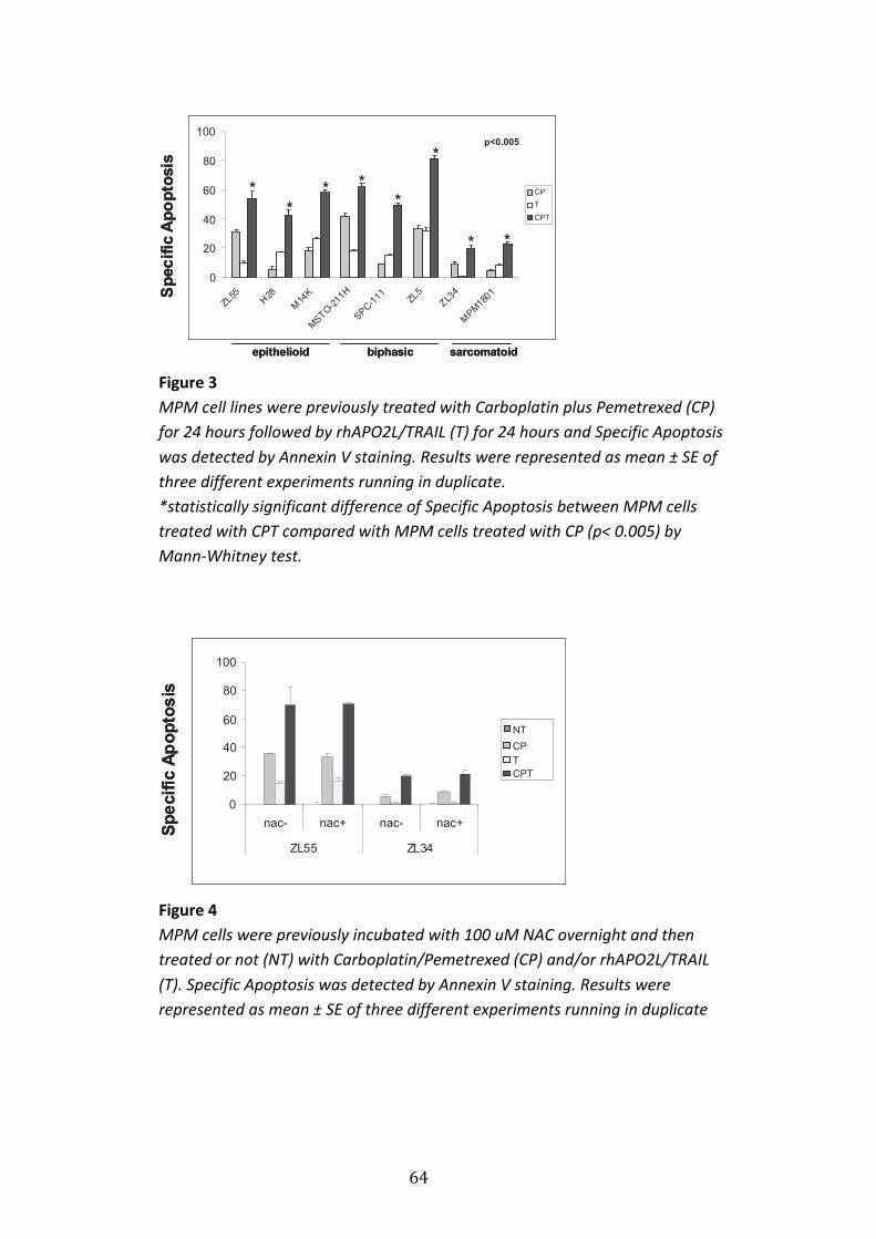
64
Figure 3 MPM cell lines were previously treated with Carboplatin plus Pemetrexed (CP) for 24 hours followed by rhAPO2L/TRAIL (T) for 24 hours and Specific Apoptosis was detected by Annexin V staining. Results were represented as mean ± SE of three different experiments running in duplicate. *statistically significant difference of Specific Apoptosis between MPM cells treated with CPT compared with MPM cells treated with CP (p< 0.005) by Mann-‐Whitney test.
Figure 4 MPM cells were previously incubated with 100 uM NAC overnight and then treated or not (NT) with Carboplatin/Pemetrexed (CP) and/or rhAPO2L/TRAIL (T). Specific Apoptosis was detected by Annexin V staining. Results were represented as mean ± SE of three different experiments running in duplicate
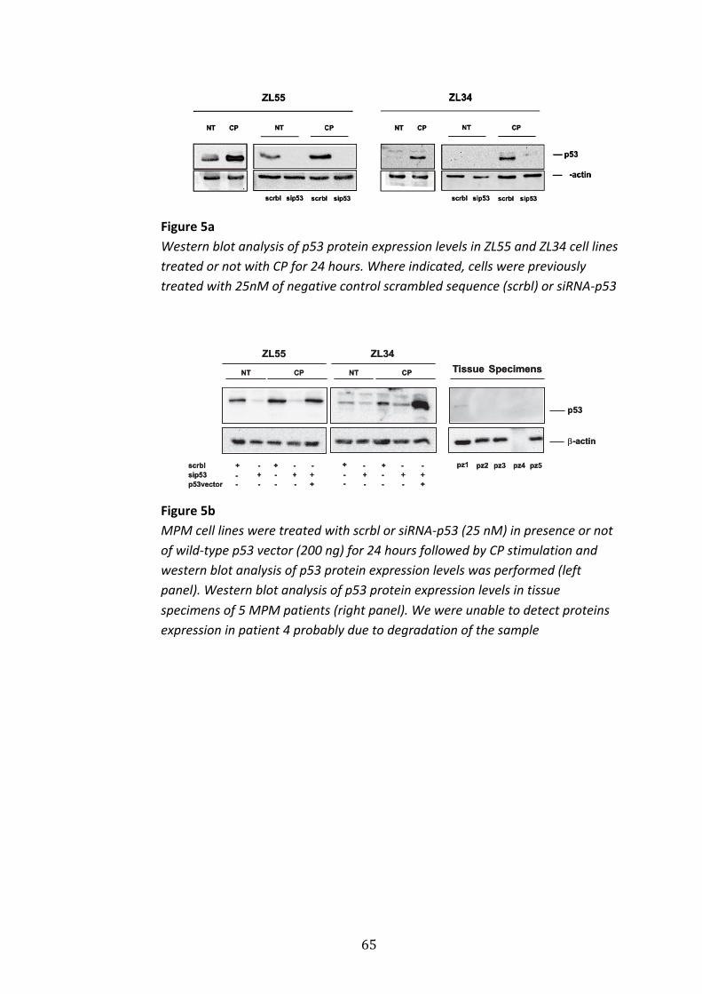
65
Figure 5a Western blot analysis of p53 protein expression levels in ZL55 and ZL34 cell lines treated or not with CP for 24 hours. Where indicated, cells were previously treated with 25nM of negative control scrambled sequence (scrbl) or siRNA-‐p53
Figure 5b MPM cell lines were treated with scrbl or siRNA-‐p53 (25 nM) in presence or not of wild-‐type p53 vector (200 ng) for 24 hours followed by CP stimulation and western blot analysis of p53 protein expression levels was performed (left panel). Western blot analysis of p53 protein expression levels in tissue specimens of 5 MPM patients (right panel). We were unable to detect proteins expression in patient 4 probably due to degradation of the sample
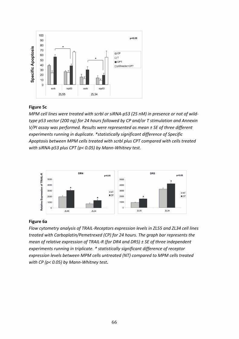
66
Figure 5c MPM cell lines were treated with scrbl or siRNA-‐p53 (25 nM) in presence or not of wild-‐type p53 vector (200 ng) for 24 hours followed by CP and/or T stimulation and Annexin V/PI assay was performed. Results were represented as mean ± SE of three different experiments running in duplicate. *statistically significant difference of Specific Apoptosis between MPM cells treated with scrbl plus CPT compared with cells treated with siRNA-‐p53 plus CPT (p< 0.05) by Mann-‐Whitney test.
Figure 6a Flow cytometry analysis of TRAIL-‐Receptors expression levels in ZL55 and ZL34 cell lines treated with Carboplatin/Pemetrexed (CP) for 24 hours. The graph bar represents the mean of relative expression of TRAIL-‐R (for DR4 and DR5) ± SE of three independent experiments running in triplicate. * statistically significant difference of receptor expression levels between MPM cells untreated (NT) compared to MPM cells treated with CP (p< 0.05) by Mann-‐Whitney test.
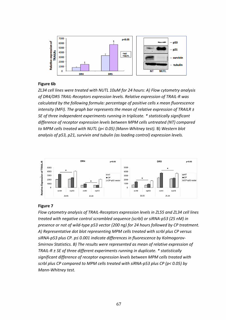
67
Figure 6b ZL34 cell lines were treated with NUTL 10uM for 24 hours: A) Flow cytometry analysis of DR4/DR5 TRAIL-‐Receptors expression levels. Relative expression of TRAIL-‐R was calculated by the following formula: percentage of positive cells x mean fluorescence intensity (MFI). The graph bar represents the mean of relative expression of TRAILR ± SE of three independent experiments running in triplicate. * statistically significant difference of receptor expression levels between MPM cells untreated (NT) compared to MPM cells treated with NUTL (p< 0.05) (Mann-‐Whitney test). B) Western blot analysis of p53, p21, survivin and tubulin (as loading control) expression levels.
Figure 7 Flow cytometry analysis of TRAIL-‐Receptors expression levels in ZL55 and ZL34 cell lines treated with negative control scrambled sequence (scrbl) or siRNA-‐p53 (25 nM) in presence or not of wild-‐type p53 vector (200 ng) for 24 hours followed by CP treatment. A) Representative dot blot representing MPM cells treated with scrbl plus CP versus siRNA-‐p53 plus CP. p≤ 0.001 indicate differences in fluorescence by Kolmogorov-‐Smirnov Statistics. B) The results were represented as mean of relative expression of TRAIL-‐R ± SE of three different experiments running in duplicate. * statistically significant difference of receptor expression levels between MPM cells treated with scrbl plus CP compared to MPM cells treated with siRNA-‐p53 plus CP (p< 0.05) by Mann-‐Whitney test.
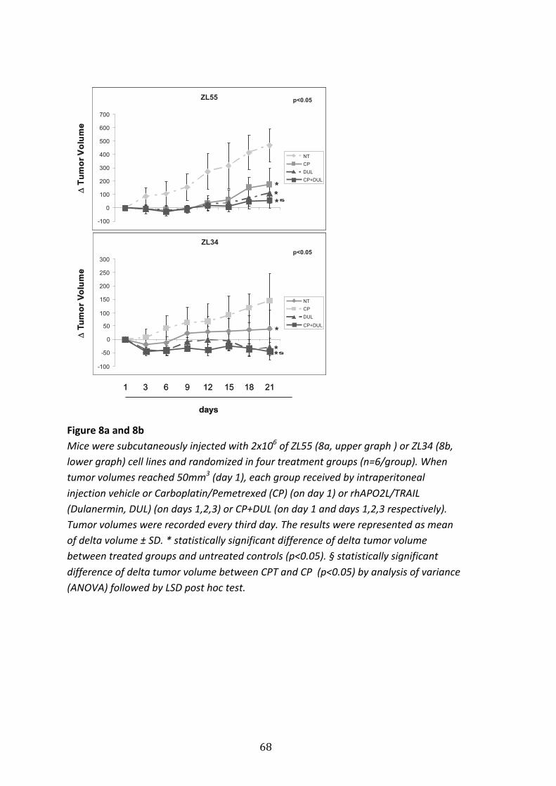
68
Figure 8a and 8b Mice were subcutaneously injected with 2x10⁶ of ZL55 (8a, upper graph ) or ZL34 (8b, lower graph) cell lines and randomized in four treatment groups (n=6/group). When tumor volumes reached 50mm3 (day 1), each group received by intraperitoneal injection vehicle or Carboplatin/Pemetrexed (CP) (on day 1) or rhAPO2L/TRAIL (Dulanermin, DUL) (on days 1,2,3) or CP+DUL (on day 1 and days 1,2,3 respectively). Tumor volumes were recorded every third day. The results were represented as mean of delta volume ± SD. * statistically significant difference of delta tumor volume between treated groups and untreated controls (p<0.05). § statistically significant difference of delta tumor volume between CPT and CP (p<0.05) by analysis of variance (ANOVA) followed by LSD post hoc test.
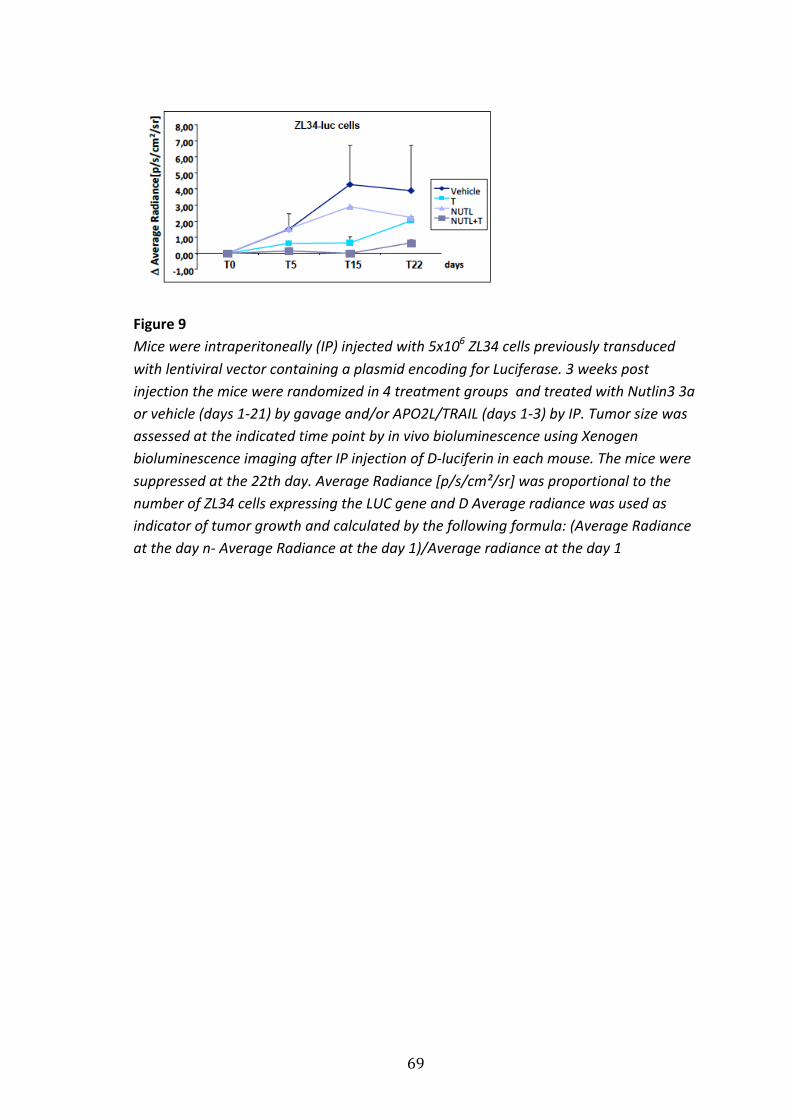
69
Figure 9 Mice were intraperitoneally (IP) injected with 5x10⁶ ZL34 cells previously transduced with lentiviral vector containing a plasmid encoding for Luciferase. 3 weeks post injection the mice were randomized in 4 treatment groups and treated with Nutlin3 3a or vehicle (days 1-‐21) by gavage and/or APO2L/TRAIL (days 1-‐3) by IP. Tumor size was assessed at the indicated time point by in vivo bioluminescence using Xenogen bioluminescence imaging after IP injection of D-‐luciferin in each mouse. The mice were suppressed at the 22th day. Average Radiance [p/s/cm²/sr] was proportional to the number of ZL34 cells expressing the LUC gene and D Average radiance was used as indicator of tumor growth and calculated by the following formula: (Average Radiance at the day n-‐ Average Radiance at the day 1)/Average radiance at the day 1

70
Figure 10. Expression levels of MDM2 in epithelioid versus sarcomatoid/biphasic samples.
Figure 11. HIF1alpha expression levels in epithelioid versus sarcomatoid/biphasic samples.
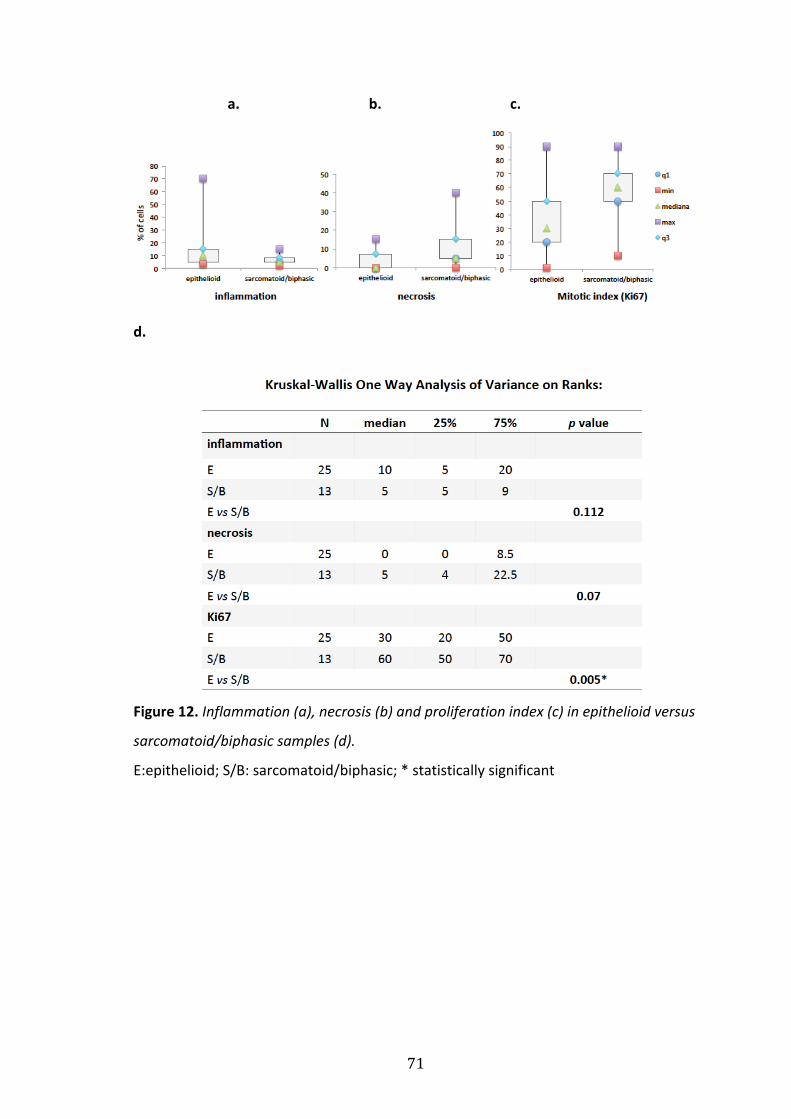
71
a. b. c.
d.
Figure 12. Inflammation (a), necrosis (b) and proliferation index (c) in epithelioid versus
sarcomatoid/biphasic samples (d).
E:epithelioid; S/B: sarcomatoid/biphasic; * statistically significant
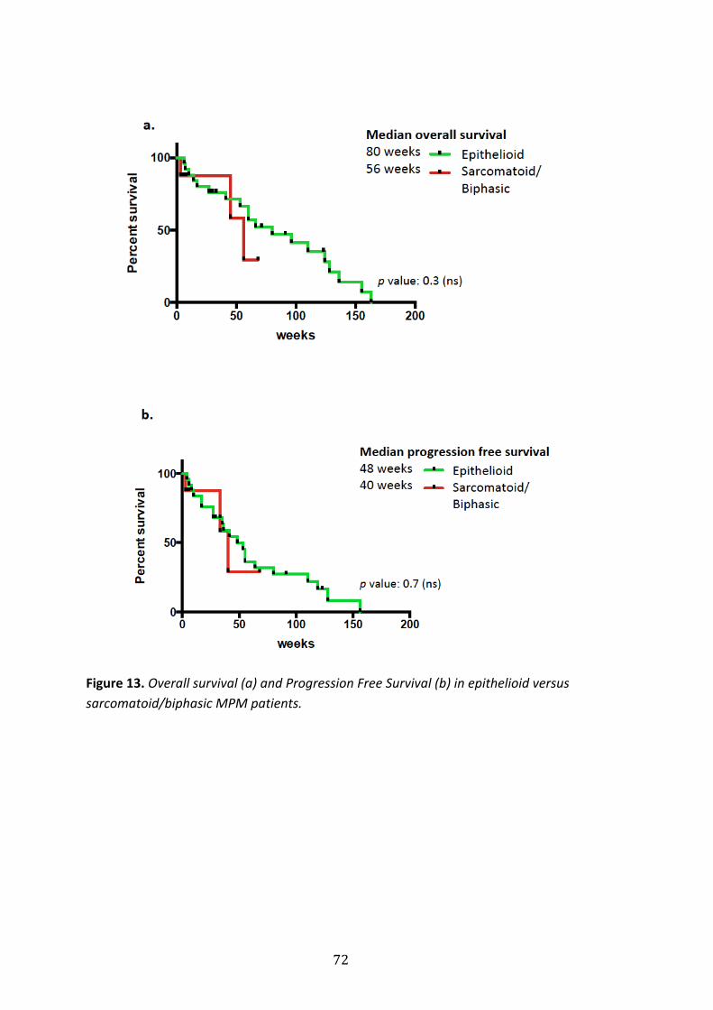
72
Figure 13. Overall survival (a) and Progression Free Survival (b) in epithelioid versus sarcomatoid/biphasic MPM patients.
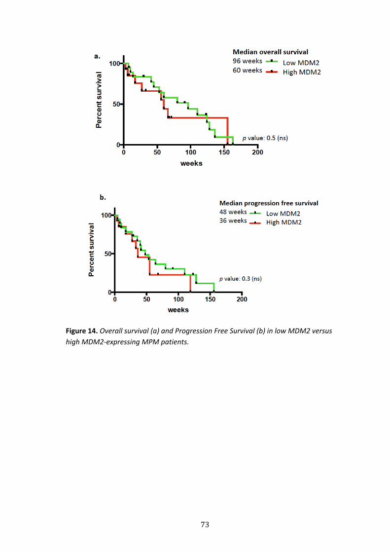
73
Figure 14. Overall survival (a) and Progression Free Survival (b) in low MDM2 versus high MDM2-‐expressing MPM patients.
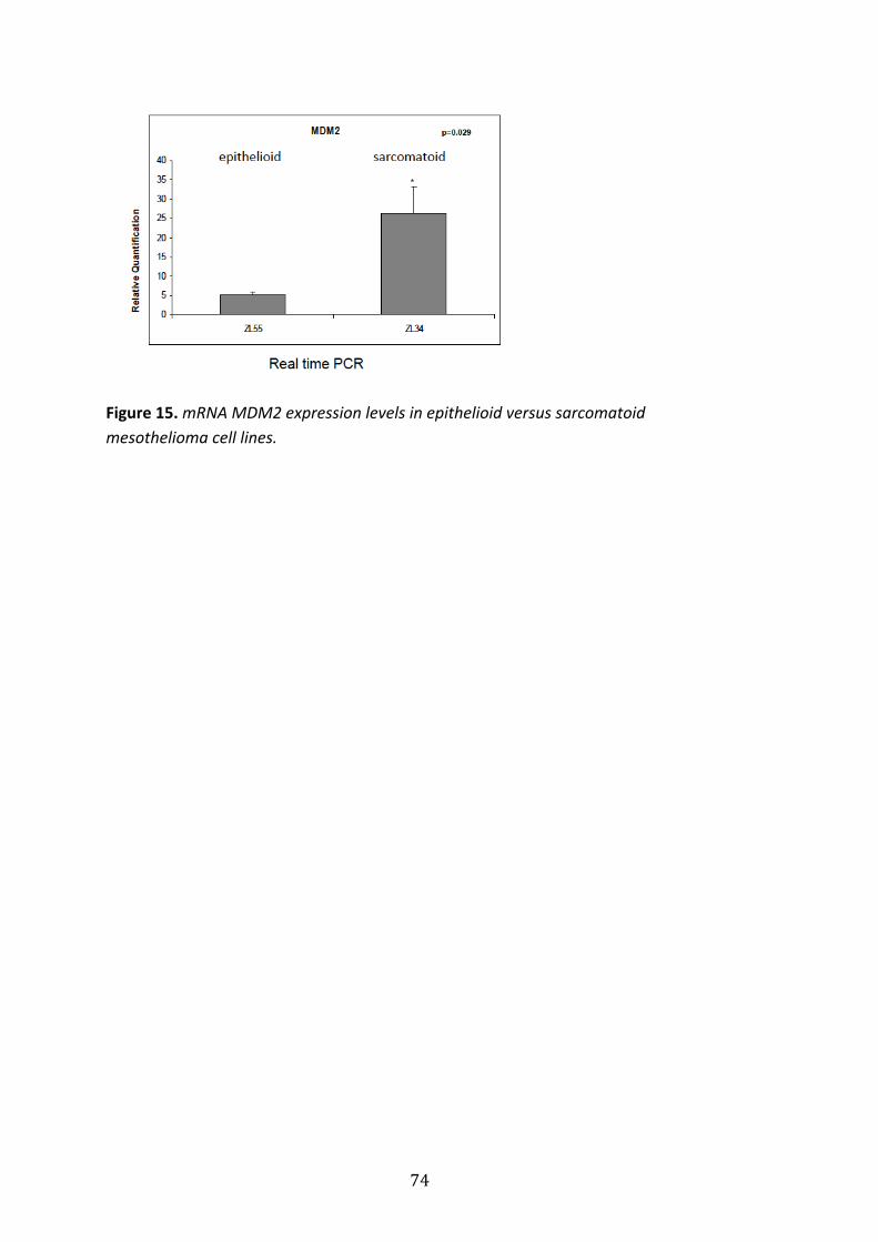
74
Figure 15. mRNA MDM2 expression levels in epithelioid versus sarcomatoid mesothelioma cell lines.
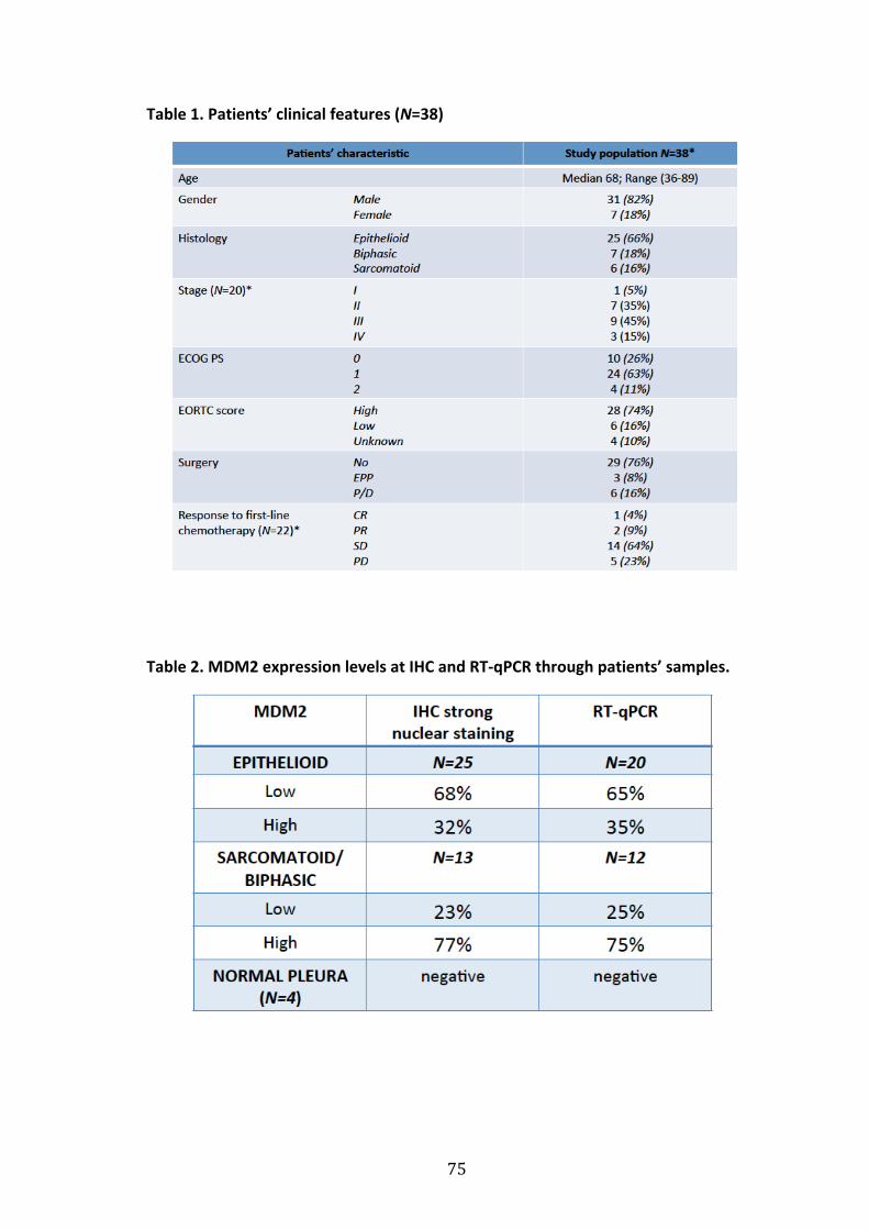
75
Table 1. Patients’ clinical features (N=38)
Table 2. MDM2 expression levels at IHC and RT-‐qPCR through patients’ samples.
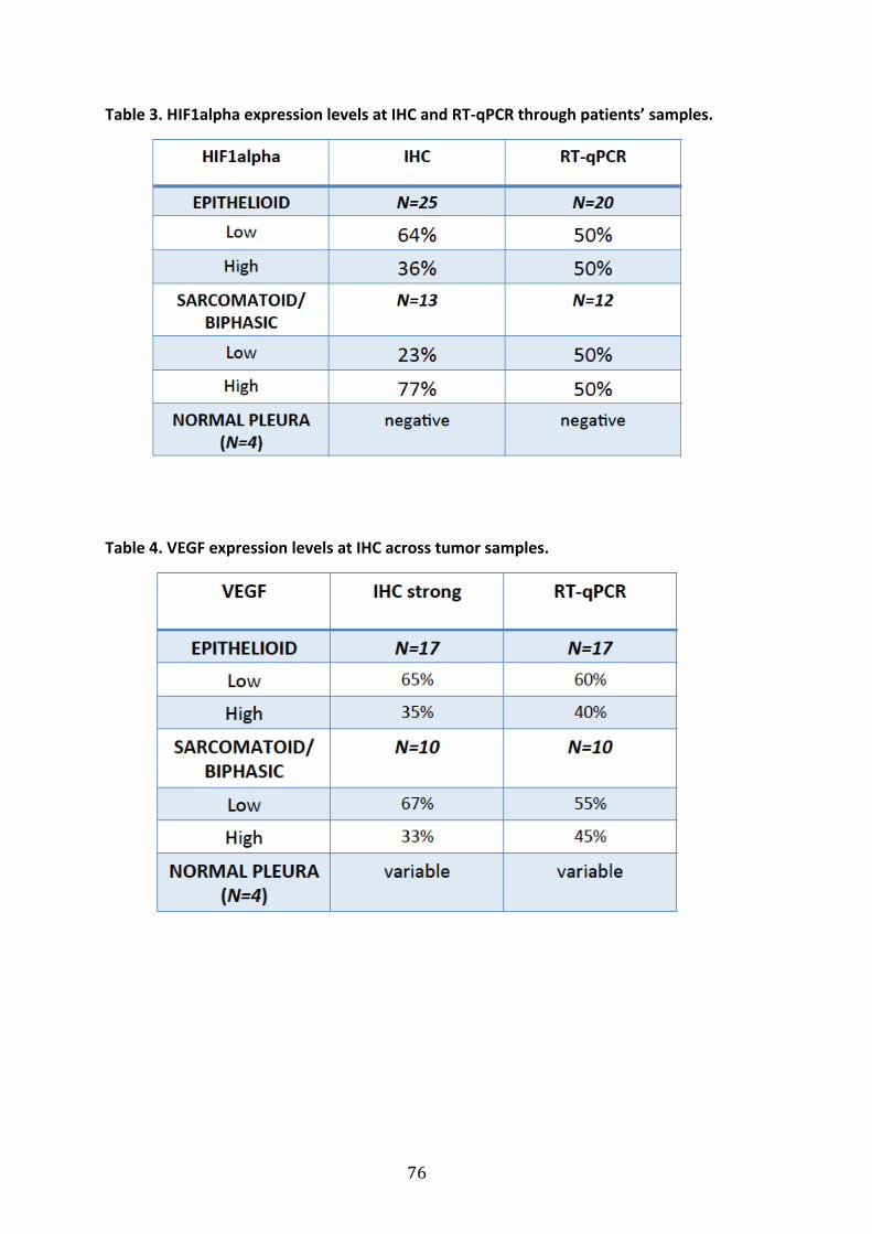
76
Table 3. HIF1alpha expression levels at IHC and RT-‐qPCR through patients’ samples.
Table 4. VEGF expression levels at IHC across tumor samples.
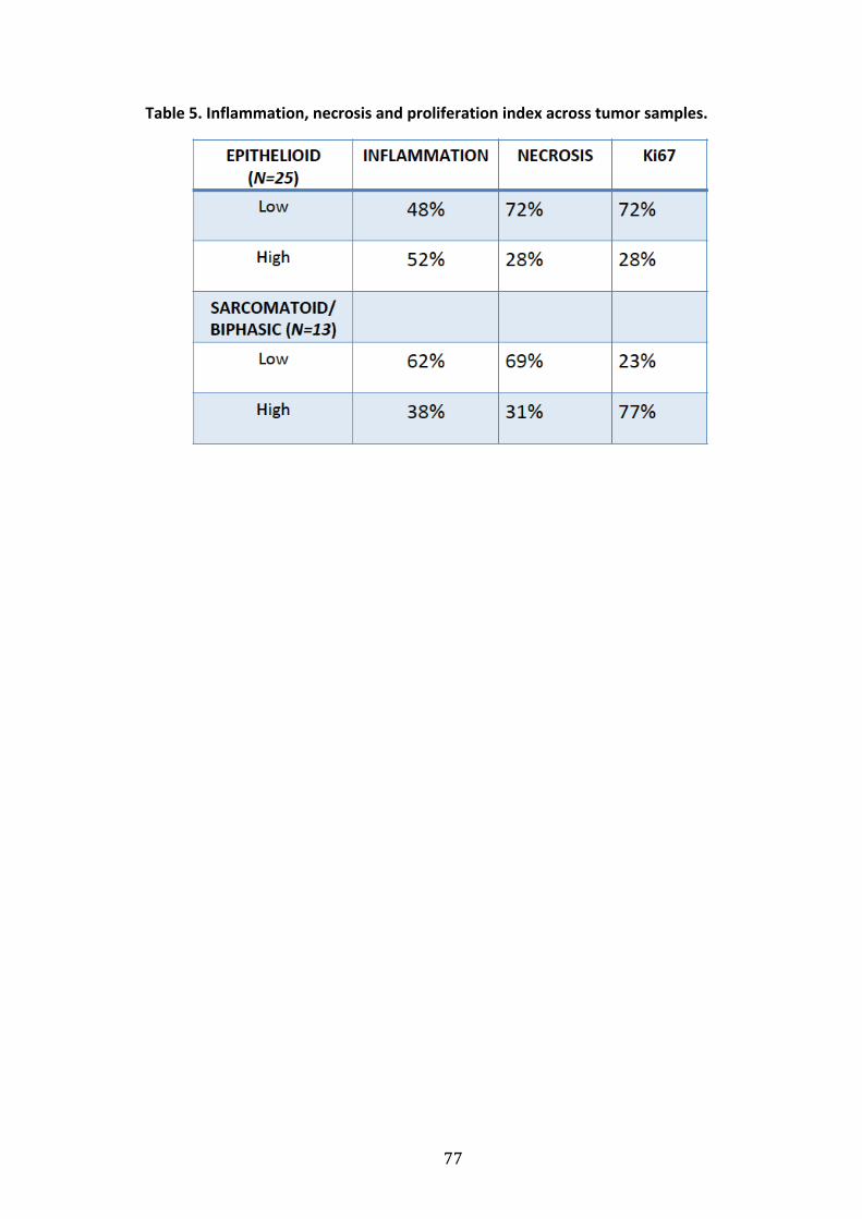
77
Table 5. Inflammation, necrosis and proliferation index across tumor samples.
78
9. References
1. Vogelzang NJ, Rusthoven JJ, Symanowski J, Denham C, Kaukel E, Ruffie P,
Gatzemeier U, Boyer M, Emri S, Manegold C, Niyikiza C, Paoletti P. Phase III
study of pemetrexed in combination with cisplatin versus cisplatin alone in
patients with malignant pleural mesothelioma. J Clin Oncol 2003;21: 2636-‐
2644.
2. van Meerbeeck JP, Gaafar R, Manegold C, Van Klaveren RJ, Van Marck EA,
Vincent M, Legrand C, Bottomley A, Debruyne C, Giaccone G, European
Organisation for R, Treatment of Cancer Lung Cancer G, National Cancer
Institute of C. Randomized phase III study of cisplatin with or without
raltitrexed in patients with malignant pleural mesothelioma: an intergroup
study of the European Organisation for Research and Treatment of Cancer Lung
Cancer Group and the National Cancer Institute of Canada. J Clin Oncol
2005;23: 6881-‐6889.
3. Bianchi C, Bianchi T. Malignant mesothelioma: global incidence and
relationship with asbestos. Ind Health 2007;45: 379-‐387.
4. Tossavainen A. National mesothelioma incidence and the past use of asbestos.
Monaldi Arch Chest Dis 2003;59: 146-‐149.
5. Tossavainen A. Global use of asbestos and the incidence of mesothelioma. Int J
Occup Environ Health 2004;10: 22-‐25.
6. Marinaccio A, Binazzi A, Cauzillo G, Chellini E, De Zotti R, Gennaro V,
Menegozzo M, Mensi C, Merler E, Mirabelli D, Musti M, Pannelli F, Romanelli A,
Scarselli A, Tosi S, Tumino R, Nesti M, Gruppo di lavoro ReNa M.
[Epidemiological surveillance of malignant mesothelioma cases in Italy:
79
incidence and asbestos exposure figures by the Italian mesothelioma registry
(ReNaM)]. Epidemiol Prev 2007;31: 23-‐26.
7. Peto J, Decarli A, La Vecchia C, Levi F, Negri E. The European mesothelioma
epidemic. Br J Cancer 1999;79: 666-‐672.
8. Pelucchi C, Malvezzi M, La Vecchia C, Levi F, Decarli A, Negri E. The
Mesothelioma epidemic in Western Europe: an update. Br J Cancer 2004;90:
1022-‐1024.
9. Hodgson JT, McElvenny DM, Darnton AJ, Price MJ, Peto J. The expected burden
of mesothelioma mortality in Great Britain from 2002 to 2050. Br J Cancer
2005;92: 587-‐593.
10. Maltoni C, Minardi F. Recent results of carcinogenicity bioassays of fibres and
other particulate materials. IARC Sci Publ 1989: 46-‐53.
11. Yarborough CM. The risk of mesothelioma from exposure to chrysotile
asbestos. Curr Opin Pulm Med 2007;13: 334-‐338.
12. Choe N, Tanaka S, Xia W, Hemenway DR, Roggli VL, Kagan E. Pleural
macrophage recruitment and activation in asbestos-‐induced pleural injury.
Environ Health Perspect 1997;105 Suppl 5: 1257-‐1260.
13. Zanella CL, Posada J, Tritton TR, Mossman BT. Asbestos causes stimulation of
the extracellular signal-‐regulated kinase 1 mitogen-‐activated protein kinase
cascade after phosphorylation of the epidermal growth factor receptor. Cancer
Res 1996;56: 5334-‐5338.
14. Weiner SJ, Neragi-‐Miandoab S. Pathogenesis of malignant pleural
mesothelioma and the role of environmental and genetic factors. J Cancer Res
Clin Oncol 2009;135: 15-‐27.
80
15. Lee AY, Raz DJ, He B, Jablons DM. Update on the molecular biology of
malignant mesothelioma. Cancer 2007;109: 1454-‐1461.
16. Bianchi AB, Mitsunaga SI, Cheng JQ, Klein WM, Jhanwar SC, Seizinger B, Kley N,
Klein-‐Szanto AJ, Testa JR. High frequency of inactivating mutations in the
neurofibromatosis type 2 gene (NF2) in primary malignant mesotheliomas. Proc
Natl Acad Sci U S A 1995;92: 10854-‐10858.
17. Sekido Y, Pass HI, Bader S, Mew DJ, Christman MF, Gazdar AF, Minna JD.
Neurofibromatosis type 2 (NF2) gene is somatically mutated in mesothelioma
but not in lung cancer. Cancer Res 1995;55: 1227-‐1231.
18. Deguen B, Goutebroze L, Giovannini M, Boisson C, van der Neut R, Jaurand MC,
Thomas G. Heterogeneity of mesothelioma cell lines as defined by altered
genomic structure and expression of the NF2 gene. Int J Cancer 1998;77: 554-‐
560.
19. Fleury-‐Feith J, Lecomte C, Renier A, Matrat M, Kheuang L, Abramowski V, Levy
F, Janin A, Giovannini M, Jaurand MC. Hemizygosity of Nf2 is associated with
increased susceptibility to asbestos-‐induced peritoneal tumours. Oncogene
2003;22: 3799-‐3805.
20. Stahel RA F-‐BE, Opitz I, Weder W. Malignant Pleural Mesothelioma. Future
Oncol 2009;5: 391-‐402.
21. Soini Y, Kinnula V, Kaarteenaho-‐Wiik R, Kurttila E, Linnainmaa K, Paakko P.
Apoptosis and expression of apoptosis regulating proteins bcl-‐2, mcl-‐1, bcl-‐X,
and bax in malignant mesothelioma. Clin Cancer Res 1999;5: 3508-‐3515.
22. Attanoos RL, Griffin A, Gibbs AR. The use of immunohistochemistry in
distinguishing reactive from neoplastic mesothelium. A novel use for desmin
81
and comparative evaluation with epithelial membrane antigen, p53, platelet-‐
derived growth factor-‐receptor, P-‐glycoprotein and Bcl-‐2. Histopathology
2003;43: 231-‐238.
23. Hopkins-‐Donaldson S, Cathomas R, Simoes-‐Wust AP, Kurtz S, Belyanskaya L,
Stahel RA, Zangemeister-‐Wittke U. Induction of apoptosis and
chemosensitization of mesothelioma cells by Bcl-‐2 and Bcl-‐xL antisense
treatment. Int J Cancer 2003;106: 160-‐166.
24. Travis W. Pathology and genetics of tumours of the lung, pleura, thymus and
heart. Lyon, France: IARC Press. 2004: World Health Organization Classification
of Tumours.
25. Steele JP. Prognostic factors for mesothelioma. Hematol Oncol Clin North Am
2005;19: 1041-‐1052, vi.
26. Spaggiari L, Marulli G, Bovolato P, Alloisio M, Pagan V, Oliaro A, Ratto GB,
Facciolo F, Sacco R, Brambilla D, Maisonneuve P, Mucilli F, Alessandrini G,
Leoncini G, Ruffini E, Fontana P, Infante M, Pariscenti GL, Casiraghi M, Rea F.
Extrapleural pneumonectomy for malignant mesothelioma: an Italian
multicenter retrospective study. Ann Thorac Surg 2014;97: 1859-‐1865.
27. Scherpereel A, Astoul P, Baas P, Berghmans T, Clayson H, de Vuyst P,
Dienemann H, Galateau-‐Salle F, Hennequin C, Hillerdal G, Le Pechoux C, Mutti
L, Pairon JC, Stahel R, van Houtte P, van Meerbeeck J, Waller D, Weder W.
Guidelines of the European Respiratory Society and the European Society of
Thoracic Surgeons for management of Malignant Pleural Mesothelioma. Eur
Respir J 2009.
82
28. Pasello G, Ceresoli GL, Favaretto A. An overview of neoadjuvant chemotherapy
in the multimodality treatment of malignant pleural mesothelioma. Cancer
Treat Rev 2013;39: 10-‐17.
29. Treasure T, Lang-‐Lazdunski L, Waller D, Bliss JM, Tan C, Entwisle J, Snee M,
O'Brien M, Thomas G, Senan S, O'Byrne K, Kilburn LS, Spicer J, Landau D,
Edwards J, Coombes G, Darlison L, Peto J, trialists M. Extra-‐pleural
pneumonectomy versus no extra-‐pleural pneumonectomy for patients with
malignant pleural mesothelioma: clinical outcomes of the Mesothelioma and
Radical Surgery (MARS) randomised feasibility study. Lancet Oncol 2011;12:
763-‐772.
30. Castagneto B, Botta M, Aitini E, Spigno F, Degiovanni D, Alabiso O, Serra M,
Muzio A, Carbone R, Buosi R, Galbusera V, Piccolini E, Giaretto L, Rebella L,
Mencoboni M. Phase II study of pemetrexed in combination with carboplatin in
patients with malignant pleural mesothelioma (MPM). Ann Oncol 2008;19: 370-‐
373.
31. Ceresoli GL, Castagneto B, Zucali PA, Favaretto A, Mencoboni M, Grossi F,
Cortinovis D, Del Conte G, Ceribelli A, Bearz A, Salamina S, De Vincenzo F,
Cappuzzo F, Marangolo M, Torri V, Santoro A. Pemetrexed plus carboplatin in
elderly patients with malignant pleural mesothelioma: combined analysis of
two phase II trials. Br J Cancer 2008;99: 51-‐56.
32. Ceresoli GL, Zucali PA, Favaretto AG, Grossi F, Bidoli P, Del Conte G, Ceribelli A,
Bearz A, Morenghi E, Cavina R, Marangolo M, Parra HJ, Santoro A. Phase II
study of pemetrexed plus carboplatin in malignant pleural mesothelioma. J Clin
Oncol 2006;24: 1443-‐1448.
83
33. Pasello G, Favaretto A. Molecular targets in malignant pleural mesothelioma
treatment. Curr Drug Targets 2009;10: 1235-‐1244.
34. LeBlanc HN, Ashkenazi A. Apo2L/TRAIL and its death and decoy receptors. Cell
Death Differ 2003;10: 66-‐75.
35. Pan G, Ni J, Wei YF, Yu G, Gentz R, Dixit VM. An antagonist decoy receptor and
a death domain-‐containing receptor for TRAIL. Science 1997;277: 815-‐818.
36. Pan G, O'Rourke K, Chinnaiyan AM, Gentz R, Ebner R, Ni J, Dixit VM. The
receptor for the cytotoxic ligand TRAIL. Science 1997;276: 111-‐113.
37. Marsters SA, Sheridan JP, Pitti RM, Huang A, Skubatch M, Baldwin D, Yuan J,
Gurney A, Goddard AD, Godowski P, Ashkenazi A. A novel receptor for
Apo2L/TRAIL contains a truncated death domain. Curr Biol 1997;7: 1003-‐1006.
38. Sheridan JP, Marsters SA, Pitti RM, Gurney A, Skubatch M, Baldwin D,
Ramakrishnan L, Gray CL, Baker K, Wood WI, Goddard AD, Godowski P,
Ashkenazi A. Control of TRAIL-‐induced apoptosis by a family of signaling and
decoy receptors. Science 1997;277: 818-‐821.
39. Emery JG, McDonnell P, Burke MB, Deen KC, Lyn S, Silverman C, Dul E,
Appelbaum ER, Eichman C, DiPrinzio R, Dodds RA, James IE, Rosenberg M, Lee
JC, Young PR. Osteoprotegerin is a receptor for the cytotoxic ligand TRAIL. J Biol
Chem 1998;273: 14363-‐14367.
40. Gura T. How TRAIL kills cancer cells, but not normal cells. Science 1997;277:
768.
41. Ashkenazi A. Targeting death and decoy receptors of the tumour-‐necrosis
factor superfamily. Nat Rev Cancer 2002;2: 420-‐430.
84
42. Zhang L, Fang B. Mechanisms of resistance to TRAIL-‐induced apoptosis in
cancer. Cancer Gene Ther 2005;12: 228-‐237.
43. Gonzalvez F, Ashkenazi A. New insights into apoptosis signaling by
Apo2L/TRAIL. Oncogene 2010;29: 4752-‐4765.
44. Nagane M, Pan G, Weddle JJ, Dixit VM, Cavenee WK, Huang HJ. Increased death
receptor 5 expression by chemotherapeutic agents in human gliomas causes
synergistic cytotoxicity with tumor necrosis factor-‐related apoptosis-‐inducing
ligand in vitro and in vivo. Cancer Res 2000;60: 847-‐853.
45. Gibson SB, Oyer R, Spalding AC, Anderson SM, Johnson GL. Increased
expression of death receptors 4 and 5 synergizes the apoptosis response to
combined treatment with etoposide and TRAIL. Mol Cell Biol 2000;20: 205-‐212.
46. Chinnaiyan AM, Prasad U, Shankar S, Hamstra DA, Shanaiah M, Chenevert TL,
Ross BD, Rehemtulla A. Combined effect of tumor necrosis factor-‐related
apoptosis-‐inducing ligand and ionizing radiation in breast cancer therapy. Proc
Natl Acad Sci U S A 2000;97: 1754-‐1759.
47. Pukac L, Kanakaraj P, Humphreys R, Alderson R, Bloom M, Sung C, Riccobene T,
Johnson R, Fiscella M, Mahoney A, Carrell J, Boyd E, Yao XT, Zhang L, Zhong L,
von Kerczek A, Shepard L, Vaughan T, Edwards B, Dobson C, Salcedo T, Albert V.
HGS-‐ETR1, a fully human TRAIL-‐receptor 1 monoclonal antibody, induces cell
death in multiple tumour types in vitro and in vivo. Br J Cancer 2005;92: 1430-‐
1441.
48. Georgakis GV, Li Y, Humphreys R, Andreeff M, O'Brien S, Younes M, Carbone A,
Albert V, Younes A. Activity of selective fully human agonistic antibodies to the
TRAIL death receptors TRAIL-‐R1 and TRAIL-‐R2 in primary and cultured
85
lymphoma cells: induction of apoptosis and enhancement of doxorubicin-‐ and
bortezomib-‐induced cell death. Br J Haematol 2005;130: 501-‐510.
49. Belyanskaya LL, Marti TM, Hopkins-‐Donaldson S, Kurtz S, Felley-‐Bosco E, Stahel
RA. Human agonistic TRAIL receptor antibodies Mapatumumab and
Lexatumumab induce apoptosis in malignant mesothelioma and act
synergistically with cisplatin. Mol Cancer 2007;6: 66.
50. Ashkenazi A, Holland P, Eckhardt SG. Ligand-‐based targeting of apoptosis in
cancer: the potential of recombinant human apoptosis ligand 2/Tumor necrosis
factor-‐related apoptosis-‐inducing ligand (rhApo2L/TRAIL). J Clin Oncol 2008;26:
3621-‐3630.
51. Herbst RS, Eckhardt SG, Kurzrock R, Ebbinghaus S, O'Dwyer PJ, Gordon MS,
Novotny W, Goldwasser MA, Tohnya TM, Lum BL, Ashkenazi A, Jubb AM,
Mendelson DS. Phase I dose-‐escalation study of recombinant human
Apo2L/TRAIL, a dual proapoptotic receptor agonist, in patients with advanced
cancer. J Clin Oncol 2010;28: 2839-‐2846.
52. Soria JC, Smit E, Khayat D, Besse B, Yang X, Hsu CP, Reese D, Wiezorek J,
Blackhall F. Phase 1b study of dulanermin (recombinant human Apo2L/TRAIL)
in combination with paclitaxel, carboplatin, and bevacizumab in patients with
advanced non-‐squamous non-‐small-‐cell lung cancer. J Clin Oncol 2010;28:
1527-‐1533.
53. Duiker EW, Mom CH, de Jong S, Willemse PH, Gietema JA, van der Zee AG, de
Vries EG. The clinical trail of TRAIL. Eur J Cancer 2006;42: 2233-‐2240.
54. Kelley SK, Harris LA, Xie D, Deforge L, Totpal K, Bussiere J, Fox JA. Preclinical
studies to predict the disposition of Apo2L/tumor necrosis factor-‐related
86
apoptosis-‐inducing ligand in humans: characterization of in vivo efficacy,
pharmacokinetics, and safety. J Pharmacol Exp Ther 2001;299: 31-‐38.
55. Ashkenazi A, Pai RC, Fong S, Leung S, Lawrence DA, Marsters SA, Blackie C,
Chang L, McMurtrey AE, Hebert A, DeForge L, Koumenis IL, Lewis D, Harris L,
Bussiere J, Koeppen H, Shahrokh Z, Schwall RH. Safety and antitumor activity of
recombinant soluble Apo2 ligand. J Clin Invest 1999;104: 155-‐162.
56. Hylander BL, Pitoniak R, Penetrante RB, Gibbs JF, Oktay D, Cheng J, Repasky EA.
The anti-‐tumor effect of Apo2L/TRAIL on patient pancreatic adenocarcinomas
grown as xenografts in SCID mice. J Transl Med 2005;3: 22.
57. Jin H, Yang R, Fong S, Totpal K, Lawrence D, Zheng Z, Ross J, Koeppen H, Schwall
R, Ashkenazi A. Apo2 ligand/tumor necrosis factor-‐related apoptosis-‐inducing
ligand cooperates with chemotherapy to inhibit orthotopic lung tumor growth
and improve survival. Cancer Res 2004;64: 4900-‐4905.
58. Soria JC, Mark Z, Zatloukal P, Szima B, Albert I, Juhasz E, Pujol JL, Kozielski J,
Baker N, Smethurst D, Hei YJ, Ashkenazi A, Stern H, Amler L, Pan Y, Blackhall F.
Randomized phase II study of dulanermin in combination with paclitaxel,
carboplatin, and bevacizumab in advanced non-‐small-‐cell lung cancer. J Clin
Oncol 2011;29: 4442-‐4451.
59. Kim KU, Wilson SM, Abayasiriwardana KS, Collins R, Fjellbirkeland L, Xu Z,
Jablons DM, Nishimura SL, Broaddus VC. A novel in vitro model of human
mesothelioma for studying tumor biology and apoptotic resistance. Am J Respir
Cell Mol Biol 2005;33: 541-‐548.
87
60. Liu Q, El-‐Deiry WS, Gazitt Y. Additive effect of Apo2L/TRAIL and Adeno-‐p53 in
the induction of apoptosis in myeloma cell lines. Exp Hematol 2001;29: 962-‐
970.
61. Tomasetti M, Rippo MR, Alleva R, Moretti S, Andera L, Neuzil J, Procopio A.
Alpha-‐tocopheryl succinate and TRAIL selectively synergise in induction of
apoptosis in human malignant mesothelioma cells. Br J Cancer 2004;90: 1644-‐
1653.
62. Zhao J, Lu Y, Shen HM. Targeting p53 as a therapeutic strategy in sensitizing
TRAIL-‐induced apoptosis in cancer cells. Cancer Lett 2012;314: 8-‐23.
63. Mor O, Yaron P, Huszar M, Yellin A, Jakobovitz O, Brok-‐Simoni F, Rechavi G,
Reichert N. Absence of p53 mutations in malignant mesotheliomas. Am J Respir
Cell Mol Biol 1997;16: 9-‐13.
64. Momand J, Zambetti GP, Olson DC, George D, Levine AJ. The mdm-‐2 oncogene
product forms a complex with the p53 protein and inhibits p53-‐mediated
transactivation. Cell 1992;69: 1237-‐1245.
65. Biderman L, Manley JL, Prives C. Mdm2 and MdmX as Regulators of Gene
Expression. Genes Cancer 2012;3: 264-‐273.
66. Hoe KK, Verma CS, Lane DP. Drugging the p53 pathway: understanding the
route to clinical efficacy. Nat Rev Drug Discov 2014;13: 217-‐236.
67. Vogelstein B, Lane D, Levine AJ. Surfing the p53 network. Nature 2000;408:
307-‐310.
68. Wade M, Li YC, Wahl GM. MDM2, MDMX and p53 in oncogenesis and cancer
therapy. Nat Rev Cancer 2013;13: 83-‐96.
88
69. Toledo F, Wahl GM. Regulating the p53 pathway: in vitro hypotheses, in vivo
veritas. Nat Rev Cancer 2006;6: 909-‐923.
70. Bartel F, Schulz J, Bohnke A, Blumke K, Kappler M, Bache M, Schmidt H, Wurl P,
Taubert H, Hauptmann S. Significance of HDMX-‐S (or MDM4) mRNA splice
variant overexpression and HDMX gene amplification on primary soft tissue
sarcoma prognosis. Int J Cancer 2005;117: 469-‐475.
71. Toguchida J, Yamaguchi T, Dayton SH, Beauchamp RL, Herrera GE, Ishizaki K,
Yamamuro T, Meyers PA, Little JB, Sasaki MS, et al. Prevalence and spectrum of
germline mutations of the p53 gene among patients with sarcoma. N Engl J
Med 1992;326: 1301-‐1308.
72. Maelandsmo GM, Berner JM, Florenes VA, Forus A, Hovig E, Fodstad O,
Myklebost O. Homozygous deletion frequency and expression levels of the
CDKN2 gene in human sarcomas-‐-‐relationship to amplification and mRNA levels
of CDK4 and CCND1. Br J Cancer 1995;72: 393-‐398.
73. Ito M, Barys L, O'Reilly T, Young S, Gorbatcheva B, Monahan J, Zumstein-‐
Mecker S, Choong PF, Dickinson I, Crowe P, Hemmings C, Desai J, Thomas DM,
Lisztwan J. Comprehensive mapping of p53 pathway alterations reveals an
apparent role for both SNP309 and MDM2 amplification in sarcomagenesis.
Clin Cancer Res 2011;17: 416-‐426.
74. Gilkes DM, Pan Y, Coppola D, Yeatman T, Reuther GW, Chen J. Regulation of
MDMX expression by mitogenic signaling. Mol Cell Biol 2008;28: 1999-‐2010.
75. Gembarska A, Luciani F, Fedele C, Russell EA, Dewaele M, Villar S, Zwolinska A,
Haupt S, de Lange J, Yip D, Goydos J, Haigh JJ, Haupt Y, Larue L, Jochemsen A,
Shi H, Moriceau G, Lo RS, Ghanem G, Shackleton M, Bernal F, Marine JC. MDM4
89
is a key therapeutic target in cutaneous melanoma. Nat Med 2012;18: 1239-‐
1247.
76. McEvoy J, Ulyanov A, Brennan R, Wu G, Pounds S, Zhang J, Dyer MA. Analysis of
MDM2 and MDM4 single nucleotide polymorphisms, mRNA splicing and
protein expression in retinoblastoma. PLoS One 2012;7: e42739.
77. Fahraeus R, Olivares-‐Illana V. MDM2's social network. Oncogene 2013.
78. Onel K, Cordon-‐Cardo C. MDM2 and prognosis. Mol Cancer Res 2004;2: 1-‐8.
79. Tagawa M, Tada Y, Shimada H, Hiroshima K. Gene therapy for malignant
mesothelioma: current prospects and challenges. Cancer Gene Ther 2013;20:
150-‐156.
80. Iwakuma T, Lozano G. MDM2, an introduction. Mol Cancer Res 2003;1: 993-‐
1000.
81. Sekido Y. Molecular pathogenesis of malignant mesothelioma. Carcinogenesis
2013;34: 1413-‐1419.
82. Hopkins-‐Donaldson S, Belyanskaya LL, Simoes-‐Wust AP, Sigrist B, Kurtz S,
Zangemeister-‐Wittke U, Stahel R. p53-‐induced apoptosis occurs in the absence
of p14(ARF) in malignant pleural mesothelioma. Neoplasia 2006;8: 551-‐559.
83. Yang CT, You L, Yeh CC, Chang JW, Zhang F, McCormick F, Jablons DM.
Adenovirus-‐mediated p14(ARF) gene transfer in human mesothelioma cells. J
Natl Cancer Inst 2000;92: 636-‐641.
84. Vu B, Wovkulich P, Pizzolato G, Lovey A, Ding Q, Jiang N, Liu JJ, Zhao C, Glenn K,
Wen Y, Tovar C, Packman K, Vassilev L, Graves B. Discovery of RG7112: A Small-‐
Molecule MDM2 Inhibitor in Clinical Development. ACS Med Chem Lett 2013;4:
466-‐469.
90
85. Tovar C, Graves B, Packman K, Filipovic Z, Higgins B, Xia M, Tardell C, Garrido R,
Lee E, Kolinsky K, To KH, Linn M, Podlaski F, Wovkulich P, Vu B, Vassilev LT.
MDM2 small-‐molecule antagonist RG7112 activates p53 signaling and regresses
human tumors in preclinical cancer models. Cancer Res 2013;73: 2587-‐2597.
86. Ohta Y, Shridhar V, Bright RK, Kalemkerian GP, Du W, Carbone M, Watanabe Y,
Pass HI. VEGF and VEGF type C play an important role in angiogenesis and
lymphangiogenesis in human malignant mesothelioma tumours. Br J Cancer
1999;81: 54-‐61.
87. Demirag F, Unsal E, Yilmaz A, Caglar A. Prognostic significance of vascular
endothelial growth factor, tumor necrosis, and mitotic activity index in
malignant pleural mesothelioma. Chest 2005;128: 3382-‐3387.
88. Strizzi L, Catalano A, Vianale G, Orecchia S, Casalini A, Tassi G, Puntoni R, Mutti
L, Procopio A. Vascular endothelial growth factor is an autocrine growth factor
in human malignant mesothelioma. J Pathol 2001;193: 468-‐475.
89. Masood R, Kundra A, Zhu S, Xia G, Scalia P, Smith DL, Gill PS. Malignant
mesothelioma growth inhibition by agents that target the VEGF and VEGF-‐C
autocrine loops. Int J Cancer 2003;104: 603-‐610.
90. Ceresoli GL, Zucali, P., Mencoboni, M., De Vincenzo, F., Botta, M., Grossi, F.,
Bidoli, P., Bajetta, E., Ardizzoni, A., Favaretto, A., Simonelli, M., Lorenzi, E.,
Gianoncelli, L., Chiti, A., Santoro, A. Phase II study of the combination of
bevacizumab plus pemetrexed and carboplatin as first-‐line therapy in patients
with malignant pleural mesothelioma (MPM). 10th International Conference of
the International Mesothelioma Interest Group Abstract Book 2010: S06-‐03.
91
91. Dowell J, Taub, R., Lan, C., Xie, Y., Dunphy, F., Blake, V., Kindler, H. A
multicenter phase II study of pemetrexed (P), cisplatin (C), and bevacizumab (B)
in patients (pts) with advanced malignant mesothelioma (MM). J Clin Oncol
2009;27: 7578.
92. Kindler HL, Karrison TG, Gandara DR, Lu C, Krug LM, Stevenson JP, Janne PA,
Quinn DI, Koczywas MN, Brahmer JR, Albain KS, Taber DA, Armato SG, 3rd,
Vogelzang NJ, Chen HX, Stadler WM, Vokes EE. Multicenter, double-‐blind,
placebo-‐controlled, randomized phase II trial of gemcitabine/cisplatin plus
bevacizumab or placebo in patients with malignant mesothelioma. J Clin Oncol
2012;30: 2509-‐2515.
93. Ceresoli GL, Zucali PA, Mencoboni M, Botta M, Grossi F, Cortinovis D, Zilembo
N, Ripa C, Tiseo M, Favaretto AG, Soto-‐Parra H, De Vincenzo F, Bruzzone A,
Lorenzi E, Gianoncelli L, Ercoli B, Giordano L, Santoro A. Phase II study of
pemetrexed and carboplatin plus bevacizumab as first-‐line therapy in
malignant pleural mesothelioma. Br J Cancer 2013;109: 552-‐558.
94. Roudier E, Forn P, Perry ME, Birot O. Murine double minute-‐2 expression is
required for capillary maintenance and exercise-‐induced angiogenesis in
skeletal muscle. FASEB J 2012;26: 4530-‐4539.
95. Carroll VA, Ashcroft M. Regulation of angiogenic factors by HDM2 in renal cell
carcinoma. Cancer Res 2008;68: 545-‐552.
96. LaRusch GA, Jackson MW, Dunbar JD, Warren RS, Donner DB, Mayo LD. Nutlin3
blocks vascular endothelial growth factor induction by preventing the
interaction between hypoxia inducible factor 1alpha and Hdm2. Cancer Res
2007;67: 450-‐454.
92
97. Zhou S, Liu L, Li H, Eilers G, Kuang Y, Shi S, Yan Z, Li X, Corson JM, Meng F, Zhou
H, Sheng Q, Fletcher JA, Ou WB. Multipoint targeting of the PI3K/mTOR
pathway in mesothelioma. Br J Cancer 2014;110: 2479-‐2488.
98. Xiong J, Yang Q, Li J, Zhou S. Effects of MDM2 inhibitors on vascular endothelial
growth factor-‐mediated tumor angiogenesis in human breast cancer.
Angiogenesis 2014;17: 37-‐50.
99. Kay BP, Hsu CP, Lu JF, Sun YN, Bai S, Xin Y, D'Argenio DZ. Intracellular-‐signaling
tumor-‐regression modeling of the pro-‐apoptotic receptor agonists dulanermin
and conatumumab. J Pharmacokinet Pharmacodyn 2012;39: 577-‐590.
100. Pasello G, Urso L, Silic-‐Benussi M, Schiavon M, Cavallari I, Marulli G, Nannini N,
Rea F, Ciminale V, Favaretto A. Synergistic antitumor activity of recombinant
human Apo2L/tumor necrosis factor-‐related apoptosis-‐inducing ligand (TRAIL)
in combination with carboplatin and pemetrexed in malignant pleural
mesothelioma. J Thorac Oncol 2014;9: 1008-‐1017.
101. Zucali PA, Ceresoli GL, Garassino I, De Vincenzo F, Cavina R, Campagnoli E,
Cappuzzo F, Salamina S, Soto Parra HJ, Santoro A. Gemcitabine and vinorelbine
in pemetrexed-‐pretreated patients with malignant pleural mesothelioma.
Cancer 2008;112: 1555-‐1561.
102. Xanthopoulos A, Bauer TT, Blum TG, Kollmeier J, Schonfeld N, Serke M.
Gemcitabine combined with oxaliplatin in pretreated patients with malignant
pleural mesothelioma: an observational study. J Occup Med Toxicol 2008;3: 34.
103. Pasello G, Nicotra S, Marulli G, Rea F, Bonanno L, Carli P, Magro C, Jirillo A,
Favaretto A. Platinum-‐based doublet chemotherapy in pre-‐treated malignant
93
pleural mesothelioma (MPM) patients: a mono-‐institutional experience. Lung
Cancer 2011;73: 351-‐355.
104. Ceresoli GL, Zucali PA, De Vincenzo F, Gianoncelli L, Simonelli M, Lorenzi E, Ripa
C, Giordano L, Santoro A. Retreatment with pemetrexed-‐based chemotherapy
in patients with malignant pleural mesothelioma. Lung Cancer 2011;72: 73-‐77.
105. Pitti RM, Marsters SA, Ruppert S, Donahue CJ, Moore A, Ashkenazi A. Induction
of apoptosis by Apo-‐2 ligand, a new member of the tumor necrosis factor
cytokine family. J Biol Chem 1996;271: 12687-‐12690.
106. Abayasiriwardana KS, Barbone D, Kim KU, Vivo C, Lee KK, Dansen TB, Hunt AE,
Evan GI, Broaddus VC. Malignant mesothelioma cells are rapidly sensitized to
TRAIL-‐induced apoptosis by low-‐dose anisomycin via Bim. Mol Cancer Ther
2007;6: 2766-‐2776.
107. Symanowski J, Vogelzang N, Zawel L, Atadja P, Pass H, Sharma S. A histone
deacetylase inhibitor LBH589 downregulates XIAP in mesothelioma cell lines
which is likely responsible for increased apoptosis with TRAIL. J Thorac Oncol
2009;4: 149-‐160.
108. Yuan BZ, Chapman J, Ding M, Wang J, Jiang B, Rojanasakul Y, Reynolds SH.
TRAIL and proteasome inhibitors combination induces a robust apoptosis in
human malignant pleural mesothelioma cells through Mcl-‐1 and Akt protein
cleavages. BMC Cancer 2013;13: 140.
109. Rippo MR, Moretti S, Vescovi S, Tomasetti M, Orecchia S, Amici G, Catalano A,
Procopio A. FLIP overexpression inhibits death receptor-‐induced apoptosis in
malignant mesothelial cells. Oncogene 2004;23: 7753-‐7760.
94
110. Hoffman WH, Biade S, Zilfou JT, Chen J, Murphy M. Transcriptional repression
of the anti-‐apoptotic survivin gene by wild type p53. J Biol Chem 2002;277:
3247-‐3257.
111. Lo Iacono Marco NS, Grosso Federica, Vattarano Simona, Righi Luisella, Papotti
Mauro, Bironzo Paolo, Monica Valentina, Scagliotti Giorgio V. Next generation
sequencing in malignant pleural mesothelioma: preliminary data from a
retrospective cohort of 123 patients. Journal Of Thoracic Oncology 2013;8:
S223.
112. Hori T, Kondo T, Kanamori M, Tabuchi Y, Ogawa R, Zhao QL, Ahmed K, Yasuda
T, Seki S, Suzuki K, Kimura T. Nutlin-‐3 enhances tumor necrosis factor-‐related
apoptosis-‐inducing ligand (TRAIL)-‐induced apoptosis through up-‐regulation of
death receptor 5 (DR5) in human sarcoma HOS cells and human colon cancer
HCT116 cells. Cancer Lett 2010;287: 98-‐108.
113. Kadota K, Suzuki K, Colovos C, Sima CS, Rusch VW, Travis WD, Adusumilli PS. A
nuclear grading system is a strong predictor of survival in epitheloid diffuse
malignant pleural mesothelioma. Mod Pathol 2012;25: 260-‐271.
114. Husai AN AV, Gallan A, McGregor S, Arif Q, Hadi, D, Alikhan MB, Vigneswaran W
and Krausz T. Necrosis and nuclear grade are predictors of overall survival in
pleural epithelioid malignant mesotheliomas (MM). International
Mesothelioma Interest Group (IMIG) Conference abstract book 2014.
95
10. European curriculum vitae
Curriculum Vitae
Europass
Personal Information
Surname(s) / Name(s) PASELLO GIULIA
Address VIA R. TOGNANA 16
31100 TREVISO (TREVISO)
Mobile 0039320698088
3
E-‐mail [email protected]
Nationality Italian
Date of birdth 17/10/1979
Gender Female
Occupational Field Medical Oncology
Work experience
Dates
Occupation or position held
Main activities and
responsabilities
Name and address of employer
Type of business or sector
Dates
07/01/2014-‐current position
Associate Medical Director
Clinical and research activity in thoracic oncology
Second Medical Oncology Unit, Istituto
Oncologico Veneto, Via Gattamelata 64, 35128
Padua (Italy)
Medical Oncology
01/02/2010-‐ 06/01/2014
96
Occupation or position held Medical Oncologist (with contract)
Main activities and
responsabilities
Clinical and research activity in thoracic oncology
Name and address of employer Second Medical Oncology Unit, Istituto Oncologico
Veneto
Via Gattamelata 64, 35128 Padua (Italy)
Type of business or sector Medical oncology
Dates 01/07/2009 -‐ 01/12/2009
Occupation or position held Fellow
Main activities and
responsabilities
Basic research about malignant pleural mesothelioma
Name and address of employer Molecular Oncology laboratories, University of Zurich
(Switzerland), Prof. Rolf Stahel
Type of business or sector Molecular Oncology
Dates 01/07/2008 -‐ 31/12/2008
Occupation or position held Resident in Medical Oncology
Main activities and
responsabilities
Clinical management of oncological pediatric patients
Name and address of employer Pediatric oncohematology, University of Padua (Italy)
Type of business or sector Pediatric Oncology and Haematology
Dates 01/07/2007 -‐ 31/12/2009
Occupation or position held Resident in Medical Oncology
Main activities and
responsabilities
Clinical management and research in Oncology
Name and address of employer Second Medical Oncology Unit, Istituto Oncologico
Veneto
Padua (Italy)
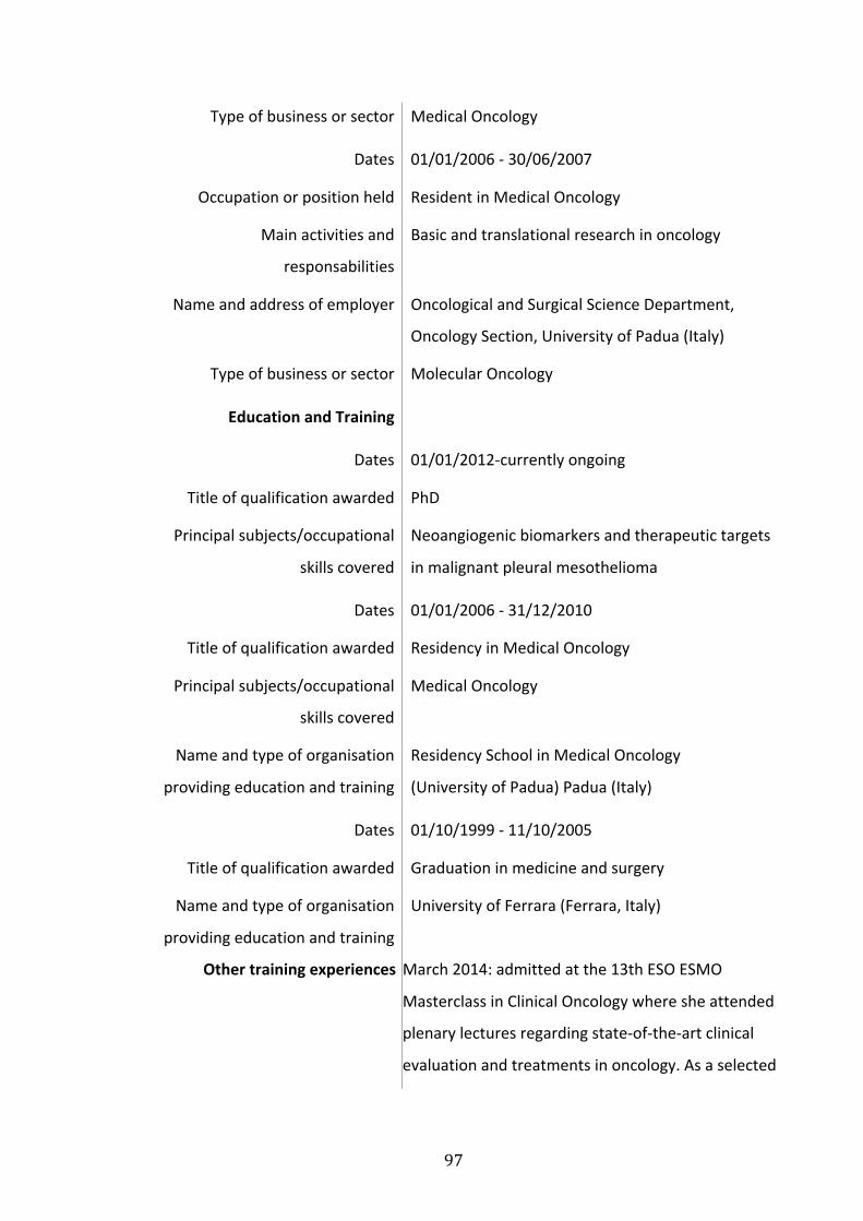
97
Type of business or sector Medical Oncology
Dates 01/01/2006 -‐ 30/06/2007
Occupation or position held Resident in Medical Oncology
Main activities and
responsabilities
Basic and translational research in oncology
Name and address of employer Oncological and Surgical Science Department,
Oncology Section, University of Padua (Italy)
Type of business or sector Molecular Oncology
Education and Training
Dates 01/01/2012-‐currently ongoing
Title of qualification awarded PhD
Principal subjects/occupational
skills covered
Neoangiogenic biomarkers and therapeutic targets
in malignant pleural mesothelioma
Dates 01/01/2006 -‐ 31/12/2010
Title of qualification awarded Residency in Medical Oncology
Principal subjects/occupational
skills covered
Medical Oncology
Name and type of organisation
providing education and training
Residency School in Medical Oncology
(University of Padua) Padua (Italy)
Dates 01/10/1999 -‐ 11/10/2005
Title of qualification awarded Graduation in medicine and surgery
Name and type of organisation
providing education and training
Other training experiences
University of Ferrara (Ferrara, Italy)
March 2014: admitted at the 13th ESO ESMO
Masterclass in Clinical Oncology where she attended
plenary lectures regarding state-‐of-‐the-‐art clinical
evaluation and treatments in oncology. As a selected
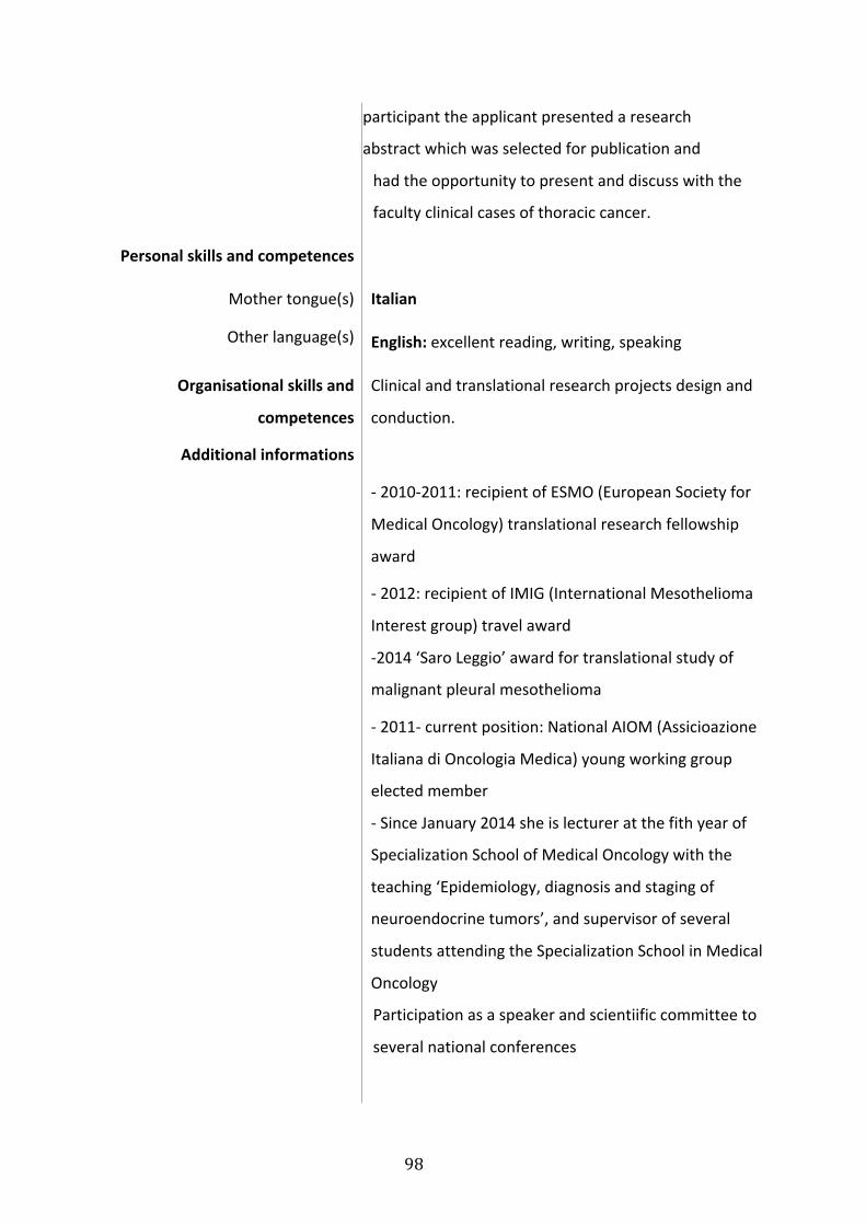
98
participant the applicant presented a research
abstract which was selected for publication and
had the opportunity to present and discuss with the
faculty clinical cases of thoracic cancer.
Personal skills and competences
Mother tongue(s) Italian
Other language(s) English: excellent reading, writing, speaking
Organisational skills and
competences
Additional informations
Clinical and translational research projects design and
conduction.
-‐ 2010-‐2011: recipient of ESMO (European Society for
Medical Oncology) translational research fellowship
award
-‐ 2012: recipient of IMIG (International Mesothelioma
Interest group) travel award
-‐2014 ‘Saro Leggio’ award for translational study of
malignant pleural mesothelioma
-‐ 2011-‐ current position: National AIOM (Assicioazione
Italiana di Oncologia Medica) young working group
elected member
-‐ Since January 2014 she is lecturer at the fith year of
Specialization School of Medical Oncology with the
teaching ‘Epidemiology, diagnosis and staging of
neuroendocrine tumors’, and supervisor of several
students attending the Specialization School in Medical
Oncology
Participation as a speaker and scientiific committee to
several national conferences
99
Summary of scientific and research activity
Several research and scientific activities have been done during the three years of PhD
program. First, the scientific literature study about the main object of the project –
malignant pleural mesothelioma-‐ lead to the study design which took into account the
main unmet medical needs and scientific questions in the specific context.
After study design and planning and working group constitution, a grant application
was performed in order to receive support for preclinical and traslational studies. The
European Society for Medical Oncology (ESMO) translational research fellowship
award was received in 2012 and renewed also for the second year. The project was
mainly divided into two aims. Preclinical studies about new drug combinations for the
treatment of the disease under study were performed during the first two years, while
the third year was focused on translational studies investigating treatment targets and
morphological features on patients samples. In July 2014 the first paper reporting data
from this project was published, and currently two more papers are under review.
Malignant mesothelioma was the main field of interest during the three years of the
PhD program, both as clinical management and research studies, and several papers
were published as single-‐center thoracic oncology group and in collaboration with
other specialized center.
Scientific publication with impact factor (2012-‐2014)
1° year
-‐ Gridelli C, Gallo C, Morabito A, Iaffaioli RV, Favaretto A, Isa L, Barbera S, Gamucci T,
Ceribelli A, Filipazzi V, Maione P, Rossi A, Barletta E, Signoriello S, De Maio E, Piccirillo
MC, Di Maio M, Rocco G, Vecchione A, Perrone F; G-‐STEP Investigators “Phase I-‐II trial
of gemcitabine-‐based first-‐line chemotherapies for small cell lung cancer in elderly
100
patients with performance status 0-‐2: the G-‐STEP trial” J Thorac Oncol. 2012
Jan;7(1):233-‐42
-‐ Di Maio M, Leighl NB, Gallo C, Feld R, Ciardiello F, Butts C, Maione P, Gebbia V,
Morgillo F, Wierzbicki R, Favaretto A, Alam Y, Cinieri S, Siena S, Bianco R, Riccardi F,
Spatafora M, Ravaioli A, Felletti R, Fregoni V, Genestreti G, Rossi A, Mancuso G, Fasano
M, Morabito A, Tsao MS, Signoriello S, Perrone F, Gridelli C; TORCH
Investigators.“Quality of life analysis of TORCH, a randomized trial testing first-‐line
erlotinib followed by second-‐line cisplatin/gemcitabine chemotherapy in advanced
non-‐small-‐cell lung cancer.” J Thorac Oncol. 2012 Dec;7 (12):1830-‐4
-‐ Pasello G, Marulli G, Polo V, Breda C, Bonanno L, Loreggian L, Rea F, Favaretto A.
“Pemetrexed plus carboplatin or cisplatin as neoadjuvant treatment of operable
malignant pleural mesothelioma (MPM)” Anticancer Res. 2012 Dec;32 (12):5393-‐9.
2° year
-‐ Pasello G, Ceresoli GL, Favaretto A “An overview of neoadjuvant chemotherapy in the
multimodality treatment of malignant pleural mesotelioma”. Cancer Treat Rev. 2013
Feb;39 (1):10-‐7
-‐ Marulli G, Di Chiara F, Braccioni F, Perissinotto E, Pasello G, Gino Favaretto A, Breda
C, Rea F. “Changes in pulmonary function tests predict radiological response to
chemotherapy in malignant pleural mesothelioma” Eur J Cardiothorac Surg. 2013 Jul;
44 (1):104-‐10
-‐Pasello G, Urso L, Conte P, Favaretto A. “Effects of Sulfonylureas on Tumor Growth: A
Review of the Literature”. Oncologist. 2013;18(10):1118-‐25
3° year
101
-‐ Pasello G, Urso L, Silic-‐Benussi M, Schiavon M, Cavallari I, Marulli G, Nannini N, Rea F,
Ciminale V, Favaretto A. “Synergistic antitumor activity of recombinant human
Apo2L/tumor necrosis factor-‐related apoptosis-‐inducing ligand (TRAIL) in combination
with carboplatin and pemetrexed in malignant pleural mesothelioma.”J Thorac Oncol.
2014 Jul;9(7):1008-‐17.
-‐ Ceresoli GL, Grosso F, Zucali PA, Mencoboni M, Pasello G, Ripa C, Degiovanni D,
Simonelli M, Bruzzone A, Dipietrantonj C, Piccolini E, Beretta GD, Favaretto AG,
Giordano L, Santoro A, Botta M.“Prognostic factors in elderly patients with malignant
pleural mesothelioma: results of a multicenter survey.”Br J Cancer. 2014 Jul
15;111(2):220-‐6.
-‐ Marulli G, Verderi E, Zuin A, Schiavon M, Battistella L, Perissinotto E, Romanello P,
Favaretto AG, Pasello G, Rea F.”Outcomes and prognostic factors of non-‐small-‐cell lung
cancer with lymph node involvement treated with induction treatment and surgical
resection.”Interact Cardiovasc Thorac Surg. 2014 Aug;19(2):256-‐62
-‐ Schiavon M, Marulli G, Nannini N, Pasello G, Lunardi F, Balestro E, Perissinotto E,
Rebusso A, Saetta M, Rea F, Calabrese F. “COPD-‐related adenocarcinoma presents low
aggressiveness morphological and molecular features compared to smoker tumours”.
Lung Cancer. 2014 Dec; 86(3):311-‐7.
-‐ Pasello G, Carli P, Canova F, Bonanno L, Polo V, Zago G, Urso L, Conte PF, Favaretto A.
“Epirubicin plus Paclitaxel regimen as second line treatment of Small-‐Cell Lung Cancer
(SCLC) patients” IN PRESS
Book chapter (2012-‐2014)
-‐ Giulia Pasello and Adolfo Favaretto. Neoadjuvant Chemotherapy in Malignant Pleural
Mesothelioma. Malignant Mesothelioma Chapter 5 December 2012.
102
http://dx.doi.org/10.5772/47880
-‐ Giovanni Luca Ceresoli, Giulia Pasello, Fiorella Calabrese.
ESMO book: Thoracic Tumours –Essentials for Clinicians: Malignant Pleural
Mesothelioma
March 2014
Conference participation (2012-‐2014)
-‐advances in medicine 23 march 2012, Padova
-‐ 2012 american society of clinical oncology (asco) meeting 1-‐5 june 2012, Chicago (il)
-‐ primo congresso italo-‐greco di oncologia medica, Padova, 10-‐11 may 2012:
“treatment of malignant pleural mesothelioma: multimodality management and new
perspectives” (speaker)
-‐aiom post asco review: updates and news from the annual meeting in chicago,
Bologna 16-‐17 june 2012: “lung cancer: asco poster review” (speaker)
-‐ endothelial progenitors cells from discovery to therapy 21 june 2012, Padova
-‐ hot topics in oncologia toracica: stato dell’arte sulla terapia medica del mesotelioma
pleurico, Bergamo 22 june 2012 (speaker)
-‐ the 11th international mesothelioma interest group (imig) conference 11-‐14
september 2012, Boston (ma) (speaker)
-‐ malignant pleural mesothelioma: clinical pathological and legal features 8 november
2012, Padova(speaker)
-‐ corso nibit: immunobioterapia nelle neoplasie toraciche, Padova 23 november 2012
(speaker)
-‐ xii congresso nazionale fonicap (forza operativa nazionale interdisciplinare contro il
cancro al polmone), Verona 30 november-‐1 december 2012 (speaker)
103
-‐ corso nazionale aiom e siapec-‐iap “marcatori biomolecolari nella terapia
personalizzata dei tumori: indicazioni cliniche e di laboratorio”, Napoli 22 march 2013
-‐ “antiangiogenic therapy: recent advances and future directions in oncology”, Padova
23 april 2013
-‐ corso educazionale macroregionale aiom giovani “metodologia della ricerca clinica in
oncologia”, Verona 3-‐4 may 2013 (speaker)
-‐ convegno aiom giovani veneto: “tumori rari: tra empirismo ed evidenza”, Padova 21
june 2013 (speaker)
-‐ convegno nazionale aiom giovani “news in oncology 2013”, Perugia 5-‐6 july 2013
(speaker)
-‐ corso di formazione del personale medico in oncologia generale, Bugando medical
centre, Mwanza, Tanzania, 30 september 2013 – 04 october 2013 (speaker)
-‐ congresso fonicap le terapie biologichenel nsclc: update dopo iaslc 2013, Verona 9
december 2013 (speaker)
-‐ 2014 esmo-‐christie lung cancer course, manchester (uk) 12-‐14 february 2014
-‐13th eso-‐esmo masterclass in clinical oncology, 8 march 2014-‐13 march 2014,
Ermatingen Switzerland
-‐ hot topics in oncologia toracica:il mesotelioma pleurico, Bergamo 24 may 2014
(speaker)
-‐ aiom post-‐asco 2014, Bologna 13-‐14 june 2014 (speaker)
-‐ convegno nazionale aiom giovani “news in oncology 2014”, Perugia 11-‐12 july 2014
(speaker)
-‐ 2014 esmo meeting, Madrid 26 september 2014-‐ 30 september 2014 (poster
presenter)
104
-‐ chest tumors: what’s new from asco-‐esmo meetings? Padova 9-‐10 october 2014
(speaker)
-‐ the 12th international mesothelioma interest group (imig) conference 21 october
2014-‐24 october 2014 Cape Town Africa (poster presenter)
-‐ corso macroregionale aiom di metodologia della ricerca, Torino 7-‐8 november 2014
(speaker)
Awards and grants:
- 2010-‐2011 and 2011-‐2012: recipient of ESMO (European Society for Medical
Oncology) translational research fellowship award
- 2012: recipient of IMIG (International Mesothelioma Interest group) travel
award
- 2014: premio per la ricerca ‘Saro Leggio’ (AIRPP)
Teaching activity
- professor with contract 5° year of residency school in medical oncology with
the teaching: epidemiology, diagnosis and staging of neuroendocrin tumors
Research activity
- -‐Coinvestigator of more than 30 clinical trials on lung cancer, pleural
mesothelioma and thymic cancer
- Principal investigator of preclinical studies on the anticancer activity of new
drugs combinations in malignant pleural mesothelioma.
- Principal investigator of retrospective studies on lung cancer and pleural
mesothelioma.

105
Thanks to:
The present research project is the result of a multidisciplinary team involved in
basic, translational and clinical research and medical management of thoracic
tumors.
With particular respect and gratitude, I would like to thank:
Prof. Federico Rea, Prof. Fiorella Calabrese
Department of Cardiac, Thoracic and Vascular Sciences (University of Padova)
Dr. Adolfo Favaretto and Dr. Vincenzo Ciminale
Istituto Oncologico Veneto
Dr. Loredana Urso
Department of Surgical, Oncological and Gastroenterological Sciences
(University of Padova)