Embed Size (px)
Citation preview
RESEARCH ARTICLE
Ex-Vivo Uterine Environment (EVE) TherapyInduced Limited Fetal Inflammation in aPremature Lamb ModelYuichiro Miura1,2*, Masatoshi Saito2, Haruo Usuda2, Eleanor Woodward1,Judith Rittenschober-Böhm1,3, Paranthaman S. Kannan4, Gabrielle C. Musk5,6,Tadashi Matsuda2, John P. Newnham1, MatthewW. Kemp1
1 School of Women's and Infants' Health, The University of Western Australia, Crawley, Western Australia,Australia, 2 Center for Perinatal and Neonatal Medicine, Tohoku University Hospital, Sendai, Miyagi, Japan,3 Division of Neonatology, Pediatric Intensive Care and Neuropediatrics, Medical University of Vienna,Vienna, Austria, 4 Division of Pulmonary Biology, Cincinnati Children's Hospital Medical Center, Cincinnati,Ohio, United States of America, 5 School of Veterinary and Life Sciences, Murdoch University, Murdoch,Western Australia, Australia, 6 Animal Care Services, The University of Western Australia, Crawley, WesternAustralia, Australia
Abstract
Introduction
Ex-vivo uterine environment (EVE) therapy uses an artificial placenta to provide gas
exchange and nutrient delivery to a fetus submerged in an amniotic fluid bath. Development
of EVE may allow us to treat very premature neonates without mechanical ventilation.
Meanwhile, elevations in fetal inflammation are associated with adverse neonatal out-
comes. In the present study, we analysed fetal survival, inflammation and pulmonary matu-
ration in preterm lambs maintained on EVE therapy using a parallelised umbilical circuit
system with a low priming volume.
Methods
Ewes underwent surgical delivery at 115 days of gestation (term is 150 days), and fetuses
were transferred to EVE therapy (EVE group; n = 5). Physiological parameters were contin-
uously monitored; fetal blood samples were intermittently obtained to assess wellbeing and
targeted to reference range values for 2 days. Age-matched animals (Control group; n = 6)
were surgically delivered at 117 days of gestation. Fetal blood and tissue samples were
analysed and compared between the two groups.
Results
Fetal survival time in the EVE group was 27.0 ± 15.5 (group mean ± SD) hours. Only one
fetus completed the pre-determined study period with optimal physiological parameters,
while the other 4 animals demonstrated physiological deterioration or death prior to the pre-
determined study end point. Significant elevations (p<0.05) in: i) inflammatory proteins in
PLOS ONE | DOI:10.1371/journal.pone.0140701 October 16, 2015 1 / 17
OPEN ACCESS
Citation: Miura Y, Saito M, Usuda H, Woodward E,Rittenschober-Böhm J, Kannan PS, et al. (2015) Ex-Vivo Uterine Environment (EVE) Therapy InducedLimited Fetal Inflammation in a Premature LambModel. PLoS ONE 10(10): e0140701. doi:10.1371/journal.pone.0140701
Editor: Rogelio Cruz-Martinez, Hospital deEspecialidades del Niño y la Mujer de Queretaro,MEXICO
Received: May 28, 2015
Accepted: September 28, 2015
Published: October 16, 2015
Copyright: © 2015 Miura et al. This is an openaccess article distributed under the terms of theCreative Commons Attribution License, which permitsunrestricted use, distribution, and reproduction in anymedium, provided the original author and source arecredited.
Data Availability Statement: All relevant data arewithin the paper.
Funding: This work was supported by grants fromthe Channel 7 Telethon Trust, http://telethon.7perth.com.au (MWK), the Financial Markets Foundation forChildren, http://www.foundationforchildren.com.au(MWK), and the National Health and MedicalResearch Council APP1049148, https://www.nhmrc.gov.au (MWK, JPN). MWK is supported by theWomen and Infants Research Foundation, http://wirf.com.au. The funders had no role in study design,
fetal plasma; ii) selected cytokine/chemokine mRNA expression levels in fetal tissues; and
iii) histological inflammatory score in fetal lung, were observed in the EVE group compared
to the Control group. There was no significant difference (p>0.05) in surfactant protein
mRNA expression level between the two groups.
Conclusion
In this study, we achieved limited fetal survival using EVE therapy. Despite this, EVE ther-
apy only induced a modest fetal inflammatory response and did not promote lung matura-
tion. These data provide additional insight into markers of treatment efficacy for the
assessment of future studies.
IntroductionPreterm birth (live delivery prior to 37 weeks’ gestation) is responsible for at least 1 millionperinatal deaths every year [1]. Despite significant advances in perinatal medicine (i.e. antena-tal corticosteroid administration, surfactant therapy, advanced ventilation strategies), a signifi-cant proportion of infants born between 22 and 25 weeks’ gestation in high-resource settingswill either die or experience life-long diseases of the cardiovascular, respiratory or neurosen-sory systems [2,3]. It is unclear if the high incidence of morbidity and mortality in this popula-tion is due to the antenatal exposures responsible for prematurity, or is as a result of forciblyadapting the highly immature physiology of a 22–25 week gestation fetus to pulmonary gasexchange in ex-uterine life. We have speculated that treating very premature babies withoutmechanical ventilation may allow them to be supported without causing or exacerbating injury.Designing treatment strategies that take advantage of the fetal-like physiology exhibited byextremely preterm infants may decrease morbidity and mortality of early preterm babies whocannot survive with existing neonatal intensive care.
Ex-vivo uterine environment (EVE) therapy is a novel treatment strategy based on the con-cepts introduced above. It uses an artificial placenta to provide extracorporeal gas exchangeand nutrient delivery to a fetus submerged in a warmed amniotic fluid bath. An artificial pla-centa is a system that allows for arterio-venous extracorporeal life support using the umbilicalvasculature. Previously, we demonstrated the efficacy of our EVE system with the pumplessartificial placenta using the preterm lamb model [4].
Systemic fetal inflammation, characterised in humans as the fetal inflammatory responsesyndrome (cord blood interleukin-6 concentration>11 pg/mL [5]) is strongly associated withfetal injury, in particular brain injury, in the setting of preterm birth. Thus, any treatments forextremely preterm infants, such as EVE therapy, must allow for the control of pathologicalfetal inflammation. Intrauterine inflammation is also associated with precocious maturation ofthe fetal lung [6,7]. It has previously been reported that extracorporeal life support causes sys-temic inflammation [8–14], and that the intensity of inflammation is altered by the blood con-tact area [15] and the coating of the surface area [16]. As such, the inflammatory responsesgenerated by fetuses maintained on EVE therapy are of significant interest. In this study, thesystemic inflammatory response of preterm lambs undergoing EVE therapy was assessed.
Our primary hypothesis was that, when successfully functioning, the EVE system in thisstudy would cause minimal inflammation to the fetuses as it incorporated a bespoke, low-vol-ume membranous oxygenator with a heparin coating. On the other hand, we also hypothesisedthat systemic inflammatory changes would become apparent in animals in which treatment
Fetal Inflammation Induced by EVE Therapy
PLOS ONE | DOI:10.1371/journal.pone.0140701 October 16, 2015 2 / 17
data collection and analysis, decision to publish, orpreparation of the manuscript.
Competing Interests: The authors have declaredthat no competing interests exist.
was unsuccessful. As such, changes in fetal inflammatory status may serve as a useful means ofassessing treatment success in real-time. Based on our primary hypothesis, we also anticipatedthat successful adaptation to EVE therapy would not induce fetal lung maturation. To testthese hypotheses, experiments were conducted using a premature lamb model. The primaryaim was to compare the concentration of inflammatory/stress markers in fetal plasma and theexpression of inflammatory cytokine/chemokine mRNA in fetal tissues between the EVE treat-ment and the Control groups, and also between successful and unsuccessful cases in the EVEtreatment group. Our secondary aim was to assess the expression of surfactant protein (SP)mRNA in fetal lung between the EVE treatment and the Control groups.
Materials and Methods
Experimental protocolAll procedures involving animals were performed in Perth, Western Australia following reviewand approval by the Animal Ethics Committee of the University of Western Australia(approval RA/3/100/1289). All surgery was performed under the anaesthesia with isoflurane,and all efforts were made to minimise suffering.
Study groupa) EVE group. 5merino-cross ewes with timed, singleton pregnancies underwent surgery
at 115 days gestational age (dGA; term = 150). Animals were fasted for 12 hours before surgerywith ad libitum access to water. Ewes were anaesthetised, intubated, and ventilated during thesurgical procedure. Intravenous fluids (0.9% NaCl) were administered at a rate of 10 mL�kg-1�hour-1. The ewe’s abdomen was clipped to expose the skin and thoroughly prepared for sur-gery with as previously described [17]. After a maternal laparotomy and hysterotomy, catheterswere placed in fetal jugular vein (4 French, Arrow Pediatric two-lumen central venous catheter,Teleflex Medical, Morrisville, NC), carotid artery (4.5 French, PediaSat Oximetry Catheter,Edwards Lifesciences, Irvine, CA), two umbilical arteries (8 French, Fem-Flex II; Edwards Life-sciences), and one umbilical vein (10 French, Argyle Trocar catheter; Covidien, Dublin, Ire-land). The fetus was connected to the artificial placenta, and then delivered and carefullysubmerged in an amniotic fluid bath. The fetuses were continuously treated with heparin(12.5 U�kg-1�hour-1) to prevent blood coagulation and prostaglandin E1 (50 ng�kg-1�min-1) toprevent closure of the ductus arteriosus; and periodically treated with cefazolin (40 mg�kg-1�day-1) to prevent infection. Activated clotting times were monitored using a Hemochron Sig-nature Elite (Accriva Diagnostics, San Diego, CA) and maintained at>180 seconds. Appropri-ate nutrient supplementations including glucose (10–12%), amino acids (1.0–1.2 g�kg-1�day-1),and lipid (0.7–1.0 g�kg-1�day-1) were delivered via the catheterised fetal jugular vein. Fetuseswere constantly monitored to ensure that adequate sedation was maintained, and intravenousbolus of midazolam (0.2 mg/kg) was administered when needed. Each fetus was supposed tomaintain in the EVE system for pre-determined 40 hours.
b) Control group. 6 ewes with timed, singleton pregnancies were surgically delivered at117 dGA. Fetuses were cannulated and blood samples collected at the time of surgical deliveryunder non-recovery anaesthesia. Fetuses were then immediately euthanised to allow for tissuesample collection.
All ewes were euthanised with an intravenous bolus of pentobarbitone (160 mg/kg) at thetime of fetal delivery. All fetuses in the Control group were euthanised with an intravenouspentobarbitone (100 mg/kg) at 117 dGA. All fetuses in the EVE group were euthanised with anintravenous pentobarbitone (100 mg/kg) at the pre-determined time point (n = 2) or acutely
Fetal Inflammation Induced by EVE Therapy
PLOS ONE | DOI:10.1371/journal.pone.0140701 October 16, 2015 3 / 17
following irreversible deterioration of key physiological variables (either total circuit bloodflow<20 mL�kg-1�min-1 or SaO2<20% for more than 10 minutes; n = 3).
EVE Componentsa) Artificial Placenta. The circuit was composed of three main components: i) outflow
tubes; ii)membranous oxygenators; and iii) an inflow tube. Two membranous oxygenatorswere placed in a parallel orientation. Heparinised polyvinyl chloride tubes were used for boththe inflow and outflow tubes. The priming volume of whole circuit was 70 mL, and the circuitwas primed with heparinised maternal blood [18]. Total membrane surface area was 0.3 m2.Circuit flow was maintained by the fetal heart. Appropriate oxygen was supplied to the mem-branous oxygenators to keep fetal PaO2 between 10–30 mmHg.
b) Amniotic fluid bath. Both the fetus and the artificial placenta circuit were submergedin a sealed bath of artificial amniotic fluid held at a constant 38.9°C. pH and electrolyte concen-tration of artificial amniotic fluid were identical to that of amniotic fluid in merino sheep (pH:7.2 ± 0.2, Na+: 122 ± 10 mEq/L, Cl-: 112 ± 10 mEq/L, all values represent groupmean ± standard deviation (SD)). Amniotic fluid was constantly filtered and UV (isolation)irradiated to prevent microbial colonisation.
Data AcquisitionData relating to the physiological parameters. Fetal heart rate and mean arterial pressure
were continuously monitored and recorded using a SurgiVet (Smiths Medical, St. Paul, MN).Fetal arterial oxygen saturation was continuously monitored and recorded using a VigileoMonitor (Edwards Lifesciences). Circuit blood flow (mL/min) was continuously monitoredusing electromagnetic flow sensors (Transonic 400-Series, Transonic Systems Inc., Ithaca, NY)which were attached to the arterial positions of the blood circuit, and recorded using a Power-Lab (ADInstruments, Dunedin, New Zealand). pH, PaO2, PaCO2, blood lactate level (SiemensRapidlab 1200, Siemens, Munich, Germany), and activated clotting time were measured inblood samples obtained from the fetal carotid artery at least every 4 hours. Blood gas data werecorrected using fetal core temperature. Fetal arterial blood for complete blood counts was col-lected in a 10 mL Vacutainer (Becton, Dickinson and Company, Franklin Lakes, NJ) contain-ing EDTA. Haematological analyses were performed by an independent pathology laboratory(Vetpath, Perth, WA, Australia).
Data relating to the assessment of inflammation. Fetal arterial blood for inflammatorycytokine/chemokine mRNA analyses was intermittently collected in a PAXgene blood RNAtube (PreAnalytiX, Hombrechtikon, Switzerland). Fetal plasma samples were also intermit-tently collected for tumour necrosis factor (TNF) –α and monocyte chemo-attractant protein(MCP) -1 protein analyses. Fetal arterial blood for liver function tests was collected in a 10 mLSST clot-activating Vacutainer (Becton, Dickinson and Company) at the end of protocol. Bio-chemical analyses were performed by Vetpath (Perth, WA, Australia).
Fetal tissues (lung right lower lobe, axilla skin, liver, and cerebral cortex) for inflammatorycytokine/chemokine mRNA analyses were collected and snap-frozen in liquid nitrogen at theend of the protocol. Fetal lung (right upper lobe) was also collected at the end of the protocol,and inflation-fixed in 10% neutral buffered formalin using constant pressure (30 cm H2O) for24 hours before being washed in phosphate buffered saline and embedded in paraffin.
Data relating to the assessment of pulmonary maturation. Fetal lung right lower lobefor surfactant protein mRNA analyses was collected and snap-frozen in liquid nitrogen at theend of the protocol.
Fetal Inflammation Induced by EVE Therapy
PLOS ONE | DOI:10.1371/journal.pone.0140701 October 16, 2015 4 / 17
Post processing sectionFetal tissues were homogenised at 6,500 rpm, 15 seconds (brain), 30 seconds (lung and liver);or 20 seconds, 2 cycles (skin) using a Precellys 24 macerator (Bertin Technologies, Montigny-le-Bretonneux, France). Total RNA was extracted using TRIzol (Life Technologies, Carlsbad,CA). Extracted RNA was treated with Turbo-DNase (Life Technologies) in accordance withmanufacturer’s instructions to remove any residual DNA. RNA template was quantified usinga Qubit 2.0 Fluorometer (Life Technologies) using a broad-range RNA quantitation kit (LifeTechnologies). RNA extracts were diluted in nuclease-free water (Life Technologies) to a finalconcentration of 25 ng/μL.
Total RNA was extracted from immunocytes in fetal whole blood using PAXgene BloodRNA Kit (PreAnalytiX) in accordance with manufacturer’s instructions. RNA quantificationand dilution were demonstrated in the same way described above.
Ovine-specific PCR primers and hydrolysis probes for SP-A, B, C, D, interleukin (IL)-1β,IL-6, IL-8, TNF-α, MCP-2, C-reactive protein (CRP), Serum amyloid A (SAA) 3, and Hepcidin(all Life Technologies) were used to perform quantitative PCR. Reactions were performedusing an EXPRESS One-Step SuperScript qRT-PCR Kit (Life Technologies) with 125 ng oftemplate fetal tissue RNA in a total volume of 20 μL as per manufacturer’s instructions. Reac-tion cycling conditions were: 15 minutes reverse transcription at 50°C and an initial denatur-ation/polymerase activation at 95°C for 20 seconds, followed by 40 cycles of 95°C for 3 secondsand 60°C for 30 seconds (data acquisition phase). All reactions were performed in the fast96-well plates on a ViiA7 real-time PCR thermocycler (Life Technologies). Target Cq valueswere normalised to 18S rRNA Cq value and expressed as fold changes relative to pooled controlvalues. Reaction efficiencies were within limits proposed in the MIQE guidelines [19]. dCq val-ues were used to perform statistical analyses for significant differences between the Controlgroup vs. the EVE group.
Quantification of TNF-α and MCP-1 protein concentrations in fetal plasma samples wasperformed using a commercial kit from Kingfisher Biotech (St. Paul, MN) with washing per-formed on a Biosan platewasher (3D-IW8, Inteliwasher, Biosan, Riga, Latvia). Standards (cali-bration curve R2>0.99) were assayed in triplicate (average coefficient of variation 7.8%) andunknown samples were assayed in duplicate. The assay limit of detection was<4 pg/mL.100 μL of each standard or sample was incubated overnight (16 hours) at 4°C. The remainderof the assay was performed in accordance with the manufacturer’s instructions, with absor-bance at 450 nm read on an Anthos 2010 Microplate Reader (Biochrom Ltd., Cambridge,United Kingdom).
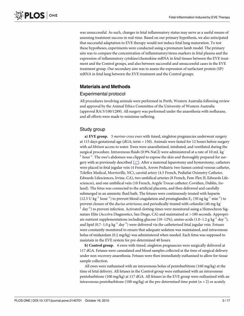
5 μm paraffin sections of 10% (pH 7.4) formalin-fixed fetal lung (right upper lobe) werestained with Meyer’s haematoxylin and eosin. Qualitative scoring of airspace infiltration wasperformed by a single investigator blinded to treatment groups. Six fields (200 x total magnifi-cation) were scored for each animal. Airspace infiltration and consolidation were graded as fol-lows: 0 = no inflammatory cells in airspace; 1 = airspace inflammatory cells, no consolidation;2 = airspace inflammatory cells + limited microconsolidation (1–4 per field) foci; 3 = airspaceinflammatory cells + numerous (>4 per field) but predominantly discrete microconsolidationfoci; and 4 = airspace inflammatory cells + confluent airspace consolidation (Fig 1).
Statistical AnalysisNormally distributed values were expressed as the group mean ± SD, while non-parametricvalues were expressed as the group median [interquartile range (IQR)]. Analyses were per-formed using IBM SPSS for Windows, Version 20.0 (IBM Corporation, Armonk, NY). Datawere tested for normality using Shapiro-Wilk test. In the comparison of two groups, for
Fetal Inflammation Induced by EVE Therapy
PLOS ONE | DOI:10.1371/journal.pone.0140701 October 16, 2015 5 / 17
normally distributed data, mean differences were tested for significance using t-test. Between-group differences in non-parametric data were tested for significance using Mann-Whitney Utest. In the comparison of more than three groups, for normally distributed data, mean differ-ences were tested for significance using one-way ANOVA. Multiple post-hoc comparisons wereperformed using Tukey’s test. Between-group differences in non-parametric data were testedfor significance using Kruskal-Wallis one-way ANOVA. Multiple post-hoc comparisons wereperformed using the rank sum test. All p values<0.05 were accepted as significant.
Results
Data relating to the physiological parametersTable 1 shows the comparison between the Control and the EVE groups. There were no unex-pected deaths in the Control group. There was no statistically significant difference in anydelivery data shown in Table 1 between the two groups.
Fig 1. Representative images for qualitative scoring of airspace infiltration. Images are representative of the indicated inflammatory scores assigned toeach field assessed (× 200 total magnification). Scale bar = 100 μm.
doi:10.1371/journal.pone.0140701.g001
Fetal Inflammation Induced by EVE Therapy
PLOS ONE | DOI:10.1371/journal.pone.0140701 October 16, 2015 6 / 17
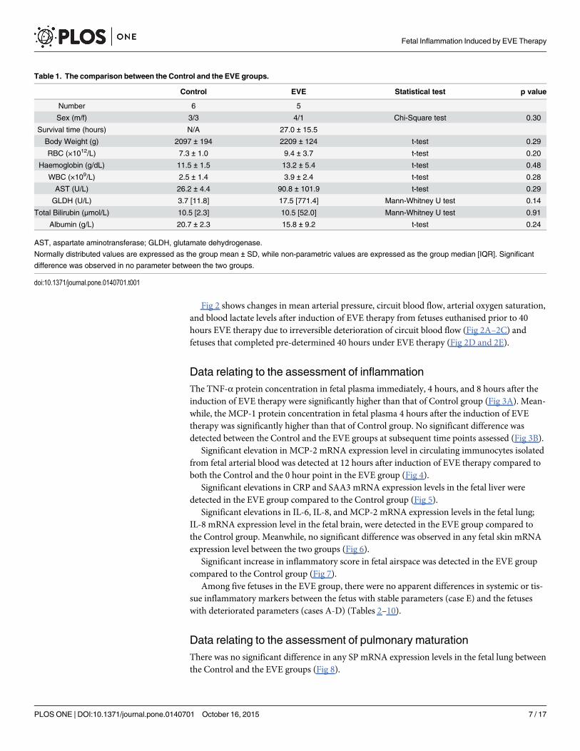
Fig 2 shows changes in mean arterial pressure, circuit blood flow, arterial oxygen saturation,and blood lactate levels after induction of EVE therapy from fetuses euthanised prior to 40hours EVE therapy due to irreversible deterioration of circuit blood flow (Fig 2A–2C) andfetuses that completed pre-determined 40 hours under EVE therapy (Fig 2D and 2E).
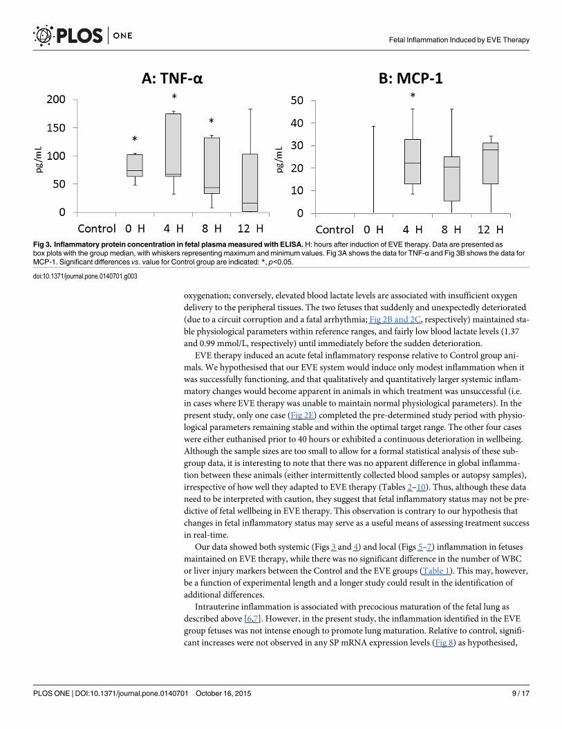
Data relating to the assessment of inflammationThe TNF-α protein concentration in fetal plasma immediately, 4 hours, and 8 hours after theinduction of EVE therapy were significantly higher than that of Control group (Fig 3A). Mean-while, the MCP-1 protein concentration in fetal plasma 4 hours after the induction of EVEtherapy was significantly higher than that of Control group. No significant difference wasdetected between the Control and the EVE groups at subsequent time points assessed (Fig 3B).
Significant elevation in MCP-2 mRNA expression level in circulating immunocytes isolatedfrom fetal arterial blood was detected at 12 hours after induction of EVE therapy compared toboth the Control and the 0 hour point in the EVE group (Fig 4).
Significant elevations in CRP and SAA3 mRNA expression levels in the fetal liver weredetected in the EVE group compared to the Control group (Fig 5).
Significant elevations in IL-6, IL-8, and MCP-2 mRNA expression levels in the fetal lung;IL-8 mRNA expression level in the fetal brain, were detected in the EVE group compared tothe Control group. Meanwhile, no significant difference was observed in any fetal skin mRNAexpression level between the two groups (Fig 6).
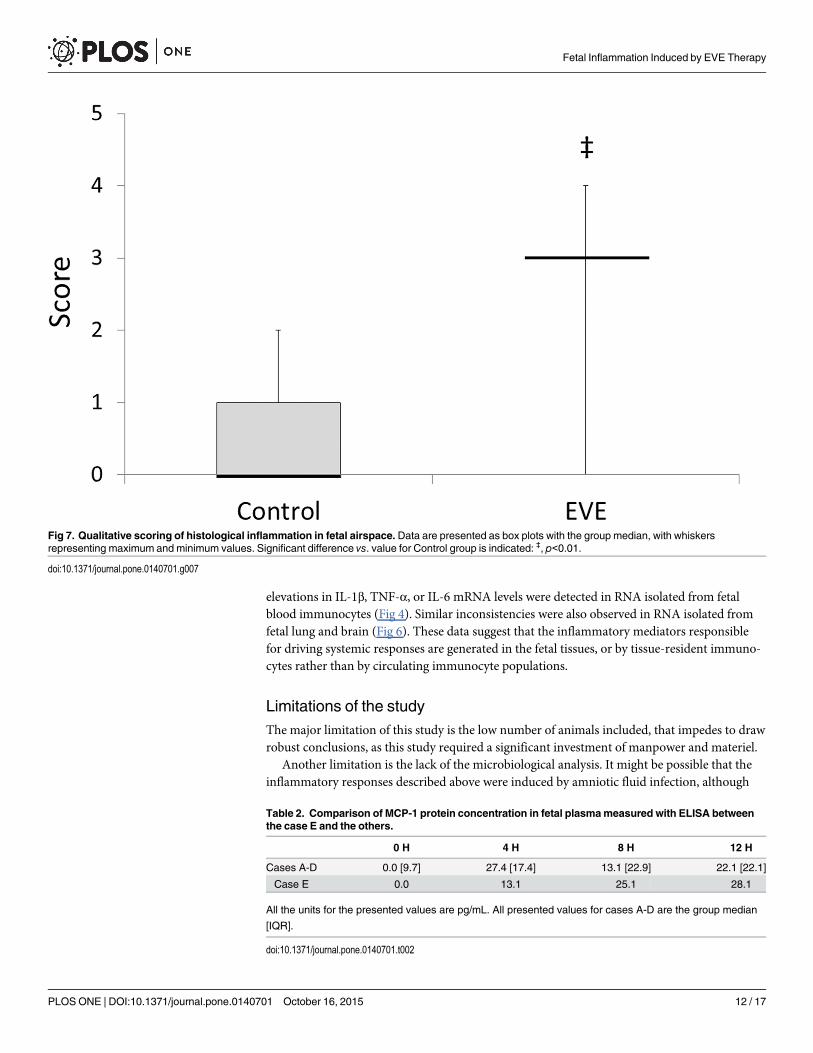
Significant increase in inflammatory score in fetal airspace was detected in the EVE groupcompared to the Control group (Fig 7).
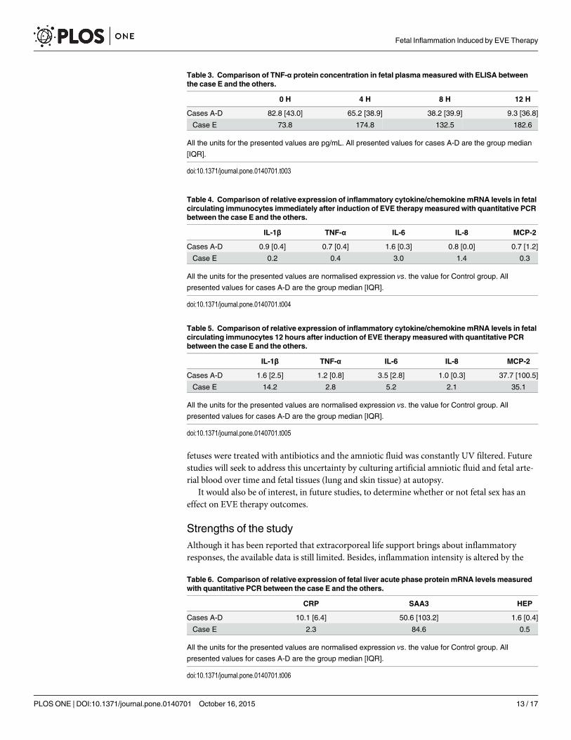
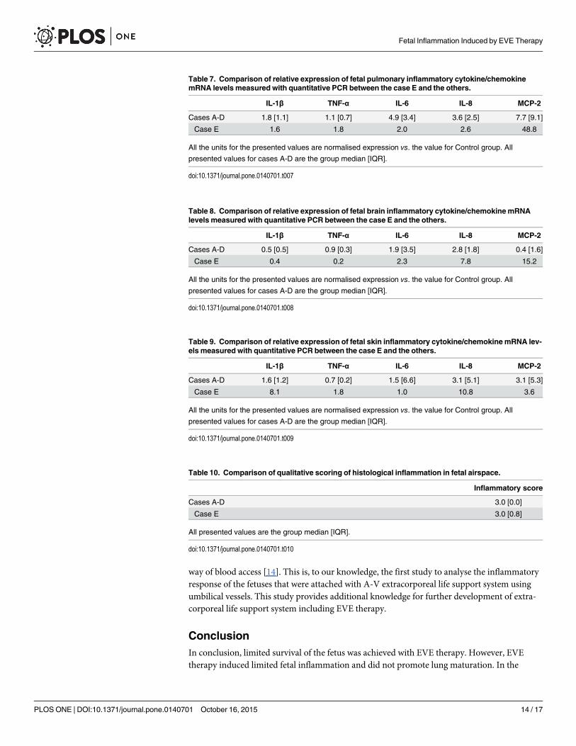
Among five fetuses in the EVE group, there were no apparent differences in systemic or tis-sue inflammatory markers between the fetus with stable parameters (case E) and the fetuseswith deteriorated parameters (cases A-D) (Tables 2–10).
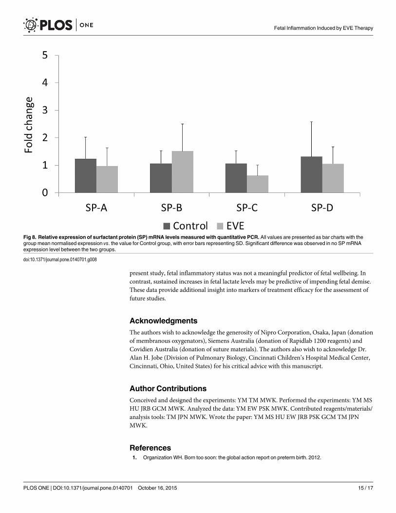
Data relating to the assessment of pulmonary maturationThere was no significant difference in any SP mRNA expression levels in the fetal lung betweenthe Control and the EVE groups (Fig 8).
Table 1. The comparison between the Control and the EVE groups.
Control EVE Statistical test p value
Number 6 5
Sex (m/f) 3/3 4/1 Chi-Square test 0.30
Survival time (hours) N/A 27.0 ± 15.5
Body Weight (g) 2097 ± 194 2209 ± 124 t-test 0.29
RBC (×1012/L) 7.3 ± 1.0 9.4 ± 3.7 t-test 0.20
Haemoglobin (g/dL) 11.5 ± 1.5 13.2 ± 5.4 t-test 0.48
WBC (×109/L) 2.5 ± 1.4 3.9 ± 2.4 t-test 0.28
AST (U/L) 26.2 ± 4.4 90.8 ± 101.9 t-test 0.29
GLDH (U/L) 3.7 [11.8] 17.5 [771.4] Mann-Whitney U test 0.14
Total Bilirubin (μmol/L) 10.5 [2.3] 10.5 [52.0] Mann-Whitney U test 0.91
Albumin (g/L) 20.7 ± 2.3 15.8 ± 9.2 t-test 0.24
AST, aspartate aminotransferase; GLDH, glutamate dehydrogenase.
Normally distributed values are expressed as the group mean ± SD, while non-parametric values are expressed as the group median [IQR]. Significant
difference was observed in no parameter between the two groups.
doi:10.1371/journal.pone.0140701.t001
Fetal Inflammation Induced by EVE Therapy
PLOS ONE | DOI:10.1371/journal.pone.0140701 October 16, 2015 7 / 17
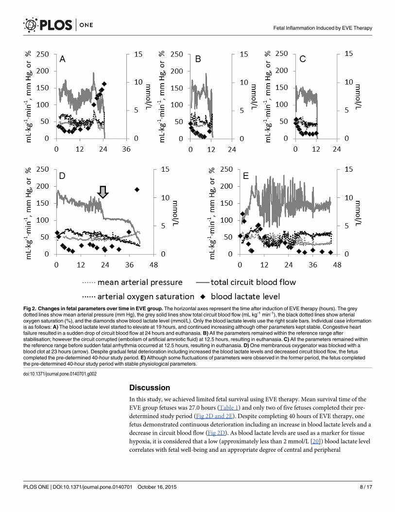
DiscussionIn this study, we achieved limited fetal survival using EVE therapy. Mean survival time of theEVE group fetuses was 27.0 hours (Table 1) and only two of five fetuses completed their pre-determined study period (Fig 2D and 2E). Despite completing 40 hours of EVE therapy, onefetus demonstrated continuous deterioration including an increase in blood lactate levels and adecrease in circuit blood flow (Fig 2D). As blood lactate levels are used as a marker for tissuehypoxia, it is considered that a low (approximately less than 2 mmol/L [20]) blood lactate levelcorrelates with fetal well-being and an appropriate degree of central and peripheral
Fig 2. Changes in fetal parameters over time in EVE group. The horizontal axes represent the time after induction of EVE therapy (hours). The greydotted lines showmean arterial pressure (mm Hg), the grey solid lines show total circuit blood flow (mL�kg-1�min-1), the black dotted lines show arterialoxygen saturation (%), and the diamonds show blood lactate level (mmol/L). Only the blood lactate levels use the right scale bars. Individual case informationis as follows: A) The blood lactate level started to elevate at 19 hours, and continued increasing although other parameters kept stable. Congestive heartfailure resulted in a sudden drop of circuit blood flow at 24 hours and euthanasia. B) All the parameters remained within the reference range afterstabilisation; however the circuit corrupted (embolism of artificial amniotic fluid) at 12.5 hours, resulting in euthanasia. C) All the parameters remained withinthe reference range before sudden fatal arrhythmia occurred at 12.5 hours, resulting in euthanasia.D)One membranous oxygenator was blocked with ablood clot at 23 hours (arrow). Despite gradual fetal deterioration including increased the blood lactate levels and decreased circuit blood flow, the fetuscompleted the pre-determined 40-hour study period. E) Although some fluctuations of parameters were observed in the former period, the fetus completedthe pre-determined 40-hour study period with stable physiological parameters.
doi:10.1371/journal.pone.0140701.g002
Fetal Inflammation Induced by EVE Therapy
PLOS ONE | DOI:10.1371/journal.pone.0140701 October 16, 2015 8 / 17
oxygenation; conversely, elevated blood lactate levels are associated with insufficient oxygendelivery to the peripheral tissues. The two fetuses that suddenly and unexpectedly deteriorated(due to a circuit corruption and a fatal arrhythmia; Fig 2B and 2C, respectively) maintained sta-ble physiological parameters within reference ranges, and fairly low blood lactate levels (1.37and 0.99 mmol/L, respectively) until immediately before the sudden deterioration.
EVE therapy induced an acute fetal inflammatory response relative to Control group ani-mals. We hypothesised that our EVE system would induce only modest inflammation when itwas successfully functioning, and that qualitatively and quantitatively larger systemic inflam-matory changes would become apparent in animals in which treatment was unsuccessful (i.e.in cases where EVE therapy was unable to maintain normal physiological parameters). In thepresent study, only one case (Fig 2E) completed the pre-determined study period with physio-logical parameters remaining stable and within the optimal target range. The other four caseswere either euthanised prior to 40 hours or exhibited a continuous deterioration in wellbeing.Although the sample sizes are too small to allow for a formal statistical analysis of these sub-group data, it is interesting to note that there was no apparent difference in global inflamma-tion between these animals (either intermittently collected blood samples or autopsy samples),irrespective of how well they adapted to EVE therapy (Tables 2–10). Thus, although these dataneed to be interpreted with caution, they suggest that fetal inflammatory status may not be pre-dictive of fetal wellbeing in EVE therapy. This observation is contrary to our hypothesis thatchanges in fetal inflammatory status may serve as a useful means of assessing treatment successin real-time.
Our data showed both systemic (Figs 3 and 4) and local (Figs 5–7) inflammation in fetusesmaintained on EVE therapy, while there was no significant difference in the number of WBCor liver injury markers between the Control and the EVE groups (Table 1). This may, however,be a function of experimental length and a longer study could result in the identification ofadditional differences.
Intrauterine inflammation is associated with precocious maturation of the fetal lung asdescribed above [6,7]. However, in the present study, the inflammation identified in the EVEgroup fetuses was not intense enough to promote lung maturation. Relative to control, signifi-cant increases were not observed in any SP mRNA expression levels (Fig 8) as hypothesised,
Fig 3. Inflammatory protein concentration in fetal plasmameasured with ELISA. H: hours after induction of EVE therapy. Data are presented asbox plots with the group median, with whiskers representing maximum and minimum values. Fig 3A shows the data for TNF-α and Fig 3B shows the data forMCP-1. Significant differences vs. value for Control group are indicated: *, p<0.05.
doi:10.1371/journal.pone.0140701.g003
Fetal Inflammation Induced by EVE Therapy
PLOS ONE | DOI:10.1371/journal.pone.0140701 October 16, 2015 9 / 17
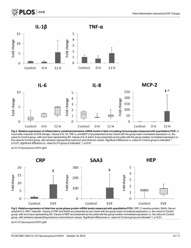
Fig 4. Relative expression of inflammatory cytokine/chemokine mRNA levels in fetal circulating immunocytes measured with quantitative PCR.H:hours after induction of EVE therapy. Values of IL-1β, TNF-α, and MCP-2 are presented as bar charts with the group mean normalised expression vs. thevalue for Control group, with error bars representing SD. Values of IL-6 and IL-8 are presented as box plots with the group median normalised expression vs.the value for Control group, with whiskers representing maximum and minimum values. Significant difference vs. value for Control group is indicated: ‡,p<0.01; significant difference vs. value for 0 H group is indicated: ^, p<0.01.
doi:10.1371/journal.pone.0140701.g004
Fig 5. Relative expression of fetal liver acute phase protein mRNA levels measured with quantitative PCR.CRP, C-reactive protein; SAA3, Serumamyloid A 3; HEP, Hepcidin. Values of CRP and SAA3 are presented as bar charts with the group mean normalised expression vs. the value for Controlgroup, with error bars representing SD. Values of HEP are presented as box plots with the group median normalised expression vs. the value for Controlgroup, with whiskers representing maximum and minimum values. Significant differences vs. value for Control group are indicated: ‡, p<0.01.
doi:10.1371/journal.pone.0140701.g005
Fetal Inflammation Induced by EVE Therapy
PLOS ONE | DOI:10.1371/journal.pone.0140701 October 16, 2015 10 / 17
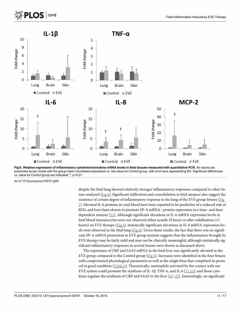
despite the fetal lung showed relatively stronger inflammatory responses compared to other tis-sues analysed (Fig 6). Significant infiltration and consolidation in fetal airspace also suggest theexistence of certain degree of inflammatory response in the lung of the EVE group fetuses (Fig7). Elevated IL-6 proteins in cord blood have been reported to be predictive of a reduced risk ofRDS, and have been shown to promote SP-A mRNA / protein expression in a time- and dose-dependent manner [21]. Although significant elevations in IL-6 mRNA expression levels infetal blood immunocytes were not observed either acutely (0 hour) or after stabilisation (12hours) on EVE therapy (Fig 4), statistically significant elevations in IL-6 mRNA expression lev-els were observed in the fetal lung (Fig 6). Given these results, the fact that there was no signifi-cant SP-A mRNA promotion in EVE group animals suggests that the inflammation brought byEVE therapy may be fairly mild and may not be clinically meaningful, although statistically sig-nificant inflammatory responses in several tissues were shown as discussed above.
The expression of CRP and SAA3 mRNA in the fetal liver was significantly elevated in theEVE group compared to the Control group (Fig 5). Increases were identified in the four fetuseswith compromised physiological parameters as well as the single fetus that completed its proto-col in good condition (Table 6). Theoretically, neutrophils activated by the contact with ourEVE system could promote the synthesis of IL-1β, TNF-α, and IL-6 [11,22]; and those cyto-kines regulate the synthesis of CRP and SAA3 in the liver [23–25]. Interestingly, no significant
Fig 6. Relative expression of inflammatory cytokine/chemokine mRNA levels in fetal tissuesmeasured with quantitative PCR. All values arepresented as bar charts with the group mean normalised expression vs. the value for Control group, with error bars representing SD. Significant differencesvs. value for Control group are indicated: ‡, p<0.01.
doi:10.1371/journal.pone.0140701.g006
Fetal Inflammation Induced by EVE Therapy
PLOS ONE | DOI:10.1371/journal.pone.0140701 October 16, 2015 11 / 17
elevations in IL-1β, TNF-α, or IL-6 mRNA levels were detected in RNA isolated from fetalblood immunocytes (Fig 4). Similar inconsistencies were also observed in RNA isolated fromfetal lung and brain (Fig 6). These data suggest that the inflammatory mediators responsiblefor driving systemic responses are generated in the fetal tissues, or by tissue-resident immuno-cytes rather than by circulating immunocyte populations.
Limitations of the studyThe major limitation of this study is the low number of animals included, that impedes to drawrobust conclusions, as this study required a significant investment of manpower and materiel.
Another limitation is the lack of the microbiological analysis. It might be possible that theinflammatory responses described above were induced by amniotic fluid infection, although
Fig 7. Qualitative scoring of histological inflammation in fetal airspace. Data are presented as box plots with the group median, with whiskersrepresenting maximum and minimum values. Significant difference vs. value for Control group is indicated: ‡, p<0.01.
doi:10.1371/journal.pone.0140701.g007
Table 2. Comparison of MCP-1 protein concentration in fetal plasmameasured with ELISA betweenthe case E and the others.
0 H 4 H 8 H 12 H
Cases A-D 0.0 [9.7] 27.4 [17.4] 13.1 [22.9] 22.1 [22.1]
Case E 0.0 13.1 25.1 28.1
All the units for the presented values are pg/mL. All presented values for cases A-D are the group median
[IQR].
doi:10.1371/journal.pone.0140701.t002
Fetal Inflammation Induced by EVE Therapy
PLOS ONE | DOI:10.1371/journal.pone.0140701 October 16, 2015 12 / 17
fetuses were treated with antibiotics and the amniotic fluid was constantly UV filtered. Futurestudies will seek to address this uncertainty by culturing artificial amniotic fluid and fetal arte-rial blood over time and fetal tissues (lung and skin tissue) at autopsy.
It would also be of interest, in future studies, to determine whether or not fetal sex has aneffect on EVE therapy outcomes.
Strengths of the studyAlthough it has been reported that extracorporeal life support brings about inflammatoryresponses, the available data is still limited. Besides, inflammation intensity is altered by the
Table 3. Comparison of TNF-α protein concentration in fetal plasmameasured with ELISA betweenthe case E and the others.
0 H 4 H 8 H 12 H
Cases A-D 82.8 [43.0] 65.2 [38.9] 38.2 [39.9] 9.3 [36.8]
Case E 73.8 174.8 132.5 182.6
All the units for the presented values are pg/mL. All presented values for cases A-D are the group median
[IQR].
doi:10.1371/journal.pone.0140701.t003
Table 4. Comparison of relative expression of inflammatory cytokine/chemokine mRNA levels in fetalcirculating immunocytes immediately after induction of EVE therapymeasured with quantitative PCRbetween the case E and the others.
IL-1β TNF-α IL-6 IL-8 MCP-2
Cases A-D 0.9 [0.4] 0.7 [0.4] 1.6 [0.3] 0.8 [0.0] 0.7 [1.2]
Case E 0.2 0.4 3.0 1.4 0.3
All the units for the presented values are normalised expression vs. the value for Control group. All
presented values for cases A-D are the group median [IQR].
doi:10.1371/journal.pone.0140701.t004
Table 5. Comparison of relative expression of inflammatory cytokine/chemokine mRNA levels in fetalcirculating immunocytes 12 hours after induction of EVE therapymeasured with quantitative PCRbetween the case E and the others.
IL-1β TNF-α IL-6 IL-8 MCP-2
Cases A-D 1.6 [2.5] 1.2 [0.8] 3.5 [2.8] 1.0 [0.3] 37.7 [100.5]
Case E 14.2 2.8 5.2 2.1 35.1
All the units for the presented values are normalised expression vs. the value for Control group. All
presented values for cases A-D are the group median [IQR].
doi:10.1371/journal.pone.0140701.t005
Table 6. Comparison of relative expression of fetal liver acute phase protein mRNA levels measuredwith quantitative PCR between the case E and the others.
CRP SAA3 HEP
Cases A-D 10.1 [6.4] 50.6 [103.2] 1.6 [0.4]
Case E 2.3 84.6 0.5
All the units for the presented values are normalised expression vs. the value for Control group. All
presented values for cases A-D are the group median [IQR].
doi:10.1371/journal.pone.0140701.t006
Fetal Inflammation Induced by EVE Therapy
PLOS ONE | DOI:10.1371/journal.pone.0140701 October 16, 2015 13 / 17
way of blood access [14]. This is, to our knowledge, the first study to analyse the inflammatoryresponse of the fetuses that were attached with A-V extracorporeal life support system usingumbilical vessels. This study provides additional knowledge for further development of extra-corporeal life support system including EVE therapy.
ConclusionIn conclusion, limited survival of the fetus was achieved with EVE therapy. However, EVEtherapy induced limited fetal inflammation and did not promote lung maturation. In the
Table 7. Comparison of relative expression of fetal pulmonary inflammatory cytokine/chemokinemRNA levels measured with quantitative PCR between the case E and the others.
IL-1β TNF-α IL-6 IL-8 MCP-2
Cases A-D 1.8 [1.1] 1.1 [0.7] 4.9 [3.4] 3.6 [2.5] 7.7 [9.1]
Case E 1.6 1.8 2.0 2.6 48.8
All the units for the presented values are normalised expression vs. the value for Control group. All
presented values for cases A-D are the group median [IQR].
doi:10.1371/journal.pone.0140701.t007
Table 8. Comparison of relative expression of fetal brain inflammatory cytokine/chemokinemRNAlevels measured with quantitative PCR between the case E and the others.
IL-1β TNF-α IL-6 IL-8 MCP-2
Cases A-D 0.5 [0.5] 0.9 [0.3] 1.9 [3.5] 2.8 [1.8] 0.4 [1.6]
Case E 0.4 0.2 2.3 7.8 15.2
All the units for the presented values are normalised expression vs. the value for Control group. All
presented values for cases A-D are the group median [IQR].
doi:10.1371/journal.pone.0140701.t008
Table 9. Comparison of relative expression of fetal skin inflammatory cytokine/chemokinemRNA lev-els measured with quantitative PCR between the case E and the others.
IL-1β TNF-α IL-6 IL-8 MCP-2
Cases A-D 1.6 [1.2] 0.7 [0.2] 1.5 [6.6] 3.1 [5.1] 3.1 [5.3]
Case E 8.1 1.8 1.0 10.8 3.6
All the units for the presented values are normalised expression vs. the value for Control group. All
presented values for cases A-D are the group median [IQR].
doi:10.1371/journal.pone.0140701.t009
Table 10. Comparison of qualitative scoring of histological inflammation in fetal airspace.
Inflammatory score
Cases A-D 3.0 [0.0]
Case E 3.0 [0.8]
All presented values are the group median [IQR].
doi:10.1371/journal.pone.0140701.t010
Fetal Inflammation Induced by EVE Therapy
PLOS ONE | DOI:10.1371/journal.pone.0140701 October 16, 2015 14 / 17
present study, fetal inflammatory status was not a meaningful predictor of fetal wellbeing. Incontrast, sustained increases in fetal lactate levels may be predictive of impending fetal demise.These data provide additional insight into markers of treatment efficacy for the assessment offuture studies.
AcknowledgmentsThe authors wish to acknowledge the generosity of Nipro Corporation, Osaka, Japan (donationof membranous oxygenators), Siemens Australia (donation of Rapidlab 1200 reagents) andCovidien Australia (donation of suture materials). The authors also wish to acknowledge Dr.Alan H. Jobe (Division of Pulmonary Biology, Cincinnati Children’s Hospital Medical Center,Cincinnati, Ohio, United States) for his critical advice with this manuscript.
Author ContributionsConceived and designed the experiments: YM TMMWK. Performed the experiments: YMMSHU JRB GCMMWK. Analyzed the data: YM EW PSK MWK. Contributed reagents/materials/analysis tools: TM JPNMWK. Wrote the paper: YMMS HU EW JRB PSK GCM TM JPNMWK.
References1. Organization WH. Born too soon: the global action report on preterm birth. 2012.
Fig 8. Relative expression of surfactant protein (SP) mRNA levels measured with quantitative PCR. All values are presented as bar charts with thegroup mean normalised expression vs. the value for Control group, with error bars representing SD. Significant difference was observed in no SP mRNAexpression level between the two groups.
doi:10.1371/journal.pone.0140701.g008
Fetal Inflammation Induced by EVE Therapy
PLOS ONE | DOI:10.1371/journal.pone.0140701 October 16, 2015 15 / 17
2. Marret S, Marchand-Martin L, Picaud JC, Hascoet JM, Arnaud C, Roze JC, et al. Brain injury in verypreterm children and neurosensory and cognitive disabilities during childhood: the EPIPAGE cohortstudy. PLoS One. 2013; 8: e62683. doi: 10.1371/journal.pone.0062683 PMID: 23658763
3. Stoinska B, Gadzinowski J. Neurological and developmental disabilities in ELBW and VLBW: follow-upat 2 years of age. J Perinatol. 2011; 31: 137–142. doi: 10.1038/jp.2010.75 PMID: 20634795
4. Miura Y, Matsuda T, Funakubo A, Watanabe S, Kitanishi R, Saito M, et al. Novel modification of an arti-ficial placenta: pumpless arteriovenous extracorporeal life support in a premature lamb model. PediatrRes. 2012; 72: 490–494. doi: 10.1038/pr.2012.108 PMID: 22885413
5. Gomez R, Romero R, Ghezzi F, Yoon BH, Mazor M, Berry SM. The fetal inflammatory response syn-drome. Am J Obstet Gynecol. 1998; 179: 194–202. PMID: 9704787
6. Jobe AH, Newnham JP, Willet KE, Moss TJ, Gore Ervin M, Padbury JF, et al. Endotoxin-induced lungmaturation in preterm lambs is not mediated by cortisol. Am J Respir Crit Care Med. 2000; 162: 1656–1661. PMID: 11069792
7. Bachurski CJ, Ross GF, Ikegami M, Kramer BW, Jobe AH. Intra-amniotic endotoxin increases pulmo-nary surfactant proteins and induces SP-B processing in fetal sheep. Am J Physiol Lung Cell Mol Phy-siol. 2001; 280: L279–285. PMID: 11159007
8. Fortenberry JD, Bhardwaj V, Niemer P, Cornish JD, Wright JA, Bland L. Neutrophil and cytokine activa-tion with neonatal extracorporeal membrane oxygenation. J Pediatr. 1996; 128: 670–678. PMID:8627440
9. Adrian K, Mellgren K, Skogby M, Friberg LG, Mellgren G, Wadenvik H. Cytokine release during long-term extracorporeal circulation in an experimental model. Artif Organs. 1998; 22: 859–863. PMID:9790084
10. Halter J, Steinberg J, Fink G, Lutz C, Picone A, Maybury R, et al. Evidence of systemic cytokine releasein patients undergoing cardiopulmonary bypass. J Extra Corpor Technol. 2005; 37: 272–277. PMID:16350379
11. McILwain RB, Timpa JG, Kurundkar AR, Holt DW, Kelly DR, Hartman YE, et al. Plasma concentrationsof inflammatory cytokines rise rapidly during ECMO-related SIRS due to the release of preformedstores in the intestine. Lab Invest. 2010; 90: 128–139. doi: 10.1038/labinvest.2009.119 PMID:19901912
12. Fujii Y, Shirai M, Inamori S, Takewa Y, Tatsumi E. Investigation of the biological effects of artificial per-fusion using rat extracorporeal circulation model. Conf Proc IEEE Eng Med Biol Soc. 2014; 2014:4483–4486. doi: 10.1109/EMBC.2014.6944619 PMID: 25570987
13. Fujii Y, Shirai M, Inamori S, Takewa Y, Tatsumi E. A novel small animal extracorporeal circulationmodel for studying pathophysiology of cardiopulmonary bypass. J Artif Organs. 2015; 18: 35–39. doi:10.1007/s10047-014-0804-y PMID: 25373368
14. Golej J, Winter P, Schoffmann G, Kahlbacher H, Stoll E, Boigner H, et al. Impact of extracorporealmembrane oxygenation modality on cytokine release during rescue from infant hypoxia. Shock. 2003;20: 110–115. PMID: 12865653
15. Gourlay T, Stefanou DC, Asimakopoulos G, Taylor KM. The effect of circuit surface area on CD11b(mac-1) expression in a rat recirculation model. Artif Organs. 2001; 25: 475–479. PMID: 11453878
16. Zimmermann AK, Weber N, Aebert H, Ziemer G, Wendel HP. Effect of biopassive and bioactive sur-face-coatings on the hemocompatibility of membrane oxygenators. J Biomed Mater Res B Appl Bioma-ter. 2007; 80: 433–439. PMID: 16850460
17. KempMW, Musk GC, Saito M. Animal Models for the Study of Infection-Associated Preterm Birth. In:Conn PM, editor. Animal Models for the Study of Human Disease. Amsterdam, Netherlands: Elsevier;2013. pp. 863–888.
18. Rasmusen BA. Blood groups in sheep. Ann N Y Acad Sci. 1962; 97: 306–319. PMID: 14490465
19. Bustin SA, Benes V, Garson JA, Hellemans J, Huggett J, Kubista M, et al. The MIQE guidelines: mini-mum information for publication of quantitative real-time PCR experiments. Clin Chem. 2009; 55: 611–622. doi: 10.1373/clinchem.2008.112797 PMID: 19246619
20. Mann LI. Effects in sheep of hypoxia on levels of lactate, pyruvate, and glucose in blood of mothers andfetus. Pediatr Res. 1970; 4: 46–54. PMID: 5416998
21. Shimoya K, Taniguchi T, Matsuzaki N, Moriyama A, Murata Y, Kitajima H, et al. Chorioamnionitisdecreased incidence of respiratory distress syndrome by elevating fetal interleukin-6 serum concentra-tion. Hum Reprod. 2000; 15: 2234–2240. PMID: 11006206
22. Heinrich PC, Castell JV, Andus T. Interleukin-6 and the acute phase response. Biochem J. 1990; 265:621–636. PMID: 1689567
Fetal Inflammation Induced by EVE Therapy
PLOS ONE | DOI:10.1371/journal.pone.0140701 October 16, 2015 16 / 17
23. Castell JV, Gomez-Lechon MJ, David M, Hirano T, Kishimoto T, Heinrich PC. Recombinant humaninterleukin-6 (IL-6/BSF-2/HSF) regulates the synthesis of acute phase proteins in human hepatocytes.FEBS Lett. 1988; 232: 347–350. PMID: 2454206
24. Castell JV, Andus T, Kunz D, Heinrich PC. Interleukin-6. The major regulator of acute-phase proteinsynthesis in man and rat. Ann N Y Acad Sci. 1989; 557: 87–99; discussion 100–101. PMID: 2472097
25. Castell JV, Gomez-Lechon MJ, David M, Andus T, Geiger T, Trullenque R, et al. Interleukin-6 is themajor regulator of acute phase protein synthesis in adult human hepatocytes. FEBS Lett. 1989; 242:237–239. PMID: 2464504
Fetal Inflammation Induced by EVE Therapy
PLOS ONE | DOI:10.1371/journal.pone.0140701 October 16, 2015 17 / 17