Embed Size (px)
Citation preview
Clin Genet 2010: 77: 119–130Printed in Singapore. All rights reserved
© 2009 John Wiley & Sons A/SCLINICAL GENETICS
doi: 10.1111/j.1399-0004.2009.01325.x
Original Article
Respiratory disease in Niemann-Pick type C2is caused by pulmonary alveolar proteinosis
Griese M, Brasch F, Aldana VR, Cabrera MM, Goelnitz U, Ikonen E,Karam BJ, Liebisch G, Linder MD, Lohse P, Meyer W, Schmitz G,Pamir A, Ripper J, Rolfs A, Schams A, Lezana FJ. Respiratory diseasein Niemann-Pick type C2 is caused by pulmonary alveolar proteinosis.Clin Genet 2010: 77: 119–130. © John Wiley & Sons A/S, 2009
Niemann-Pick diseases are hereditary neurovisceral lysosomal lipid storagedisorders, of which the rare type C2 almost uniformly presents withrespiratory distress in early infancy. In the patient presented here, theNPC2 exon 4 frameshift mutation c.408 409delAA caused reduced NPC2protein levels in serum and lung lavage fluid and the synthesis of anaberrant, larger sized protein of around 28 kDa. Protein expression wasstrongly reduced also in alveolar macrophages. The infant developedfailure to thrive and tachypnea. Lung lavage, computer tomography, andhistology showed typical signs of pulmonary alveolar proteinosis with anabnormal intraalveolar accumulation of surfactant as well as macrophages.An NPC2-hypomorph animal model also showed pulmonary alveolarproteinosis and accumulation of macrophages in the lung, liver, and spleenlong before the mice died. Due to the elevation of cholesterol, thesurfactant had an abnormal composition and function. Despite the removalof large amounts of surfactant from the lungs by therapeutic lung lavages,this treatment was only temporarily successful and the infant died ofrespiratory failure. Our data indicate that respiratory distress in NPC2disease is associated with a loss of normal NPC2 protein expression inalveolar macrophages and the accumulation of functionally inactivesurfactant rich in cholesterol.
M Griesea, F Braschb,VR Aldanac, MM Cabrerad,U Goelnitze, E Ikonenf,BJ Karamc, G Liebischg,MD Linderf, P Lohseh,W Meyeri, G Schmitzg,A Pamira, J Rippera, A Rolfse,j,A Schamsa and FJ Lezanac
aDr von Haunersches Kinderspital,University of Munich, Lindwurmstr,Munich, Germany, bDepartment ofPathology, Klinikum Bremen Mitte,Bremen, Germany, cDepartment ofPneumology and Respiratory Physiology,dDepartment of Pathology, HospitalInfantil de Mexico Federico Gomez,Mexico City, Mexico, eUniversity ofRostock, Albrecht-Kossel Institute forNeuroregeneration, Medical Faculty,Rostock, Germany, fInstitute ofBiomedicine/Anatomy, Haartmaninkatu8, 00014 University of Helsinki, Finland,gKlinikum der Universitat RegensburgInstitut fur Klinische Chemie,Franz-Josef-Strauß-Allee 11, 93053Regensburg, Germany, hDepartment ofClinical Chemistry, Grosshadern,University of Munich, Munich, Germany,iQueen Mary University of London, St.Bartholomew’s and the Royal LondonSchool of Medicine, London, UK, andjCentogene GmbH, Schillingallee 68,18057 Rostock, Germany
Key words: infant – Niemann-Pick typeC2 – pulmonary alveolar proteinosis –tachypnea – therapeutic lung lavage
Corresponding author: Dr MatthiasGriese, Dr von HaunerschesKinderspital, University of Munich,Lindwurmstraße 4, D-80337Munich, Germany.Tel.: +49 89 5160 7871;fax: +49 89 5160 7872;e-mail:[email protected]
Received 21 June 2009, revised andaccepted for publication 30 September2009
119
Griese et al.
Niemann-Pick diseases represent a family ofneurovisceral lysosomal lipid storage disorders,many of which lead to severe neurodegenera-tion and early death. Types A (OMIM #257200)and B (OMIM #607616) result from an inheriteddeficiency of lysosomal acid sphingomyelinaseactivity. Niemann-Pick type C1 disease (OMIM#257220) is caused by mutations in the NPC1gene (1), which codes for the NPC1 protein. Thisis an integral membrane protein that is primar-ily localised to late endosomes/lysosomes andalso cycles through the Golgi apparatus (2, 3).Niemann-Pick type C2 disease (OMIM #607625)is caused by mutations in the NPC2 gene (4). Itencodes the NPC2 protein, in its mature form a132 amino-acid, soluble, ubiquitously expressed,mannose 6-phosphorylated lysosomal glycopro-tein with highest levels in testes, kidney, andliver. NPC2 protein binds unesterified choles-terol with high affinity (5, 6) and transportsit to phospholipid vesicles (7, 8). Patients withNPC2 mutations represent the smallest group ofNiemann-Pick patients. Up to now, 27 differentfamilies with a defined mutational status havebeen described (9–12). In contrast to the othertypes of Niemann-Pick disorders, many of thesepatients present with respiratory distress in theirearly infancy (9). There are only 18 infantile caseswith genetically proven NPC2 disease. Informa-tion on respiratory symptoms within the first sixmonths of life was only available in 14 patients,all of which had developed significant symptoms.The median age of death from respiratory insuffi-ciency was 8 months (range 2.5–54). The patho-physiological mechanisms of respiratory distress inNiemann-Pick type C2 disease are still unclear.
In a recent report of two infants with Niemann-Pick disease type C2, recurrent bronchitis andbronchiolitis with respiratory failure were suspec-ted (13). Histopathology of autopsy lungs docu-mented increasing pulmonary fibrosis, progressivelung interstitial involvement, and lipoid pneumo-nia (9, 14–16), whereas in biopsy specimens a his-tological pattern of pulmonary alveolar proteinosispredominated (17, 18).
Alveolar proteinosis is a rare pulmonary diseasecharacterised by an intraalveolar accumulation ofsurfactant. Pulmonary surfactant is a complex mix-ture of proteins and lipids that normally coversthe alveolar surface to reduce surface tension (19).Until now, four surfactant-associated proteins(SP-A, SP-B, SP-C, and SP-D) have been char-acterised. Dipalmitoyl phosphatidylcholine, phos-phatidylglycerol, and cholesterol are the principallipid components. The most frequent form of alve-olar proteinosis is caused by autoantibodies against
granulocyte-macrophage colony-stimulating factor(GM-CSF), which is a haematologic growth fac-tor regulating the uptake of surfactant by alveolarmacrophages (20). In cases of primary alveolarproteinosis in infants, mutations of the surfactantproteins B or C or of the alpha chain of theGM-CSF receptor were found (21–23). Secondarypulmonary alveolar proteinosis develops in associ-ation with inhalation of inorganic dusts like silicaor titanium, some forms of leukaemia, and certaininfections (20). The role of alveolar proteinosisin the pathophysiology of respiratory distress inNiemann-Pick disease type C2, however, needs tobe clarified.
Case, materials and methods
Case report
The parents of the patient were unrelated, thepregnancy was normal, and the female child wasborn at term. She had a 5 year old sister in goodhealth. The mother noticed progressive tachypnea,failure to thrive, and poor feeding since the infant’ssecond month of life. Within four months, shewas admitted to the Hospital Infantil de Mexico,Mexico City, Mexico with dyspnea, peripheralcyanosis, fever, and non-productive cough. Shewas diagnosed with bronchiolitis and cardiacfailure. The initial chest X-ray (Fig. 1c) showedbilateral micronodular infiltrates particularly in thelower left lung and apical right lobe. The lungswere overinflated, and basal fine crackles werenoticed. Liver and spleen were enlarged to 4 and5 cm, respectively. Clubbing of the fingers waspresent. Neurology and neurological developmentwere normal.
Pneumonia was then diagnosed and she wastreated with Cefuroxime and Claritromycin. Micro-biologic investigations, a sweat test, immunoglob-ulins as well as blood and urine metabolicscreening were normal. Serum triglycerides weremoderately elevated [380 mg/dl (normal <150)],while the cholesterol concentration was normal[169 mg/dl (normal <170)] with a slight elevationin LDL cholesterol [115 mg/dl (normal <110)]and a slight decrease in HDL cholesterol [31 mg/dl(normal >35)]. The abdominal CT scan showedhepatosplenomegaly. Chest CT scan and broncho-alveolar lavage (BAL) pointed towards a diagnosisof alveolar proteinosis. Open liver, spleen, andlung biopsies were performed, and the diagnosisof Niemann-Pick type C disease and pulmonaryalveolar proteinosis was made.
In an attempt to treat the deteriorating respira-tory function with the prominent feature of alveolar
120
Alveolar proteinosis and Niemann-Pick C2
a
.
b
c
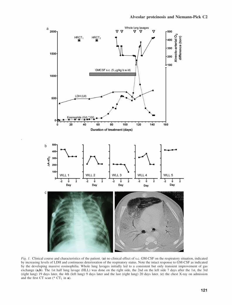
Fig. 1. Clinical course and characteristics of the patient. (a) no clinical effect of s.c. GM-CSF on the respiratory situation, indicatedby increasing levels of LDH and continuous deterioration of the respiratory status. Note the intact response to GM-CSF as indicatedby the developing massive eosinophilia. Whole lung lavages initially led to a consistent but only transient improvement of gasexchange (a,b). The 1st half lung lavage (HLL) was done on the right side, the 2nd on the left side 7 days after the 1st, the 3rd(right lung) 19 days later, the 4th (left lung) 9 days later and the last (right lung) 20 days later. (c) the chest X-ray on admissionand the first CT scan (* CT1 in a).
121
Griese et al.
proteinosis, subcutaneous treatment with GM-CSFwas initiated, but without clinical effect. Standardtreatment with half lung lavages was then intro-duced to treat the pulmonary alveolar proteinosis.This was initially successful (Fig. 1), but after thefifth lavage the patient developed a pneumothorax.She deteriorated rapidly and died the followingday. The parents did not consent to perform a post-mortem examination.
Half lung lavages
Under general anesthesia and endotracheal intuba-tion, a 3.5 mm diameter fiberoptic bronchoscopewas inserted via a tracheostomy. With the airwaysafe, the fiberoptic bronchoscope was wedged inthe lobar bronchi of one side of the lungs foreach half lung lavage. Each lobe was washed with15-ml aliquots of pre-warmed isotonic saline solu-tion until a clear aspirate was obtained. During theprocedure, positional and assisted clearance usingmanual chest physiotherapy was performed.
Samples from control or comparison subjects
Serum and lavage fluid was used from both anadult patient with autoimmune alveolar proteinosiscaused by antibodies against GM-CSF and thepatient with congenital SP-C deficiency due to theSFTPC I73T mutation (21). In addition, normalcontrol sera from lab personnel were obtained. Toassess surface tension, natural bovine surfactantwas used as control in in vitro experiments (24).
Preparation of lavage and surfactant fractions
To sediment the cells, lavage fluid was centrifugedat 200 g for 10 min. The supernatant was eitherused directly for Western blotting and biochemicalanalyses or further centrifuged at 40,000 × g for30 min in order to separate the surfactant intothe large surfactant aggregate fraction (pellet) andthe small surfactant aggregates (supernatant) toperform surface tension measurements using thepulsating bubble surfactometer (24).
Mutation analysis and biochemical assays
Genomic DNA was isolated from whole bloodand the genes of Niemann-Pick diseases (SMPD1,NPC1, NPC2 ) and of the pulmonary surfactant-associated proteins B and C (SFTPB and SPTPC )were sequenced following standard protocols (25).
Acid sphingomyelinase activity in peripheralleukocytes of the patient was normal (3.9 nmol/mgper hour; normal controls 3.3–6.4). The level of
GM-CSF autoantibodies in serum was determinedas described previously (26). For Western blotting,proteins were separated under reducing conditions[NuPage10% Bis-Tris gels (Novex, San Diego,CA)] and incubated with antibodies against pro-SP-B, SP-C, SP-B (27), or NPC2 (28). ELISAs forconcentrations of surfactant proteins (SP) A and Dwere performed as described, allowing for minormodifications (24). Lavage or homogenized cellswere extracted, and lipid classes and subspecieswere determined by electrospray ionization tandemmass spectrometry (ESI-MS/MS) analysis (29).
Analysis of surfactant activity in the pulsating bubblesurfactometer
Surface tension of surfactant isolated from lavageswas determined at phospholipid concentrations of1 and 3 mg/ml (24). Inhibition of surface activ-ity by cholesterol (Sigma, Taufkirchen, Germany)or ceramide [(N -Nervonoyl-d-erythro-Sphingosine(C24:1 Ceramide), Avanti, Polar lipids, Alabaster,AL] was tested after admixture of the lipids tolyophilized large aggregate surfactant and reconsti-tution in bubble buffer [140 mM NaCl, 10 mM N -2-hydroxyethylpiperazine-N -ethane sulfonic acid(HEPES), 0.5 mM ethylenediamimetetraacetic acid(EDTA), 3.5 mM CaCl2, pH 6.9].
Histology and immunohistochemistry of biopsymaterial and of NPC2-deficient mice
Lung biopsy specimens were fixed in 4% bufferedformaldehyde and immunostained using alkalinephosphatase (30). Tissues from female NPC2-deficient mice of mixed genetic background(129/C57Bl6/BALBC, Nr 9645) and littermatecontrols (Nr 9644) were provided by the Lobellaboratory (31). Day 41 animals were analysed. Atthis point of time, mortality was zero, the naturallife cycle of such NPC2 deficient mice being pre-mature death. The first mice died aged 56 days.The median survival for this genotype is 110 days.The control animals were healthy and developedno symptoms. The tissue of two animals from eachgroup was assessed by a pathologist blinded to thegroup assignment.
Ethics
Written informed consent was obtained from allsubjects involved. The procedures during treatmentand the usage of case data and materials wereapproved by the ethics review boards of theparticipating institutions.
122
Alveolar proteinosis and Niemann-Pick C2
Results
Novel homozygous Niemann-Pick C2 mutationc.408 409delAA in an infant with chronic respiratorydistress
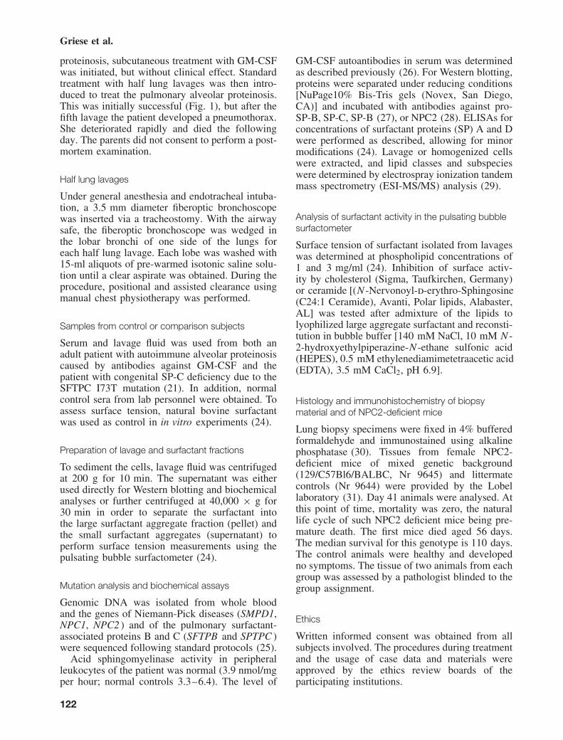
By light microscopy, large macrophages with afoamy cytoplasm (arrows) were found within theliver (Fig. 2a) and spleen (Fig. 2d). Since foamymacrophages are easily overlooked in the liver due
to increased vaculation and decreased eosinophiliaof hepatocytes (arrowheads), Niemann-Pick typeC was suggested. Proof of lipid inclusions consist-ing of concentrically laminated myelin-like figureswithin foamy macrophages at the ultrastruc-tural level was confirmatory (Fig. 2a,d, insets).Sequencing of the NPC2 gene demonstrated thehomozygous deletion of two adenosines at aminoacid position 136/137 in exon 4 (c.408 409delAA)
Fig. 2. Comparison of human (left panels a, d, g, j) and mouse [middle (b, e, h, k) and right panels (c, f, i, l)] histology of liver,spleen, and lung (panels a to i all HE stain, bar 100 μm) from an open lung biopsy of the infant with Niemann-Pick type C2disease, from the NPC2-hypomorph animal model of Niemann–Pick type C2 disease (on day 41), and from the correspondingcontrol mice. The inserted electron micrographs demonstrate the typical histology of Niemann-Pick C disease (panels a, d withinsets each). The lower three panels (j, k, l) show lung tissue stained with the typical findings of pulmonary alveolar proteinosis andlung tissue of a healthy control mouse. The inset in panel k (bar 50 μm) illustrates oval bodies’ characteristic of pulmonary alveolarproteinosis.
123
Griese et al.
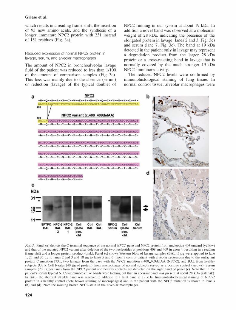
which results in a reading frame shift, the insertionof 93 new amino acids, and the synthesis of alonger, immature NPC2 protein with 231 insteadof 151 residues (Fig. 3a).
Reduced expression of normal NPC2 protein inlavage, serum, and alveolar macrophages
The amount of NPC2 in bronchoalveolar lavagefluid of the patient was reduced to less than 1/100of the amount of comparison samples (Fig. 3c).This loss was mainly due to the absence (serum)or reduction (lavage) of the typical doublet of
NPC2 running in our system at about 19 kDa. Inaddition a novel band was observed at a molecularweight of 28 kDa, indicating the presence of theelongated protein in lavage (lanes 2 and 3, Fig. 3c)and serum (lane 7, Fig. 3c). The band at 19 kDadetected in the patient only in lavage may representa degradation product from the larger 28 kDaprotein or a cross-reacting band in lavage that isnormally covered by the much stronger 19 kDaNPC2 immunoreactivity.
The reduced NPC2 levels were confirmed byimmunohistological staining of lung tissue. Innormal control tissue, alveolar macrophages were
Fig. 3. Panel (a) depicts the C-terminal sequence of the normal NPC2 gene and NPC2 protein from nucleotide 403 onward (yellow)and that of the mutated NPC2 variant after deletion of the two nucleotides at positions 408 and 409 in exon 4, resulting in a readingframe shift and a longer protein product (pink). Panel (c) shows Western blots of lavage samples (BAL, 5 μg were applied to lane1, 25 and 35 μg to lanes 2 and 3 and 10 μg to lanes 5 and 6) from a control patient with alveolar proteinosis due to the surfactantprotein C mutation I73T, two lavages from the case with the NPC2 mutation c.408 409delAA (NPC-2), and BAL from healthysubjects (Ctrl). Cell lysates (40 μg of protein) from macrophages of normal subjects served as a positive control (arrows). Serumsamples (20 μg per lane) from the NPC2 patient and healthy controls are depicted on the right hand of panel (c). Note that in thepatient’s serum typical NPC2-immunoreactive bands were lacking but that an aberrant band was present at about 28 kDa (asterisk).In BAL, the aberrant 28 kDa band was reactive in addition to a faint band at 19 kDa. Immunohistochemical staining of NPC-2protein in a healthy control (note brown staining of macrophages) and in the patient with the NPC2 mutation is shown in Panels(b) and (d). Note the missing brown NPC2-stain in the alveolar macrophages.
124
Alveolar proteinosis and Niemann-Pick C2
primarily stained (Fig. 3b,d), as confirmed bycolocalization of the staining pattern with theCD68 stain (macrophages), but not with theMNF116 stain, which stains epithelial cells (notshown).
The chronic respiratory distress is due to pulmonaryalveolar proteinosis
The characteristic clinical course with the insid-ious development of dyspnoea exaggerated bya respiratory tract infection with fever, the ini-tial diagnostic BAL with minimal inflammatorycells, no cultural growth except for a few coloniesof Stenotrophomonas maltophilia, opaque andmilky fluid lavage fluid with plenty of oval bod-ies and eosinophilic amorphous periodic acid-Schiff (PAS)-positive material microscopically(not shown), and enlarged foamy and vacuolatedalveolar macrophages, and the CT scan were typ-ical of a pulmonary alveolar proteinosis (Fig. 1c).
Open lung biopsy showed histological patternstypical of pulmonary alveolar proteinosis on onehand (Fig. 2, panels g, j) and typical Niemann-Pickdisease on the other hand (Fig. 2, panels a, d, g).The alveolar spaces were filled with a granular,eosinophilic material (Fig. 2, panel g) that wasPAS stain-positive (Fig. 2, panel j) and diastase-resistant (not shown). Furthermore, a hyperplasiaof type II pneumocytes and intraalveolar as wellas interstitial accumulation of foamy macrophageswere present.
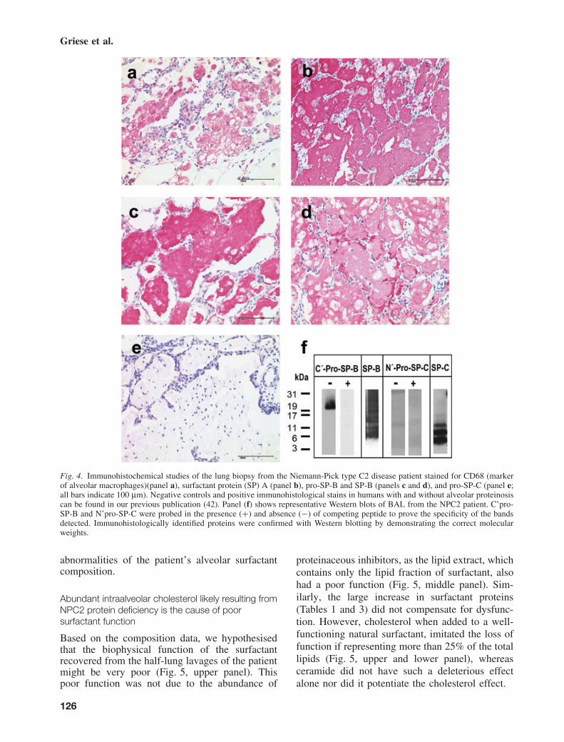
Special stains for the pulmonary surfactantproteins characterised the surfactant metabolism.Intraalveolar accumulation of SP-A, which is
pathognomonic for alveolar proteinosis (Fig. 4,panel b), was confirmed by biochemical analysisof lavage fluid for SP-A, and extended to SP-D(Table 1). Precursors of SP-B and SP-B were alsovery abundant in the alveolar space (Fig. 4, panelsc, d), whereas precursors of SP-C were restricted totype II pneumocytes (Fig. 4, panels e and f). SP-C was abundantly present in the alveolar space,both with normal molecular weight (about 4.7 and10 kDa) and with higher molecular weight formswhich are typical for alveolar proteinosis (Fig. 4,panel f).
Alveolar surfactant is abnormal in lipid speciescomposition and surfactant proteins
The lipid composition of the alveolar surfactantmaterial recovered was significantly different fromthat of a normal alveolar surfactant (Table 1).The percentage of the surface-active lipids phos-phatidylcholine, phosphatidylglycerol, and phos-phatidylinositol was reduced, whereas cholesterol,glucosylceramide, ceramide, and sphingomyelinwere increased several fold (Table 1).
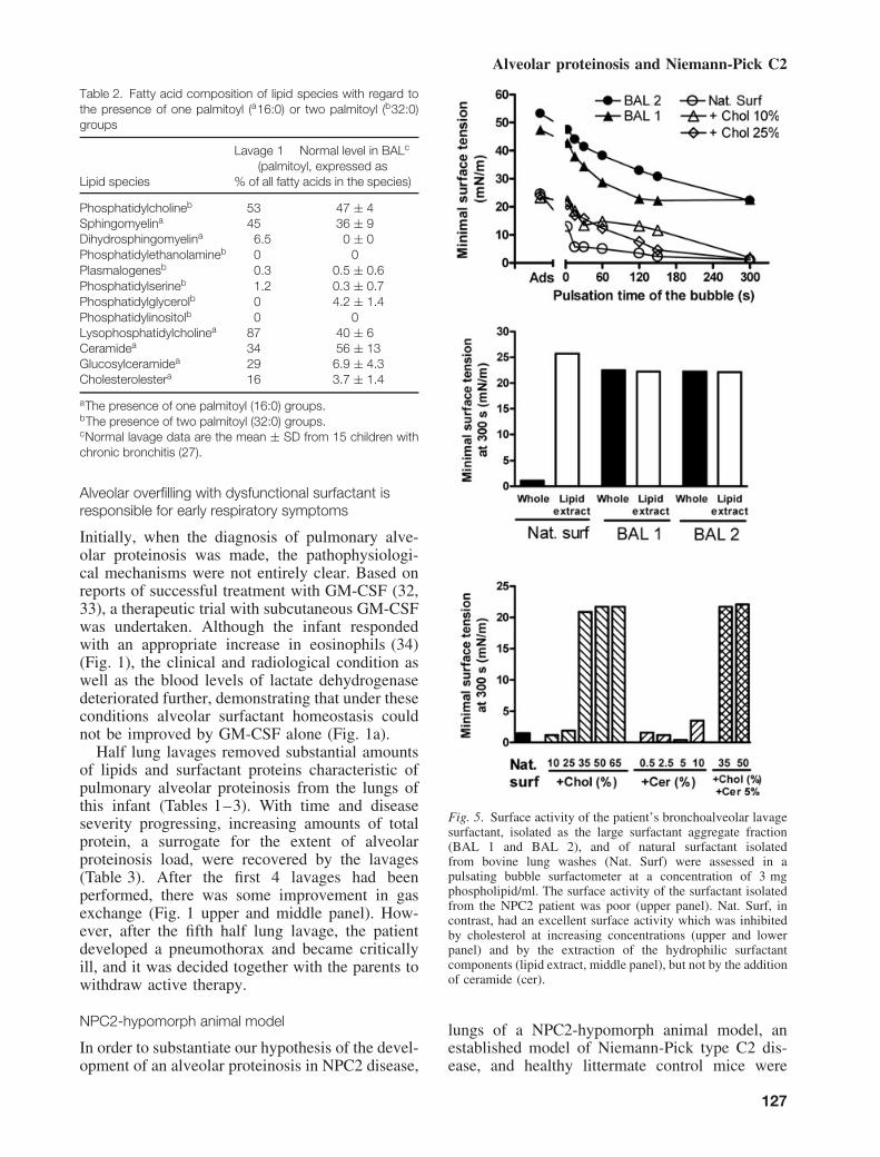
Interestingly, alterations of the fatty acid com-position of the lipids were minor. Representativefor the large number of fatty acids investigated, theresults for the most important and abundant palmi-toyl species are given (Table 2). Dipalmitoylphos-phatidylcholine, the major surfactant phospholipid,was present in the same abundance as in controls(Table 2). In addition to the huge increase in SP-A, the increase of cholesterol to about 50% oftotal surfactant lipid mass and the more than 70-fold increase of ceramide were the most striking
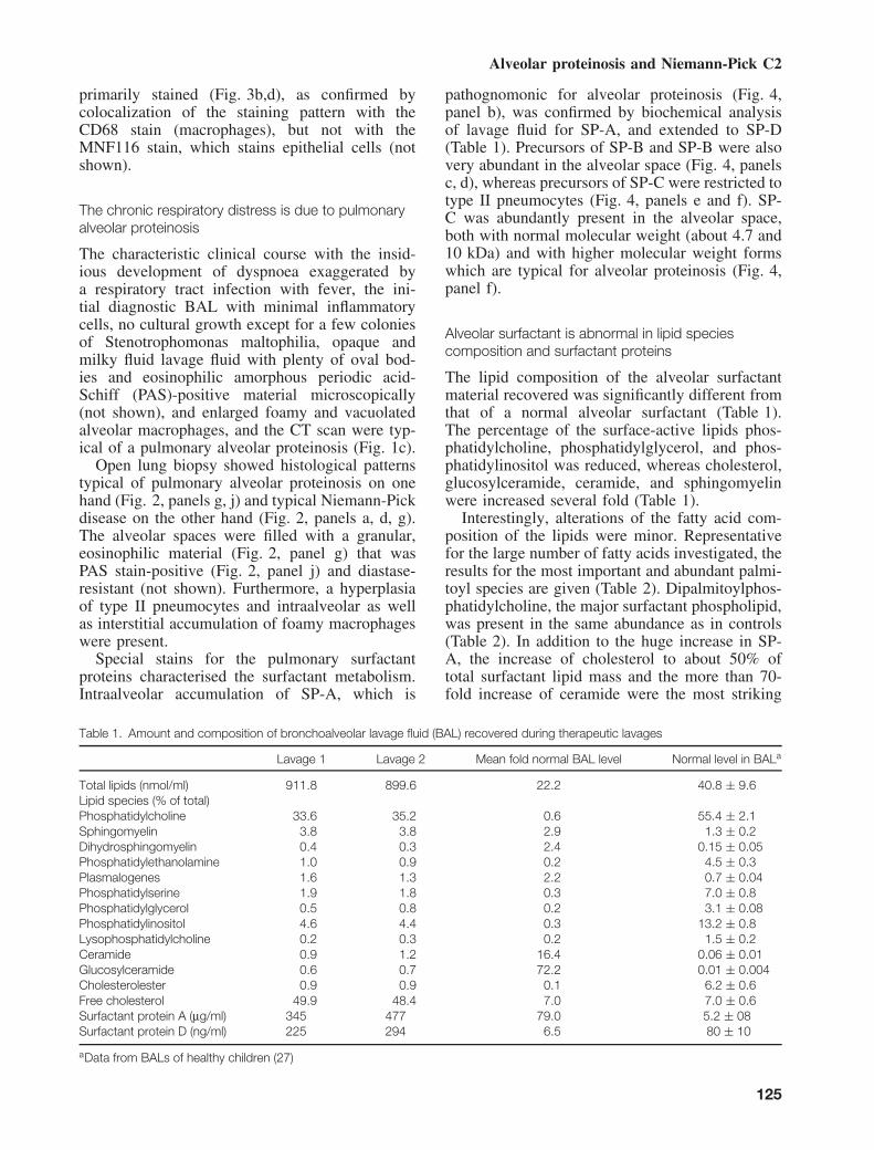
Table 1. Amount and composition of bronchoalveolar lavage fluid (BAL) recovered during therapeutic lavages
Lavage 1 Lavage 2 Mean fold normal BAL level Normal level in BALa
Total lipids (nmol/ml) 911.8 899.6 22.2 40.8 ± 9.6Lipid species (% of total)Phosphatidylcholine 33.6 35.2 0.6 55.4 ± 2.1Sphingomyelin 3.8 3.8 2.9 1.3 ± 0.2Dihydrosphingomyelin 0.4 0.3 2.4 0.15 ± 0.05Phosphatidylethanolamine 1.0 0.9 0.2 4.5 ± 0.3Plasmalogenes 1.6 1.3 2.2 0.7 ± 0.04Phosphatidylserine 1.9 1.8 0.3 7.0 ± 0.8Phosphatidylglycerol 0.5 0.8 0.2 3.1 ± 0.08Phosphatidylinositol 4.6 4.4 0.3 13.2 ± 0.8Lysophosphatidylcholine 0.2 0.3 0.2 1.5 ± 0.2Ceramide 0.9 1.2 16.4 0.06 ± 0.01Glucosylceramide 0.6 0.7 72.2 0.01 ± 0.004Cholesterolester 0.9 0.9 0.1 6.2 ± 0.6Free cholesterol 49.9 48.4 7.0 7.0 ± 0.6Surfactant protein A (μg/ml) 345 477 79.0 5.2 ± 08Surfactant protein D (ng/ml) 225 294 6.5 80 ± 10
aData from BALs of healthy children (27)
125
Griese et al.
Fig. 4. Immunohistochemical studies of the lung biopsy from the Niemann-Pick type C2 disease patient stained for CD68 (markerof alveolar macrophages)(panel a), surfactant protein (SP) A (panel b), pro-SP-B and SP-B (panels c and d), and pro-SP-C (panel e;all bars indicate 100 μm). Negative controls and positive immunohistological stains in humans with and without alveolar proteinosiscan be found in our previous publication (42). Panel (f) shows representative Western blots of BAL from the NPC2 patient. C’pro-SP-B and N’pro-SP-C were probed in the presence (+) and absence (−) of competing peptide to prove the specificity of the bandsdetected. Immunohistologically identified proteins were confirmed with Western blotting by demonstrating the correct molecularweights.
abnormalities of the patient’s alveolar surfactantcomposition.
Abundant intraalveolar cholesterol likely resulting fromNPC2 protein deficiency is the cause of poorsurfactant function
Based on the composition data, we hypothesisedthat the biophysical function of the surfactantrecovered from the half-lung lavages of the patientmight be very poor (Fig. 5, upper panel). Thispoor function was not due to the abundance of
proteinaceous inhibitors, as the lipid extract, whichcontains only the lipid fraction of surfactant, alsohad a poor function (Fig. 5, middle panel). Sim-ilarly, the large increase in surfactant proteins(Tables 1 and 3) did not compensate for dysfunc-tion. However, cholesterol when added to a well-functioning natural surfactant, imitated the loss offunction if representing more than 25% of the totallipids (Fig. 5, upper and lower panel), whereasceramide did not have such a deleterious effectalone nor did it potentiate the cholesterol effect.
126
Alveolar proteinosis and Niemann-Pick C2
Table 2. Fatty acid composition of lipid species with regard tothe presence of one palmitoyl (a16:0) or two palmitoyl (b32:0)groups
Lavage 1 Normal level in BALc
Lipid species(palmitoyl, expressed as
% of all fatty acids in the species)
Phosphatidylcholineb 53 47 ± 4Sphingomyelina 45 36 ± 9Dihydrosphingomyelina 6.5 0 ± 0Phosphatidylethanolamineb 0 0Plasmalogenesb 0.3 0.5 ± 0.6Phosphatidylserineb 1.2 0.3 ± 0.7Phosphatidylglycerolb 0 4.2 ± 1.4Phosphatidylinositolb 0 0Lysophosphatidylcholinea 87 40 ± 6Ceramidea 34 56 ± 13Glucosylceramidea 29 6.9 ± 4.3Cholesterolestera 16 3.7 ± 1.4
aThe presence of one palmitoyl (16:0) groups.bThe presence of two palmitoyl (32:0) groups.cNormal lavage data are the mean ± SD from 15 children withchronic bronchitis (27).
Alveolar overfilling with dysfunctional surfactant isresponsible for early respiratory symptoms
Initially, when the diagnosis of pulmonary alve-olar proteinosis was made, the pathophysiologi-cal mechanisms were not entirely clear. Based onreports of successful treatment with GM-CSF (32,33), a therapeutic trial with subcutaneous GM-CSFwas undertaken. Although the infant respondedwith an appropriate increase in eosinophils (34)(Fig. 1), the clinical and radiological condition aswell as the blood levels of lactate dehydrogenasedeteriorated further, demonstrating that under theseconditions alveolar surfactant homeostasis couldnot be improved by GM-CSF alone (Fig. 1a).
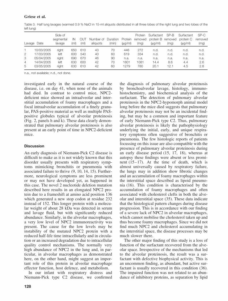
Half lung lavages removed substantial amountsof lipids and surfactant proteins characteristic ofpulmonary alveolar proteinosis from the lungs ofthis infant (Tables 1–3). With time and diseaseseverity progressing, increasing amounts of totalprotein, a surrogate for the extent of alveolarproteinosis load, were recovered by the lavages(Table 3). After the first 4 lavages had beenperformed, there was some improvement in gasexchange (Fig. 1 upper and middle panel). How-ever, after the fifth half lung lavage, the patientdeveloped a pneumothorax and became criticallyill, and it was decided together with the parents towithdraw active therapy.
NPC2-hypomorph animal model
In order to substantiate our hypothesis of the devel-opment of an alveolar proteinosis in NPC2 disease,
Fig. 5. Surface activity of the patient’s bronchoalveolar lavagesurfactant, isolated as the large surfactant aggregate fraction(BAL 1 and BAL 2), and of natural surfactant isolatedfrom bovine lung washes (Nat. Surf) were assessed in apulsating bubble surfactometer at a concentration of 3 mgphospholipid/ml. The surface activity of the surfactant isolatedfrom the NPC2 patient was poor (upper panel). Nat. Surf, incontrast, had an excellent surface activity which was inhibitedby cholesterol at increasing concentrations (upper and lowerpanel) and by the extraction of the hydrophilic surfactantcomponents (lipid extract, middle panel), but not by the additionof ceramide (cer).
lungs of a NPC2-hypomorph animal model, anestablished model of Niemann-Pick type C2 dis-ease, and healthy littermate control mice were
127
Griese et al.
Table 3. Half lung lavages (warmed 0.9 % NaCl in 15-ml aliquots distributed in all three lobes of the right lung and two lobes of theleft lung)
Lavage Date
Side ofsegmental
lavageIN
(ml)OUT(ml)
Number ofaliquots
Duration(min)
Protein(μg/ml)
Proteinremoved
(mg)
Surfactantprotein B(μg/ml)
SP-Bremoved
(mg)
Surfactantprotein C(μg/ml)
SP-Cremoved
(mg)
1 10/03/2005 right 650 610 43 70 446 272 n.d. n.d. n.d. n.d.2 17/03/2005 left 600 540 40 60 619 334 n.d. n.d. n.d. n.d.3 05/04/2005 right 690 670 46 90 n.a. n.a. n.a. n.a. n.a. n.a.4 14/04/2005 left 630 600 42 70 1801 1081 14.4 8.6 4.4 2.65 03/05/2005 right 630 610 42 60 1279 780 20.1 12.1 4.5 2.8
n.a., not available; n.d., not done.
investigated early in the natural course of thedisease, i.e. on day 41, when none of the animalshad died. In contrast to control mice, NPC2-deficient mice showed an intraalveolar and inter-stitial accumulation of foamy macrophages and afocal intraalveolar accumulation of a finely granu-lar, PAS-positive material as well as multiple PAS-positive globules typical of alveolar proteinosis(Fig. 2, panels h and k). These data clearly demon-strated that pulmonary alveolar proteinosis is alsopresent at an early point of time in NPC2-deficientmice.
Discussion
An early diagnosis of Niemann-Pick C2 disease isdifficult to make as it is not widely known that thisdisorder usually presents with respiratory symp-toms mimicking bronchitis or pneumonia withassociated failure to thrive (9, 10, 14, 15). Further-more, neurological symptoms are less prominentor may not have developed yet, as happened inthis case. The novel 2 nucleotide deletion mutationdescribed here results in an elongated NPC2 pro-tein due to a frameshift at amino acid position 136which generated a new stop codon at residue 232instead of 152. This longer protein with a molecu-lar weight of about 28 kDa was detected in serumand lavage fluid, but with significantly reducedabundance. Similarly, in the alveolar macrophages,a very low level of NPC2 immunoreactivity waspresent. The cause for the low levels may beinstability of the mutated NPC2 protein with areduced half-life time intracellularly and in circula-tion or an increased degradation due to intracellularquality control mechanisms. The normally veryhigh abundance of NPC2 in the lung and, in par-ticular, in alveolar macrophages as demonstratedhere, on the other hand, might suggest an impor-tant role of this protein in alveolar macrophageeffector function, host defence, and metabolism.
In our infant with respiratory distress andNiemann-Pick type C2 disease, we confirmed
the diagnosis of pulmonary alveolar proteinosisby bronchoalveolar lavage, histology, immuno-histochemistry, and biochemical analysis of thesurfactant. The detection of pulmonary alveolarproteinosis in the NPC2-hypomorph animal modellong before the mice died suggests that pulmonaryalveolar proteinosis may not be an incidental find-ing, but may be a common and important featureof early Niemann-Pick type C2. Thus, pulmonaryalveolar proteinosis is likely the pathophysiologyunderlying the initial, early, and unique respira-tory symptoms often suggestive of bronchitis orpneumonia. The few histologic reports of patientsfocussing on this issue are also compatible with thepresence of pulmonary alveolar proteinosis duringan early disease period (15, 17, 18), whereas atautopsy these findings were absent or less promi-nent (15–17). At the time of death, which isalmost universally caused by respiratory failure,the lungs may in addition show fibrotic changesand an accumulation of foamy macrophages withinthe interstitial space described as lipoid pneumo-nia (16). This condition is characterised by theaccumulation of foamy macrophages and oftenassociated with cholesterol clefts in both the alve-olar and interstitial space (35). These data indicatethat the histological pattern changes during diseaseprogression. This is in accordance with our findingof a severe lack of NPC2 in alveolar macrophages,which cannot mobilise the cholesterol taken up andthus become foamy macrophages. Since we did notfind much NPC2 and cholesterol accumulating inthe interstitial space, the disease processes may bemuch slower there.
The other major finding of this study is a loss offunction of the surfactant recovered from the alve-olar space. Irrespective of the mechanisms that ledto the alveolar proteinosis, the result was a sur-factant with defective biophysical activity. This isan uncommon finding, as abundant, but active sur-factant is usually recovered in this condition (36).The impaired function was not related to an abun-dance of inhibitory proteins, as separation by lipid
128
Alveolar proteinosis and Niemann-Pick C2
extraction did not improve the surface activity ofthe lavages. The extremely high content of SP-Ain the patient’s surfactant even may have providedsome resistance to its biophysical inhibition. Wefound that the increased content of cholesterol inthe surfactant contributed to its impaired surfaceactivity, whereas ceramide was not inhibitory atand above the concentrations found in lavages ofthe patient. These results are in agreement withearlier studies that demonstrated in vitro inhibi-tion of surfactant function by cholesterol (37). Inthe patient, the cholesterol content was as high as50% of total surfactant lipids. Compared to usuallyabout 7% in healthy controls (Table 1), it is likelythat the increased cholesterol concentration wasmainly responsible for the biophysically defec-tive surfactant. In addition, a relative reduction ofsurface active phosphatidylcholine (Table 1) andof the anionic phospholipids phosphatidylglyceroland phosphatidylinositol may have contributed todisease pathogenesis (38).
Our data clearly demonstrate aberration andinhibition of the pulmonary surfactant system inNPC2 disease. This information is of value notonly for future diagnostic reference, but also forthe development of therapeutic concepts for earlylung disease in NPC2 patients. This means thatpediatricians should anticipate the development ofpulmonary alveolar proteinosis and prepare fortherapeutic lavages very early on. We as well asthe other two groups, who reported infants withNPC2-associated alveolar proteinosis, suggest thatuntil now this treatment option was used too late inthe course of the disease in order to be sufficientlyeffective, especially in view of the technically verydemanding conditions for whole lung lavages invery young infants (17, 18, 39). This procedurehas been successfully used in some older patientswith other lysosomal storage diseases (35, 39, 40).Alternate strategies include the stimulation of alve-olar macrophage phagocytosis of surfactant andare investigated in autoimmune pulmonary alveo-lar proteinosis (20, 41). In our infant, subcutaneousGM-CSF in pharmacological doses given for morethan two months had no effect on the severityof the pulmonary alveolar proteinosis, whereas aprofound effect on bone marrow, i.e. induction ofeosinophilia was noted, indicating normal functionof the GM-CSF transduction pathway.
In conclusion, the differential diagnosis ofthe respiratory symptoms of bronchiolitis orpneumonia in infants not responding to com-mon treatments should include pulmonary alve-olar proteinosis due to NPC2 disease. The fea-tures of this condition are recapitulated in theNPC2-hypomorph mouse model, where the typical
histology also developed long before the animalsdied. It therefore represents a highly attractive ani-mal model to study novel therapeutic interventionsfor infants with NPC2 disease.
Conflicts of interest
The authors state that they do not have competinginterests.
Acknowledgements
We thank Dr Peter Lobel and his laboratory for providingthe 129/C57Bl6/BALBC NPC2-deficient mice, Mrs Azevedo(Hannover) for measuring the surfactant samples in the bubblesurfactometer, and Dr Markus Woischnik for his help with thebiochemical analysis of the samples. These studies were supportedby grants from the German Research Council to M. Griese (DFG970/7-3) and from the BMBF (Gold.net).
References
1. Carstea ED, Morris JA, Coleman KG et al. Niemann-PickC1 disease gene: homology to mediators of cholesterolhomeostasis. Science 1997: 277 (5323): 228–231.
2. Neufeld EB, Wastney M, Patel S et al. The Niemann-Pick C1protein resides in a vesicular compartment linked to retrogradetransport of multiple lysosomal cargo. J Biol Chem 1999: 274(14): 9627–9635.
3. Higgins ME, Davies JP, Chen FW, Ioannou YA. Niemann-Pick C1 is a late endosome-resident protein that transientlyassociates with lysosomes and the trans-Golgi network. MolGenet Metab 1999: 68 (1): 1–13.
4. Naureckiene S, Sleat DE, Lackland H et al. Identification ofHE1 as the second gene of Niemann-Pick C disease. Science2000: 290: 2298–2301.
5. Okamura N, Kiuchi S, Tamba M et al. A porcine homologof the major secretory protein of human epididymis, HE1,specifically binds cholesterol. Biochim Biophys Acta 1999:1438 (3): 377–387.
6. Friedland N, Liou HL, Lobel P, Stock AM. Structure of acholesterol-binding protein deficient in Niemann-Pick type C2disease. Proc Natl Acad Sci U S A 2003: 100 (5): 2512–2517.
7. Cheruku SR, Xu Z, Dutia R, Lobel P, Storch J. Mechanismof cholesterol transfer from the Niemann-Pick type C2 proteinto model membranes supports a role in lysosomal cholesteroltransport. J Biol Chem 2006: 281 (42): 31594–31604.
8. Babalola JO, Wendeler M, Breiden B et al. Development ofan assay for the intermembrane transfer of cholesterol byNiemann-Pick C2 protein. Biol Chem 2007: 388 (6): 617–626.
9. Verot L, Chikh K, Freydiere E, Honore R, Vanier MT, Mil-lat G. Niemann-Pick C disease: functional characterization ofthree NPC2 mutations and clinical and molecular update onpatients with NPC2. Clin Genet 2007: 71 (4): 320–330.
10. Park WD, O’Brien JF, Lundquist PA et al. Identification of 58novel mutations in Niemann-Pick disease Type C: correlationwith biochemical phenotype and importance of PTC1-likedomains in NPC1. Hum Mutat 2003: 22 (4): 313–325.
11. Vanier MT, Millat G. Structure and function of the NPC2protein. Biochim Biophys Acta 2004: 1685 (1–3): 14–21.
12. Fancello T, Dardis A, Rosano C et al. Molecular analysis ofNPC1 and NPC2 gene in 34 Niemann-Pick C Italian patients:identification and structural modeling of novel mutations.Neurogenetics 2009: 10 (3): 229–239.
129
Griese et al.
13. Guillemot N, Troadec C, de Villemeur TB, Clement A, Fau-roux B. Lung disease in Niemann-Pick disease. Pediatr Pul-monol 2007: 42 (12): 1207–1214.
14. Millat G, Chikh K, Naureckiene S et al. Niemann-Pick dis-ease type C: spectrum of HE1 mutations and genotype/pheno-type correlations in the NPC2 group. Am J Hum Genet 2001:69 (5): 1013–1021.
15. Elleder M, Houstkova H, Zeman J, Ledvinova J, Poupetova H.Pulmonary storage with emphysema as a sign of Niemann-Picktype C2 disease (second complementation group). Report of acase. Virchows Arch 2001: 439 (2): 206–211.
16. Nicholson AG, Florio R, Hansell DM et al. Pulmonary invol-vement by Niemann-Pick disease. A report of six cases.Histopathology 2006: 48 (5): 596–603.
17. Bjurulf B, Spetalen S, Erichsen A, Vanier MT, Strom EH,Stromme P. Niemann-Pick disease type C2 presenting as fatalpulmonary alveolar lipoproteinosis: morphological findingsin lung and nervous tissue. Med Sci Monit 2008: 14 (8):CS71–CS75.
18. Lindemann R, Rajka T, Henrichsen T et al. Bronchioalveolarlavage with perfluorochemical liquid during conventionalventilation. Pediatr Crit Care Med 2007: 8 (5): 486–488.
19. Griese M. Pulmonary surfactant in health and human lungdiseases: state of the art. Eur Respir J 1999: 13: 1455–1476.
20. Trapnell BC, Whitsett JA, Nakata K. Pulmonary alveolarproteinosis. N Engl J Med 2003: 349: 2527–2539.
21. Brasch F, Griese M, Tredano M et al. Interstitial lung diseasein a baby with a de novo mutation in the SFTPC gene. EurRespir J 2004: 24: 30–39.
22. Suzuki T, Sakagami T, Rubin BK et al. Familial pulmonaryalveolar proteinosis caused by mutations in CSF2RA. J ExpMed 2008: 205 (12): 2703–2710.
23. Tredano M, Griese M, de Blic J et al. Analysis of 40 sporadicor familial neonatal and pediatric cases with severe unex-plained respiratory distress: relationship to SFTPB. Am J MedGenet 2003: 119A: 324–339.
24. Griese M, Birrer P, Demirsoy A. Pulmonary surfactant incystic fibrosis. Eur Respir J 1997: 10: 1983–1988.
25. Rolfs A, Bottcher T, Zschiesche M et al. Prevalence of Fabrydisease in patients with cryptogenic stroke: a prospectivestudy. Lancet 2005: 366 (9499): 1794–1796.
26. Latzin P, Tredano M, Wust Y et al. Anti-GM-CSF antibodiesin pediatric pulmonary alveolar proteinosis. Thorax 2005: 60:39–44.
27. Tafel O, Latzin P, Paul K, Winter T, Woischnik M, Griese M.Surfactant proteins SP-B and SP-C and their precursors inbronchoalveolar lavages from children with acute and chronicinflammatory airway disease. BMC Pulm Med 2008: 8: 6.
28. Blom TS, Linder MD, Snow K et al. Defective endocytictrafficking of NPC1 and NPC2 underlying infantile Niemann-Pick type C disease. Hum Mol Genet 2003: 12 (3): 257–272.
29. Schwudke D, Liebisch G, Herzog R, Schmitz G, Shevc-henko A. Shotgun lipidomics by tandem mass spectrometry
under data-dependent acquisition control. Methods Enzymol2007: 433:175–191.
30. Brasch F, Ochs M, Kahne T et al. Involvement of Napsin Ain the C- and N-terminal processing of surfactant protein B inTyp-II pneumocytes of the human lung. J Biol Chem 2003:278: 49006–49014.
31. Sleat DE, Wiseman JA, El Banna M et al. Genetic evidencefor non-redundant functional cooperativity between NPC1and NPC2 in lipid transport. Proc Natl Acad Sci U S A 2004:101 (16): 5886–5891.
32. Kavuru MS, Sullivan EJ, Piccin R, Thomassen MJ, Stoller J.Exogenous Granulocyte-Macrophage colony-stimulating fac-tor administration for pulmonary alveolar proteinosis. Am JRespir Crit Care Med 2000: 161: 1143–1148.
33. Venkateshiah SB, Yan TD, Bonfield TL et al. An open-labeltrial of granulocyte macrophage colony stimulating factor ther-apy for moderate symptomatic pulmonary alveolar proteinosis.Chest 2006: 130 (1): 227–237.
34. Seymour JF, Begley CG, Dirksen U et al. Attenuated hemato-poietic response to Granulocyte-Macrophage Colony-Stimula-ting factor in patients with acquired pulmonary alveolarproteinosis. Blood 1998: 92: 2657–2667.
35. Nicholson AG, Wells AU, Hooper J, Hansell DM, Kelle-her A, Morgan C. Successful treatment of endogenous lipoidpneumonia due to Niemann-Pick type B disease with whole-lung lavage. Am J Respir Crit Care Med 2002: 165 (1):128–131.
36. Akino T, Okano G, Ohno K. Alveolar phospholipids in pul-monary alveolar proteinosis. Tohoku J Exp Med 1978: 126(1): 51–62.
37. Yu SH, Possmayer F. Effect of pulmonary surfactant protein-A (Sp-A) and calcium on the adsorption of cholesterol andfilm stability. Biochim et Biophys Acta-Lipids and LipidMetabolism 1994: 1211 (3): 350–358.
38. Kuronuma K, Mitsuzawa H, Takeda K et al. Anionic pul-monary surfactant phospholipids inhibit inflammatory respon-ses from alveolar macrophages and U937 cells by binding thelipopolysaccharide interacting proteins CD14 and MD2. J BiolChem 2009: 284: 25488–25500.
39. Palmeri S, Tarugi P, Sicurelli F et al. Lung involvement inNiemann-Pick disease type C1: improvement with bron-choalveolar lavage. Neurol Sci 2005: 26 (3): 171–173.
40. Ceruti M, Rodi G, Stella GM et al. Successful whole lunglavage in pulmonary alveolar proteinosis secondary to lysinuricprotein intolerance: a case report. Orphanet J Rare Dis2007: 2: 14.
41. Seymour JF, Presneill JJ, Schoch OD et al. Therapeutic effi-cacy of granulocyte-macrophage colony-stimulating factor inpatients with idiopathic acquired alveolar proteinosis. Am JRespir Crit Care Med 2001: 163 (2): 524–531.
42. Brasch F, Birzele J, Ochs M et al. Surfactant proteins inpulmonary alveolar proteinosis in adults. Eur Respir J 2004:24: 426–435.
130


![GÖQ - tip.kocaeli.edu.trtip.kocaeli.edu.tr/.../NIEMANN-PICKTIPC.pdf · x Genetik inceleme Niemann Pick d] Z ofRfvf }R µo v fX. Niemann Pick , ofRf (NPH) Niemann Pick , ofRfV ol](https://img.pdfslide.net/doc/110x75/5c6c994f09d3f2fe088b4cea/goeq-tip-x-genetik-inceleme-niemann-pick-d-z-ofrfvf-r-o-v-fx-niemann.jpg)