Embed Size (px)
Citation preview
Respiratory presentation of PIDs
Dr Somwe Wa Somwe Paediatrician Department of Paediatrics and Child Health UTH
The heterogeneity of the PIDs, the variability of their clinical manifestations, inconsistence between genotype and phenotype, and involvement of whatever organ or tissue supports the interdisciplinary character of these diseases, which requires multidisciplinary approach in their management.
In children, respiratory symptoms are typical initial presentation of various PIDs.
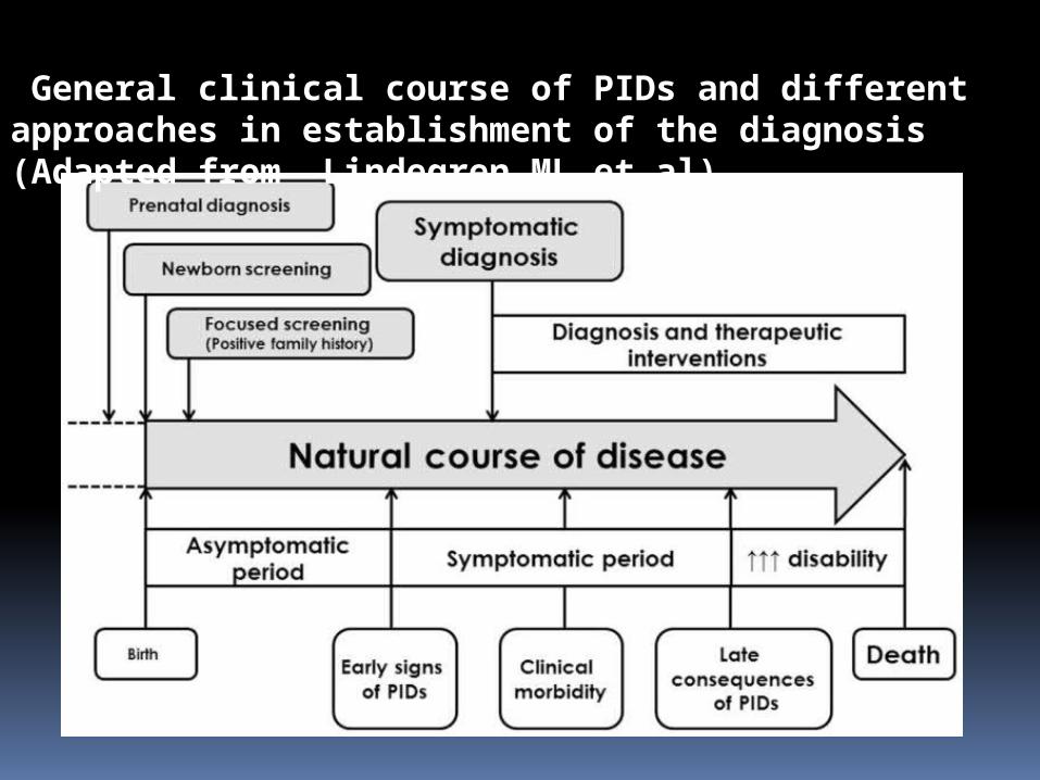
General clinical course of PIDs and different approaches in establishment of the diagnosis (Adapted from Lindegren ML et al)
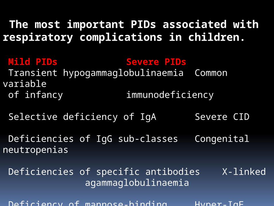
The most important PIDs associated with respiratory complications in children.
Mild PIDs Severe PIDs Transient hypogammaglobulinaemia Common variable of infancy immunodeficiency
Selective deficiency of IgA Severe CID
Deficiencies of IgG sub-classes Congenital neutropenias
Deficiencies of specific antibodies X-linked
agammaglobulinaemia
Deficiency of mannose-binding Hyper-IgE syndromes lectin DNA-repair defects
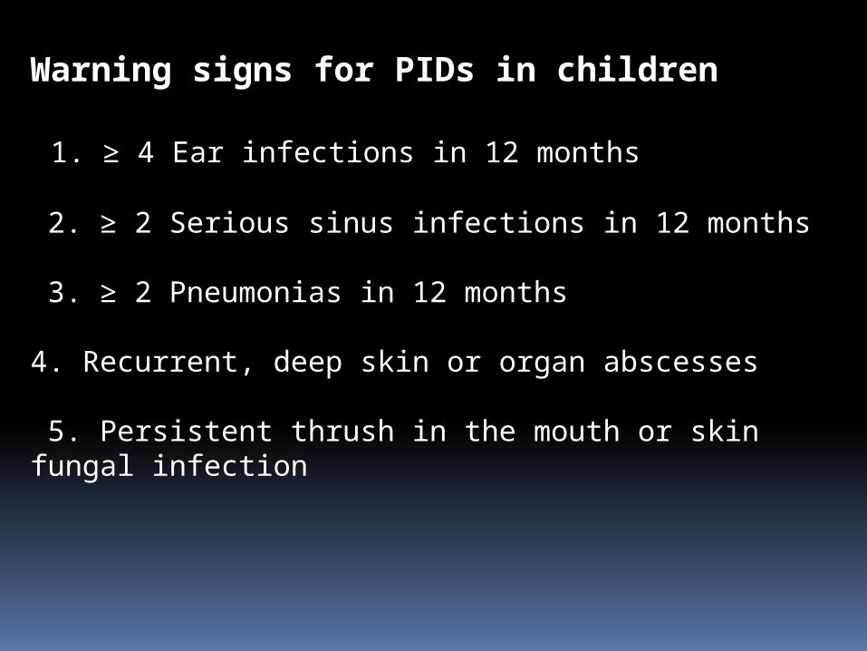
Warning signs for PIDs in children
1. ≥ 4 Ear infections in 12 months
2. ≥ 2 Serious sinus infections in 12 months
3. ≥ 2 Pneumonias in 12 months
4. Recurrent, deep skin or organ abscesses
5. Persistent thrush in the mouth or skin fungal infection
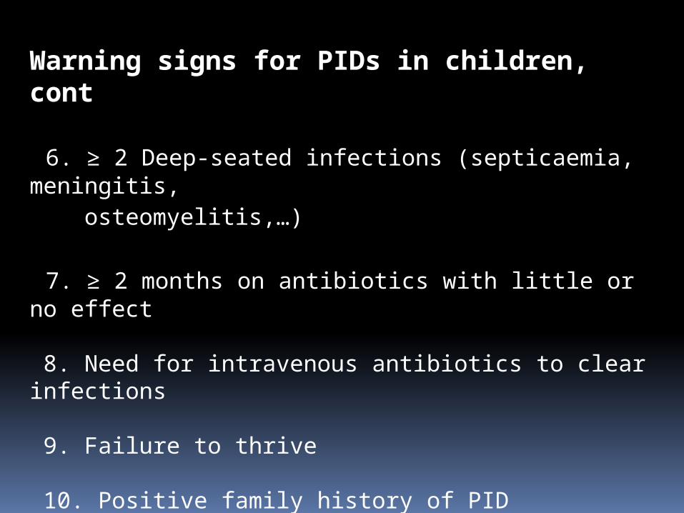
Warning signs for PIDs in children, cont
6. ≥ 2 Deep-seated infections (septicaemia, meningitis, osteomyelitis,…)
7. ≥ 2 months on antibiotics with little or no effect
8. Need for intravenous antibiotics to clear infections
9. Failure to thrive
10. Positive family history of PID
(Adapted according to Arkwright and Gennery, 2011)
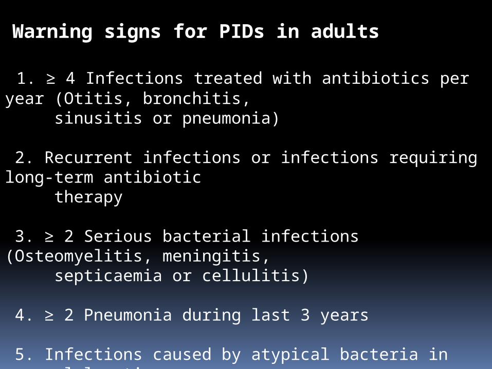
Warning signs for PIDs in adults
1. ≥ 4 Infections treated with antibiotics per year (Otitis, bronchitis, sinusitis or pneumonia)
2. Recurrent infections or infections requiring long-term antibiotic therapy
3. ≥ 2 Serious bacterial infections (Osteomyelitis, meningitis, septicaemia or cellulitis)
4. ≥ 2 Pneumonia during last 3 years
5. Infections caused by atypical bacteria in unusual location
6. Positive family history of PID
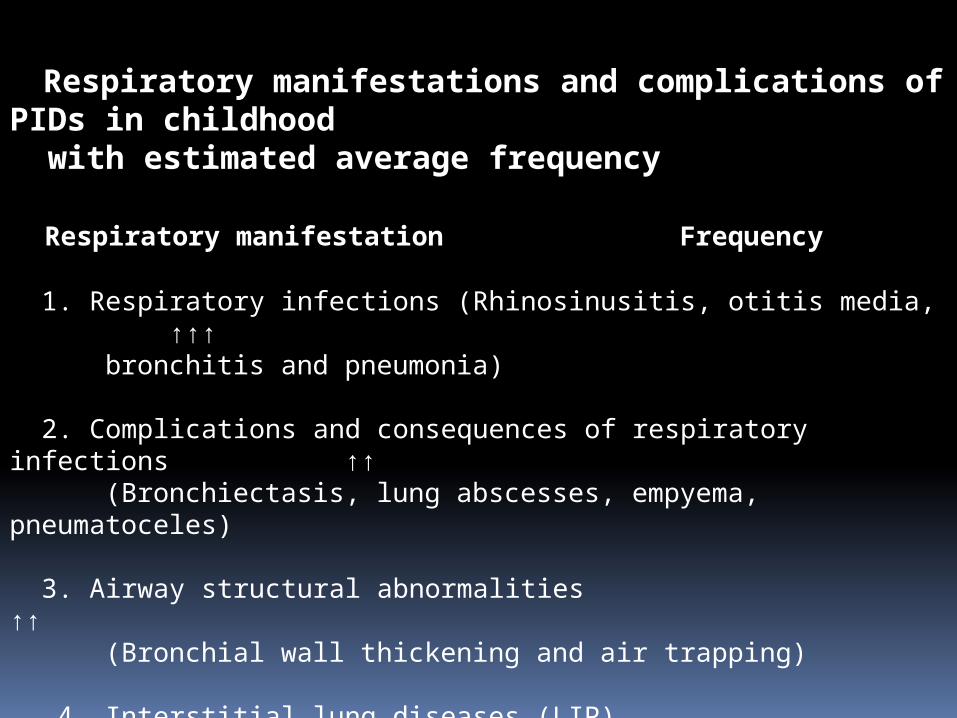
Respiratory manifestations and complications of PIDs in childhood with estimated average frequency
Respiratory manifestationFrequency
1. Respiratory infections (Rhinosinusitis, otitis media, ↑↑↑ bronchitis and pneumonia)
2. Complications and consequences of respiratory infections ↑↑
(Bronchiectasis, lung abscesses, empyema, pneumatoceles)
3. Airway structural abnormalities ↑↑ (Bronchial wall thickening and air trapping)
4. Interstitial lung diseases (LIP) ↑
5. Lymphoproliferative diseases (Lymphoma, benign Rare lymphoproliferative diseases and lymphadenopathy)
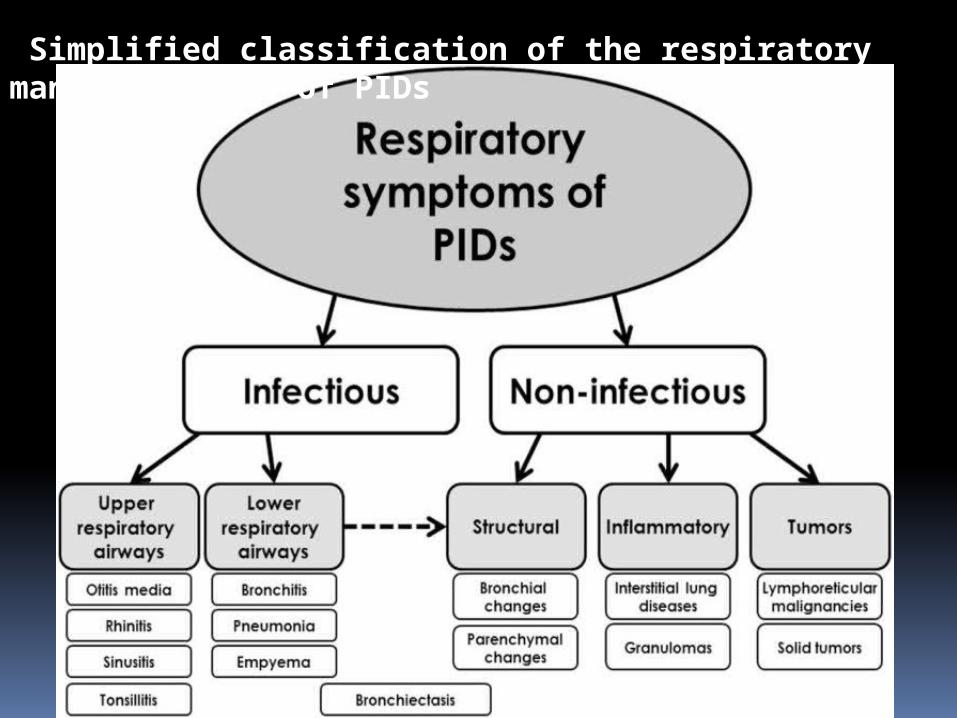
Simplified classification of the respiratory manifestations of PIDs
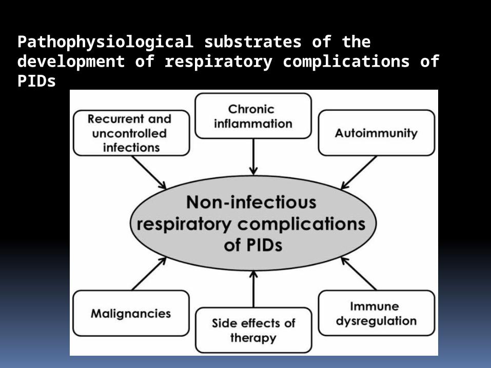
Pathophysiological substrates of the development of respiratory complications of PIDs
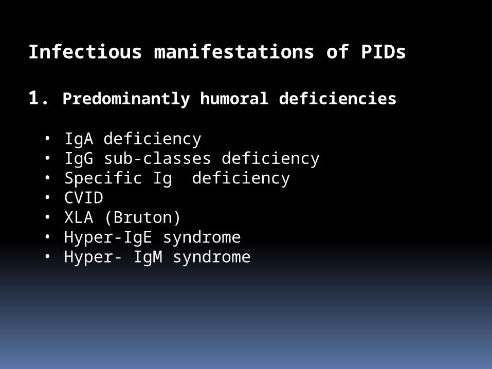
Infectious manifestations of PIDs
1. Predominantly humoral deficiencies
• IgA deficiency• IgG sub-classes deficiency• Specific Ig deficiency• CVID• XLA (Bruton)• Hyper-IgE syndrome• Hyper- IgM syndrome
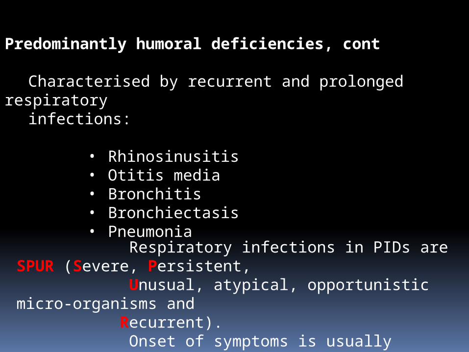
Predominantly humoral deficiencies, cont
Characterised by recurrent and prolonged respiratory
infections:
• Rhinosinusitis• Otitis media• Bronchitis• Bronchiectasis• Pneumonia
Respiratory infections in PIDs are SPUR (Severe, Persistent, Unusual, atypical, opportunistic micro-organisms and Recurrent). Onset of symptoms is usually beyond 6 months of age after maternal IgG have disappeared.
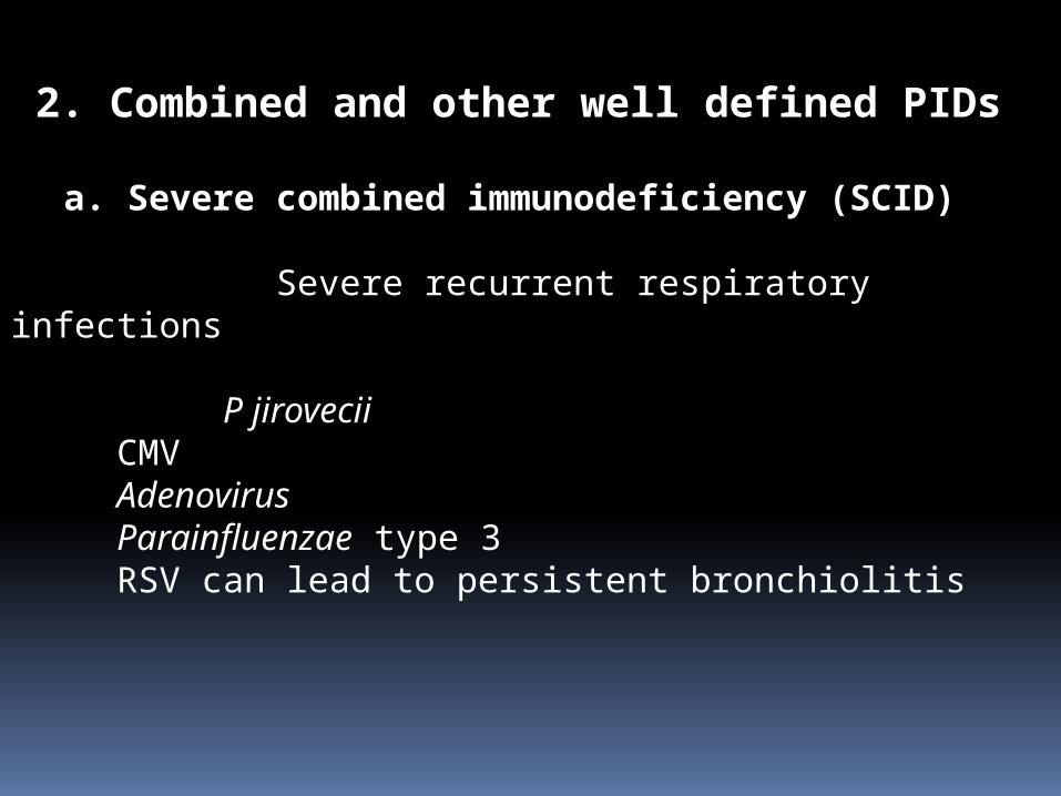
2. Combined and other well defined PIDs a. Severe combined immunodeficiency (SCID)
Severe recurrent respiratory infections
P jiroveciiCMVAdenovirusParainfluenzae type 3RSV can lead to persistent bronchiolitis
Combined and other well defined PIDs, cont
b. Hyper-IgE syndrome (Job syndrome)
Recurrent sino-pulmonary infections
S aureusS pneumoniaeH influenzae
May lead to bronchiectasis (P aeruginosa)
Pneumatoceles are another complication (Aspergillus
and scedosporium spp)
Combined and other well defined PIDs, cont
c. Hyper-IgE syndrome
Recurrent pneumonias
Encapsulated bacteriaCMVHistoplasmaP jiroveciiC albicansC neoformans
3. Phagocytic immunodeficiencies
Predominantly chronic granulomatous disease and congenital neutropenias
Common pathogens
S aureus P aeruginosaK pneumoniae Aspergillus sppAerobacter spp C albicans
Others are Burkholderia and Serratia spp
Difficult to treat, slow to resolve, recurrent pneumonias.
4. Complement immunodeficiencies
These diseases are generally underdiagnosed and underreported.
C1 C4 deficiencies lead to pyogenic infections
C5 C9 deficiencies predispose to Neisseria spp infections
C3 deficiency will particularly cause recurrent pneumonias.
There is an association with hereditary angioedema which is potentially life-threatening when occuring in the larynx.
Non-infectious respiratory complications of PIDs
Recurrent bacterial pulmonary infections may cause the following
Air trapping Atelectasis Bronchial wall thickening Bronchiectasis
1. Bronchiectasis
Major causes
• Cystic fibrosis• Primary ciliary dyskinesia• Primary immunodeficiencies
CVID up to 73% XLA from 17 to 76% Selective IgA deficiency IgG sub-classes deficiency
Mainly cylindrical type, bilateral and diffuse Affecting middle and lower lobes Proximal bronchi
Early detection and aggressive management are required.
2. Interstitial lung disease and PIDs
CVID may cause granulomatous lymphocytic interstitial lung disease (GLILD) which may present as
Lymphocytic interstitial pneumonia Follicular bronchiolitis Granulomatous lung disease Organising pneumonia Pulmonary lymphoid hyperplasia
Development of GLILD may be associated with infections such as
HHV8EBVCMV
Interstitial lung disease and PIDs, cont
ILD usually results from immune dysregulation.It has a poor prognosis.There is increased prevalence of
lymphoproliferative disorders.ILD is asymptomatic at the initial stage, hence
screening at risk patients is very important.
3. Respiratory tract tumours in PIDs
There is a strong association with EBV. CVID and Wiskott-Aldrich syndrome patients are at greater
risk.
Some of the tumours areLeiomyomaPulmonary adenocarcinomaThymomaLymphoma (NHL)
These tumours must be differentiated from benign
lymphoproliferative diseases.
4. Other respiratory complications of PIDs
Allergies Primary pulmonary hypertension Pulmonary alveolar proteinosis Cor pulmonale Respiratory insufficiency
Examination and investigations in a child with recurrent, severe and prolonged respiratory infections
Examination:
• Plot weight and height on growth chart
• Respiratory examination including clubbing and chest wall deformity
• Look for lymphadenopathy and/or hepatosplenomegaly
• Examine mouth/mucous membranes? candidal infection
• ENT and full systemic examination, including skin
Investigations to consider:
First line:
FBC and WBC differential IgG and IgG subclasses, IgA, IgM Specific antibodies to Tetanus, HiB and pneumococcus
(repeat levels one month after any boosters)
Second line:
Lymphocyte subsets Neutrophil oxidative burst if indicated Simple spirometry (if more than 5 years) Plain chest radiograph High resolution computerized tomography of chest Bronchoscopy
References
1. Milos J et al. Pulmonary manifestations of primary immunodefciency disorders in children. Frontiers in Pediatrics,July 2014, Volume 2, Article 77.
2. Woodsford M A M, Spencer D A and Cant A J. Symposium: Respiratory Medicine. Immune deficiency and the lung. Paediatrics and Child Health, 2010, 21:5, pp 213-218.