Embed Size (px)
Citation preview
Review ArticleSAJS
22 VOL 44, NO. 1, FEBRUARY 2006 SAJS
Coagulation physiologyThe integrity of the circulation is maintained through the provision of a rapid, potent, but tightly localised coagulation response to vascular damage. There is, however, one extraordinary problem in the regulation of haemostasis – blood flows. Normal haemostasis is the ability of the haemostatic system to control activation of clot formation and clot lysis in order to prevent haemorrhage without causing thrombosis. It classically involves vasoconstriction, platelet adhesion and aggregation at the site of injury, leading to a plug formation. This is followed by fibrin formation consolidating the plug and rendering it stable. All of this occurs without clotting occurring elsewhere. Procoagulant products, whether clotting factors or platelets, must therefore be spatially localised to the site of damage, and/or kinetically controlled so that they are inactive when distant from the damaged vessel. The blood coagulation cascade is usually initiated when sub-endothelial tissue factor is exposed/expressed to the blood flow following either damage or activation of the endothelium.1-3 As a result, the haemostatic mechanism is invoked through a complex series of regulated events, involving interactions of blood components and tissue proteins, resulting in a spectrum from haemorrhage, through controlled haemostasis, to thrombosis (Fig. 1).
A major insight into the functioning of the process of coagulation is that the tissue factor/factor VIIa complex of the classic ‘extrinsic pathway’ not only activates factor X of the ‘common pathway’ but also directly activates factor IX4,5 of the classic ‘intrinsic pathway’, making factor XII redundant. The entire response of clot formation has been characterised as a cascade or waterfall of enzymatic reactions, which result in a-thrombin converting soluble plasma fibrinogen to the
insoluble fibrin polymer,6,7 making the coagulation process a series of reactions in which the sequentially derived products have the capacity to amplify small stimuli to result in the rapid generation of large amounts of a-thrombin.8 Tissue factor, factor Xa and thrombin are pivotal, together with physiological controls (positive and negative feedback loops, and natural anticoagulants) that first enhance thrombin generation but then preserve vessel patency by limiting haemostatic plug formation to areas of injury. Abnormalities in these mechanisms can increase thrombosis risk.9
The classic pathways of blood coagulation
The ‘extrinsic pathway’ is the primary route of thrombin formation, through which the physiological haemostatic response occurs. It occurs through contact with a non-blood protein source. The physiological initiator of coagulation is tissue factor, which comes into contact with the blood at the site of vascular injury and combines with plasma serine protease, factor VII.10,11 Being a trans-membrane protein, it is ipso facto localised.12 The tissue factor/factor VIIa (TF/VIIa) complex activates factor X to factor Xa. It can, however, also activate factor IX to IXa.4,5
Tissue factor, the intrinsic membrane protein, is present on the surface of many cell types that are not normally in contact with the circulation (Fig. 2). However, it is exposed to blood when vascular damage occurs. Vascular damage will therefore lead to increasing levels of tissue factor at the site of injury. In association with the serine protease factor VIIa,13 it initiates the blood coagulation cascade, requiring no calcium.10-12 The direct factor IX activation, while slower, forms exponentially increasing amounts of thrombin. Tissue factor is not released into the circulation in a soluble form during normal haemostasis. If there is a major injury there is a potential for it to be swept into the circulation, which may lead to disseminated intravascular coagulation (DIC).14 However, at the site of damage platelets interact with
Coagulation for the clinicianTOm RUTTmANN, Ph.D., F.C.A.
Department of Anaesthesia, University of Cape Town
OVERVIEW
The integrity of the circulation is maintained through the provision of a rapid, potent, but tightly
localised coagulation response to vascular damage. There is, however, one extraordinary
problem in the regulation of haemostasis - blood flows. Normal haemostasis is the ability of
the haemostatic system to control activation of clot formation and clot lysis in order to prevent
haemorrhage without causing thrombosis. It classically involves vasoconstriction, platelet
adhesion and aggregation at the site of injury, leading to a plug formation. This is followed by
fibrin formation consolidating the plug and rendering it stable. All of this occurs without clotting
occurring elsewhere. Procoagulant products, whether clotting factors or platelets, must
therefore be spatially localised to the site of damage, and/or kinetically controlled so that they
are inactive when distant from the damaged vessel.
The blood coagulation cascade is usually initiated when sub-endothelial tissue factor is
exposed/expressed to the blood flow following either the damage or activation of the
endothelium1-3
. As a result, the haemostatic mechanism is invoked through a complex series
of regulated events, involving interactions of blood components and tissue proteins, resulting
in a spectrum from haemorrhage, through controlled haemostasis, to thrombosis. (Figure 1)
Figure 1. The Coagulation Cascade
A major insight into the functioning of the process of coagulation is that the Tissue
Factor/Factor VIIa complex of the classic “Extrinsic Pathway” not only activates Factor X of
the “Common Pathway”, but also directly activates Factor IX4,5
of the classic “Intrinsic
Pathway”, making Factor XII redundant. The entire response of clot formation has been
characterised as a cascade or waterfall of enzymatic reactions, which result in !-thrombin
Fig. 1. The coagulation cascade.
converting soluble plasma fibrinogen to the insoluble fibrin polymer6,7
, making the coagulation
process a series of reactions in which the sequentially derived products have the capacity to
amplify small stimuli to result in the rapid generation of large amounts of !-thrombin8. Tissue
factor, factor Xa, and thrombin are pivotal; together with physiological controls (positive and
negative feedback loops, and natural anticoagulants) that first enhance thrombin generation
but then preserve vessel patency by limiting haemostatic plug formation to areas of injury.
Abnormalities in these mechanisms can increase thrombosis risk9.
THE CLASSIC PATHWAYS OF BLOOD COAGULATION
The “extrinsic pathway” is the primary route of thrombin formation, through which the
physiological haemostatic response occurs. It occurs through contact with a non-blood protein
source. The physiological initiator of coagulation is tissue factor, which comes into contact
with the blood at the site of vascular injury and combines with plasma serine protease, factor
VII10,11
. Being a trans-membrane protein, it is ipso facto localised12
. The tissue factor/factor
VIIa (TF/VIIa) complex activates factor X to factor Xa. It can, however, also activate factor IX
to IXa4,5
.
Figure 2. The Coagulation Cascade – Initiation Phase
Tissue factor, the intrinsic membrane protein, is present on the surface of many cell types that
are not normally in contact with the circulation. (Figure 2) However, it is exposed to blood
when vascular damage occurs. Thus, vascular damage will lead to increasing levels of tissue
factor at the site of injury. In association with the serine protease factor VIIa13
, it initiates the
blood coagulation cascade, requiring no calcium10-12
. The direct factor IX activation, while
slower, forms exponentially increasing amounts of thrombin. Tissue factor is not released into
the circulation in a soluble form during normal haemostasis. If there is a major injury, there is
a potential for it to be swept into the circulation, which may lead to disseminated intravascular
coagulation (DIC)14
. However, at the site of damage, platelets interact with components of the
Fig. 2. The coagulation cascade – initiation phase.
pg 22-37.indd 22 3/6/06 10:19:31 Am

pg 22-37.indd 23 3/6/06 10:19:32 Am
SAJS
24 VOL 44, NO. 1, FEBRUARY 2006 SAJS
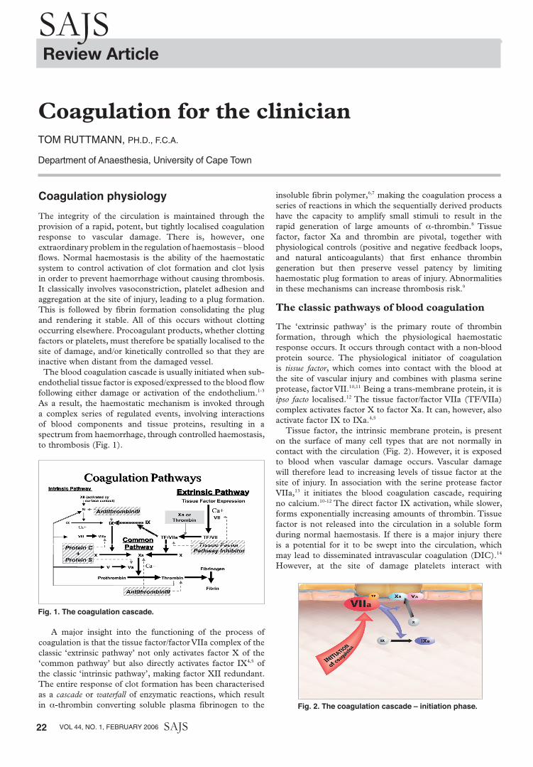
components of the vessel wall at the same time that plasma factor VII comes into contact with tissue factor. The platelet plug thus prevents the unchecked release of the TF/VIIa complex. The limited amounts of the serine protease factor Xa produced generate minute concentrations of thrombin, which partially activates platelets and cleaves the pro-cofactors factor V and factor VIII, generating the active cofactors factor Va and factor VIIIa, respectively.15,16 The extrinsic pathway catalyst is therefore responsible for factor X activation through both pathways.17,18 Furthermore, the efficiency of factor X activation by the factor VIIIa/IXa complex is significantly greater than the efficiency of the TF/VIIa complex. It is relevant that the factor IX-dependent route, in contrast to the direct activation of factor X by TF/VIIa, involves two enzyme-catalysed reactions in sequence and therefore provides extra amplification over the direct activation of factor X, but will take longer.19 The reactions occur on the platelet surface, but only on those platelets that are activated and involved at the site of vascular injury (see below); the response is therefore localised to the site of vascular damage. Multiple control mechanisms guard against procoagulant materials entering the general circulation and causing disseminated intravascular coagulation.20 Obviously, since the latter condition exists, these safeguards can on occasion be overrun. The pathway classically referred to as the ‘intrinsic pathway’ starts with the formation of factor XIIa.21 This can cleave prekallikrein to provide kallikrein,22 which in turn reciprocally activates factor XII.23 Factor XIIa, in the presence of high-molecular-weight kininogen, converts factor XI to factor XIa, which in turn converts factor IX to factor IXa.24 Factor IXa binds with its cofactor protein factor VIIIa, in the presence of calcium and the appropriate membrane surface, and activates factor X to factor Xa.25 Factor VIIIa forms the intrinsic factor Xase complex with the serine protease, factor IXa, on a membrane surface provided by platelets, microparticles and endothelial and other cells26 and activates factor X at a 50 - 100-fold higher rate than the factor VIIa-tissue factor complex,4 in turn creating a ‘thrombin burst’ that activates fibrin (Fig. 3).
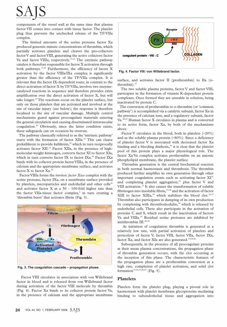
Factor VIII circulates in association with von Willebrand factor in blood and is released from von Willebrand factor during activation of the factor VIII molecule by thrombin (Fig. 4). Factor Xa binds to its cofactor protein factor Va, in the presence of calcium and the appropriate membrane
surface, and activates factor II (prothrombin) to IIa (a-thrombin).27 The two soluble plasma proteins, factor V and factor VIII, participate in the formation of vitamin K-dependent protein complexes. Once formed they are unstable in solution, being inactivated by protein C. The conversion of prothrombin to a-thrombin (or ‘common pathway’) is accomplished via a catalytic subunit, factor Xa in the presence of calcium ions, and a regulatory subunit, factor Va.28,29 Human factor X circulates in plasma and is converted to its active form, factor Xa, by both of the mechanisms above. Factor V circulates in the blood, both in platelets (~20%) and as the soluble plasma protein (~80%). Since a deficiency of platelet factor V is associated with decreased factor Xa binding and a bleeding diathesis,30 it is clear that the platelet pool of this protein plays a major physiological role. The factor Xa/Va complex activates prothrombin on an anionic phospholipid membrane, the platelet surface. Thrombin generation is the central biochemical reaction in both normal haemostasis and thrombosis. The thrombin produced further amplifies its own generation through other important coagulation events such as activating factor XI31 and completing platelet aggregation,32 plus factor V and VIII activation.15 It also causes the transformation of soluble fibrinogen into insoluble fibrin,33,34 and the activation of factor XIII to factor XIIIa,35 which stabilises the blood clot.36,37 Thrombin also participates in damping of its own production by complexing with thrombomodulin,38 which is released by endothelial cells. These also participate in the activation of proteins C and S, which result in the inactivation of factors Va and VIIIa.39 Residual serine proteases are inhibited by antithrombin III.40-45
At initiation of coagulation thrombin is generated at a relatively low rate, with partial activation of platelets and proteolysis of factor V, factor VIII, factor VIIa, factor IXa, factor Xa, and factor XIa are also generated.15,16,46
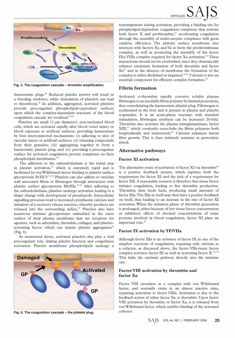
Subsequently, in the presence of all procoagulant proteins at their mean plasma concentrations, the propagation phase of thrombin generation occurs, with the clot occurring at the inception of this phase. The characteristic features of the propagation phase are a prothrombin conversion at a high rate, completion of platelet activation, and solid clot formation15,16,37,46,47 (Fig. 5).
Platelets
Platelets form the platelet plug, playing a pivotal role in haemostasis with platelet membrane glycoproteins mediating binding to subendothelial tissue and aggregation into
vessel wall at the same time that plasma factor VII comes into contact with tissue factor. The
platelet plug thus prevents the unchecked release of the TF/VIIa complex.
The limited amounts of the serine protease factor Xa produced generate minute
concentrations of thrombin, which partially activates platelets and cleaves the pro-cofactors
factor V and factor VIII generating the active cofactors--factor Va and factor VIIIa,
respectively15,16
. Thus the extrinsic pathway catalyst is responsible for factor X activation
through both pathways17,18
. Furthermore, the efficiency of factor X activation by the factor
VIIIa/IXa complex is significantly greater than the efficiency of the TF/VIIa complex. It is
relevant that the factor IX-dependent route, in contrast to the direct activation of factor X by
TF/VIIa, involves two enzyme-catalysed reactions in sequence and therefore provides extra
amplification over the direct activation of factor X, but will take longer19
. The reactions occur
on the platelet surface, but only on those platelets that are activated and involved at the site
of vascular injury (see below), thus the response is localised to the site of vascular damage.
Multiple control mechanisms guard against procoagulant materials entering the general
circulation and causing disseminated intravascular coagulation20
. Obviously, since the latter
condition exists, these safeguards can on occasion be overrun.
The pathway classically referred to as the “Intrinsic Pathway” starts with the formation of
factor XIIa21
, which can cleave prekallikrein to provide kallikrein22
, which in turn reciprocally
activates factor XII23
. Factor XIIa, in the presence of high-molecular-weight kininogen,
converts factor XI to factor XIa, which in turn converts factor IX to factor IXa24
. Factor IXa
binds with its cofactor protein factor VIIIa, in the presence of calcium and the appropriate
membrane surface, and activates factor X to factor Xa25
.
Factor VIIIa forms the intrinsic factor Xase complex with the serine protease, factor IXa, on a
membrane surface provided by platelets, microparticles, and endothelial and other cells26
and
activates factor X at a 50-100-fold higher rate than the factor VIIa-tissue factor complex4
- in
turn creating a “thrombin burst” that activates fibrin. (Figure 3)
Fig. 3. The coagulation cascade – propagation phase.
Figure 3. The Coagulation Cascade – Propagation Phase
Factor VIII circulates in association with von Willebrand factor in blood and is released from
von Willebrand factor during activation of the factor VIII molecule by thrombin (Figure 4).
Factor Xa binds to its cofactor protein factor Va, in the presence of calcium and the
appropriate membrane surface, and activates factor II (prothrombin) to IIa (!-thrombin)27
.
vWFvWFPltPlt
ReceptorReceptor
COLLAGENCOLLAGENcoagulant proteincoagulant protein -- VIII : CVIII : C
vWFvWFPltPlt
ReceptorReceptor
COLLAGENCOLLAGENcoagulant proteincoagulant protein -- VIII : CVIII : C
Figure 4. Factor VIII:von Willebrand Factor
The two soluble plasma proteins, factor V and factor VIII, participate in the formation of
vitamin K-dependent protein complexes. Once formed, they are unstable in solution, being
inactivated by protein C.
The conversion of prothrombin to !-thrombin (or “Common Pathway“) is accomplished via a
catalytic subunit, factor Xa in the presence of calcium ions, and a regulatory subunit, factor
Va28,29
. Human factor X circulates in plasma, and is converted to its active form, factor Xa, by
both of the mechanism above.
Factor V circulates in the blood, both in platelet (~20%), and as the soluble plasma protein
(~80%). Since a deficiency of platelet factor V is associated with decreased factor Xa binding
and a bleeding diathesis30
, it is clear that the platelet pool of this protein plays a major
physiological role. The factor Xa/Va complex activates prothrombin on an anionic
phospholipid membrane: the platelet surface.
Thrombin generation is the central biochemical reaction in both normal haemostasis and
thrombosis. The thrombin produced further amplifies its own generation through other
important coagulation events like activating factor XI31
, completing platelet aggregation32
, plus
factor V and VIII activation15
. It also causes the transformation of soluble fibrinogen into
insoluble fibrin33,34
, and the activation of factor XIII to factor XIIIa35
, which stabilises the blood
clot36,37
. Thrombin also participates in damping of its own production by complexing with
thrombomodulin,38
which is released by endothelial cells. These also participate in the
activation of proteins C and S, which result in the inactivation of factors Va and VIIIa39
.
Residual serine proteases are inhibited by antithrombin III40-45
.
Fig. 4. Factor VIII: von Willebrand factor.
pg 22-37.indd 24 3/6/06 10:19:41 Am
SAJSARTICLES
SAJS VOL 44, NO. 1, FEBRUARY 2006 25
haemostatic plugs.48 Reduced platelet activity will result in a bleeding tendency, while stimulation of platelets can lead to thrombosis.49 In addition, aggregated, activated platelets provide procoagulant phospholipid-equivalent surfaces upon which the complex-dependent reactions of the blood coagulation cascade are localised.50,51
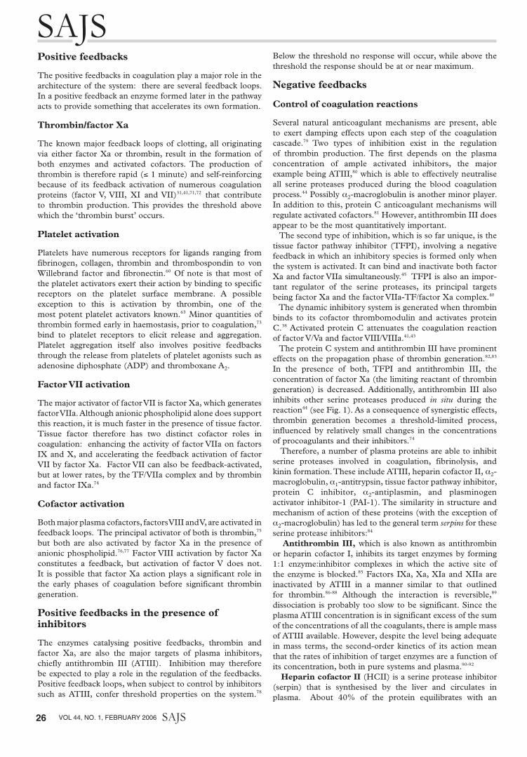
Platelets are small (2 µm diameter), non-nucleated blood cells, which are activated rapidly after blood vessel injury or blood exposure to artificial surfaces, providing haemostasis by four interconnected mechanisms: (i) adhering to sites of vascular injury or artificial surfaces; (ii) releasing compounds from their granules; (iii) aggregating together to form a haemostatic platelet plug; and (iv) providing a procoagulant surface for activated coagulation protein complexes on their phospholipid membranes.52
The adhesion to the subendothelium is the initial step in platelet activation,53 which is extremely rapid and is facilitated by von Willebrand factor binding to platelet surface glycoprotein Ib/IX/V.54-56 Platelets can also adhere to vascular wall associated fibrin or fibrinogen through interaction with platelet surface glycoprotein IIb/IIIa.57,58 After adhering to the subendothelium, platelets undergo activation leading to a shape change with development of pseudopods. Intracellular signalling processes lead to increased cytoplasmic calcium and initiation of a secretory release reaction, whereby products are released into the surrounding milieu.59 Platelets also have numerous intrinsic glycoproteins embedded in the outer surface of their plasma membrane that are receptors for agonists, such as adrenaline, thrombin, collagen, and platelet-activating factor, which can initiate platelet aggregation60 (Fig. 6). As mentioned above, activated platelets also play a vital procoagulant role, linking platelet function and coagulation activation. Platelet membrane phospholipids undergo a
rearrangement during activation, providing a binding site for phospholipid-dependent coagulation complexes that activate both factor X and prothrombin,61 accelerating coagulation through the assembly of multi-enzyme complexes with great catalytic efficiency. The platelet surface membrane also interacts with factors Xa and Va to form the prothrombinase complex, as well as promoting the assembly of the factor IXa-VIIIa complex required for factor Xa activation.62 These interactions should not be overlooked, since they dramatically enhance enzymatic formation of both thrombin and factor Xa63 and in the absence of membrane the formation of the complex is either abolished or impaired.64,65 Calcium is also an essential component for efficient complex formation.66
Fibrin formation
Activated a-thrombin rapidly converts soluble plasma fibrinogen to an insoluble fibrin polymer by limited proteolysis, thus consolidating the haemostatic platelet plug. Fibrinogen is synthesised in the liver and is present in plasma and platelet a-granules. It is an acute-phase reactant: with maximal stimulation, fibrinogen synthesis can be increased 20-fold. Thrombin also activates the plasma transglutaminase, factor XIII,67 which covalently cross-links the fibrin polymers both longitudinally and transversely.68 Calcium enhances lateral fibre growth. This is then relatively resistant to proteolytic attack.
Alternative pathways
Factor XI activation
The alternative route of activation of factor XI via thrombin69 is a positive feedback system, which explains both the requirement for factor XI and the lack of a requirement for factor XII. A reasonable scenario is therefore that tissue factor initiates coagulation, leading to fast thrombin production. Thrombin then feeds back, producing small amounts of factor XIa. The XIa in itself may then have a positive feedback on itself, thus leading to an increase in the rate of factor XI activation. When the initiation phase of thrombin generation is prolonged, either because of low tissue factor concentration or inhibitory effects of elevated concentrations of some proteins involved in blood coagulation, factor XI plays an important role.47
Factor IX activation by TF/VIIa
Although factor XIa is an activator of factor IX in one of the simplest reactions of coagulation, requiring only calcium as a cofactor, as discussed above, the factor VIIa-tissue factor complex activates factor IX as well as activating factor X.4,5,70 This links the extrinsic pathway directly into the intrinsic one.
Factor VIII activation by thrombin and factor Xa
Factor VIII circulates as a complex with von Willebrand factor, and normally exists in an almost inactive state, requiring activation to factor VIIIa. Activation is due to the feedback action of either factor Xa or thrombin. Upon factor VIII activation by thrombin or factor Xa, it is released from von Willebrand factor, which enables binding of the activated cofactor.
At initiation of coagulation thrombin is generated at a relatively low rate, with partial activation
of platelets and proteolysis of factor V, factor VIII, factor VIIa, factor IXa, factor Xa, and factor
XIa are also generated15,16,46
.
Figure 5. The Coagulation Cascade – Thrombin amplification
Subsequently, in the presence of all procoagulant proteins at their mean plasma
concentrations, the propagation phase of thrombin generation occurs, with the clot
occurring at the inception of this phase. The characteristic features of the propagation
phase are a prothrombin conversion at a high rate, completion of platelet activation, and
solid clot formation15,16,37,46,47
(Figure 5).
PLATELETS
Platelets form the platelet plug, playing a pivotal role in haemostasis with platelet membrane
glycoproteins mediating binding to subendothelial tissue and aggregation into haemostatic
plugs48
. Reduced platelet activity will result in a bleeding tendency, while stimulation of
platelets can lead to thrombosis49
. In addition, aggregated, activated platelets provide
procoagulant phospholipid-equivalent surfaces upon which the complex-dependent reactions
of the blood coagulation cascade are localised50,51
.
Platelets are small (2-µm-diameter), non-nucleated blood cells, which are activated rapidly
after blood vessel injury or blood exposure to artificial surfaces, providing haemostasis by 4
interconnected mechanisms: (1) adhering to sites of vascular injury or artificial surfaces, (2)
releasing compounds from their granules, (3) aggregating together to form a haemostatic
platelet plug, and (4) providing a procoagulant surface for activated coagulation protein
complexes on their phospholipid membranes52
.
Fig. 5. The coagulation cascade – thrombin amplification.
FF
Figure 6. The Coagulation Cascade – The Platelet Plug
The adhesion to the subendothelium is the initial step in platelet activation53
, which is
extremely rapid and is facilitated by vWF binding to platelet surface glycoprotein Ib/IX/V54-56
.
Platelets can also adhere to vascular wall associated fibrin or fibrinogen through interaction
with platelet surface glycoprotein IIb/IIIa57,58
. After adhering to the subendothelium, platelets
undergo activation leading to a shape change with development of pseudopods. Intracellular
signalling processes lead to increased cytoplasmic calcium and initiation of a secretory
release reaction, whereby products are released into the surrounding milieu59
. Platelets also
have numerous intrinsic glycoproteins embedded in the outer surface of their plasma
membrane that are receptors for agonists, such as adrenaline, thrombin, collagen, and
platelet-activating factor, which can initiate platelet aggregation60
(Figure 6).
As mentioned above, activated platelets also play a vital procoagulant role, linking platelet
function and coagulation activation. Platelet membrane phospholipids undergo a
rearrangement during activation, providing a binding site for phospholipid-dependent
coagulation complexes that activate both factor X and prothrombin61
, accelerating coagulation
through the assembly of multi-enzyme complexes with great catalytic efficiency. The platelet
surface membrane also interacts with factors Xa and Va to form the prothrombinase complex,
as well as promoting the assembly of the factor IXa-VIIIa complex required for factor Xa
activation62
. These interactions should not be overlooked, since they dramatically enhance
enzymatic formation of both thrombin and factor Xa63
and in the absence of membrane the
formation of the complex is either abolished or impaired64,65
. Calcium is also an essential
component for efficient complex formation66
.
GP
1b
Damaged
endotheliumvWF
Activated
Platelet
GP
IIb/IIIa
Fig. 6. The coagulation cascade – the platelet plug.
pg 22-37.indd 25 3/6/06 10:19:52 Am
SAJS
26 VOL 44, NO. 1, FEBRUARY 2006 SAJS
Positive feedbacks
The positive feedbacks in coagulation play a major role in the architecture of the system: there are several feedback loops. In a positive feedback an enzyme formed later in the pathway acts to provide something that accelerates its own formation.
Thrombin/factor Xa
The known major feedback loops of clotting, all originating via either factor Xa or thrombin, result in the formation of both enzymes and activated cofactors. The production of thrombin is therefore rapid (≤ 1 minute) and self-reinforcing because of its feedback activation of numerous coagulation proteins (factor V, VIII, XI and VII)31,41,71,72 that contribute to thrombin production. This provides the threshold above which the ‘thrombin burst’ occurs.
Platelet activation
Platelets have numerous receptors for ligands ranging from fibrinogen, collagen, thrombin and thrombospondin to von Willebrand factor and fibronectin.60 Of note is that most of the platelet activators exert their action by binding to specific receptors on the platelet surface membrane. A possible exception to this is activation by thrombin, one of the most potent platelet activators known.63 Minor quantities of thrombin formed early in haemostasis, prior to coagulation,73 bind to platelet receptors to elicit release and aggregation. Platelet aggregation itself also involves positive feedbacks through the release from platelets of platelet agonists such as adenosine diphosphate (ADP) and thromboxane A2.
Factor VII activation
The major activator of factor VII is factor Xa, which generates factor VIIa. Although anionic phospholipid alone does support this reaction, it is much faster in the presence of tissue factor. Tissue factor therefore has two distinct cofactor roles in coagulation: enhancing the activity of factor VIIa on factors IX and X, and accelerating the feedback activation of factor VII by factor Xa. Factor VII can also be feedback-activated, but at lower rates, by the TF/VIIa complex and by thrombin and factor IXa.74
Cofactor activation
Both major plasma cofactors, factors VIII and V, are activated in feedback loops. The principal activator of both is thrombin,75 but both are also activated by factor Xa in the presence of anionic phospholipid.76,77 Factor VIII activation by factor Xa constitutes a feedback, but activation of factor V does not. It is possible that factor Xa action plays a significant role in the early phases of coagulation before significant thrombin generation.
Positive feedbacks in the presence of inhibitors
The enzymes catalysing positive feedbacks, thrombin and factor Xa, are also the major targets of plasma inhibitors, chiefly antithrombin III (ATIII). Inhibition may therefore be expected to play a role in the regulation of the feedbacks. Positive feedback loops, when subject to control by inhibitors such as ATIII, confer threshold properties on the system.78
Below the threshold no response will occur, while above the threshold the response should be at or near maximum.
Negative feedbacks
Control of coagulation reactions
Several natural anticoagulant mechanisms are present, able to exert damping effects upon each step of the coagulation cascade.79 Two types of inhibition exist in the regulation of thrombin production. The first depends on the plasma concentration of ample activated inhibitors, the major example being ATIII,80 which is able to effectively neutralise all serine proteases produced during the blood coagulation process.44 Possibly a2-macroglobulin is another minor player. In addition to this, protein C anticoagulant mechanisms will regulate activated cofactors.81 However, antithrombin III does appear to be the most quantitatively important. The second type of inhibition, which is so far unique, is the tissue factor pathway inhibitor (TFPI), involving a negative feedback in which an inhibitory species is formed only when the system is activated. It can bind and inactivate both factor Xa and factor VIIa simultaneously.45 TFPI is also an impor- tant regulator of the serine proteases, its principal targets being factor Xa and the factor VIIa-TF/factor Xa complex.40
The dynamic inhibitory system is generated when thrombin binds to its cofactor thrombomodulin and activates protein C.38 Activated protein C attenuates the coagulation reaction of factor V/Va and factor VIII/VIIIa.41,43 The protein C system and antithrombin III have prominent effects on the propagation phase of thrombin generation.82,83 In the presence of both, TFPI and antithrombin III, the concentration of factor Xa (the limiting reactant of thrombin generation) is decreased. Additionally, antithrombin III also inhibits other serine proteases produced in situ during the reaction44 (see Fig. 1). As a consequence of synergistic effects, thrombin generation becomes a threshold-limited process, influenced by relatively small changes in the concentrations of procoagulants and their inhibitors.74
Therefore, a number of plasma proteins are able to inhibit serine proteases involved in coagulation, fibrinolysis, and kinin formation. These include ATIII, heparin cofactor II, a2-macroglobulin, a1-antitrypsin, tissue factor pathway inhibitor, protein C inhibitor, a2-antiplasmin, and plasminogen activator inhibitor-1 (PAI-1). The similarity in structure and mechanism of action of these proteins (with the exception of a2-macroglobulin) has led to the general term serpins for these serine protease inhibitors:84
Antithrombin III, which is also known as antithrombin or heparin cofactor I, inhibits its target enzymes by forming 1:1 enzyme:inhibitor complexes in which the active site of the enzyme is blocked.85 Factors IXa, Xa, XIa and XIIa are inactivated by ATIII in a manner similar to that outlined for thrombin.86-88 Although the interaction is reversible,89 dissociation is probably too slow to be significant. Since the plasma ATIII concentration is in significant excess of the sum of the concentrations of all the coagulants, there is ample mass of ATIII available. However, despite the level being adequate in mass terms, the second-order kinetics of its action mean that the rates of inhibition of target enzymes are a function of its concentration, both in pure systems and plasma.90-92 Heparin cofactor II (HCII) is a serine protease inhibitor (serpin) that is synthesised by the liver and circulates in plasma. About 40% of the protein equilibrates with an
pg 22-37.indd 26 3/6/06 10:19:53 Am

pg 22-37.indd 27 3/6/06 10:19:53 Am
SAJS
28 VOL 44, NO. 1, FEBRUARY 2006 SAJS
extravascular compartment and it has been detected in the intima of normal human arteries. It inhibits thrombin, but has no activity against other proteases involved in coagulation or fibrinolysis.93
a2-Macroglobulin can inhibit numerous serine proteases, including thrombin, factor Xa, plasmin and kallikrein, albeit at a relatively modest rate compared with other inhibitors.81
Tissue factor pathway inhibitor is a serum component that has been shown to form a quaternary complex with factor Xa, factor VIIa and tissue factor in the presence of calcium and thereby suppresses the activity of the extrinsic cascade.94,95
Protein C. A blood coagulation inhibitor, autoprothrombin IIa, is generated when thrombin is converted to prothrombin. The vitamin K-dependent protein, which was termed protein C, was subsequently found to be the same protein as autoprothrombin IIa.96 Plasminogen activator inhibitor-1 plays the major physiological part in suppressing the function of tissue plasminogen activator as well as urokinase, thus preventing the activation of the fibrinolytic pathway. In contrast to most other serine protease inhibitors in plasma, the normal level of PAI-1 is quite low. PAI-1 can also inhibit other coagulation system proteases including plasmin, activated protein C and thrombin.97,98
Monitoring of coagulation abnormalities Normal haemostasis is the ability of the haemostatic system to control activation of clot formation and clot lysis in order to prevent haemorrhage without causing thrombosis. All this occurs with no clotting elsewhere. This basic definition of normal haemostasis, depending on several finely balanced mechanisms, shows that: 1. There are three systems involved simultaneously in haemostasis: the formation of clots, involving platelets and the coagulation pathways; this is opposed by the anticoagulant system, and thirdly, the fibrinolysis of clots once they have been formed. 2. The absence or presence of haemorrhage or thrombosis depends on a delicate balance between the activated procoagulants and anticoagulant systems, while the clot maturity, or early breakdown once formed, will depend on the fibrinolytic system. In other words, an excess of activated pro-coagulants will primarily result in thrombosis. On the other hand, an increased anticoagulant potential (antithrombin, protein C, etc.), or decrease in activation of procoagulant factors will result in haemorrhage. In addition, enhanced fibrinolysis will also lead to bleeding. 3. Given the intricate interactions between the three systems, any instrument measuring coagulation endpoints may only be able to identify single points, rather than the interaction between systems. This is because coagulation is a dynamic process dependent on the interaction of an array of factors rather than one determined by actual values of individual agents. As the understanding of the normal mechanisms of coagulation grows, so does the ability to monitor haemostasis, becoming more specific and accurate as instrumentation techniques become simpler and more efficient, facilitating the monitoring of haemostasis in the clinical setting.99 The haemostatic process of coagulation is extremely complex, with multiple interactions between factors, including the coagulation and fibrinolytic proteins, platelets, activators and
inhibitors. This is in order to avoid uncontrolled action in either direction, and is accomplished by numerous positive and negative inhibiting and activating feedback mechanisms. As a result, the actual quantity or level of an individual factor does not necessarily reflect its functional status. With this complicated and interactive dynamic system, the measurement of static endpoints will not reflect the dynamics of the system. Any medical coagulopathy, as opposed to a surgical bleed, needs the actual underlying cause determined in order to direct the therapy appropriately. Traditionally, haematological tests like the prothrombin time (PT) and its derived value, the international normalised ratio (INR), activated partial thromboplastin time (aPTT), bleeding time and activated clotting time (ACT) (see below) have been done. However, while they are useful endpoint markers to identify the lack of a given factor, they do not measure the dynamic interaction between the different systems, as blood coagulation occurs efficiently on cell surfaces such as activated platelets, monocytes, and fibroblasts. Both PT and aPTT are highly artificial in vitro systems with major limitations.100
Standard tests of coagulation
Most of the routine tests of coagulation are performed on blood that has had the coagulation cascade suspended through the addition of sodium citrate 3.8%, which removes the calcium from the blood. The sample is then spun down leaving only plasma, with almost no platelets. In order to measure the various tests of coagulation endpoints, calcium is added in abundance, as are the various factors used to initiate coagulation. As a result, these tests are generally useful for measuring absolute values, or endpoints. However, they will not measure the interaction between pro- and anticoagulant factors, because of the addition of excess initiating factors.
Prothrombin time (PT/INR)101
This test measures the overall efficiency of the extrinsic system. It was introduced as an assay of prothrombin in plasma before many of the factors participating in the coagulation reactions were identified. It is now used as a screening tool of the activities of fibrinogen, prothrombin and factors V, VII, and X. It is also used to monitor the oral anticoagulants, since these lower the levels of prothrombin, VII and X (Fig. 7). The test is initiated by recalcifying citrated plasma in the presence of thromboplastin. Thromboplastin is added far in excess of normal levels and is a source of tissue factor in a
the interaction between pro- and anticoagulant factors, because of the addition of excess
initiating factors.
Prothrombin time (PT / INR)101
This test measures the overall efficiency of the extrinsic system. It was introduced as an
assay of prothrombin in plasma before many of the factors participating in the coagulation
reactions were identified. It is now used as a screening tool of the activities of fibrinogen,
prothrombin and factors V, VII, and X. It is also used to monitor the oral anticoagulants, since
these lower the levels of prothrombin, VII and X (Figure 7).
The test is initiated by recalcifying citrated plasma in the presence of thromboplastin.
Thromboplastin is added far in excess of normal levels and is a source of tissue factor in a
suspension of phospholipids. The time that elapses until the plasma clots is the PT and is
predominantly determined by: i) factor VIIa/tissue factor in the presence of phospholipids and
calcium activating factor X; ii) factor Xa/Va in the presence of phospholipids and calcium
activating prothrombin; iii) thrombin cleaving fibrinogen; and iv) fibrin monomers polymerising.
Ca+Ca+
XX
VIIIVIII VIIIaVIIIa
Intrinsic PathwayIntrinsic Pathway
XII (activated byXII (activated by
surface contact)surface contact)
XIXI
IXIX IXaIXa
CommonCommon
PathwayPathway
FibrinogenFibrinogen
FibrinFibrin
VV VaVa CaCa22++
Prothrombin IIProthrombin II ThrombinThrombin IIaIIa
IXIX
Tissue Factor Expression
Extrinsic Pathway
TF/VIIaTF/VIIa TF/VIITF/VII
VIIVIIXa or ThrombinXa or Thrombin
Ca+Ca+
XaXa XX
Ca+Ca+
XX
VIIIVIII VIIIaVIIIa
Intrinsic PathwayIntrinsic Pathway
XII (activated byXII (activated by
surface contact)surface contact)
XIXI
IXIX IXaIXa
Ca+Ca+
XX
VIIIVIII VIIIaVIIIa
Intrinsic PathwayIntrinsic Pathway
XII (activated byXII (activated by
surface contact)surface contact)
XIXI
IXIX IXaIXa
CommonCommon
PathwayPathway
FibrinogenFibrinogen
FibrinFibrin
VV VaVa CaCa22++
Prothrombin IIProthrombin II ThrombinThrombin IIaIIa
CommonCommon
PathwayPathway
FibrinogenFibrinogen
FibrinFibrin
VV VaVa CaCa22++
Prothrombin IIProthrombin II ThrombinThrombin IIaIIaProthrombin IIProthrombin II ThrombinThrombin IIaIIa
IXIXIXIX
Tissue Factor Expression
Extrinsic Pathway
TF/VIIaTF/VIIa TF/VIITF/VII
VIIVIIXa or ThrombinXa or Thrombin
Ca+Ca+
XaXa XX
Tissue Factor Expression
Extrinsic Pathway
TF/VIIaTF/VIIa TF/VIITF/VII
VIIVIIXa or ThrombinXa or Thrombin
Ca+Ca+
XaXa XX
Figure 7. The Prothrombin Time
The test has to be standardised and consists of taking blood in a ratio of 9 parts blood to 1
part 3.8% citrated blood. Inter-assay variability is increased by the variability of methods used
to detect clot formation.
In order to interpret this, the prothrombin time ratio (PTR) needs to be calculated, i.e. the
patient’s result compared with the mean reference range. It then needs to be corrected for the
slope of response of the assay to oral anti-coagulants. This is the international sensitivity
index (ISI). The International Normalised Ratio (INR) is then calculated as INR = PTRISI
. “INR
shorter than the reference range are uncommon and have not been consistently correlated
with any clinical situation.” Calculation of the INR improves standardisation, but is still limited
Fig. 7. The prothrombin time.
pg 22-37.indd 28 3/6/06 10:19:58 Am
SAJSARTICLES
SAJS VOL 44, NO. 1, FEBRUARY 2006 29
suspension of phospholipids. The time that elapses until the plasma clots is the PT and is predominantly determined by: (i) factor VIIa/tissue factor in the presence of phospholipids and calcium activating factor X; (ii) factor Xa/Va in the presence of phospholipids and calcium activating prothrombin; (iii) thrombin cleaving fibrinogen; and (iv) fibrin monomers polymerising. The test has to be standardised and consists of taking blood in a ratio of 9 parts blood to 1 part 3.8% citrated blood. Inter-assay variability is increased by the variability of methods used to detect clot formation. In order to interpret this, the prothrombin time ratio (PTR) needs to be calculated, i.e. the patient’s result compared with the mean reference range. It then needs to be corrected for the slope of response of the assay to oral anticoagulants. This is the international sensitivity index (ISI). The INR is then calculated as INR = PTRISI. INRs shorter than the reference range are uncommon and have not been consistently correlated with any clinical situation. Calculation of the INR improves standardisation, but is still limited by variations in reagents and methods. As a measure of anticoagulation, the INR only has meaning for patients on a stable dose of anticoagulants; it therefore cannot be used in patients who have not been taking their anticoagulants for at least a week or in patients who have abnormal PTs for reasons such as liver disease. However, in practice the INR is used to test for intrinsic liver function in hepatitis, as factor VII is produced in the liver. The ACL 200 analyser simultaneously determines the PT and fibrinogen using the PT-fibrinogen recombinant (PT-FIB) reagent. The normal INR range is 1.0 to 1.3.
Activated partial thromboplastin time102,103
This is usually used as a screening test to identify acquired or inherited deficiencies or inhibitors of factors VIII, IX, and XI. Reduced activation of fibrinogen, prothrombin, or factors V and X can prolong the aPTT. It is also used to monitor heparin therapy (Fig. 8). Again the test is initiated with citrated platelet-poor plasma being incubated and recalcified after the addition of a source of platelets or platelet substitute together with a contact activator and phospholipid to ‘activate’ coagulation. The time that elapses until the plasma clots is the aPTT. While the normal has been difficult to establish as there is great variability, hence the ‘controlled activation’ of citrated blood to give an endpoint, it is again important to note that the variability of coagulation results from subtle changes
occurring in the dynamic coagulation system and that these are lost in the aPTT. The aPTT is sensitive to heparin and is usually used to monitor anticoagulant therapy. It is noted that some patients with disseminated intravascular disease (DIC) or malignancies may have a shortened aPTT reflecting the in vivo state; however, a shortened aPTT or PT rarely point to significant findings of enhanced coagulation. The ACL 200 analyser determines the activated partial thromboplastin time using the APTT reagent and calcium, giving the printout aPTT result in seconds, with the normal range being 26 to 40 seconds.
Red blood cell count (RBC)/platelet count104
Using fresh EDTA anticoagulated blood, both red blood cell and platelet counts are analysed by a single optical cytometer after appropriate dilution of the blood sample with ADVIA 120 RBC/PLT reagent. The red blood cells are isovolumetrically sphered and lightly fixed with glutaraldehyde to preserve the spherical shape. Red cells and platelets are counted from the signals from a common detector with two different gain settings, with the platelet signals being amplified considerably more than the RBC signals. The haemoglobin concentration is calculated from the RBC count and the MCV. The test is performed on the ADVIA® 120.
Bleeding time (BT)105
This is a screening test for inherited or acquired disorders of platelet function and for von Willebrand’s disease. It is measured as the time required for a standardised skin wound to stop bleeding. It is thought to be determined by the rate of platelet plug formation. It should only be performed by experienced operators and each centre should have its own reference range (~2.5 - 10 minutes). A lower platelet count can prolong the bleeding time and it may be affected by multiple other factors. It is not possible to reliably demonstrate abnormal bleeding in anyone with an acquired platelet dysfunction. The test has a poor sensitivity and specificity. The major conclusions of a position article on bleeding time are: (i) in the absence of a history of a bleeding disorder, the bleeding time is not a useful predictor of the risk of haemorrhage associated with surgical procedures; (ii) a normal bleeding time does not exclude the possibility of excessive haemorrhage associated with invasive procedures; and (iii) the bleeding time cannot be used to reliably identify patients who may have recently ingested aspirin or non-steroidal anti-inflammatory agents.106
Activated clotting time107
This is mostly used to monitor heparin anticoagulation. The result is available within minutes of starting the test, and it is therefore often used in a clinical situation. It is conceptually similar to the aPTT, except that fresh whole blood is tested rather than citrated plasma. Whole blood is added into a tube containing an activator, and the time that elapses until clot is formed is the ACT. This assay therefore depends on the same reactions as the aPTT plus the effects of platelets and any other cells that can influence coagulation. In this regard it reflects in vivo conditions much better. However, it only measures an endpoint and does not identify where the source of a coagulation abnormality rests.
by variations in reagents and methods. As a measure of anticoagulation, the INR only has
meaning for patients on a stable dose of anticoagulants; therefore this cannot be used in
patients who have not been taking their anti-coagulants for at least a week or in patients who
have abnormal prothrombin times for reasons such as liver disease. However, in practice, the
INR is used to test for intrinsic liver function in hepatitis, as factor VII is produced in the liver.
The ACL 200 analyser simultaneously determines the prothrombin time and fibrinogen using
the PT-Fibrinogen Recombinant (PT-FIB) reagent. The normal INR range is: 1,0 to 1,3.
Activated partial thromboplastin time (aPTT)102,103
This is usually used as a screening test to identify acquired or inherited deficiencies or
inhibitors of factors VIII, IX, and XI. Reduced activation of fibrinogen, prothrombin, or factors V
and X can prolong the aPTT. It is also used to monitor heparin therapy (Figure 8).
Ca+Ca+
XX
VIIIVIII VIIIaVIIIa
Intrinsic PathwayIntrinsic Pathway
XII (activated byXII (activated by
surface contact)surface contact)
XIXI
IXIX IXaIXa
CommonCommon
PathwayPathway
FibrinogenFibrinogen
FibrinFibrin
VV VaVa CaCa22++
Prothrombin IIProthrombin II ThrombinThrombin IIaIIa
IXIX
Tissue Factor Expression
Extrinsic Pathway
TF/VIIaTF/VIIa TF/VIITF/VII
VIIVIIXa or ThrombinXa or Thrombin
Ca+Ca+
XaXa XX
Ca+Ca+
XX
VIIIVIII VIIIaVIIIa
Intrinsic PathwayIntrinsic Pathway
XII (activated byXII (activated by
surface contact)surface contact)
XIXI
IXIX IXaIXa
Ca+Ca+
XX
VIIIVIII VIIIaVIIIa
Intrinsic PathwayIntrinsic Pathway
XII (activated byXII (activated by
surface contact)surface contact)
XIXI
IXIX IXaIXa
CommonCommon
PathwayPathway
FibrinogenFibrinogen
FibrinFibrin
VV VaVa CaCa22++
Prothrombin IIProthrombin II ThrombinThrombin IIaIIa
CommonCommon
PathwayPathway
FibrinogenFibrinogen
FibrinFibrin
VV VaVa CaCa22++
Prothrombin IIProthrombin II ThrombinThrombin IIaIIaProthrombin IIProthrombin II ThrombinThrombin IIaIIa
IXIXIXIX
Tissue Factor Expression
Extrinsic Pathway
TF/VIIaTF/VIIa TF/VIITF/VII
VIIVIIXa or ThrombinXa or Thrombin
Ca+Ca+
XaXa XX
Tissue Factor Expression
Extrinsic Pathway
TF/VIIaTF/VIIa TF/VIITF/VII
VIIVIIXa or ThrombinXa or Thrombin
Ca+Ca+
XaXa XX
Figure 8. The activated Partial Thromboplastin Time (aPTT)
Again the test is initiated with citrated platelet-poor plasma being incubated and recalcified
after the addition of a source of platelets or platelet substitute together with a contact activator
and phospholipid to “activate” coagulation. The time that elapses until the plasma clots is the
aPTT. While the normal has been difficult to establish as there is great variability, and hence
the “controlled activation” of citrated blood to give an endpoint, it is again important to note
that the variability of coagulation results from subtle changes occurring in the dynamic
coagulation system and that these are lost in the aPTT.
The aPTT is sensitive to heparin and is usually used to monitor anticoagulant therapy. It is
noted that some patients with disseminated intravascular disease (DIC) or malignancies may
have a shortened aPTT reflecting the in vivo state, however, a shortened aPTT or PT rarely
point to significant findings of enhanced coagulation.
Fig. 8. The activated partial thromboplastin time (aPTT).
pg 22-37.indd 29 3/6/06 10:20:04 Am
SAJS
30 VOL 44, NO. 1, FEBRUARY 2006 SAJS
Platelet aggregation108
Platelet-rich plasma (PRP) is stirred and a suspension of collagen is added to it. As a result some of the platelets adhere to the collagen coating the fibres. After the adherence has occurred, the free platelets begin to swell and stick together, with the reaction accelerating until large platelet masses have formed. This is the process of platelet aggregation; it is distinct from the adherence to collagen although initiated by it. Platelet aggregation occurs when aggregation agents are added to PRP, which is continually being stirred. As aggregation proceeds, platelet clumping occurs. In the plasma, this reduces the optical density of the PRP allowing more light (infra red) to pass through. If all the platelets clumped, the resultant optical density would be equivalent to that of the platelet-poor plasma (PPP). The light transmittance through the PPP represents 100% and that of the PRP 0% aggregation. The aggregometer develops a voltage proportional to the transmittance of light through the plasma. This voltage is recorded on a strip chart recorder as a function of time. Since the optical channel is automated, a voltage representing the difference in transmittance between the PRP and PPP cuvettes is automatically developed and this voltage is registered on the strip chart recorder. Tests on the whole blood aggregometer optical channel detect platelet aggregation in a 0.45 ml sample of PRP by measuring infrared light transmission through the sample. Calculations. Provided that the recorder is run long enough to permit a plateau or a reversal of the responses to occur, a plot of the maximum aggregation achieved during the time verses the logarithm of the agonist concentration gives a characteristic sigmoidal curve. Normal range. The platelets of normal subjects usually produce a single reversible primary wave with 1 µmol/l ADP or less, a biphasic aggregation with 2.5 µmol/l, and a single irreversible wave at 5 - 10 µmol/l. A single-phase response is observed after a lag phase lasting not more than 1 minute with 1 and 4 µg/ml of collagen. A single phase or biphasic response is seen with 1.2 mg/ml of ristocetin and biphasic aggregation is observed with 2 - 10 µmol/l of adrenaline. Interpretation of results. The length of time between the addition of the aggregating agent and the onset of the recorder’s pen deflection provides a clue as to how the aggregation agent works: whether through directly aggregating platelets or stimulating the release of ADP. The slope of the curve allows estimation of the speed of aggregation while the course, maximum height and return to baseline provides information on the responsiveness of the platelets, from different sources, to the same stimulus. ADP. Low concentrations of ADP (0.5 - 2.5 µmol/l) cause primary or reversible aggregation. At very low concentrations of ADP, platelets may disaggregate after the first phase. In the presence of higher concentrations of ADP an irreversible secondary wave of aggregation is associated with the release of dense and a-granules due to activation of the arachidonic acid pathway. Failure to aggregate with ADP is caused by aspirin consumption, storage pool disease (no a- or dense granules released), or thrombasthenia (due to a missing or defective platelet membrane glycoprotein). Collagen. The aggregation response to collagen is preceded by a short ‘lag’ phase lasting 10 - 60 seconds. The duration of the lag phase is inversely proportional to the concentration
of the collagen used and to the responsiveness of the platelets tested. This phase is succeeded by a single wave of aggregation due to activation of the arachidonic pathway. Higher doses of collagen can cause sudden increase in intra-platelet calcium concentration, thus bypassing the prostaglandin pathway. The following causes defective or no response to collagen aggregating agents: aspirin consumption, thrombasthenia thromboxane synthetase deficiency or cyclo-oxygenase deficiency. Ristocetin. Ristocetin reacts with von Willebrand factor and the membrane receptor to induce platelets to clump together (agglutination). It does not activate any of the three aggregation pathways or cause the initial granule release. Defective aggregation is caused by a glycoprotein deficiency or defect and is associated with disease. Adrenaline. No shape change precedes aggregation, but the response thereafter resembles the ADP response. Such a response is usually obtained with concentrations of 2 - 10 µmol/ml. Some clinically normal people will fail to aggregate with adrenaline.
Antithrombin III (AT III)109
AT III or heparin cofactor I is the major inhibitor of blood coagulation, and as such a deficiency is associated with a high risk of thromboembolic disorders. It is also essential for effective heparin therapy. By inhibiting the coagulation proteases, especially thrombin, factors Xa and IXa, AT III prevents uncontrolled coagulation and thrombosis. The assay is based on a chromogenic substrate and on factor Xa inactivation, with the method being consequence specific and not influenced by heparin cofactor II. Antithrombin levels in patient plasma are measured automatically on the ACL Futura in two stages: (i) incubation of the plasma with factor Xa reagent in the presence of an excess of heparin; and (ii) quantification of the residual factor Xa activity with a synthetic chromogenic substrate. The paranitroaniline released, monitored at 405 nm, is inversely proportional to the antithrombin III level in the test sample. The result is reported in percentage activity and is automatically calculated, with the method being linear from 10% to 150%. The normal range is 85 - 120%.
Thrombin-antithrombin complex (TAT)110
This is an enzyme immunoassay for the determination of human thrombin/antithrombin III complex. Enzygnost®
TAT micro is an enzyme immunoassay for the quantitative determination of thromblin/antithromblin III complex in human plasma and is used for the diagnosis of disturbances in blood coagulation, which are associated with changes in the activity of the coagulation system. Persons predisposed to thrombosis and DIC are found to have elevated concentrations of TAT.
D-dimer (XDP)111
D-dimer-containing moieties are formed by plasmin degradation of factor XIIIa cross-linked fibrin, with elevated levels being found in clinical conditions such as deep-vein thrombosis (DVT), pulmonary embolism (PE) and DIC. For quantitative analysis using the ACL Futura the degree of agglutination is directly proportional to the concentration
pg 22-37.indd 30 3/6/06 10:20:05 Am

pg 22-37.indd 31 3/6/06 10:20:05 Am
SAJS
32 VOL 44, NO. 1, FEBRUARY 2006 SAJS
of D-dimer in the sample and is determined by measuring the decrease of the transmitted light at 405 nm caused by the aggregates (turbidimetric immunoassay). Quantitative test. The QXDP test is used and results are reported in ng/ml, with the interpretation of results being that agglutination will occur within 180 seconds for samples containing more than 250 ng/ml D-dimer. The mean level of D-dimer (XDP) in the healthy population is between 8 and 135 ng/ml and neat plasma from normal healthy individuals should not agglutinate. If no agglutination is observed a thrombotic condition is unlikely. The circulatory half-life of D-dimers is about 12 hours and therefore elevated levels can persist for some time after the active process has ceased.
Fibrinogen
This is measured using an IL Test™ PT-Fibrinogen Recombinant kit. The test is based on a combined assessment of the prothrombin time and fibrinogen level by the addition of recombinant rabbit thromboplastin to the plasma. The fibrinogen is quantified by relating the absorbance or light scatter during clotting to a calibrator
Catecholamines
Heparinised samples are immediately centrifuged at 4 000 rpm for 10 minutes, after which plasma is frozen at –70°C for batching. Plasma catecholamine concentrations are measured by electrochemical detection after separation with reverse-phase high-pressure liquid chromatography (using dihydrobenzylamine as internal standard). The coefficient of variation is 7.9% for noradrenaline and 8.7% for adrenaline; the limit of sensitivity is 20 pg/ml.
Whole-blood dynamic tests of coagulation
There are two tests using whole blood to measure clot formation rather than measuring end-points as is done in the standard tests.
Sonoclot
Sonoclot (Sonoclot R Coagulation Analyser, Sienco, Inc.) analysis provides a simple test of cellular and plasmatic coagulation properties.112 It and the thrombelastograph (TEG) (see below) assess the coagulation process in whole blood and may therefore be physiologically more relevant than assays of isolated haemostatic components. Hypercoagulability is detected in a high proportion by both techniques, with the clotting rates of the Thrombelastograph and Sonoclot being significantly correlated.113,114 In addition, Sonoclot coagulation analyses correlate better with blood loss than aPTT and platelet count in the routine coagulation analyses.115
Thromboelastography (TEG)
The Thrombelastograph Haemostasis Analyser is a non-invasive diagnostic instrument designed to analyse the coagulation state of a blood sample in order to assess haemostasis conditions from haemorrhagic through thrombotic.
TEG design and operating principles
The TEG analyser’s approach to the monitoring of patient haemostasis is based on two facts: (i) the end result of the
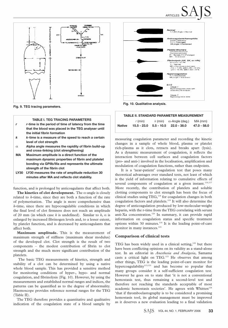
haemostasis process is a single product – the clot; and (ii) the clot’s physical properties (rate, strength, and stability), which will determine whether the patient will have normal haemostasis, will haemorrhage or will develop thrombosis. The TEG analyser involves two mechanical parts to measure the physical properties of the clot, a cup and a pin. The special stationary cylindrical cup holds the blood (0.36 ml) and is oscillated through an angle of 4°45´ at 37°C. Each rotation cycle lasts 10 seconds. The pin is suspended in the blood sample by a torsion wire and is monitored for motion, which is either transduced mechanically to a heated stylus, moving across heat-sensitive graph paper on a chart recorder, or in the newer models converted by a mechanical-electrical transducer to an electrical signal, which can be monitored by a computer. When no clot exists, the motion of the cuvette does not affect the pin and a straight line is recorded. The torque of the rotating cup is only transmitted to the immersed pin after fibrin-platelet bonding has linked the cup and pin together. The strength of these fibrin-platelet bonds affects the magnitude of the pin motion, such that strong clots move the pin directly in phase with the cup motion. The magnitude of the output is therefore directly related to the strength of the formed clot. As the clot retracts or lyses, these bonds are broken and the transfer of cup motion is diminished. The resulting haemostasis profile is a measure of the time it takes for the first fibrin strand to be formed, the kinetics of clot formation, the strength of the clot and dissolution of clot.
TEG assessment
The coagulation profile evaluated by the TEG is a graphic method of displaying a measure of the kinetics of clot formation and dissolution as well as of clot quality. It monitors shear elasticity, a physical property of a blood clot, and is therefore sensitive to all the interacting cellular and plasmatic components in the blood that affect the rate or structure of a clotting sample and its breakdown. The clot’s ability to perform useful mechanical work (the work of haemostasis) is a function of the net result of the interactive coagulation proteins and cellular elements involved in the process of haemostasis. In essence, the TEG analyser measures the ability of the clot to perform mechanical work throughout its structural development (Fig. 9). A recent advance is computerisation of the TEG, which essentially gives greater accuracy and consistency than manual measurements by removing the human error from the measurements. The TEG pattern is divided into component variables. The various parameters of the TEG are explained below and reflected in Table I. Reaction time. The time from the start of a sample run until the first significant levels of detectable clot formation (amplitude = 2 mm in the TEG tracing). This is the point at which most traditional coagulation assays reach their end-points. r-time is prolonged by anticoagulants and factor deficiencies and shortened by hypercoagulable conditions. Achievement of a certain clot firmness. The time from the measurement of r (beginning of clot formation) until a fixed level of clot firmness is reached (amplitude = 20 mm). Therefore, k-time is a measure of the speed or clot kinetics to reach a certain level of clot strength. k is shortened by increased fibrinogen level and, to a lesser extent, by platelet
pg 22-37.indd 32 3/6/06 10:20:06 Am
SAJSARTICLES
SAJS VOL 44, NO. 1, FEBRUARY 2006 33
function, and is prolonged by anticoagulants that affect both. The kinetics of clot development. The a-angle is closely related to k-time, since they are both a function of the rate of polymerisation. The angle is more comprehensive than k-time, since there are hypocoagulable conditions in which the final level of clot firmness does not reach an amplitude of 20 mm (in which case k is undefined). Similar to k, a is enlarged by increased fibrinogen levels and, to a lesser extent, by platelet function, and is decreased by anticoagulants that affect both. Maximum amplitude. This is the measurement of maximum strength of stiffness (maximum shear modulus) of the developed clot. Clot strength is the result of two components – the modest contribution of fibrin to clot strength and the much more significant contribution of the platelets. The basic TEG measurements of kinetics, strength and stability of a clot can be determined by using a native whole blood sample. This has provided a sensitive method for monitoring conditions of hyper-, hypo- and normal coagulation, and fibrinolysis (Fig. 10). However, by using the measurements and established normal ranges and indices, the patterns can be quantified as to the degree of abnormality. Haemoscope provides reference normal ranges for the TEG (Table II). The TEG therefore provides a quantitative and qualitative indication of the coagulation state of a blood sample by
measuring coagulation parameter and recording the kinetic changes in a sample of whole blood, plasma or platelet rich-plasma as it clots, retracts and breaks apart (lysis). As a dynamic measurement of coagulation, it reflects the interaction between cell surfaces and coagulation factors (pro- and anti-) involved in the localisation, amplification and modulation of coagulation functions, rather than endpoints. It is a ‘near-patient’ coagulation test that poses many theoretical advantages over standard tests, not least of which is the yield of information relating to cumulative effects of several components of coagulation at a given instant.116,117 More recently, the contribution of platelets and soluble clotting components to clot strength has been the focus of clinical studies using TEG,118 for coagulation changes in both coagulation factors and platelets.119 It will also determine the degree of anticoagulation produced by low-molecular-weight heparin, with the r-time from the TEG correlating with serum anti-Xa concentration.120 In summary, it can provide rapid information on coagulation status and specific treatment options within 30 minutes.116 It is the leading point-of-care monitor in many instances.121
Comparison of clinical tests
TEG has been widely used in a clinical setting,122 but there have been conflicting opinions on its validity as a stand-alone test. In an editorial in Anesthesia and Analgesia, Samama casts a critical light on TEG.123 He observes that among other things, TEG is the leading point-of-care monitor for hypercoagulability113,121 and has become so popular that many groups consider it a self-sufficient coagulation test. However he goes on to state that ‘it is not a conventional hemostasis test, thus remaining a second-level test and therefore not reaching the standards acceptable of most academic hemostasis societies’. He agrees with Whitten124 ‘that if thromboelastography is to be considered a promising hemostasis tool, its global management must be improved as it deserves a new evaluation leading to a final validation
TAble I. TeG TRAcInG pARAmeTeRsr r-time is the period of time of latency from the time
that the blood was placed in the TeG analyser until the initial fibrin formation
k k-time is a measure of the speed to reach a certain level of clot strength
a Alpha angle measures the rapidity of fibrin build-up and cross-linking (clot strengthening)
mA maximum amplitude is a direct function of the maximum dynamic properties of fibrin and platelet bonding via GpIIb/IIIa and represents the ultimate strength of the fibrin clot
lY30 lY30 measures the rate of amplitude reduction 30 minutes after MA and reflects clot stability.
Fig 10. Qualitative Analysis
The basic TEG measurements of kinetics, strength and stability of a clot can be determined
by using a native whole blood sample. This has provided a sensitive method for monitoring
conditions of hyper-, hypo-, normal coagulation, and fibrinolysis (Figure 10). However, by
using the measurements and established normal ranges and indices, the patterns can be
quantified as to the degree of abnormality. Haemoscope provides reference normal ranges for
the TEG (Table 2).
R (min) K (min) !-Angle (deg) MA (mm)
Native 15.5-23.0 5.5-10.5 22.0-38.0 47.0-58.0
Table 2. Standard Parameter Measurement
The TEG therefore provides a quantitative and qualitative indication of the coagulation state
of a blood sample by measuring coagulation parameter and recording the kinetic changes in a
sample of whole blood, plasma or platelet rich-plasma as it clots, retracts, and breaks apart
(lysis). As a dynamic measurement of coagulation, it reflects the interaction between cell
surfaces and coagulation factors (pro- and anti-) involved in the localisation, amplification and
modulation of coagulation functions, rather than endpoints.
It is a 'near patient' coagulation test, which poses many theoretical advantages to standard
tests, not least of which is the yield of information relating to cumulative effects of several
components of coagulation at a given instant116,117
. More recently, the contribution of platelets
and soluble clotting components to clot strength has been the focus of clinical studies using
thromboelastography118
, for coagulation changes in both coagulation factors and platelets119
.
Fig. 10. Qualitative analysis.
TAble II. sTAndARd pARAmeTeR meAsuRemenT
r(min) k(min) a-Angle(deg.) MA(mm)native 15.5 - 23.0 5.5 - 10.5 22.0 - 38.0 47.0 - 58.0
Fig. 9. TEG tracing parameters.
pg 22-37.indd 33 3/6/06 10:20:12 Am
SAJS
34 VOL 44, NO. 1, FEBRUARY 2006 SAJS
or it will remain an expensive, non-validated point-of-care monitor’.123
Conversely, TEG has been quoted as a proven useful research tool and compared with six common tests (the haematocrit, platelet count, fibrinogen, PT, aPTT and fibrin split products) by Zuckerman.125 The results indicated that, although there is a strong relationship between the thromboelastographic variables and these common laboratory tests, thromboelastographic variables contain additional information on the haemostatic process. TEG has been used to confirm normal coagulation in the face of an abnormal prothrombin time.126 Kang et al.127 found the relationship between TEG and standard laboratory tests of coagulation to be similar to that of other researchers,125,128 with the greatest correlation occurring between r-time and aPTT, while McNicol et al.129 found the relationship between standard tests of coagulation and TEG to be an inconsistent correlate. However, the authors comment that this is not surprising given that TEG variables are interdependent, measuring the interaction of the coagulation cascade and platelets in whole blood rather than specific endpoints in centrifuged plasma samples. The dynamic data provided by TEG allow far more appropriate replacement therapy.130 TEG is a real-time monitor of global haemostasis, enabling readily available analysis of coagulation in a shorter period of time than laboratory coagulation tests would take under ideal circumstances, thus providing useful and prompt identification of coagulation disorders.131 It allows rapid intraoperative diagnosis and specific management,117 covering the entire field of haemostasis in the perioperative setting, from platelet function, to fibrinolysis. Davis and Chandler132 found in their study that of various assays tested, only abnormal TEG values, a-angle (p < 0.01) and k-time (p < 0.04), were associated with an increased risk of bleeding. Bleeding times were not predictive of an increased risk of post-biopsy bleeding. All prothrombin time, partial thromboplastin time and platelet count abnormalities were mild and none of these assays predicted bleeding. It was concluded that TEG was the best assay for detecting mild coagulation abnormalities associated with an increased risk of bleeding. With the advent of computerisation in the past decade, TEG has evolved from a research laboratory tool into a compact processor, providing global information on the entire coagulation process. The battery of traditional coagulation tests is based on the isolated, static laboratory end-points and as a result they do not take into account the interactions within the clotting cascade and platelets in whole blood. Haemostasis is an integrated, interactive, dynamic, and extremely complex process involving coagulation proteins, activators, cellular elements and inhibitors, which is recorded by TEG,133 while standard tests of coagulation stop measuring at the first onset of clot formation – in other words, at static endpoints of blood coagulation. This provides no information on the dynamics of clot formation, strength and stability. As a result, it is difficult to assess what the exact dynamic process for inhibition of clotting is. In the clinical setting, TEG may be used to rapidly assess coagulation abnormalities in the initial assessment of coagulation in trauma patients.134 When compared with platelet count and PT/aPTT only TEG was predictive of early transfusion (p < 0.05), leading to the conclusion that TEG is a rapid, simple test that can broadly determine coagulation abnormalities. TEG and Sonoclot analysis were 100% accurate in predicting bleeding after cardiopulmonary
bypass and significantly better predictors of postoperative haemorrhage than routine coagulation tests.135 Essell et al.136 tested the utility of TEG versus platelet studies (BT, platelet count, mean platelet volume) and standard coagulation tests (PT, aPTT, fibrinogen). The sensitivities of the BT (71.4%) and platelet count (100%) were similar to TEG (71.4%), but the specificity (89.3%) of TEG in predicting postoperative bleeding was greater than that of the bleeding time (78.5%) and platelet count (53.6%), suggesting that patients with a normal TEG who have a post-bypass bleed should not be given blood products empirically, but that a surgical cause should rather be excluded. Kang et al.127 found TEG to be a reliable and rapid monitoring system during liver transplantation and Martin et al.137 validate the role of TEG during paediatric liver surgery, finding a 100% sensitivity (8/8) and 73% specificity (8/11) in predicting increased postoperative bleeding. In a study comparing the sensitivity of the ACT, aPTT and TEG, the ACT was less sensitive to residual heparin anticoagulation than aPTT or TEG.138 In addition TEG using heparinase cups to analyse anticoagulated blood during cardiac surgery is useful to guide treatment with haemostatic components. Twelve-hour chest tube losses were not different between groups despite the threefold reduction in the use of haemostatic products, showing that intra-operative monitoring of coagulation in the anticoagulated patient can be used to guide treatment.139 TEG, by providing information on the interaction of all the coagulation precursors, gives more clinically useful information on coagulation than that available from the coagulation profile or the activated clotting time alone. TEG was a significantly better predictor (87% accuracy) of postoperative haemorrhage and need for re-operation than was the ACT (30%) or coagulation profile (51%),140 with TEG providing diagnostic data within 30 minutes of blood sampling. TEG is also useful in assessing platelet function in the presence of thrombocytopenia141 and pre-eclampsia/eclampsia.142
In screening for hypercoagulability, standard laboratory tests that may indicate hypercoagulability include a high platelet count, high levels of coagulation factors VII, VIII, IX, X and XI, or fibrinogen levels, low levels of protein S, protein C, antithrombin, and the presence of a lupus anticoagulant, while the TEG indicators of hypercoagulability are a short r-time and k-time, a large MA or an a-angle that is large and rapidly increasing. This has been noted with increased frequency in a variety of patient groups who are considered to be at increased risk of thrombosis, with the predictive value of the TEG being higher than other systems. Hypercoagulability may not be reflected by standard coagulation monitoring and may be predominantly influenced by increased platelet reactivity,143 which needs to be measured as an dynamic test and not as an end-point.
ConclusionCoagulation is a dynamic and intricate process that becomes more complicated to understand as more is discovered about the process. On the other hand, the clinical end-point is still ‘Will the patient clot or not?’ With the many interactions taking place concomitantly there is not one simple answer to establish the exact state of the coagulation system, but the dynamic coagulation tests like the TEG are definitely getting closer to providing an understanding. Certainly rapid bedside, or theatre, information on coagulation status with rapid analysis of appropriate therapy will go far in managing
pg 22-37.indd 34 3/6/06 10:20:12 Am

SAJSARTICLES
SAJS VOL 44, NO. 1, FEBRUARY 2006 35
the coagulation status, especially when taken in conjunction with the clinical findings.
Much of this work was based on my Ph.D. thesis and a chapter on ‘Fluids and coagulation’ currently in press. Some of the diagrams were courtesy of Mike James (based on slides from Novo Nordisc) and Eli Cohen (Haemoscope).
REFERENCES
1. Camera M et al. Cooperation between VEGF and TNF-alpha is necessary for exposure of active tis-sue factor on the surface of human endothelial cells. Arterioscler Thromb Vasc Biol 1999; 19: 531-537.
2. Weiss HJ, Turitto VT, Baumgartner HR, Nemerson Y, Hoffmann T. Evidence for the presence of tissue factor activity on subendothelium. Blood 1989; 73: 968-975.
3. Yang HL, Lu FJ, Wung SL, Chiu HC. Humic acid induces expression of tissue factor by cultured endothe-lial cells: regulation by cytosolic calcium and protein kinase C. Thromb Haemost 1994; 71: 325-330.
4. Lawson JH, Mann KG. Cooperative activation of human factor IX by the human extrinsic pathway of blood coagulation. J Biol Chem 1991; 266: 11317-11327.
5. Osterud B, Rapaport SI. Activation of factor IX by the reaction product of tissue factor and factor VII: addi-tional pathway for initiating blood coagulation. Proc Natl Acad Sci USA 1977; 74: 5260-5264.
6. Luchtman-Jones L, Broze GJ jun. The current status of coagulation. Ann Med 1995; 27: 47-52.
7. Macfarlane RG. A clotting scheme for 1964. Thromb Diath Haemorrh Suppl 1965; 17: 45-52.
8. Ratnoff OD. The development of knowledge about haemostasis. In: Bloom AL, Forbes CD, Thomas DP, Tuddenham EGD, eds. Haemostasis and Thrombosis. New York: Churchill Livingstone, 1994: 3-28.
9. Gallus AS, Lee LH, Coghlan DW. New aspects of the blood coagulation cascade, anticoagulants and vein thrombosis in Asia. Ann Acad Med Singapore 2002; 31: 685-696.
10. Eichinger S, et al. Determinants of plasma factor VIIa levels in humans. Blood 1995; 86: 3021-3025.
11. Morrissey JH, Macik BG, Neuenschwander PF, Comp PC. Quantitation of activated factor VII levels in plas-ma using a tissue factor mutant selectively deficient in promoting factor VII activation. Blood 1993; 81: 734-744.
12. Broze GJ. The tissue factor pathway of coagulation: Factor VII, tissue factor, and tissue factor pathway inhibitor. In: Bloom AL, Forbes CD, Thomas DP, Tuddenham EGD, eds. Haemostasis and Thrombosis. New York: Churchill Livingstone, 1994: 349-377.
13. Rao LV, Rapaport SI. Activation of factor VII bound to tissue factor: a key early step in the tissue factor pathway of blood coagulation. Proc Natl Acad Sci USA 1988; 85: 6687-6691.
14. Iijima K, Fukuda C, Nakamura K. Measurements of tissue factor-like activity in plasma of patients with DIC. Thromb Res 1991; 61: 29-38.
15. Butenas S, van ‘, V, Mann KG. Evaluation of the ini-tiation phase of blood coagulation using ultrasensitive assays for serine proteases. J Biol Chem 1997; 272: 21527-21533.
16. Rand MD, Lock JB, van’t Veer C, Gaffney DP, Mann KG. Blood clotting in minimally altered whole blood. Blood 1996; 88: 3432-3445.
17. Jesty J, Silverberg SA. Kinetics of the tissue factor-dependent activation of coagulation Factors IX and X in a bovine plasma system. J Biol Chem 1979; 254: 12337-12345.
18. Zur M, Nemerson Y. Kinetics of factor IX activation via the extrinsic pathway. Dependence of Km on tissue factor. J Biol Chem 1980; 255: 5703-5707.
19. Repke D et al. Hemophilia as a defect of the tissue fac-tor pathway of blood coagulation: effect of factors
VIII and IX on factor X activation in a continuous-flow reactor. Proc Natl Acad Sci USA 1990; 87: 7623-7627.20. Jesty J, Nêmerson Y. The pathways of blood coagula-
tion. In: Beutler E, Lichtman MA, Coller BS, Kipps TJ, eds. Williams Hematology. New York: McGraw-Hill, 1995: 1227-1238.
21. Pixley RA, Colemann RW. Factor XII: Hagemann factor. In: Lorand L, Mann KG, eds. Methods in Enzymology, Part A. San Diego: Academic, 1993: 51-64.
22. Mandle R jun., Kaplan AP. Hageman factor substrates. Human plasma prekallikrein: mechanism of activation by Hageman factor and participation in Hageman factor-dependent fibrinolysis. J Biol Chem 1977; 252: 6097-6104.
23. Revak SD, Cochrane CG, Griffin JH. The binding and cleavage characteristics of human Hageman factor dur-ing contact activation. A comparison of normal plasma with plasmas deficient in factor XI, prekallikrein, or high molecular weight kininogen. J Clin Invest 1977; 59: 1167-1175.
24. Fujikawa K, Legaz ME, Kato H, Davie EW. The mech-anism of activation of bovine factor IX (Christmas factor) by bovine factor XIa (activated plasma throm-boplastin antecedent). Biochemistry 1974; 13: 4508-4516.
25. Hultin MB, Nemerson Y. Activation of factor X by fac-tors IXa and VIII; a specific assay for factor IXa in the presence of thrombin-activated factor VIII. Blood 1978; 52: 928-940.
26. Tans G, et al. Comparison of anticoagulant and proco-agulant activities of stimulated platelets and platelet-derived microparticles. Blood 1991; 77: 2641-2648.
27. Jackson CM. Physiology and biochemistry of pro-thrombin. In: Bloom AL, Forbes CD, Thomas DP, Tuddenham EGD, eds. Haemostasis and Thrombosis. New York: Churchill Livingstone, 1994: 397-438.
28. Kalafatis M, Swords NA, Rand MD, Mann KG. Membrane-dependent reactions in blood coagulation: role of the vitamin K-dependent enzyme complexes. Biochim Biophys Acta 1994; 1227: 113-129.
29. Nesheim ME, Taswell JB, Mann KG. The contribution of bovine factor V and factor Va to the activity of pro-thrombinase. J Biol Chem 1979; 254: 10952-10962.
30. Tracy PB et al. Factor V (Quebec): a bleeding diathesis associated with a qualitative platelet Factor V deficien-cy. J Clin Invest 1984; 74: 1221-1228.
31. Gailani D, Broze GJ jun. Factor XI activation in a revised model of blood coagulation. Science 1991; 253: 909-912.
32. Vu TK, Wheaton VI, Hung DT, Charo I, Coughlin SR. Domains specifying thrombin-receptor interaction. Nature 1991; 353: 674-677.
33. Doolittle RF. Fibrinogen and fibrin. Annu Rev Biochem 1984; 53: 195-229.
34. Mosesson MW. The roles of fibrinogen and fibrin in hemostasis and thrombosis. Semin Hematol 1992; 29: 177-188.
35. Naski MC, Lorand L, Shafer JA. Characterization of the kinetic pathway for fibrin promotion of alpha-thrombin-catalyzed activation of plasma factor XIII. Biochemistry 1991; 30: 934-941.
36. McDonagh J. Structure and function of factor XIII. In: Coleman RW, Hirsh J, Marder VJ, Salzman EW, eds. Hemostasis and Thrombosis: Basic Principles and Clinical Practice. Philadelphia: Lippincott, 1994: 301-313.
37. Brummel KE, Butenas S, Mann KG. An integrated study of fibrinogen during blood coagulation. J Biol Chem 1999; 274: 22862-22870.
38. Esmon CT, Esmon NL, Le Bonniec BF, Johnson AE. Protein C activation. Methods Enzymol 1993; 222: 359-385.
39. Esmon NL, Carroll RC, Esmon CT. Thrombomodulin blocks the ability of thrombin to activate platelets. J Biol Chem 1983; 258: 12238-12242.
40. Broze GJ jun, et al. The lipoprotein-associated coagula-tion inhibitor that inhibits the factor VII-tissue factor complex also inhibits factor Xa: insight into its possible mechanism of action. Blood 1988; 71: 335-343.
pg 22-37.indd 35 3/6/06 10:20:20 Am
SAJS
36 VOL 44, NO. 1, FEBRUARY 2006 SAJS
41. Eaton D, Rodriguez H, Vehar GA. Proteolytic processing of human factor VIII. Correlation of specific cleavages by thrombin, factor Xa, and acti-vated protein C with activation and inactivation of factor VIII coagulant activity. Biochemistry 1986; 25: 505-512.
42. Esmon CT. The regulation of natural anticoagulant pathways. Science 1987; 235: 1348-1352.
43. Hockin MF, Kalafatis M, Shatos M, Mann KG. Protein C activation and factor Va inactivation on human umbilical vein endothelial cells. Arterioscler Thromb Vasc Biol 1997; 17: 2765-2775.
44. Olson ST, Bjork I, Shore JD. Kinetic characterization of heparin-catalyzed and uncatalyzed inhibition of blood coagulation proteinases by anti-thrombin. Methods Enzymol 1993; 222: 525-559.
45. Rapaport SI. The extrinsic pathway inhibitor: a regulator of tissue factor-dependent blood coagulation. Thromb Haemost 1991; 66: 6-15.
46. Lawson JH, Kalafatis M, Stram S, Mann KG. A model for the tissue fac-tor pathway to thrombin. I. An empirical study. J Biol Chem 1994; 269: 23357-2366.
47. Cawthern KM, et al. Blood coagulation in hemophilia A and hemophilia C. Blood 1998; 91: 4581-4592.
48. George JN. Platelets. Lancet 2000; 355: 1531-1439.49. Deckmyn H, Vanhoorelbeke K, Peerlinck K. Inhibitory and activating
human antiplatelet antibodies. Baillieres Clin Haematol 1998; 11: 343-359.50. Rosing J, et al. The role of activated human platelets in prothrombin and
factor X activation. Blood 1985; 65: 319-332.51. Walsh PN. Platelet coagulant activities and hemostasis: a hypothesis. Blood
1974; 43: 597-605.52. Kottke-Marchant K, Corcoran G. The laboratory diagnosis of platelet dis-
orders. Arch Pathol Lab Med 2002; 126: 133-146.53. Marcus AJ. Platelets and their disorders. In: Ratnoff OC, Forbes CD, eds.
Disorders of Hemostasis. Philadelphia, Pa: WB Saunders, 1996: 79-137.54. Meyer D, Baumgartner HR. Role of von Willebrand factor in platelet
adhesion to the subendothelium. Br J Haematol 1983; 54: 1-9.55. Siedlecki CA, et al. Shear-dependent changes in the three-dimensional
structure of human von Willebrand factor. Blood 1996; 88: 2939-2950.56. Tracy PR. Role of platelets and leukocytes in coagulation. In: Colman
RW, Hirsh J, Marder VJ, Clowes AW, George JN, eds. Hemostasis and Thrombosis: Basic Principles and Clinical Practice. Philadelphia, Pa: JB Lippincott, 2001: 575-596.
57. Ruggeri ZM, Savage B. Biological functions of von Willebrand factor. In: Ruggeri ZM, ed. Von Willebrand Factor and the Mechanisms of Platelet Function. Berlin: Springer-Verlag, 1998: 79-109.
58. Savage B, Saldivar E, Ruggeri ZM. Initiation of platelet adhesion by arrest onto fibrinogen or translocation on von Willebrand factor. Cell 1996; 84: 289-297.
59. Fukami MH, Holmsen H, Kowalski MA, Niewiarowski S. Platelet secretion. In: Colman RW, Hirsh J, Marder VJ, Clowes AW, George JN, eds. Hemostasis and Thrombosis: Basic Principles and Clinical Practice. Phladelphia, Pa: JB Lippincott Co, 2001: 561-574.
60. Peerschke EI. Platelet membrane glycoproteins. Functional characteriza-tion and clinical applications. Am J Clin Pathol 1992; 98: 455-463.
61. Zwaal RF, Comfurius P, Bevers EM. Mechanism and function of changes in membrane-phospholipid asymmetry in platelets and erythrocytes. Biochem Soc Trans 1993; 21: 248-253.
62. Zwaal RF. Membrane and lipid involvement in blood coagulation. Biochim Biophys Acta 1978; 515: 163-205.
63. Zwaal RFA, Bevers EM, Comfurius P. Platelets and coagulation. In: Zwaal RFA, Hemker HC, eds. Blood Coagulation. New York: Elsevier Science Publishers, 1986: 141-169.
64. Mann KG, Krishnaswamy S, Lawson JH. Surface-dependent hemostasis. Semin Hematol 1992; 29: 213-226.
65. Tracy PB, Nesheim ME, Mann KG. Coordinate binding of factor Va and factor Xa to the unstimulated platelet. J Biol Chem 1981; 256: 743-751.
66. Nelsestuen GL, Kisiel W, Di Scipio RG. Interaction of vitamin K depend-ent proteins with membranes. Biochemistry 1978; 17: 2134-2138.
67. Greenberg CS, Achyuthan KE, Fenton JW. Factor XIIIa formation pro-moted by complexing of alpha-thrombin, fibrin, and plasma factor XIII. Blood 1987; 69: 867-871.
68. Lewis SD, Janus TJ, Lorand L, Shafer JA. Regulation of formation of fac-tor XIIIa by its fibrin substrates. Biochemistry 1985; 24: 6772-6777.
69. Naito K, Fujikawa K. Activation of human blood coagulation factor XI independent of factor XII. Factor XI is activated by thrombin and factor XIa in the presence of negatively charged surfaces. J Biol Chem 1991; 266: 7353-7358.
70. Komiyama Y, Pedersen AH, Kisiel W. Proteolytic activation of human factors IX and X by recombinant human factor VIIa: effects of calcium, phospholipids, and tissue factor. Biochemistry 1990; 29: 9418-9425.
71. Mann KG, Jenny RJ, Krishnaswamy S. Cofactor proteins in the assembly and expression of blood clotting enzyme complexes. Annu Rev Biochem
1988; 57: 915-956.72. Radcliffe R, Nemerson Y. Activation and control of factor VII by activated
factor X and thrombin. Isolation and characterization of a single chain form of factor VII. J Biol Chem 1975; 250: 388-395.
73. Shuman MA, Majerus PW. The measurement of thrombin in clotting blood by radioimmunoassay. J Clin Invest 1976; 58: 1249-1258.
74. Butenas S, Mann KG. Blood coagulation. Biochemistry (Mosc) 2002; 67: 3-12.
75. Neuenschwander P, Jesty J. A comparison of phospholipid and platelets in the activation of human factor VIII by thrombin and factor Xa, and in the activation of factor X. Blood 1988; 72: 1761-1770.
76. Giddings JC, Bloom AL. Factor-V activation by thrombin and its role in prothrombin conversion. Br J Haematol 1975; 29: 349-364.
77. Baruch D, Hemker HC, Lindhout T. Kinetics of thrombin-induced release and activation of platelet factor V. Eur J Biochem 1986; 154: 213-218.
78. Jesty J, Beltrami E, Willems G. Mathematical analysis of a proteolytic posi-tive-feedback loop: dependence of lag time and enzyme yields on the ini-tial conditions and kinetic parameters. Biochemistry 1993; 32: 6266-6274.
79. Rosenberg RD. Regulation of the hemostatic mechanism. In: Stamatoyannopoulos G, Nienhuis AW, Leder P, Majerus PW, eds. The Molecular Basis of Blood Diseases. Philadelphia: Saunders, 1987: 534-574.
80. Rosenberg RD, Rosenberg JS. Natural anticoagulant mechanisms. J Clin Invest 1984; 74: 1-6.
81. Esmon CT. The roles of protein C and thrombomodulin in the regulation of blood coagulation. J Biol Chem 1989; 264: 4743-4746.
82. van ‘, V, Mann KG. Regulation of tissue factor initiated thrombin genera-tion by the stoichiometric inhibitors tissue factor pathway inhibitor, anti-thrombin-III, and heparin cofactor-II. J Biol Chem 1997; 272: 4367-4377.
83. van ‘, V, Golden NJ, Kalafatis M, Mann KG. Inhibitory mechanism of the protein C pathway on tissue factor-induced thrombin generation. Synergistic effect in combination with tissue factor pathway inhibitor. J Biol Chem 1997; 272: 7983-7994.
84. Travis J, Salvesen GS. Human plasma proteinase inhibitors. Annu Rev Biochem 1983; 52: 655-709.
85. Rosenberg RD, Damus PS. The purification and mechanism of action of human antithrombin-heparin cofactor. J Biol Chem 1973; 248: 6490-6505.
86. Kurachi K, Fujikawa K, Schmer G, Davie EW. Inhibition of bovine factor IXa and factor Xabeta by antithrombin III. Biochemistry 1976; 15: 373-377.
87. Rosenberg JS, McKenna PW, Rosenberg RD. Inhibition of human factor IXa by human antithrombin. J Biol Chem 1975; 250: 8883-8838.
88. Stead N, Kaplan AP, Rosenberg RD. Inhibition of activated factor XII by antithrombin-heparin cofactor. J Biol Chem 1976; 251: 6481-6488.
89. Jesty J. The kinetics of formation and dissociation of the bovine thrombin.antithrombin III complex. J Biol Chem 1979; 254: 10044-10050.
90. Downing MR, Bloom JW, Mann KG. Comparison of the inhibition of thrombin by three plasma protease inhibitors. Biochemistry 1978; 17: 2649-2653.
91. Jesty J. The kinetics of inhibition of alpha-thrombin in human plasma. J Biol Chem 1986; 261: 10313-10318.
92. Jesty J. Measurement of the kinetics of inhibition of activated coagulation factor X in human plasma: the effect of plasma and inhibitor concentra-tion. Anal Biochem 1986; 152: 402-411.
93. Tollefsen DM. Heparin cofactor II deficiency. Arch Pathol Lab Med 2002; 126: 1394-1400.
94. Rao LV, Rapaport SI. Studies of a mechanism inhibiting the initiation of the extrinsic pathway of coagulation. Blood 1987; 69: 645-651.
95. Sanders NL, Bajaj SP, Zivelin A, Rapaport SI. Inhibition of tissue factor/factor VIIa activity in plasma requires factor X and an additional plasma component. Blood 1985; 66: 204-212.
96. Seegers WH, Novoa E, Henry RL, Hassouna HI. Relationship of ‘new’ vitamin K-dependent protein C and ‘old’ autoprothrombin II-a. Thromb Res 1976; 8: 543-552.
97. Ehrlich HJ, et al. Alteration of serpin specificity by a protein cofactor. Vitronectin endows plasminogen activator inhibitor 1 with thrombin inhibitory properties. J Biol Chem 1990; 265: 13029-13035.
98. Ehrlich HJ, Keijer J, Preissner KT, Gebbink RK, Pannekoek H. Functional interaction of plasminogen activator inhibitor type 1 (PAI-1) and heparin. Biochemistry 1991; 30: 1021-1028.
99. Whalen J, Tuman KJ. Monitoring hemostasis. Int Anesthesiol Clin 1996; 34: 195-213.
100. Bajaj SP, Joist JH. New insights into how blood clots: implications for the use of APTT and PT as coagulation screening tests and in monitoring of anticoagulant therapy. Semin Thromb Hemost 1999; 25: 407-418.
101. Miletich JP. Prothrombin time. In: Beutler E, Lichtman MA, Coller BS, Kipps TJ, eds. Williams Hematology. New York: McGraw-Hill, 1995: L82-L84.
pg 22-37.indd 36 3/6/06 10:20:21 Am
SAJSARTICLES
SAJS VOL 44, NO. 1, FEBRUARY 2006 37
102. Kitchens CS. Prolonged activated partial thrombo-plastin time of unknown etiology: a prospective study of 100 consecutive cases referred for consultation. Am J Hematol 1988; 27: 38-45.
103. Miletich JP. Activated partial thromboplastin time. In: Beutler E, Lichtman MA, Coller BS, Kipps TJ, eds. Williams Hematology. New York: McGraw-Hill, 1995: L85-L86.
104. Blackbeard, J. Full Blood Counts and Differential Counts on the Adiva 120. Hindley, N. MB046.R00. 2002. Department of Clinical Laboratory Sciences, University of Cape Town.
105. Miletich JP. Bleeding time. In: Beutler E, Lichtman MA, Coller BS, Kipps TJ, eds. Williams Hematology. New York: McGraw-Hill, 1995: L111-L112.
106. Peterson P, et al. The preoperative bleeding time test lacks clinical benefit: College of American Pathologists’ and American Society of Clinical Pathologists’ position article. Arch Surg 1998; 133: 134-139.
107. Miletich JP. Activated clotting time. In: Beutler E, Lichtman MA, Coller BS, Kipps TJ, eds. Williams Hematology. New York: McGraw-Hill, 1995: L86.
108. Hindley, N. Platelet Aggregation. Williamson, A. C022.R01. 2003. Department of Clinical Laboratory Sciences, University of Cape Town.
109. Hindley, N. Antithrombin III. Williamson, A. C023.R01. 2002. Department of Clinical Laboratory Sciences, University of Cape Town.
110. Dade Behring. Enzygnost TAT micro . June 1998. 1998.
111. Hindley, N. D-Dimers (XDP). Williamson, A. C008.R01. 2003. Department of Clinical Laboratory Sciences, University of Cape Town.
112. Konrad C, Markl T, Schuepfer G, Gerber H, Tschopp M. The effects of in vitro hemodilution with gela-tin, hydroxyethyl starch, and lactated Ringer’s solu-tion on markers of coagulation: an analysis using SONOCLOT. Anesth Analg 1999; 88: 483-488.
113. Francis JL, Francis DA, Gunathilagan GJ. Assessment of hypercoagulability in patients with cancer using the Sonoclot Analyzer and thromboelastography. Thromb Res 1994; 74: 335-346.
114. Steer PL, Krantz HB. Thromboelastography and Sonoclot analysis in the healthy parturient. J Clin Anesth 1993; 5: 419-424.
115. Ekback G, Schott U, Axelsson K, Carlberg M. Perioperative autotransfusion and functional coagula-tion analysis in total hip replacement. Acta Anaesthesiol Scand 1995; 39: 390-395.
116. Salooja N, Perry DJ. Thrombelastography. Blood Coagul Fibrinolysis 2001; 12: 327-337.
117. Tuman KJ, Spiess BD, McCarthy RJ, Ivankovich AD. Effects of progressive blood loss on coagulation as measured by thrombelastography. Anesth Analg 1987; 66: 856-863.
118. Nielsen VG, Geary BT, Baird MS. Evaluation of the contribution of platelets to clot strength by throm-boelastography in rabbits: the role of tissue factor and cytochalasin D. Anesth Analg 2000; 91: 35-39.
119. Oshita K, Az-ma T, Osawa Y, Yuge O. Quantitative measurement of thromboelastography as a function of platelet count. Anesth Analg 1999; 89: 296-299.
120. Klein SM et al. Thromboelastography as a periop-erative measure of anticoagulation resulting from low molecular weight heparin: a comparison with anti-Xa concentrations. Anesth Analg 2000; 91: 1091-1095.
121. Gibbs NM, Crawford GP, Michalopoulos N. Thrombelastographic patterns following abdominal aortic surgery. Anaesth Intensive Care 1994; 22: 534-538.
122. Mallett SV, Cox DJ. Thrombelastography. Br J Anaesth 1992; 69: 307-313.
123. Samama CM. Thromboelastography: the next step. Anesth Analg 2001; 92: 563-564.
124. Whitten CW, Greilich PE. Thromboelastography: past, present, and future. Anesthesiology 2000; 92: 1223-1225.
125. Zuckerman L, Cohen E, Vagher JP, Woodward E, Caprini JA. Comparison of thrombelastography with common coagulation tests. Thromb Haemost 1981; 46: 752-756.
126. Pivalizza EG, Henderson KL, Craig AL. Discrepancy between thromboelastography and prothrombin time. Anesthesiology 1996; 84: 1262.
127. Kang YG et al. Intraoperative changes in blood coagu-lation and thrombelastographic monitoring in liver transplantation. Anesth Analg 1985; 64: 888-896.
128. Howland WS, Schweizer O, Gould P. A comparison of intraoperative measurements of coagulation. Anesth Analg 1974; 53: 6576-6563.
129. McNicol PL et al. Patterns of coagulopathy during liver transplantation: experience with the first 75 cases using thrombelastography. Anaesth Intensive Care 1994; 22: 659-665.
130. Chapin JW, Peters KR, Winslow J, Becker GL, Skinner H. Circulating heparin during liver transplantation. Transplant Proc 1993; 25: 1803.
131. Pivalizza EG, Abramson DC. Thromboelastography: another point of view. Anesth Analg 2001; 93: 517-518.
132. Davis CL, Chandler WL. Thromboelastography for the prediction of bleeding after transplant renal biopsy. J Am Soc Nephrol 1995; 6: 1250-1255.
133. Srinivasa V, Gilbertson LI, Bhavani-Shankar K. Thromboelastography: where is it and where is it heading? Int Anesthesiol Clin 2001; 39: 35-49.
134. Kaufmann CR, Dwyer KM, Crews JD, Dols SJ, Trask AL. Usefulness of thrombelastography in assessment of trauma patient coagulation. J Trauma 1997; 42: 716-720.
135. Tuman KJ, Spiess BD, McCarthy RJ, Ivankovich AD. Comparison of viscoelastic measures of coagulation after cardiopulmonary bypass. Anesth Analg 1989; 69: 69-75.
136. Essell JH, Martin TJ, Salinas J, Thompson JM, Smith VC. Comparison of thromboelastography to bleeding time and standard coagulation tests in patients after cardiopulmonary bypass. J Cardiothorac Vasc Anesth 1993; 7: 410-415.
137. Martin P, Horkay F, Rajah SM, Walker DR. Monitoring of coagulation status using thrombelastog-raphy during paediatric open heart surgery. Int J Clin Monit Comput 1991; 8: 183-187.
138. Murray DJ et al. Heparin detection by the activated coagulation time: a comparison of the sensitivity of coagulation tests and heparin assays. J Cardiothorac Vasc Anesth 1997; 11: 24-28.
139. Royston D, von Kier S. Reduced haemostatic factor transfusion using heparinase-modified thrombelastog-raphy during cardiopulmonary bypass. Br J Anaesth 2001; 86: 575-578.
140. Spiess BD, et al. Thromboelastography as an indicator of post-cardiopulmonary bypass coagulopathies. J Clin Monit 1987; 3: 25-30.
141. Wong CA, Liu S, Glassenberg R. Comparison of thrombelastography with common coagulation tests in preeclamptic and healthy parturients. Reg Anesth 1995; 20: 521-527.
142. Orlikowski CE, et al. Thrombelastography changes in pre-eclampsia and eclampsia. Br J Anaesth 1996; 77: 157-161.
143. Mahla E, et al. Thromboelastography for monitoring prolonged hypercoagulability after major abdominal surgery. Anesth Analg 2001; 92: 572-577.
pg 22-37.indd 37 3/6/06 10:20:25 Am