Embed Size (px)
Citation preview
Review ArticleClinical Relevance of the Advanced Microbiologic andBiochemical Investigations in Periodontal Diagnosis:A Critical Analysis
Vishakha Grover,1 Anoop Kapoor,2 Ranjan Malhotra,1 and Gagandeep Kaur1
1 Department of Periodontics & Oral Implantology, National Dental College & Hospital, Gulabgarh, Derabassi, District SAS Nagar,Mohali, Punjab 140507, India
2Department of Periodontics & Oral Implantology, M.N.D.A.V. Dental College & Hospital, Solan, Himachal Pradesh 173223, India
Correspondence should be addressed to Vishakha Grover; vishakha [email protected]
Received 5 May 2014; Accepted 16 October 2014; Published 17 November 2014
Academic Editor: Gul Atilla
Copyright © 2014 Vishakha Grover et al. This is an open access article distributed under the Creative Commons AttributionLicense, which permits unrestricted use, distribution, and reproduction in any medium, provided the original work is properlycited.
New approaches to periodontal diagnosis, including advanced microbiologic, biochemical, and genetic tests, have been shownto provide the clinician with the information not available by traditional means. The purpose of a diagnostic test is to confirm,exclude, classify, or monitor disease to guide treatment.Their clinical value depends on whether the information they provide leadsto improved patient outcomes.This can be assessed by randomized trials, which compare patient outcomes from the new diagnostictest versus the old test strategy. Being nonmandatory for marketing approval, such trials are not always feasible because of largesample sizes requirements. So, many diagnostic tests enter the practice without being critically analysed for any additional benefits.Effective diagnosis is just as essential as the selection of effective treatments for the success of periodontal therapy. So, the currentpaper aims to focus on the practical utility of this rapidly emerging plethora of periodontal diagnostic tools, emphasizing the criticalissues surrounding the clinical application of microbiologic and biochemical investigations, employed for periodontal diagnosis.
1. Introduction
“Periodontal diagnosis” is an important tag that a clinicianties on the periodontal disease condition of the patient, cap-turing all his past experience with the condition in question.The entire constellation of signs and symptoms, along with adetailed history, is elicited, documented, and interpreted toreach at a diagnosis. Most often an accurate diagnosis is thevery first concrete step towards the planning and executionof an appropriate individualized treatment plan, contributingsignificantly towards the success of the therapy [1].
Clinical diagnostic parameters thatwere introducedmorethan half a century ago continue to function as the basicmodel for periodontal diagnosis in current clinical practice aswell. A periodontal diagnostic tool, in general, provides per-tinent information for differential diagnosis, localization ofdisease, and severity of infection. They include various dis-ease characteristics such as probing pocket depths, bleeding
on probing, clinical attachment levels, plaque index, andradiographs quantifying alveolar bone levels [2, 3]. Althoughthere have been significant advances in the understanding ofthe etiopathogenesis of periodontal disease over the past 4-5 decades, the traditional methods by which clinicians diag-nose periodontal disease have remained virtually unchanged[4].
These diagnostics were called in to question during theearly 1980s, when longitudinal clinical studies demonstratedthat long-held concepts concerning the natural history ofperiodontal disease required modification [5]. More recentparadigms for periodontitis diagnosis include the possibilityof several disease types, based primarily on the rate of diseaseprogression, the distribution of the disease within themouth,and the chronological age of the patient as well as active andinactive stages of the disease. Since then, clinicians got to beinterested in assessment tools that should give them informa-tion in the following three areas:
Hindawi Publishing CorporationJournal of Oral DiseasesVolume 2014, Article ID 785615, 11 pageshttp://dx.doi.org/10.1155/2014/785615
2 Journal of Oral Diseases
(i) diagnostic tests that could determine whether theperiodontal disease process is currently active (pro-gressive loss of attachment) with accuracy above whatcan be determined by traditional clinical indicators;
(ii) risk assessment, by which clinicians could identifypatients or specific sites that are at higher risk fordisease onset;
(iii) prognosis assessment, by which clinicians could pre-dict the course of disease with or without treatment[6, 7].
Traditional clinical assessments do not enable a practi-tioner performing a single routine periodontal examinationto determine whether active tissue destruction is occurring,for example, no definitive method to determine that gingivalinflammation in a successfully treated case of periodontitisrepresents early recurrent disease or gingivitis on a stablebut reduced periodontium [4, 8]. Albeit easy to use, cost-effective, and relatively noninvasive, clinical attachment lossevaluation using the periodontal probe measures damagefrom past episodes of destruction but requires a 2-3mmthreshold change before a site with significant breakdown canbe identified. Demonstrating progressive loss of periodontalsupport requires longitudinal assessment. Current diagnosticmethodologies do not enable us to accurately predict whichperiodontal sites, teeth, or individuals are susceptible tofurther periodontal breakdown [2, 9, 10]. Given the limita-tions of current diagnostic tools, researchers are continuouslyworking to develop techniques that focus on the earlydetection, disease activity, and host susceptibility of disease[4, 8].
With the advent of so many new diagnostic tests devel-oped in past few decades, we must not mislead ourselvesto the belief that everything we can measure will alwaysbe helpful. Many traditionally taught methods have neverbeen scrutinized for their precise benefits, and new testsare made available without any properly documented utility.To determine the diagnostic utility (the quality of being ofpractical use), detailed information is needed on how a testor diagnostic algorithm performs in a specific setting andwhat the consequences of a positive or negative test might be[8]. Detection of periodontal disease is seldom the principalproblem in periodontics. One and the same test can have vari-able utility depending on the information already availablebefore the test is done. The ideal diagnostic test should be[10, 11]
(i) highly specific,(ii) highly sensitive,(iii) reproducible,(iv) quantitative,(v) simple to perform,(vi) rapid,(vii) a one-stage or a two-stage procedure,(viii) noninvasive,(ix) versatile in terms of sample handling, storage, and
transport,
(x) amenable to chairside use,(xi) economical,(xii) dependent upon simple and robust instrumentation.The clinical value further largely depends on finding truly
new information, any treatment alternative, cost effective-ness, and safety profile of the newly developed test protocol.Additional information (for, e.g., the cost of test, time taken,and patient acceptance) also should be sorted out to analysethe practical utility and actual impact of the test on the qualityof care offered to the patient [8].
It is now worth taking a moment to critically analysethese newly emerged methods for their practical utility, withthe purposes redefined. The diagnostic tests should not onlyprovide details about the past disease activity but also beable to detect current disease status and predict the futuresusceptibility.The following section focuses on the microbio-logic and biochemical investigations employed in periodontaldiagnosis.
2. Microbiological Investigations
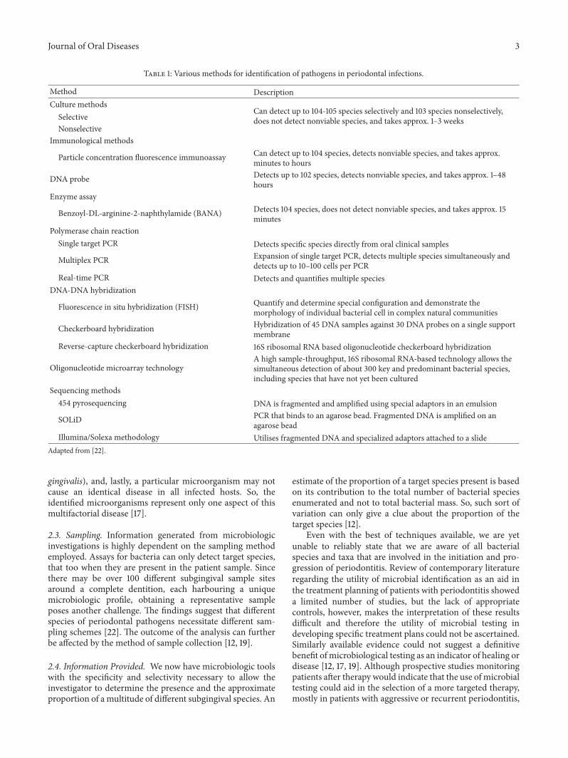
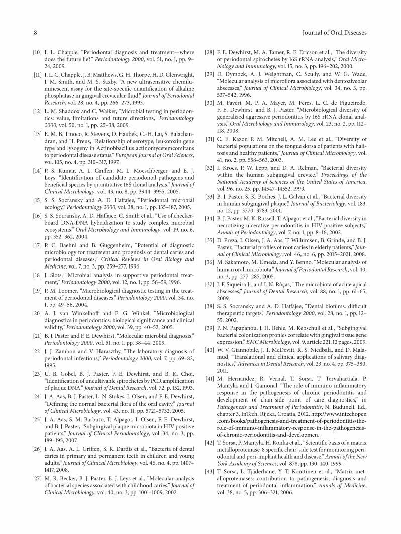
Recent developments in molecular biology techniques haveenabled investigators to detect a much wider variety of bacte-rial species closely associated with periodontal disease [12–16]. Detailed information on the individual microbiologictechniques is beyond the scope of the present paper; anoverview of the available techniques is summarized in Table 1[17–22]. There are some issues which are certainly associatedwith these techniques in general and might affect theirclinical applicability and limit the practical usefulness of themicrobiologic diagnostic techniques.
2.1. The Uncultivables. The techniques in molecular biologyhave obviated the necessity for bacterial culture of den-tal plaque samples [22, 23]. Most predominant bacterialspecies in the oral cavity have been identified using culture-independent molecular methods based on sequence analysisof the 16S ribosomal RNA genes [24–37]. Collectively speak-ing, there are about 620 predominant oral bacterial species,of which about 35% have not yet been cultured in vitro [21].The need for study of these uncultivables to understand theirexact position in the oral ecology and in the pathogenesisof periodontal disease keeps us far from deciphering thecomplete scenario.
2.2. “What Is Being Measured?” The microbiologic tests thatmeasure periodontal pathogens do not necessarily measureperiodontal disease. Bacterial pathogens can be present evenin high numbers in periodontal pockets without loss of con-nective tissue attachment or alveolar bone. Therefore, assaysfor periodontal pathogens are not, of themselves, diagnosticfor periodontal disease [22]. Mere presence of the suspectedpathogens cannot be directly interpreted as disease, as mostof the putative periodontal pathogens are found colonizingthe healthy gingival sulci as well. All identified isolatesof a particular bacterial species are not necessarily equallypathogenic or detrimental, for example, Aggregatibacter acti-nomycetemcomitans (A.a) and Porphyromonas gingivalis (P.
Journal of Oral Diseases 3
Table 1: Various methods for identification of pathogens in periodontal infections.
Method DescriptionCulture methods
Can detect up to 104-105 species selectively and 103 species nonselectively,does not detect nonviable species, and takes approx. 1–3 weeksSelective
NonselectiveImmunological methods
Particle concentration fluorescence immunoassay Can detect up to 104 species, detects nonviable species, and takes approx.minutes to hours
DNA probe Detects up to 102 species, detects nonviable species, and takes approx. 1–48hours
Enzyme assay
Benzoyl-DL-arginine-2-naphthylamide (BANA) Detects 104 species, does not detect nonviable species, and takes approx. 15minutes
Polymerase chain reactionSingle target PCR Detects specific species directly from oral clinical samples
Multiplex PCR Expansion of single target PCR, detects multiple species simultaneously anddetects up to 10–100 cells per PCR
Real-time PCR Detects and quantifies multiple speciesDNA-DNA hybridization
Fluorescence in situ hybridization (FISH) Quantify and determine special configuration and demonstrate themorphology of individual bacterial cell in complex natural communities
Checkerboard hybridization Hybridization of 45 DNA samples against 30 DNA probes on a single supportmembrane
Reverse-capture checkerboard hybridization 16S ribosomal RNA based oligonucleotide checkerboard hybridization
Oligonucleotide microarray technologyA high sample-throughput, 16S ribosomal RNA-based technology allows thesimultaneous detection of about 300 key and predominant bacterial species,including species that have not yet been cultured
Sequencing methods454 pyrosequencing DNA is fragmented and amplified using special adaptors in an emulsion
SOLiD PCR that binds to an agarose bead. Fragmented DNA is amplified on anagarose bead
Illumina/Solexa methodology Utilises fragmented DNA and specialized adaptors attached to a slideAdapted from [22].
gingivalis), and, lastly, a particular microorganism may notcause an identical disease in all infected hosts. So, theidentified microorganisms represent only one aspect of thismultifactorial disease [17].
2.3. Sampling. Information generated from microbiologicinvestigations is highly dependent on the sampling methodemployed. Assays for bacteria can only detect target species,that too when they are present in the patient sample. Sincethere may be over 100 different subgingival sample sitesaround a complete dentition, each harbouring a uniquemicrobiologic profile, obtaining a representative sampleposes another challenge. The findings suggest that differentspecies of periodontal pathogens necessitate different sam-pling schemes [22]. The outcome of the analysis can furtherbe affected by the method of sample collection [12, 19].
2.4. Information Provided. We now have microbiologic toolswith the specificity and selectivity necessary to allow theinvestigator to determine the presence and the approximateproportion of a multitude of different subgingival species. An
estimate of the proportion of a target species present is basedon its contribution to the total number of bacterial speciesenumerated and not to total bacterial mass. So, such sort ofvariation can only give a clue about the proportion of thetarget species [12].
Even with the best of techniques available, we are yetunable to reliably state that we are aware of all bacterialspecies and taxa that are involved in the initiation and pro-gression of periodontitis. Review of contemporary literatureregarding the utility of microbial identification as an aid inthe treatment planning of patients with periodontitis showeda limited number of studies, but the lack of appropriatecontrols, however, makes the interpretation of these resultsdifficult and therefore the utility of microbial testing indeveloping specific treatment plans could not be ascertained.Similarly available evidence could not suggest a definitivebenefit of microbiological testing as an indicator of healing ordisease [12, 17, 19]. Although prospective studies monitoringpatients after therapy would indicate that the use of microbialtesting could aid in the selection of a more targeted therapy,mostly in patients with aggressive or recurrent periodontitis,
4 Journal of Oral Diseases
again, the lack of clinical trials with adequate controlsprevents fromdemonstrating the real value ofmicrobial diag-nosis. Therefore, the available evidence does not fully provethe utility of microbiological testing in periodontitis patients.
Periodontal diseases are infections caused by microor-ganisms that colonize the tooth surface at or below the gin-gival margin and accumulate as dental plaque. The biofilms(dental plaque) that colonize the tooth surface are extremelycomplex and remarkably resistant to host defense mech-anisms and antimicrobial agents. Therefore, the mechani-cal plaque control (i.e., removal of supra- and subgingivalplaque) remains the cornerstone in periodontal therapy suchas self-performed oral hygiene, scaling and root planning,or periodontal surgery [38]. So microbial analysis cannot beregarded as a routine first visit investigation for all patients,but it can be reserved for specific clinical situations, such as toidentify (by knowing specific microbial profiles) for targetedand effective antimicrobial therapy for managing susceptiblepatients. Another evidence emphasizing the significanceof microbial analysis in therapeutic management is nowemerging. Papapanou et al. suggested that the microbialcontent of the periodontal pocket is a determinant of geneexpression in the gingival tissues and can potentially identifysusceptible sites in terms of additional periodontal break-down or unfavorable response to therapy [39].These findingscan serve as basis of subsequent studies for exploring the roleof microbial testing. At present, only the rationale usage ofmicrobial diagnostic techniques might benefit our patients,by saving on the time, pain, labour, and cost of repeatmechanical therapy, antibiotic usage, and surgical trauma, ifwe are able to formulate a better tailored treatment planwhichis based on the diagnostic information obtained.
3. Biochemical Analysis
Biomarkers, whether produced bynormal healthy individualsor by individuals affected by specific systemic diseases, aretell-tale molecules that could be used to monitor healthstatus, disease onset, treatment response, and outcome [2].Informative biomarkers can serve as early sentinels of disease.A huge body of literature was generated in the 1990s on theutility and value of individual biomarkers of periodontal dis-ease activity, measured within gingival crevicular fluid underthe following categories:
(i) markers of the presence or absence of periodontalpathogens,
(ii) markers of gingival and periodontal inflammation,(iii) markers of the host’s inflammatory-immune response
to certain pathogenic species,(iv) markers of host tissue destruction [10].
The principal biological media within which biomarkerswere sought including saliva, serum, subgingival plaque,tissue biopsies, and gingival crevicular fluid,mouth-rinse [40,41]. Gingival crevicular fluid became the analytical fluid ofchoice as it was the most specific to the periodontal tissues,could be collected noninvasively, and allowed site-specific
analyses. However, molecular analysis of GCF elution wastime consuming and laboratory based, technically demand-ing collection of sample leading to a small volume of the fluid(1–5 𝜇L). Despite these apparent diagnostic and technicaldisadvantages, GCF was still considered as a candidatepotential oral fluid for the development of adjunctive non-invasive chair-side point-of-care diagnostic technology [42–44] especially because tissue destructive MMPs and theirbioactive regulators can conveniently bemeasured by distinctcatalytic and noncatalytic immunoassays from GCF [41, 45].A plethora of biomarkers and diagnostic tests were developedthereafter, several of which demonstrated high levels ofsensitivity, specificity, and diagnostic accuracywith respect toidentifying and/or predicting disease activity at the site level[10, 46]. In particular, Loos and Tjoa [47] undertook a criticalreview of biomarkers in gingival crevicular fluid and foundthat only eight of 94 in the literature of the time fulfilled anyof the criteria for biomarker status. These eight biomarkerswere alkaline phosphatase [48–54], 𝛽-glucuronidase [52, 55–63], cathepsin B [64–69], MMP-8 and MMP-9 [52, 70–82], dipeptidyl peptidases II and IV [65, 66, 68, 83], andneutrophil elastase [46, 52, 61, 63–66, 76, 84–86].
A number of diagnostic kits emerged based upon individ-ual biomarkers within gingival crevicular fluid, but marketresearch had not been performed that actively and thetests did not popularize much among practicing dentistsdue to several reasons: (1) time-consuming and laboriousto perform; (2) difficult to interpret and understand; (3)site specific and the choice of site being problematic; (4)the results not materializing to alterations in therapeuticintervention; (5) expensive for routine use. As compared toGCF, collection of salivary andmouth-rinse sampleswas con-sidered more convenient, practical, rapid, and noninvasiveand requires neither professional stuff nor specific materials.Saliva and mouth-rinse represented a pooled sample fromall periodontal sites providing an overall assessment ofperiodontal disease and health at subject level [41]. Whilstit was firmly established that gingival crevicular fluid wasthe most appropriate diagnostic medium to use in analyses,it became clear that whole-mouth analysis was far morepractical, simpler, and cheaper, and thus saliva became themedium of choice in the 21st century [87]. Saliva had manyadvantages as a diagnostic fluid in that it was simple tocollect using noninvasive techniques and provides a whole-mouth summary analysis. Whole saliva could be affected bymolecular constituents and cellular remnants from other oralniches, as well as systemic conditions [88, 89] which couldhave bearing on its diagnostic applications.
Principally it remains a surrogate fluid for gingival crevic-ular fluid and therefore assays need to be highly sensitive.In addition, saliva biochemistry varies with its origin (wholesaliva or specific gland secretions), which are in turn affectedby environmental and psychological stimuli. Therefore, it isnot possible to fully quantify markers within saliva usingchairside technologies, and qualitative analyses, or at bestsemiquantification, are all that can be reliably achieved [10].
MMP-8 or collagenase-2/neutrophil-collagenase wasworked on extensively, being the major type of interstitialcollagenase present in human periodontitis-affected gingival
Journal of Oral Diseases 5
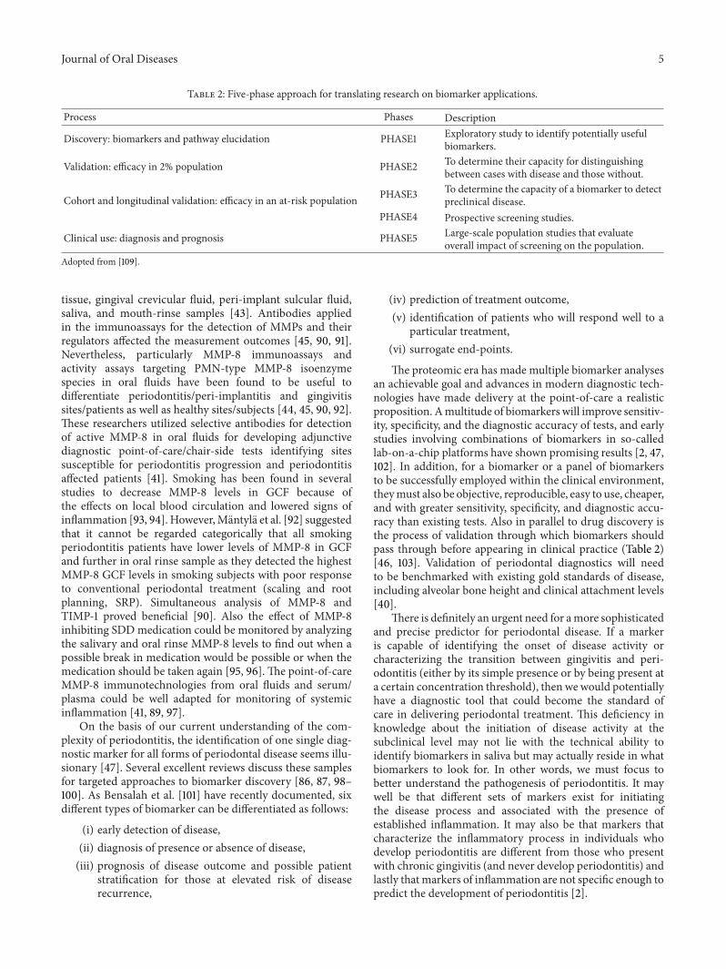
Table 2: Five-phase approach for translating research on biomarker applications.
Process Phases Description
Discovery: biomarkers and pathway elucidation PHASE1 Exploratory study to identify potentially usefulbiomarkers.
Validation: efficacy in 2% population PHASE2 To determine their capacity for distinguishingbetween cases with disease and those without.
Cohort and longitudinal validation: efficacy in an at-risk population PHASE3 To determine the capacity of a biomarker to detectpreclinical disease.
PHASE4 Prospective screening studies.
Clinical use: diagnosis and prognosis PHASE5 Large-scale population studies that evaluateoverall impact of screening on the population.
Adopted from [109].
tissue, gingival crevicular fluid, peri-implant sulcular fluid,saliva, and mouth-rinse samples [43]. Antibodies appliedin the immunoassays for the detection of MMPs and theirregulators affected the measurement outcomes [45, 90, 91].Nevertheless, particularly MMP-8 immunoassays andactivity assays targeting PMN-type MMP-8 isoenzymespecies in oral fluids have been found to be useful todifferentiate periodontitis/peri-implantitis and gingivitissites/patients as well as healthy sites/subjects [44, 45, 90, 92].These researchers utilized selective antibodies for detectionof active MMP-8 in oral fluids for developing adjunctivediagnostic point-of-care/chair-side tests identifying sitessusceptible for periodontitis progression and periodontitisaffected patients [41]. Smoking has been found in severalstudies to decrease MMP-8 levels in GCF because ofthe effects on local blood circulation and lowered signs ofinflammation [93, 94]. However,Mantyla et al. [92] suggestedthat it cannot be regarded categorically that all smokingperiodontitis patients have lower levels of MMP-8 in GCFand further in oral rinse sample as they detected the highestMMP-8 GCF levels in smoking subjects with poor responseto conventional periodontal treatment (scaling and rootplanning, SRP). Simultaneous analysis of MMP-8 andTIMP-1 proved beneficial [90]. Also the effect of MMP-8inhibiting SDDmedication could be monitored by analyzingthe salivary and oral rinse MMP-8 levels to find out when apossible break in medication would be possible or when themedication should be taken again [95, 96]. The point-of-careMMP-8 immunotechnologies from oral fluids and serum/plasma could be well adapted for monitoring of systemicinflammation [41, 89, 97].
On the basis of our current understanding of the com-plexity of periodontitis, the identification of one single diag-nostic marker for all forms of periodontal disease seems illu-sionary [47]. Several excellent reviews discuss these samplesfor targeted approaches to biomarker discovery [86, 87, 98–100]. As Bensalah et al. [101] have recently documented, sixdifferent types of biomarker can be differentiated as follows:
(i) early detection of disease,(ii) diagnosis of presence or absence of disease,(iii) prognosis of disease outcome and possible patient
stratification for those at elevated risk of diseaserecurrence,
(iv) prediction of treatment outcome,(v) identification of patients who will respond well to a
particular treatment,(vi) surrogate end-points.
The proteomic era has made multiple biomarker analysesan achievable goal and advances in modern diagnostic tech-nologies have made delivery at the point-of-care a realisticproposition. Amultitude of biomarkers will improve sensitiv-ity, specificity, and the diagnostic accuracy of tests, and earlystudies involving combinations of biomarkers in so-calledlab-on-a-chip platforms have shown promising results [2, 47,102]. In addition, for a biomarker or a panel of biomarkersto be successfully employed within the clinical environment,theymust also be objective, reproducible, easy to use, cheaper,and with greater sensitivity, specificity, and diagnostic accu-racy than existing tests. Also in parallel to drug discovery isthe process of validation through which biomarkers shouldpass through before appearing in clinical practice (Table 2)[46, 103]. Validation of periodontal diagnostics will needto be benchmarked with existing gold standards of disease,including alveolar bone height and clinical attachment levels[40].
There is definitely an urgent need for amore sophisticatedand precise predictor for periodontal disease. If a markeris capable of identifying the onset of disease activity orcharacterizing the transition between gingivitis and peri-odontitis (either by its simple presence or by being present ata certain concentration threshold), then we would potentiallyhave a diagnostic tool that could become the standard ofcare in delivering periodontal treatment. This deficiency inknowledge about the initiation of disease activity at thesubclinical level may not lie with the technical ability toidentify biomarkers in saliva but may actually reside in whatbiomarkers to look for. In other words, we must focus tobetter understand the pathogenesis of periodontitis. It maywell be that different sets of markers exist for initiatingthe disease process and associated with the presence ofestablished inflammation. It may also be that markers thatcharacterize the inflammatory process in individuals whodevelop periodontitis are different from those who presentwith chronic gingivitis (and never develop periodontitis) andlastly thatmarkers of inflammation are not specific enough topredict the development of periodontitis [2].
6 Journal of Oral Diseases
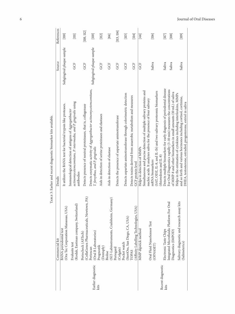
Table3:Ea
rlier
andrecent
diagno
sticbiom
arkerk
itsavailable.
Com
mercialkit
Details
Source
References
Earlier
diagno
stic
kits
BANAperio
dontaltest
(Ora
TecC
orpo
ratio
nManassas,USA
)Itutilizesthe
BANAtestforb
acteria
ltrypsin
likep
roteases.
Subgingivalplaqu
esam
ple
[110]
Evalusite
test
(Kod
ak,E
astm
ancompany,Switzerland
)
Immun
ologicaldetectionof
antig
enso
fAggregatib
acter
actin
omycetem
comita
ns,P.intermedia,and
P.gingivalisusing
antib
odies
GCF
[111]
Perio
check(A
STech)
(CollaGenex
Pharmaceutic
als,New
town,
PA)
Detectspresence
ofneutralproteinases,thatis,collagenase
GCF
[110,112]
Perio
scan
(OralB
Labo
ratorie
s)Detectsenzymaticactiv
ityof
Aggregatibactera
ctinom
ycetem
comita
ns,
T.forsythu
s,andP.gingivalis
Subgingivalplaqu
esam
ple
[110]
Progno
stik
(Dentsp
ly)
Aidsindetectionof
serin
eproteinases
andela
stases
GCF
[112]
Biolise
(SLT
-Labinstruments,
Crailsh
eim,G
ermany)
Aidsindetectionof
elastase
GCF
[84]
Perio
gard
(Colgate)
Detectsthep
resenceo
faspartateam
inotransferase
GCF
[113,114]
Pocketwatch
(SteriO
ss,San
Diego,C
A,U
SA)
Detectsaspartatea
minotransferase
throug
hcolorim
etric
detection
GCF
[115]
TOPA
S(A
ffinityLabelling
Techno
logies,U
SA)
Detectstoxins
deriv
edfro
manaerobicm
etabolism
andmeasures
GCF
proteinlevel
GCF
[114]
Recent
diagno
stic
kits
MMPdipstickmetho
dHelp
sindetectionof
MMPs
GCF
[44]
OralF
luid
NanoSensorT
est
(OFN
ASE
T)
Simultaneou
sand
precise
detectionof
multip
lesalivaryproteins
and
nucle
icacids.Itanalyzes
salivafor
thep
resenceo
ffou
rsalivary
mRN
Abiom
arkers
(SAT
,ODZ,
IL-8,and
IL-1b)
andtwosalivaryproteomicbiom
arkers
(thioredo
xinandIL-8)
Saliva
[116]
Electro
nicT
asteCh
ips
Detectsmultip
lebiom
arkersfore
arlydiagno
sisof
perio
dontaldisease
Saliva
[117]
Integrated
Microflu
idicPlatform
ForO
ral
Diagn
ostics(IM
POD)
ForO
ralD
iagn
ostic
srapidly(3–10m
in)m
easuresthe
concentrations
ofMMP-8andotherb
iomarkersin
smallamou
nts(10mL)
ofsaliva
Saliva
[118]
Salivarydiagno
sticandresearch
assaykits
(Salim
eterics)
Help
sinthee
stim
ationof
cytokinesincluding
interle
ukins,MMPs
andso
forthandvario
usho
rmon
esinclu
ding
cortiso
l,cortinine,
DHEA
,testoste
rone,estradiol,progeste
rone,estrio
linsaliva
Saliva
[119]
Journal of Oral Diseases 7
The key challenge is to elucidate a panel of biomark-ers that differentiate health from periodontitis and, moreimportantly, gingivitis from periodontitis. This calls forperforming studies in which gingivitis and periodontitis areinduced experimentally in animal models. All markers thatare potentially associated with initiating the disease processare monitored longitudinally (form health to the full-blowndisease state) by salivary genomics, proteomics, and otherstate-of-the-art diagnostic techniques. A similar methodol-ogy could be adopted to analyse the treatment of disease,correlating periodontal healing and stability with the absenceof such markers. The complexity of the associated microfloraand the critical role of the host further heighten the issuesfor conducting the research regarding specific biomarkersfor periodontal disease and might point towards exploringnovel simulated animal models for studying these specificinvestigations. In human trials, for prospective investigationsa combination of surrogate endpoints can be analysed forenhancing specificity while evaluating the biomarker.
It is highly unlikely that a single biomarker may proveto be a stand-alone measure for predicting periodontaldisease activity. A combined analysis of proteomic, genomic,microbial, and other indicators is required to identify the setof biomarkers with themost favourable combination of sensi-tivity, specificity, reproducibility, and correlations with estab-lished disease diagnostic criteria. Emerging clinical applica-tions of lab-on-a-chip (LOC) technologies as point-of-care(POC) diagnostics developed for systemic diseases are nowbeing readily applied to periodontology.Many diagnostic kitshave been commercialized and are being marketed (Table 3).The field of periodontology is now able to detect apanel of salivary biomarkers to predict disease, includ-ing matrix metalloproteinase-8 (MMP-8), microbial factors,and proinflammatory cytokines such as IL-1 beta [40].These salivary biomarker detectors can be used in the officeof a dentist or another healthcare provider for point-of-caredisease screening and detection. The dental community isnot generally familiar with mass screening of populations fororal and systemic diseases [40]. If more efficient periodontaltherapy can be delivered, clinicians will be more likely toutilize new diagnostic approaches. Dentists will have greaterinvolvement in the identification monitoring of oral andsystemic disorders in the not too distant future.
Prospective healthcare is a new approach that incorpo-rates all the power of current disease-orientedmedicine but isbased on the concept of strategic health planning, a proactive,prospective approach to care. In this system, individualsare evaluated to determine their baseline risk for a specificdisease, their current health status, and their likelihood ofdeveloping specific clinical problems given their risks [102,104, 105]. As mentioned before, allocation of resources toprevent periodontitis/peri-implantitis would be optimizedandmay help to reduce costs if diagnostic information wouldassist in identifying susceptible patients and help providingmore specific prevention/treatment strategies for high-riskand low-risk patients. Saliva as a diagnostic and/or prognostictool can improve and ease treatment planning in periodonticsand implant dentistry, thus resulting in more predictabletreatment outcomes and cost savings [2, 106]. Periodontal
oral POC devices will also enable masses to be screened;particularly underserved communities and resource limitedareas as in developing nations may be accessed more effi-ciently. such applications might serve better for identificationof at-risk groups and increase access to treatment for thosemost in need, improving public health in periodontology andthe oral health field in general [40].
A careful analysis is mandatory, before adopting anynewly emerged diagnostic test in the current clinical protocol.The novel test must be weighed against the conventionalcriteria of diagnosis in its sensitivity, specificity, validity, andreliability [107]. The benefit of having a particular piece ofdiagnostic information must not only outweigh the effort toobtain it on the level of each individual; however, the impactof a new diagnostic procedure should also be evaluated at amore global level, to maximize the overall benefit of the totalinvestment in health care [8]. Adequate guidelines for theuse of diagnostic routines should be issued and implementedfrom regulatory bodies in health care. Nevertheless, as newprocedures are introduced in periodontology during thesetimes of cost containment in health care, practitioners mustuse caution in deciding which particular patients wouldbenefit from a comprehensive evaluation [8, 108].
Conflict of Interests
The authors declare that there is no conflict of interestsregarding the publication of this paper.
References
[1] G. C. Armitage, “Periodontal diagnoses and classification ofperiodontal diseases,” Periodontology 2000, vol. 34, pp. 9–21,2004.
[2] W. V. Giannobile, T. Beikler, J. S. Kinney, C. A. Ramseier,T. Morelli, and D. T. Wong, “Saliva as a diagnostic tool forperiodontal disease: current state and future directions,” Peri-odontology 2000, vol. 50, no. 1, pp. 52–64, 2009.
[3] G. C. Armitage, “The complete periodontal examination,” Peri-odontology 2000, vol. 34, pp. 22–33, 2004.
[4] D. L. Wolf and I. B. Lamster, “Contemporary concepts inthe diagnosis of periodontal disease,” Dental Clinics of NorthAmerica, vol. 55, no. 1, pp. 47–61, 2011.
[5] I. B. Lamster and J. T. Grbic, “Diagnosis of periodontal diseasebased on analysis of the host response,” Periodontology 2000,vol. 7, pp. 83–99, 1995.
[6] J. D. Beck, “Issues in assessment of diagnostic tests and risk forperiodontal diseases,” Periodontology 2000, vol. 7, pp. 100–108,1995.
[7] Research Science and Therapy Committee of the AmericanAcademy of Periodontology, “Diagnosis of periodontal dis-eases,” Journal of Periodontology, vol. 74, no. 8, pp. 1237–1247,2003.
[8] A. Mombelli, “Critical issues in periodontal diagnosis,” Peri-odontology 2000, vol. 39, pp. 9–12, 2005.
[9] J. M. Goodson, “Diagnosis of periodontitis by physical mea-surement: interpretation from episodic disease hypothesis,”Journal of Periodontology, vol. 63, no. 4s, pp. 373–382, 1992.
8 Journal of Oral Diseases
[10] I. L. Chapple, “Periodontal diagnosis and treatment—wheredoes the future lie?” Periodontology 2000, vol. 51, no. 1, pp. 9–24, 2009.
[11] I. L. C. Chapple, J. B.Matthews, G.H.Thorpe, H.D. Glenwright,J. M. Smith, and M. S. Saxby, “A new ultrasensitive chemilu-minescent assay for the site-specific quantification of alkalinephosphatase in gingival crevicular fluid,” Journal of PeriodontalResearch, vol. 28, no. 4, pp. 266–273, 1993.
[12] L. M. Shaddox and C. Walker, “Microbial testing in periodon-tics: value, limitations and future directions,” Periodontology2000, vol. 50, no. 1, pp. 25–38, 2009.
[13] E. M. B. Tinoco, R. Stevens, D. Haubek, C.-H. Lai, S. Balachan-dran, and H. Preus, “Relationship of serotype, leukotoxin genetype and lysogeny in Actinobbacillus actinomycetemcomitansto periodontal disease status,” European Journal of Oral Sciences,vol. 105, no. 4, pp. 310–317, 1997.
[14] P. S. Kumar, A. L. Griffen, M. L. Moeschberger, and E. J.Leys, “Identification of candidate periodontal pathogens andbeneficial species by quantitative 16S clonal analysis,” Journal ofClinical Microbiology, vol. 43, no. 8, pp. 3944–3955, 2005.
[15] S. S. Socransky and A. D. Haffajee, “Periodontal microbialecology,” Periodontology 2000, vol. 38, no. 1, pp. 135–187, 2005.
[16] S. S. Socransky, A. D. Haffajee, C. Smith et al., “Use of checker-board DNA-DNA hybridization to study complex microbialecosystems,” Oral Microbiology and Immunology, vol. 19, no. 6,pp. 352–362, 2004.
[17] P. C. Baehni and B. Guggenheim, “Potential of diagnosticmicrobiology for treatment and prognosis of dental caries andperiodontal diseases,” Critical Reviews in Oral Biology andMedicine, vol. 7, no. 3, pp. 259–277, 1996.
[18] J. Slots, “Microbial analysis in supportive periodontal treat-ment,” Periodontology 2000, vol. 12, no. 1, pp. 56–59, 1996.
[19] P. M. Loomer, “Microbiological diagnostic testing in the treat-ment of periodontal diseases,” Periodontology 2000, vol. 34, no.1, pp. 49–56, 2004.
[20] A. J. van Winkelhoff and E. G. Winkel, “Microbiologicaldiagnostics in periodontics: biological significance and clinicalvalidity,” Periodontology 2000, vol. 39, pp. 40–52, 2005.
[21] B. J. Paster and F. E. Dewhirst, “Molecular microbial diagnosis,”Periodontology 2000, vol. 51, no. 1, pp. 38–44, 2009.
[22] J. J. Zambon and V. Haraszthy, “The laboratory diagnosis ofperiodontal infections,” Periodontology 2000, vol. 7, pp. 69–82,1995.
[23] U. B. Gobel, B. J. Paster, F. E. Dewhirst, and B. K. Choi,“Identification of uncultivahle spirochetes byPCRamplificationof plaque DNA,” Journal of Dental Research, vol. 72, p. 152, 1993.
[24] J. A. Aas, B. J. Paster, L. N. Stokes, I. Olsen, and F. E. Dewhirst,“Defining the normal bacterial flora of the oral cavity,” Journalof Clinical Microbiology, vol. 43, no. 11, pp. 5721–5732, 2005.
[25] J. A. Aas, S. M. Barbuto, T. Alpagot, I. Olsen, F. E. Dewhirst,and B. J. Paster, “Subgingival plaque microbiota in HIV positivepatients,” Journal of Clinical Periodontology, vol. 34, no. 3, pp.189–195, 2007.
[26] J. A. Aas, A. L. Griffen, S. R. Dardis et al., “Bacteria of dentalcaries in primary and permanent teeth in children and youngadults,” Journal of Clinical Microbiology, vol. 46, no. 4, pp. 1407–1417, 2008.
[27] M. R. Becker, B. J. Paster, E. J. Leys et al., “Molecular analysisof bacterial species associated with childhood caries,” Journal ofClinical Microbiology, vol. 40, no. 3, pp. 1001–1009, 2002.
[28] F. E. Dewhirst, M. A. Tamer, R. E. Ericson et al., “The diversityof periodontal spirochetes by 16S rRNA analysis,” Oral Micro-biology and Immunology, vol. 15, no. 3, pp. 196–202, 2000.
[29] D. Dymock, A. J. Weightman, C. Scully, and W. G. Wade,“Molecular analysis of microflora associated with dentoalveolarabscesses,” Journal of Clinical Microbiology, vol. 34, no. 3, pp.537–542, 1996.
[30] M. Faveri, M. P. A. Mayer, M. Feres, L. C. de Figueiredo,F. E. Dewhirst, and B. J. Paster, “Microbiological diversity ofgeneralized aggressive periodontitis by 16S rRNA clonal anal-ysis,”Oral Microbiology and Immunology, vol. 23, no. 2, pp. 112–118, 2008.
[31] C. E. Kazor, P. M. Mitchell, A. M. Lee et al., “Diversity ofbacterial populations on the tongue dorsa of patients with hali-tosis and healthy patients,” Journal of Clinical Microbiology, vol.41, no. 2, pp. 558–563, 2003.
[32] I. Kroes, P. W. Lepp, and D. A. Relman, “Bacterial diversitywithin the human subgingival crevice,” Proceedings of theNational Academy of Sciences of the United States of America,vol. 96, no. 25, pp. 14547–14552, 1999.
[33] B. J. Paster, S. K. Boches, J. L. Galvin et al., “Bacterial diversityin human subgingival plaque,” Journal of Bacteriology, vol. 183,no. 12, pp. 3770–3783, 2001.
[34] B. J. Paster,M. K. Russell, T. Alpagot et al., “Bacterial diversity innecrotizing ulcerative periodontitis in HIV-positive subjects,”Annals of Periodontology, vol. 7, no. 1, pp. 8–16, 2002.
[35] D. Preza, I. Olsen, J. A. Aas, T. Willumsen, B. Grinde, and B. J.Paster, “Bacterial profiles of root caries in elderly patients,” Jour-nal of Clinical Microbiology, vol. 46, no. 6, pp. 2015–2021, 2008.
[36] M. Sakamoto, M. Umeda, and Y. Benno, “Molecular analysis ofhumanoralmicrobiota,” Journal of Periodontal Research, vol. 40,no. 3, pp. 277–285, 2005.
[37] J. F. Siqueira Jr. and I. N. Rocas, “The microbiota of acute apicalabscesses,” Journal of Dental Research, vol. 88, no. 1, pp. 61–65,2009.
[38] S. S. Socransky and A. D. Haffajee, “Dental biofilms: difficulttherapeutic targets,” Periodontology 2000, vol. 28, no. 1, pp. 12–55, 2002.
[39] P. N. Papapanou, J. H. Behle, M. Kebschull et al., “Subgingivalbacterial colonization profiles correlatewith gingival tissue geneexpression,”BMCMicrobiology, vol. 9, article 221, 12 pages, 2009.
[40] W. V. Giannobile, J. T. McDevitt, R. S. Niedbala, and D. Mala-mud, “Translational and clinical applications of salivary diag-nostics,”Advances inDental Research, vol. 23, no. 4, pp. 375–380,2011.
[41] M. Hernandez, R. Vernal, T. Sorsa, T. Tervahartiala, P.Mantyla, and J. Gamonal, “The role of immuno-inflammatoryresponse in the pathogenesis of chronic periodontitis anddevelopment of chair-side point of care diagnostics,” inPathogenesis and Treatment of Periodontitis, N. Buduneli, Ed.,chapter 3, InTech, Rijeka, Croatia, 2012, http://www.intechopen.com/books/pathogenesis-and-treatment-of-periodontitis/the-role-of-immuno-inflammatory-response-in-the-pathogenesis-of-chronic-periodontitis-and-developmen.
[42] T. Sorsa, P. Mantyla, H. Ronka et al., “Scientific basis of a matrixmetalloproteinase-8 specific chair-side test formonitoring peri-odontal and peri-implant health and disease,”Annals of the NewYork Academy of Sciences, vol. 878, pp. 130–140, 1999.
[43] T. Sorsa, L. Tjaderhane, Y. T. Konttinen et al., “Matrix met-alloproteinases: contribution to pathogenesis, diagnosis andtreatment of periodontal inflammation,” Annals of Medicine,vol. 38, no. 5, pp. 306–321, 2006.
Journal of Oral Diseases 9
[44] P.Mantyla, M. Stenman, D. F. Kinane et al., “Gingival crevicularfluid collagenase-2 (MMP-8) test stick for chair-sidemonitoringof periodontitis,” Journal of Periodontal Research, vol. 38, no. 4,pp. 436–439, 2003.
[45] T. Sorsa, M. Hernandez, J. Leppilahti, S. Munjal, L. Netuschil,and P. Mantyla, “Detection of gingival crevicular fluid MMP-8 levels with different laboratory and chair-side methods,” OralDiseases, vol. 16, no. 1, pp. 39–45, 2010.
[46] M. M. Grant, “What do ’omic technologies have to offer peri-odontal clinical practice in the future?” Journal of PeriodontalResearch, vol. 47, no. 1, pp. 2–14, 2012.
[47] B. G. Loos and S. Tjoa, “Host-derived diagnostic markers forperiodontitis: do they exist in gingival crevice fluid?” Peri-odontology 2000, vol. 39, pp. 53–72, 2005.
[48] I. L. Chapple, H. D. Glenwright, J. B. Matthews, G. H. Thorpe,and P. J. Lumley, “Site-specific alkaline phosphatase levels ingingival crevicular fluid in health and gingivitis: cross-sectionalstudies,” Journal of Clinical Periodontology, vol. 21, no. 6, pp.409–414, 1994.
[49] H. J.Wright, I. L. Chapple, and J. B.Matthews, “Levels of TGF𝛽1in gingival crevicular fluid during a 21-day experimental modelof gingivitis,” Oral Diseases, vol. 9, no. 2, pp. 88–94, 2003.
[50] I. L. C. Chappie, “Chemiluminescent assay of alkaline phos-phatase in human gingival crevicular fluid: investigations withan experimental gingivitis model and studies on the source ofthe enzyme within crevicular fluid,” Journal of Clinical Peri-odontology, vol. 23, no. 6, pp. 587–594, 1996.
[51] I. L. Chapple, I. Garner, M. S. Saxby, H. Moscrop, and J. B.Matthews, “Prediction and diagnosis of attachment loss byenhanced chemiluminescent assay of crevicular fluid alkalinephosphatase levels,” Journal of Clinical Periodontology, vol. 26,no. 3, pp. 190–198, 1999.
[52] T. Hanioka, K. Takaya, Y. Matsumori, R. Matsuse, and S.Shizukuishi, “Relationship of the substance P to indicators ofhost response in human gingival crevicular fluid,” Journal ofClinical Periodontology, vol. 27, no. 4, pp. 262–266, 2000.
[53] K. Nakashima, C. Giannopoulou, E. Andersen et al., “A longitu-dinal study of various crevicular fluid components asmarkers ofperiodontal disease activity,” Journal of Clinical Periodontology,vol. 23, no. 9, pp. 832–838, 1996.
[54] K. Nakashima, N. Roehrich, and G. Cimasoni, “Osteocalcin,prostaglandin E
2and alkaline phosphatase in gingival crevicu-
lar fluid: their relations to periodontal status,” Journal of ClinicalPeriodontology, vol. 21, no. 5, pp. 327–333, 1994.
[55] J. T. Grbic, I. B. Lamster, J. B. Fine et al., “Changes in gingivalcrevicular fluid levels of immunoglobulin a following therapy:associationwith attachment loss,” Journal of Periodontology, vol.70, no. 10, pp. 1221–1227, 1999.
[56] J. T. Grbic, R. E. Singer, H. H. Jans, R. S. Celenti, and I. B. Lam-ster, “Immunoglobulin isotypes in gingival crevicular fluid: pos-sible protective role of IgA,” Journal of Periodontology, vol. 66,no. 1, pp. 55–61, 1995.
[57] I. B. Lamster, “Evaluation of components of gingival crevicularfluid as diagnostic tests,” Annals of Periodontology, vol. 2, no. 1,pp. 123–137, 1997.
[58] I. B. Lamster, R. Celenti, and J. L. Ebersole, “The relationshipof serum IgG antidbody titers to periodontal pathogens toindicators of the host response in crevicular fluid,” Journal ofClinical Periodontology, vol. 17, no. 7, pp. 419–425, 1990.
[59] I. B. Lamster, D. S. Harper, L. A. Fiorello, R. L. Oshrain, R. S.Celenti, and J.M. Gordon, “Lysosomal and cytoplasmic enzyme
activity, crevicular fluid volume, and clinical parameters char-acterizing gingival sites with shallow to intermediate probingdepths,” Journal of Periodontology, vol. 58, no. 9, pp. 614–621,1987.
[60] I. B. Lamster, L. G. Holmes, K. B. Gross et al., “The relationshipof beta-glucuronidase activity in crevicular fluid to probingattachment loss in patients with adult periodontitis. Findingsfrom a multicenter study,” Journal of Clinical Periodontology,vol. 22, no. 1, pp. 36–44, 1995.
[61] I. B. Lamster, R. L. Oshrain, D. S. Harper, R. S. Celenti, C. A.Hovliaras, and J. M. Gordon, “Enzyme activity in crevicularfluid for detection and prediction of clinical attachment lossin patients with chronic adult periodontitis. Six month results,”Journal of Periodontology, vol. 59, no. 8, pp. 516–523, 1988.
[62] I. B. Lamster, S.Wallenstein, S. Sengupta, andT.Duffy, “Within-mouth correlations for indicators of the host response ingingival crevicular fluid,” Archives of Oral Biology, vol. 35, no.10, pp. 779–783, 1990.
[63] L. F. Wolff, N. J. Koller, Q. T. Smith, A. Mathur, and D. Aeppli,“Subgingival temperature: relation to gingival crevicular fluidenzymes, cytokines, and subgingival plaque micro-organisms,”Journal of Clinical Periodontology, vol. 24, no. 12, pp. 900–906,1997.
[64] H. Y. Chen, S. W. Cox, and B. M. Eley, “Cathepsin B, 𝛼2-macroglobulin and cystatin levels in gingival crevicular fluidfrom chronic periodontitis patients,” Journal of Clinical Peri-odontology, vol. 25, no. 1, pp. 34–41, 1998.
[65] S. W. Cox and B. M. Eley, “Cathepsin B/L-, elastase-, tryptase-,trypsin- and dipeptidyl peptidase IV-like activities in gingivalcrevicular fluid. A comparison of levels before and after basicperiodontal treatment of chronic periodontitis patients,” Jour-nal of Clinical Periodontology, vol. 19, no. 5, pp. 333–339, 1992.
[66] B. M. Eley and S. W. Cox, “The relationship between gingivalcrevicular fluid cathepsin B activity and periodontal attachmentloss in chronic periodontitis patients: a 2-year longitudinalstudy,” Journal of Periodontal Research, vol. 31, no. 6, pp. 381–392, 1996.
[67] E. Ichimaru, M. Tanoue, M. Tani et al., “Cathepsin B in gingivalcrevicular fluid of adult periodontitis patients: identificationby immunological and enzymological methods,” InflammationResearch, vol. 45, no. 6, pp. 277–282, 1996.
[68] C. N. Kennett, S. W. Cox, and B. M. Eley, “Investigations intothe cellular contribution to host tissue proteases and inhibitorsin gingival crevicular fluid,” Journal of Clinical Periodontology,vol. 24, no. 6, pp. 424–431, 1997.
[69] K. Kunimatsu, K. Yamamoto, E. Ichimaru, Y. Kato, and I. Kato,“Cathepsins B, H and L activities in gingival crevicular fluidfrom chronic adult periodontitis patients and experimentalgingvitis subjects,” Journal of Periodontal Research, vol. 25, no.2, pp. 69–73, 1990.
[70] N. Buduneli, S. Vardar, G. Atilla, T. Sorsa, H. Luoto, and H.Baylas, “Gingival crevicular fluid matrix metalloproteinase-8levels following adjunctive use of meloxicam and initial phaseof periodontal therapy,” Journal of Periodontology, vol. 73, no. 1,pp. 103–109, 2002.
[71] L. M. Golub, K. Siegel, N. S. Ramamurthy, and I. D. Mandel,“Some characteristics of collagenase activity in gingival crevic-ular fluid and its relationship to gingival diseases in humans,”Journal of Dental Research, vol. 55, no. 6, pp. 1049–1057, 1976.
[72] M. Kiili, S. W. Cox, H. W. Chen et al., “Collagenase-2 (MMP-8)and collagenase-3 (MMP-13) in adult periodontitis: molecular
10 Journal of Oral Diseases
forms and levels in gingival crevicular fluid and immunolocali-sation in gingival tissue,” Journal of Clinical Periodontology, vol.29, no. 3, pp. 224–232, 2002.
[73] D. F. Kinane, I. B. Darby, S. Said et al., “Changes in gingi-val crevicular fluid matrix metalloproteinase-8 levels duringperiodontal treatment andmaintenance,” Journal of PeriodontalResearch, vol. 38, no. 4, pp. 400–404, 2003.
[74] W. Lee, S. Aitken, G. Kulkarni et al., “Collagenase activity inrecurrent periodontitis: relationship to disease progression anddoxycycline therapy,” Journal of Periodontal Research, vol. 26,no. 6, pp. 479–485, 1991.
[75] W. Lee, S. Aitken, J. Sodek, and C. A.McCulloch, “Evidence of adirect relationship between neutrophil collagenase activity andperiodontal tissue destruction in vivo: role of active enzyme inhuman periodontitis,” Journal of Periodontal Research, vol. 30,no. 1, pp. 23–33, 1995.
[76] C.-M. Liu and L.-T. Hou, “Collagenase activity in the gingivalcrevicular fluid of periodontal patients,” Journal of the FormosanMedical Association, vol. 92, no. 2, pp. 157–164, 1993.
[77] S. Mancini, R. Romanelli, C. A. Laschinger, C. M. Overall, J.Sodek, and C. A. G. McCulloch, “Assessment of a novel screen-ing test for neutrophil collagenase activity in the diagnosis ofperiodontal diseases,” Journal of Periodontology, vol. 70, no. 11,pp. 1292–1302, 1999.
[78] T. Nakamura, J.-I. Kido, R. Kido et al., “The association ofcalprotectin level in gingival crevicular fluid with gingival indexand the activities of collagenase and aspartate aminotransferasein adult periodontitis patients,” Journal of Periodontology, vol.71, no. 3, pp. 361–367, 2000.
[79] T. Nomura, A. Ishii, Y. Oishi, H. Kohma, and K. Hara, “Tissueinhibitors ofmetalloproteinases level and collagenase activity ingingival crevicular fluid: the relevance to periodontal diseases,”Oral Diseases, vol. 4, no. 4, pp. 231–240, 1998.
[80] L. Persson, J. Bergstrom, and A. Gustafsson, “Effect of tobaccosmoking on neutrophil activity following periodontal surgery,”Journal of Periodontology, vol. 74, no. 10, pp. 1475–1482, 2003.
[81] R. Romanelli, S.Mancini, C. Laschinger, C.M.Overall, J. Sodek,and C. A. McCulloch, “Activation of neutrophil collagenase inperiodontitis,” Infection and Immunity, vol. 67, no. 5, pp. 2319–2326, 1999.
[82] Y. T. Teng, J. Sodek, and C. A. McCulloch, “Gingival crevicularfluid gelatinase and its relationship to periodontal disease inhuman subjects,” Journal of Periodontal Research, vol. 27, no. 5,pp. 544–552, 1992.
[83] M. I. Gazi, S. W. Cox, D. T. Clark, and B. M. Eley, “Comparisonof host tissue and bacterial dipeptidyl peptidases in humangingival crevicular fluid by analytical isoelectric focusing,”Archives of Oral Biology, vol. 40, no. 8, pp. 731–736, 1995.
[84] G. C. Armitage, M. K. Jeffcoat, D. E. Chadwick et al., “Longitu-dinal evaluation of elastase as a marker for the progression ofperiodontitis,” Journal of Periodontology, vol. 65, no. 2, pp. 120–128, 1994.
[85] Q. T. Smith, L. Harriman, G. S. Au et al., “Neutrophil elastase increvicular fluid: comparison of a middle-aged general popula-tion with healthy and periodontitis groups,” Journal of ClinicalPeriodontology, vol. 22, no. 12, pp. 935–941, 1995.
[86] I. B. Lamster, E. Kaufman, J. T. Grbic, L. J. Winston, and R.E. Singer, “𝛽-glucuronidase activity in saliva: relationship toclinical periodontal parameters,” Journal of Periodontology, vol.74, no. 3, pp. 353–359, 2003.
[87] L. Zhang, B. S. Henson, P. M. Camargo, and D. T. Wong, “Theclinical value of salivary biomarkers for periodontal disease,”Periodontology 2000, vol. 51, no. 1, pp. 25–37, 2009.
[88] E. Buduneli, P. Mantyla, G. Emingil et al., “Acute myocardialinfarction is reflected in salivary matrix metalloproteinase-8activation level,” Journal of Periodontology, vol. 82, no. 5, pp.716–725, 2011.
[89] N. Buduneli and D. F. Kinane, “Host-derived diagnostic mark-ers related to soft tissue destruction and bone degradation inperiodontitis,” Journal of Clinical Periodontology, vol. 38, no. 11,pp. 85–105, 2011.
[90] J. M. Leppilahti, M.-M. Ahonen, M. Hernandez et al., “OralrinseMMP-8 point-of-care immuno test identifies patients withstrong periodontal inflammatory burden,”Oral Diseases, vol. 17,no. 1, pp. 115–122, 2011.
[91] M. Gursoy, E. Kononen, U. K. Gursoy, T. Tervahartiala, R.Pajukanta, and T. Sorsa, “Periodontal status and neutrophilicenzyme levels in gingival crevicular fluid during pregnancy andpostpartum,” Journal of Periodontology, vol. 81, no. 12, pp. 1790–1796, 2010.
[92] P. Mantyla, M. Stenman, D. Kinane et al., “Monitoring peri-odontal disease status in smokers and nonsmokers using agingival crevicular fluid matrix metalloproteinase-8-specificchair-side test,” Journal of Periodontal Research, vol. 41, no. 6,pp. 503–512, 2006.
[93] B. Soder, “Neutrophil elastase activity, levels of prostaglandinE2, and matrix metalloproteinase-8 in refractory periodontitissites in smokers and non-smokers,” Acta Odontologica Scandi-navica, vol. 57, no. 2, pp. 77–82, 1999.
[94] B. Soder, L. J. Jin, and S. Wickholm, “Granulocyte elastase,matrix metalloproteinase-8 and prostaglandin E
2in gingival
crevicular fluid in matched clinical sites in smokers and non-smokers with persistent periodontitis,” Journal of Clinical Peri-odontology, vol. 29, no. 5, pp. 384–391, 2002.
[95] L. M. Golub, M. L. Hsi, J. A. Stoner et al., “Subantimicrobial-dose doxycycline modulates gingival crevicular fluid biomark-ers of periodontitis in postmenopausal osteopenic women,”Journal of Periodontology, vol. 79, no. 8, pp. 1409–1418, 2008.
[96] R. A. Reinhardt, J. A. Stoner, L. M. Golub et al., “Association ofgingival crevicular fluid biomarkers during periodontal main-tenance with subsequent progressive periodontitis,” Journal ofPeriodontology, vol. 81, no. 2, pp. 251–259, 2010.
[97] A.M. Tuomainen, K. Nyyssonen, J. A. Laukkanen et al., “Serummatrix metalloproteinase-8 concentrations are associated withcardiovascular outcome in men,” Arteriosclerosis, Thrombosis,and Vascular Biology, vol. 27, no. 12, pp. 2722–2728, 2007.
[98] F. G. Oppenheim, E. Salih, W. L. Siqueira, W. Zhang, and E.J. Helmerhorst, “Salivary proteome and its genetic polymor-phisms,” Annals of the New York Academy of Sciences, vol. 1098,pp. 22–50, 2007.
[99] N. Ozmeric, “Advances in periodontal disease markers,” ClinicaChimica Acta, vol. 343, no. 1-2, pp. 1–16, 2004.
[100] M. Taba Jr., J. Kinney, A. S. Kim, and W. V. Giannobile, “Diag-nostic biomarkers for oral and periodontal diseases,” DentalClinics of North America, vol. 49, no. 3, pp. 551–571, 2005.
[101] K. Bensalah, F.Montorsi, and S. F. Shariat, “Challenges of cancerbiomarker profiling,” European Urology, vol. 52, no. 6, pp. 1601–1609, 2007.
[102] R. Snyderman and J. Langheier, “Prospective health care: thesecond transformation of medicine,”Genome Biology, vol. 7, no.2, article 104, 2006.
Journal of Oral Diseases 11
[103] B. M. Eley and J. D. Manson, Periodontics, Wright, Edinburgh,UK, 5th edition, 2004.
[104] R. Snyderman and R. S. Williams, “Prospective medicine: thenext health care transformation,” Academic Medicine, vol. 78,no. 11, pp. 1079–1084, 2003.
[105] R. Snyderman and Z. Yoedlono, “Pospective care: a personal-ized, preventative approach to medicine,” Pharmacogenomics,vol. 7, no. 1, pp. 5–9, 2006.
[106] C. A. Ramseier, T. Morelli, J. S. Kineey et al., “Periodontaldisease: salivary diagnostics,” in Salivary Diagnostics, pp. 156–168, Wiley Blackwell, Ames, Iowa, USA, 2008.
[107] S. J. Lord, L. Irwig, and R. J. Simes, “When is measuringsensitivity and specificity sufficient to evaluate a diagnostic test,and when do we need randomized trials?” Annals of InternalMedicine, vol. 144, no. 11, pp. 850–855, 2006.
[108] I. B. Lamster, “In-office diagnostic tests and their role insupportive periodontal treatment,” Periodontology 2000, vol. 12,no. 1, pp. 49–55, 1996.
[109] B. J. Baum, J. R. Yates III, S. Srivastava, D. T. W. Wong, and J. E.Melvin, “Scientific frontiers: emerging technologies for salivarydiagnostics,” Advances in Dental Research, vol. 23, no. 4, pp.360–368, 2011.
[110] K.W.Hemmings, G. S. Griffiths, and J. S. Bulman, “Detection ofneutral protease (Periocheck) and BANA hydrolase (Perioscan)compared with traditional clinical methods of diagnosis andmonitoring of chronic inflammatory periodontal disease,” Jour-nal of Clinical Periodontology, vol. 24, no. 2, pp. 110–114, 1997.
[111] B. P. Boyer, C. C. Ryerson, H. S. Reynolds, J. J. Zambon, R.J. Genco, and B. Snyder, “Colonization by Actinobacillus acti-nomycetemcomitans, Porphyromonas gingivalis and Prevotellaintermedia in adult periodontitis patients as detected by theantibody-based Evalusite Test,” Journal of Clinical Periodontol-ogy, vol. 23, no. 5, pp. 477–484, 1996.
[112] R. C. Page, “Host response tests for diagnosing periodontaldiseases,” Journal of Periodontology, vol. 63, no. 4, pp. 356–366,1992.
[113] G. R. Persson,M. Alves, D. Chambers et al., “Amulticenter clin-ical trial of PerioGard in distinguishing between diseased andhealthy periodontal sites. (I). Study design, methodology andtherapeutic outcome,” Journal of Clinical Periodontology, vol.22, no. 10, pp. 794–803, 1995.
[114] K. Shimada, T. Mizuno, K. Ohshio, M. Kamaga, S. Murai,and K. Ito, “Analysis of aspartate aminotransferase in gingivalcrevicular fluid assessed by using PocketWatch: a longitudinalstudy with initial therapy,” Journal of Clinical Periodontology,vol. 27, no. 11, pp. 819–823, 2000.
[115] B. M. Eley and S. W. Cox, “Advances in periodontal diagnosis.8. Commercial diagnostic kits based on GCF proteolytic andhydrolytic enzyme levels,” British Dental Journal, vol. 184, no. 8,pp. 373–376, 1998.
[116] UCLA Collaborative Oral Fluids Diagnostic Research Center,2006, http://hspp.ucla.edu/.
[117] N. Christodoulides, S. Mohanty, C. S. Miller et al., “Applicationof microchip assay system for the measurement of C-reactiveprotein in human saliva,” Lab on a Chip, vol. 5, no. 3, pp. 261–269, 2005.
[118] A. E. Herr, A. V. Hatch, D. J. Throckmorton et al., “Microfluidicimmunoassays as rapid saliva-based clinical diagnostics,” Pro-ceedings of the National Academy of Sciences of the United Statesof America, vol. 104, no. 13, pp. 5268–5273, 2007.
[119] “Salivary research and diagnostic kits,” https://www.salimetrics.com/assay-kits.

Submit your manuscripts athttp://www.hindawi.com
Hindawi Publishing Corporationhttp://www.hindawi.com Volume 2014
Oral OncologyJournal of
DentistryInternational Journal of
Hindawi Publishing Corporationhttp://www.hindawi.com Volume 2014
Hindawi Publishing Corporationhttp://www.hindawi.com Volume 2014
International Journal of
Biomaterials
Hindawi Publishing Corporationhttp://www.hindawi.com Volume 2014
BioMed Research International
Hindawi Publishing Corporationhttp://www.hindawi.com Volume 2014
Case Reports in Dentistry
Hindawi Publishing Corporationhttp://www.hindawi.com Volume 2014
Oral ImplantsJournal of
Hindawi Publishing Corporationhttp://www.hindawi.com Volume 2014
Anesthesiology Research and Practice
Hindawi Publishing Corporationhttp://www.hindawi.com Volume 2014
Radiology Research and Practice
Environmental and Public Health
Journal of
Hindawi Publishing Corporationhttp://www.hindawi.com Volume 2014
The Scientific World JournalHindawi Publishing Corporation http://www.hindawi.com Volume 2014
Hindawi Publishing Corporationhttp://www.hindawi.com Volume 2014
Dental SurgeryJournal of
Drug DeliveryJournal of
Hindawi Publishing Corporationhttp://www.hindawi.com Volume 2014
Hindawi Publishing Corporationhttp://www.hindawi.com Volume 2014
Oral DiseasesJournal of
Hindawi Publishing Corporationhttp://www.hindawi.com Volume 2014
Computational and Mathematical Methods in Medicine
ScientificaHindawi Publishing Corporationhttp://www.hindawi.com Volume 2014
PainResearch and TreatmentHindawi Publishing Corporationhttp://www.hindawi.com Volume 2014
Preventive MedicineAdvances in
Hindawi Publishing Corporationhttp://www.hindawi.com Volume 2014
EndocrinologyInternational Journal of
Hindawi Publishing Corporationhttp://www.hindawi.com Volume 2014
Hindawi Publishing Corporationhttp://www.hindawi.com Volume 2014
OrthopedicsAdvances in