Embed Size (px)
Citation preview
Am J Stem Cells 2014;3(1):1-20www.AJSC.us /ISSN:2160-4150/AJSC1401005
Review ArticleDecellularized matrices for cardiovascular tissue engineering
Francesco Moroni1, Teodelinda Mirabella2
1Universita’ Vita-Salute San Raffaele, Milan, 20132, Italy; 2Cardiovascular Research Center, Yale Medical School, New Haven, CT, 06511, US
Received January 22, 2014; Accepted February 6, 2014; Epub March 13, 2014; Published March 30, 2014
Abstract: Cardiovascular disease (CVD) is one of the leading causes of death in the Western world. The replacement of damaged vessels and valves has been practiced since the 1950’s. Synthetic grafts, usually made of bio-inert materials, are long-lasting and mechanically relevant, but fail when it comes to “biointegration”. Decellularized matrices, instead, can be considered biological grafts capable of stimulating in vivo migration and proliferation of endothelial cells (ECs), recruitment and differentiation of mural cells, finally, culminating in the formation of a biointegrated tissue. Decellularization protocols employ osmotic shock, ionic and non-ionic detergents, proteolitic digestions and DNase/RNase treatments; most of them effectively eliminate the cellular component, but show limi-tations in preserving the native structure of the extracellular matrix (ECM). In this review, we examine the current state of the art relative to decellularization techniques and biological performance of decellularized heart, valves and big vessels. Furthermore, we focus on the relevance of ECM components, native and resulting from decellular-ization, in mediating in vivo host response and determining repair and regeneration, as opposed to graft corruption.
Keywords: Cardiovascular, decellularization, matrix, progenitors, tissue engineering
Introduction
Organ transplantation is still the ultimate treat-ment for end-stage organ failure. Even if donor organs were not in short supply, the transplant recipient would still be at risk of chronic immune rejection and lifelong immunosuppression treatment. The change of paradigm that tissue engineering, combined to regenerative medi-cine, has introduced is in providing exogenously fabricated “biological supports”, i.e. cells, bio-materials, growth factors, or combination of them, which could boost the “endogenous bio-fabrication” of new tissues. To date, although numerous modern technologies, such as the use of bioprinters, bioreactors and induced plu-ripotent stem cells, have been employed to fab-ricate tissues, the generation of a functional whole organ has not yet been accomplished. This is due, in part, to a lacking knowledge of mechanisms of organ development, and also to logistic issues.
The approach of combining biomaterials with cells and growth factors is not sufficient to recapitulate the complexity of tissue regenera-
tion. The use of decellularized matrices, at least, would overcome the need for the tissue engineer to artificially recreate the conditions for ECM deposition [1, 2]. Decellularized matri-ces, if properly prepared, would offer a microen-vironment naturally dense of molecular cues able to drive endogenous biofabrication of a new patent tissue. Several drawbacks might be encountered when a native matrix is processed. Alterations in the ECM composition could result in mis-repopulation of the decellularized matrix once implanted in vivo.
Decellularized matrix for heart engineering
Heart failure (HF) is defined as an abnormality of cardiac structure or function leading to fail-ure to deliver oxygen at a rate commensurate to metabolic needs of tissues. It can be caused by several conditions affecting the heart, such as ischemic heart disease, valvular heart disease, hypertension or cardiomyopathies [3].
An estimated 83.6 million American adults (>1 in 3) have one or more types of cardiovascular disease (CVD), 5.1 million Americans ≥20 years
Decellularized matrices for cardiovascular tissue engineering
2 Am J Stem Cells 2014;3(1):1-20
of age suffer from HF. HF incidence approaches 10 per 1000 population after 65 years of age. The 2009 overall any-mention death rate for HF was 82.3 [4].
Despite the advances in clinical evaluation and management, heart transplantation is still the mainstay for end-stage HF [5, 6]. The gap between the supply and the demand for donor organs [7], as well as the consequences for the patient of lifelong immunosuppression and chronic rejection, make the implantation of a bioartificial heart highly desirable alternative to allo-transplantation. While the regeneration of a functional organ has not been accomplished yet, tissue engineering and regenerative medi-cine research have obtained promising results for heart regeneration. The cardiac tissue engi-neering traditional approach relies mainly on the use of synthetic or biological matrix materi-als and heart cells. Usually scaffold materials such as gelatin, collagen, alginate, or synthetic polymers are seeded in vitro with cardiac cells to reconstitute contractile cardiac muscle-like patches. Tissue coherent contractions, low dia-stolic tension, and syncytial propagation of action potentials are then tested in vivo once the patch is implanted [8, 9]. Insufficient cell migration into the scaffold and an inflammatory reaction due to scaffold biodegradation are often encountered in vivo and can be remedied by using Okano’s cell sheet technology, which layers cell sheets to construct 3-D functional tissues without any artificial scaffold [10]. However, the effective support of a severely compromised heart requires the fabrication of hearts or heart patches with proper size, prov-en contractile features and vascular provision. Parallel channels and artificial oxygen carriers have been investigated to provide appropriate metabolic exchange to engineered heart patch-es [11-13].
Biocompatible three dimensional ECM-based scaffolds with preserved geometry and vascu-lar tree can be generated from the decellular-ization of cadaveric hearts [14]. Decellularized hearts might be suitable to engineer or regen-erate the entire organ and can be used for whole-organ transplantation or as a source of myocardial tissue parts. Xenogeneic ECMs have already been used successfully to replace/repair numerous tissues and organs in both preclinical animal studies and human clinical applications. In particular, the ECM derived
from the porcine small intestinal submucosa (SIS) and urinary bladder submucosa (UBS), have been employed as a vascular graft [15-18]. Ott et al. first described a method to decel-lularize hearts by coronary perfusion [19]. In this procedure, the aorta of a rat heart was can-nulated for retrograde heart perfusion with ionic detergents. The decellularization pre-served the underlying extracellular matrix and produced an acellular, perfusable vascular architecture, competent acellular valves and intact chamber geometry. The constructs were then reseeded with cardiac and endothelial cells and maintained for up to 28 days in a bio-reactor simulating coronary perfusion, physio-logical load and electrical stimulation. The cul-tured organoid was able to generate contractions. The perfusion-decellularization approach is particularly efficient for whole organ decellularization since it reduces the dif-fusion distance required for decellularizing agents to reach cells. It also takes advantage of convective forces to facilitate tissue removal of cellular material [20]. Several protocols have been employed for the generation of acellular cardiac scaffolds from whole hearts or myocar-dial tissue (see Table 1). The quality of each resulting cardiac ECM can be subjected to vari-ables like the age and the pathological condi-tions of the donor. Moreover, unless the cardiac ECM is solubilized in a hydrogel or used as a small cardiac patch, the evaluation of its per-formance in vivo in models of whole heart transplantation is not yet practically feasible.
Decellularized matrix for heart valve engineer-ing
Heart valves are responsible for unidirectional blood flow from atria to ventricles and from ven-tricles to cardiac arteries. Several pathologies, such as rheumatic fever or infective endocardi-tis, can lead to alteration of heart valve func-tion. Congenital heart defects, including tetral-ogy of Fallot and Patent Ductus Arteriosus, can also affect valves. Valvular heart diseases are common in the general population and can lead to HF and arrhythmias [21]. About 2.5% of US population [21] is affected, and prevalence increases with age, reaching over 13% for those 75 and older [22]. Currently, optimal treatment for valvular heart diseases is either surgical repair or replacement [23]. Mechanical valves, the most commonly used prosthesis, have excellent durability but carry lifetime risks of
Decellularized matrices for cardiovascular tissue engineering
3 Am J Stem Cells 2014;3(1):1-20
thromboembolic and hemorrhagic events [24, 25]. Bioprosthetic valves are often porcine aor-tic valves mounted on a stent or a Dacron sup-port, sometimes they are made of bovine peri-cardium. Pericardium is usually bovine in origin, and pericardial valves are almost invariably stented. These valves are fixed in glutaralde-hyde which crosslinks collagen fibers and reduces tissue antigenicity, and anti-mineral-ization treatments applied to the last genera-tion valves reduces the risk of calcification [26, 27]. However, the still unavoidable in vivo structural degeneration of xeno-bioprosthesis accounts for the higher risk of reoperation when compared to mechanical valve replace-ment [28]. In the future, tissue engineering is expected to provide enduring and non- immu-nogenic heart valves, possibly able to grow and remodel as the age of the patient advances [29, 30]. In a traditional tissue engineering approach, a fundamental requirement for heart valve engineering is a three dimensional scaf-fold with appropriate mechanical properties which is seeded with appropriate cell types [31]. Examples of decellularized grafts include aortic homografts [32] and porcine valves and pericardium [33]. ECM can be obtained by using different protocols, all involving the pro-cessing of the tissue in a decellularization solu-tion, containing alternatively ionic and/or non-ionic detergents or enzymatic digestion buffers, with hypotonic or hypertonic washings (see Table 2).
Results from the few clinical studies that have been performed are conflicting. The SynerGraft valve, which was developed as an acellular (nonglutaraldehyde-fixed) porcine aortic pros-thetic valve [34], is the prototype of the decel-lularized valves used in the Ross procedure. CryoValve SynerGraft, as named after CryoLife patented the decellularization technology, has been tested in many clinical studies. In 2003, three children implanted with decellularized porcine heart valve SynerGraft died because of valve rupture or early severe degeneration, fol-lowed by the post-mortem observation that the xenogenic collagen matrix of the Synergraft valve elicits a strong inflammatory response. Also the grafts showed poor cellularization and fibrosis [35]. A subsequent report, comparing a new CryoValve SynerGraft decellularized pul-monary allograft to a standard cryopreserved allograft (SCA) in patients aged in a range of 4 months to 58 years, showed a similar rate of reoperation in patients undergoing the Ross procedure, while the quantification of valve regurgitation was in favour of Synergraft [36]. In conclusion, the early clinical and hemody-namic results were encouraging although not significantly different from the SCA. Another decellularized porcine pulmonary heart valve, Matrix P (AutoTissue GmbH), has been tested for the reconstruction of the right ventricular outflow tract (RVOT) during repair of congenital or acquired heart disease or to replace the pul-monary valve during the Ross procedure.
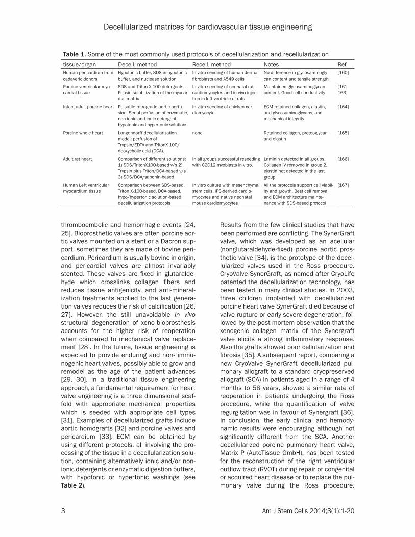
Table 1. Some of the most commonly used protocols of decellularization and recellularizationtissue/organ Decell. method Recell. method Notes RefHuman pericardium from cadaveric donors
Hypotonic buffer, SDS in hypotonic buffer, and nuclease solution
In vitro seeding of human dermal fibroblasts and A549 cells
No difference in glycosaminogly-can content and tensile strength
[160]
Porcine ventricular myo-cardial tissue
SDS and Triton X-100 detergents. Pepsin-solubilization of the myocar-dial matrix
In vitro seeding of neonatal rat cardiomyocytes and in vivo injec-tion in left ventricle of rats
Maintained glycosaminoglycan content. Good cell-conductivity
[161-163]
Intact adult porcine heart Pulsatile retrograde aortic perfu-sion. Serial perfusion of enzymatic, non-ionic and ionic detergent, hypotonic and hypertonic solutions
In vitro seeding of chicken car-diomyocyte
ECM retained collagen, elastin, and glycosaminoglycans, and mechanical integrity
[164]
Porcine whole heart Langendorff decellularization model: perfusion ofTrypsin/EDTA and TritonX 100/deoxycholic acid (DCA).
none Retained collagen, proteoglycan and elastin
[165]
Adult rat heart Comparison of different solutions: 1) SDS/TritonX100-based v/s 2) Trypsin plus Triton/DCA-based v/s 3) SDS/DCA/saponin-based
In all groups successful reseeding with C2C12 myoblasts in vitro.
Laminin detected in all groups. Collagen IV removed in group 2, elastin not detected in the last group
[166]
Human Left ventricular myocardium tissue
Comparison between SDS-based, Triton X-100-based, DCA-based, hypo/hypertonic solution-based decellularization protocols
In vitro culture with mesenchymal stem cells, iPS-derived cardio-myocytes and native neonatal mouse cardiomyocytes
All the protocols support cell viabil-ity and growth. Best cell removal and ECM architecture mainte-nance with SDS-based protocol
[167]
Decellularized matrices for cardiovascular tissue engineering
4 Am J Stem Cells 2014;3(1):1-20
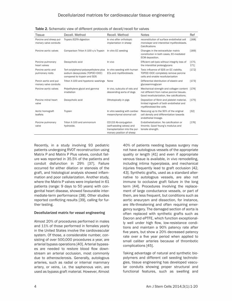
Table 2. Schematic view of different protocols of decell/recell for valvesTissue Decell. Method Recell. Method Notes RefPorcine and sheep pul-monary valve conduits
Trypsin/EDTA digestion In vivo after orthotopic implantation in sheep
reconstitution of surface endothelial cell monolayer and interstitial myofibroblasts. Calcifications
[168]
Porcine aortic valves Comparison Triton X-100 v/s Trypsin In vitro EC seeding Changes in the extracellular matrix constitution in both cases, EC-mediated ECM deposition.
[169]
Porcine pulmonary heart valves
Deoxycholic acid In vivo Efficient cell-lysis without integrity loss of the interstitial proteoglycans
[170, 171]
Porcine aortic and pulmonary roots
Tert-octylphenyl-polyoxyethylene plus sodium deoxycolate (TOPOE+DOC) compared to trypsin and SDS
In vitro seeding with human ECs and myofibroblasts
Toxic influence of SDS on EC viability. TOPOE+DOC completely remove porcine cells and enable recellularization
[172]
Porcin aortic and pul-monary valve conduits
Triton X-100 and hypotonic washings None Differential distribution of elastin and glycosaminoglycan
[173]
Porcine aortic valves Polyethylene glycol and gamma irradiation
In vivo, subcutis of rats and descending aorta of dogs
Mechanical strength and collagen content not different from native porcine tissues. Good recellularization, few calcifications.
[174]
Porcine mitral heart valve
Deoxycholic acid Othotopically in pigs Deposition of fibrin and platelet material. limited ingrowth of both endothelial and myofibroblast-like cells
[175]
Aortic homograft leaflets
Trypsin In vitro seeding with cardiac mesenchymal stromal cell
Rescuing up to the 90% of the original cell density and differentiation towards endothelial lineage.
[32]
Porcine pulmonary valve
Triton X-100 and ammonium hydroxide.
CD133 Ab-conjugation (self-seeding valves) and transplantation into the pul-monary position of sheep
Endothelialization. No calcification or thrombi. Good Young’s modulus and tensile strength.
[176]
Recently, in a study involving 93 pediatric patients undergoing RVOT reconstruction using Matrix P and Matrix P Plus valves, conduit fail-ure was reported in 35.5% of the patients and conduit disfunction in 29% [37]. Failure occurred for either dilation or stenosis of the graft, and histological analysis showed inflam-mation and poor cellularization. Another study, where the Matrix P valves were implanted in 61 patients (range: 9 days to 50 years) with con-genital heart disease, showed favourable inter-mediate-term performance [38]. Other studies reported conflicting results [39], calling for fur-ther testing.
Decellularized matrix for vessel engineering
Almost 20% of procedures performed in males and 11% of those performed in females yearly in the United States involve the cardiovascular system. Of those, a considerable number, con-sisting of over 500,000 procedures a year, are arterial bypass operations [40]. Arterial bypass-es are needed to restore blood flow down-stream an arterial occlusion, most commonly due to atherosclerosis. Generally, autologous arteries, such as radial or internal mammary artery, or veins, i.e. the saphenous vein, are used as bypass graft material. However, Almost
40% of patients needing bypass surgery may not have autologous vessels of the appropriate quality or length [41] and even if appropriate venous tissue is available, in vivo remodelling, including intima hyperplasia, and mechanical injuries frequently lead to graft occlusion [42, 43]. Synthetic grafts, used as a standard alter-native to autologous vessels, are also not immune to occlusive graft failure in the long term [44]. Procedures involving the replace-ment of large conductance vessels, or part of them, are less frequent, but conditions such as aortic aneurysm and dissection, for instance, are life-threatening and often requiring emer-gency surgery. The damaged section of aorta is often replaced with synthetic grafts such as Dacron and ePTFE, which function exceptional-ly well under high flow, low-resistance condi-tions and maintain a 90% patency rate after five years, but show a 20% decreased patency rate over a five year period when applied to small caliber arteries because of thrombotic complications [45].
Taking advantage of natural and synthetic bio-polymers and different cell seeding technolo-gies, tissue engineering has developed vascu-lar conduits showing proper structural and functional features, such as swelling and
Decellularized matrices for cardiovascular tissue engineering
5 Am J Stem Cells 2014;3(1):1-20
stretching properties, suture-retention and cell conductivity [46-52]. However, the limited pro-liferative rate of adult smooth muscle cells [53], as well as the senescence they undergo in culture, accounts for the poor in vivo mechani-cal performance of tissue engineered blood vessels. Inter alia, the biofabrication of blood vessels is still costly and time consuming [54].
The pioneering research of Malone et al. [55] and Lalka et al. [56] first reported that implant-ed cell-free arterial allografts do not undergo immunologic alterations. The simple treatment with SDS resulted in the formation of an ECM tube with morphologically intact elastin and collagen network, that was easy to suture and immediately blood perfused after in vivo graft-ing. Decellularization technologies were not advanced yet, when these scientists intro-duced a simple and powerful concept: reducing allograft/xenograft antigenicity as opposed to immunosuppressing the graft host!
Similarly to synthetic grafts, decellularized matrices would be readily available. Unlike syn-thetic grafts, they would provide the proper microenvironment for supporting cell invasion, growth and differentiation. A future goal for the tissue engineering is to identify decellulariza-tion techniques that can provide vascular grafts with both mechanical properties of native ves-sels and immuno-privileged characteristics of autologous vessels. In a recent study by Fitzpatrick et al., different protocols were applied to decellularize segments of porcine aorta and it was shown that the TritonX-100/sodium-deoxycholate treatment is a more
effective option than TritonX-100/EDTA and SDS treatments since it effectively lyses VSMCs and results in less variability in mechanical behavior at in vivo stretch ratios [44]. In anoth-er report both SDS and Triton X-100 treatments were able to remove cells effectively from por-cine aorta and the major ECM structure was preserved, while trypsin treatment disrupted the cross-linked network of collagen and elas-tin fibers [57]. Dimuzio’s group has decellular-ized the human saphenous vein by using SDS, showing that decellularized veins have a burst and suture-holding strength similar to fresh veins, as well as unchanged collagen morphol-ogy [58]. The same group reported a canine model of bilateral carotid interposition of a decellularized jugular vein allograft: the decel-lularized allograft exhibited satisfactory stre- ngth, reduced antigenicity compared to fresh allograft, and supported cellular repopulation [59].
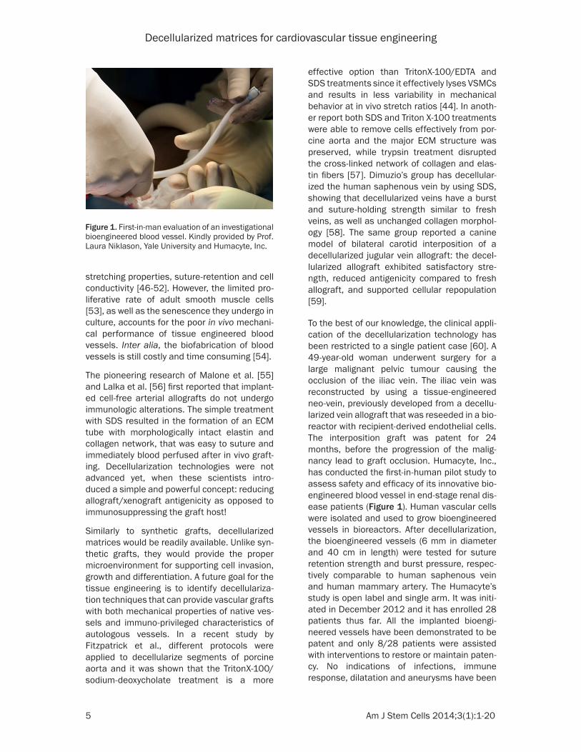
To the best of our knowledge, the clinical appli-cation of the decellularization technology has been restricted to a single patient case [60]. A 49-year-old woman underwent surgery for a large malignant pelvic tumour causing the occlusion of the iliac vein. The iliac vein was reconstructed by using a tissue-engineered neo-vein, previously developed from a decellu-larized vein allograft that was reseeded in a bio-reactor with recipient-derived endothelial cells. The interposition graft was patent for 24 months, before the progression of the malig-nancy lead to graft occlusion. Humacyte, Inc., has conducted the first-in-human pilot study to assess safety and efficacy of its innovative bio-engineered blood vessel in end-stage renal dis-ease patients (Figure 1). Human vascular cells were isolated and used to grow bioengineered vessels in bioreactors. After decellularization, the bioengineered vessels (6 mm in diameter and 40 cm in length) were tested for suture retention strength and burst pressure, respec-tively comparable to human saphenous vein and human mammary artery. The Humacyte’s study is open label and single arm. It was initi-ated in December 2012 and it has enrolled 28 patients thus far. All the implanted bioengi-neered vessels have been demonstrated to be patent and only 8/28 patients were assisted with interventions to restore or maintain paten-cy. No indications of infections, immune response, dilatation and aneurysms have been
Figure 1. First-in-man evaluation of an investigational bioengineered blood vessel. Kindly provided by Prof. Laura Niklason, Yale University and Humacyte, Inc.
Decellularized matrices for cardiovascular tissue engineering
6 Am J Stem Cells 2014;3(1):1-20
observed (Abstracts from the American Heart Association’s Emerging Science Series April 24, 2013).
Surgeons have been using cryopreserved vas-cular allografts successfully for many years to treat arterial occlusive disease and to repair arterial aneurysms. Vascular allografts demon-strate high patency rates but contain viable cells, which may evoke an immune rejection. Decellularization techniques efficiently remove cells and can be optimized to guarantee the maintenance of the microarchitecture and the biomechanical properties of native vessels (see Table 3).
ECM components of cardiovascular tissues
The extracellular matrix (ECM) mediates the interaction between the cell and the surround-ing microenvironment in a model of dynamic reciprocity, in which cells secrete ECM compo-nents and ECM proteins regulate cell prolifera-tion and differentiation to finally determine tis-sue morphogenesis and homeostasis in development and disease [61]. Far from being a merely structural component of any tissue,
the ECM represents a huge reservoir of bio-physical stimuli and signalling molecules. The regulation of cell fate mediated by ECM is also essential during tissue repair, wherein a very delicate balance in the amount, composition and spatial organization of the newly produced matrix marks the border between a regenera-tion process and scar formation [62, 63]. A pro-found knowledge of the structure and the sig-nalling mediated by the ECM of cardiovascular tissues is needed in order to rationalize the use of decellularized vessels, valves and even entire hearts. Modifications in matrix proteins, depending on decellularization techniques, might account for in vivo fibrosis, calcification, poor endothelialisation, and ultimately for the failure of the implanted patches.
ECM in the heart
ECM is crucial for heart development [64]; the cardiac jelly, an ECM-rich acellular space between the endocardium and myocardium, is particularly important for the proper formation of the endocardial cushions at the atrioventric-ular (AV) junction. In adult life, the coupling of vessel endothelium and cardiomyocytes, as
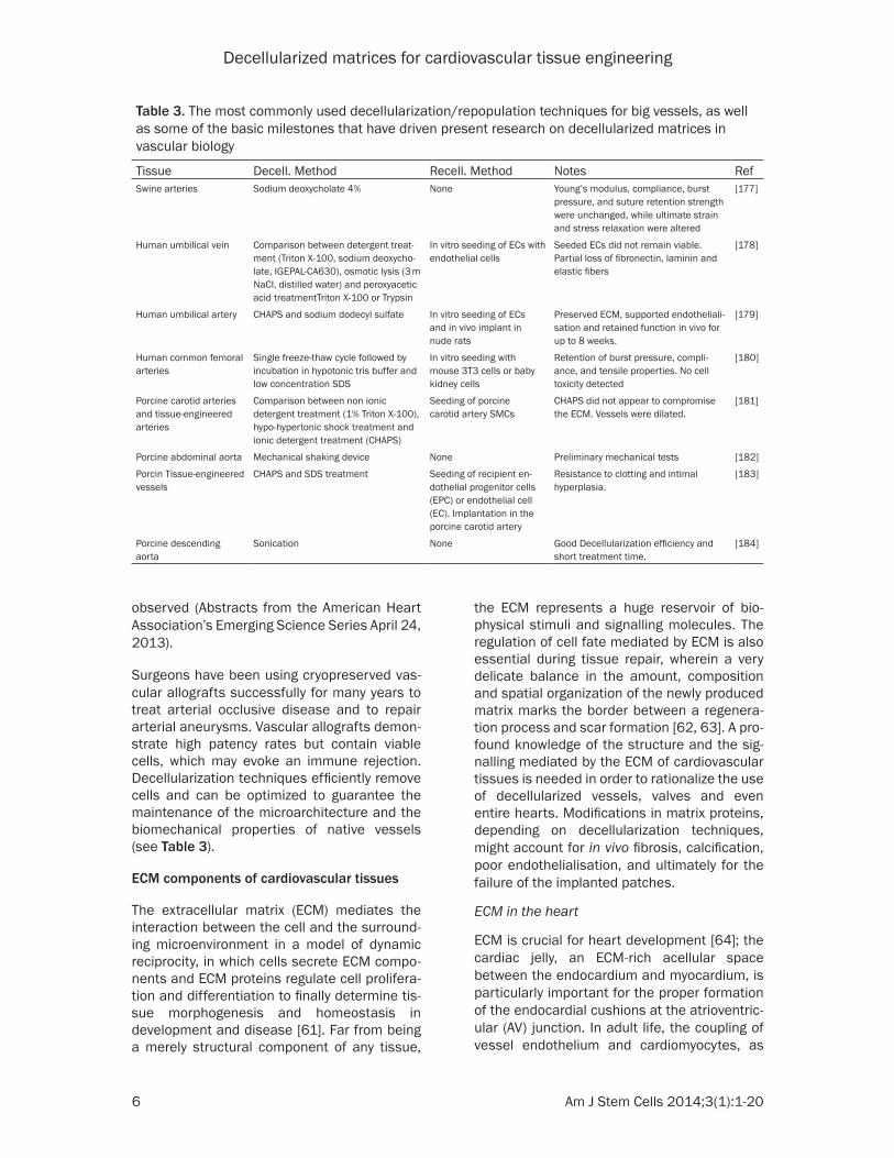
Table 3. The most commonly used decellularization/repopulation techniques for big vessels, as well as some of the basic milestones that have driven present research on decellularized matrices in vascular biology
Tissue Decell. Method Recell. Method Notes RefSwine arteries Sodium deoxycholate 4% None Young’s modulus, compliance, burst
pressure, and suture retention strength were unchanged, while ultimate strain and stress relaxation were altered
[177]
Human umbilical vein Comparison between detergent treat-ment (Triton X-100, sodium deoxycho-late, IGEPAL-CA630), osmotic lysis (3 m NaCl, distilled water) and peroxyacetic acid treatmentTriton X-100 or Trypsin
In vitro seeding of ECs with endothelial cells
Seeded ECs did not remain viable. Partial loss of fibronectin, laminin and elastic fibers
[178]
Human umbilical artery CHAPS and sodium dodecyl sulfate In vitro seeding of ECs and in vivo implant in nude rats
Preserved ECM, supported endotheliali-sation and retained function in vivo for up to 8 weeks.
[179]
Human common femoral arteries
Single freeze-thaw cycle followed by incubation in hypotonic tris buffer and low concentration SDS
In vitro seeding with mouse 3T3 cells or baby kidney cells
Retention of burst pressure, compli-ance, and tensile properties. No cell toxicity detected
[180]
Porcine carotid arteries and tissue-engineered arteries
Comparison between non ionic detergent treatment (1% Triton X-100), hypo-hypertonic shock treatment and ionic detergent treatment (CHAPS)
Seeding of porcinecarotid artery SMCs
CHAPS did not appear to compromise the ECM. Vessels were dilated.
[181]
Porcine abdominal aorta Mechanical shaking device None Preliminary mechanical tests [182]
Porcin Tissue-engineered vessels
CHAPS and SDS treatment Seeding of recipient en-dothelial progenitor cells (EPC) or endothelial cell (EC). Implantation in the porcine carotid artery
Resistance to clotting and intimal hyperplasia.
[183]
Porcine descending aorta
Sonication None Good Decellularization efficiency and short treatment time.
[184]
Decellularized matrices for cardiovascular tissue engineering
7 Am J Stem Cells 2014;3(1):1-20
well as the coordination of cardiomyocyte con-traction and relaxation is largely dependent on ECM [65]. In myocardial fibrosis, for instance, an altered secretion of ECM components by myofibroblasts causes a “mis-remodelling” of the tissue leading to cardiac muscle stiffness and contractile dysfunction [66, 67]. Main com-ponents of heart ECM [64, 66] include GAGs and proteoglycans as important structural mol-ecules for creating loose and hydrated matri-ces during key events in development and dis-ease. Some of them are:
- Hyaluronan (HA), a GAG which is synthesized at the plasma membrane and does not become linked to a core protein. It promotes cellular proliferation and motility in the cardiac jelly of developing heart [68].
- Chondroitin sulfate proteoglycans, such as versican, which is essential for the formation of endocardial cushion mesenchyme by epitheli-al–mesenchymal transformation (EMT), heart chamber specification and valvulogenesis [69, 70].
- Heparan Sulfate Proteoglycans, such as per-lecan which is important in the formation of the cardiomyocyte basement membrane and in maintaining the integrity of the ventricular wall [71].
Many different collagens are expressed in the heart, both in ventricular myocardium (type I, III, V) and heart valves (I, II, IV, XI, and XIII) [64]. Collagens provide elasticity and structural integrity to cardiac tissue. Fibronectin interacts with integrins, proteoglycans and collagens to mediate cellular adhesion. Fibronectin null embryos do not survive beyond embryonic day 10 (E10) due to cardiovascular (failure of heart tube formation in the most severely affected mutants) and vascular defects [72]. The remod-elling of the ECM, important for the release of mediators (growth factors, cytokines, small peptides), is mainly carried out by matrix metal-loproteases (MMPs) [65]. Disease states such as hypertension, excessive activation of the angiotensin aldosterone system, diabetes and hypoxia can lead to an over-activation of MMPs and consequent ECM degradation, impaired angiogenesis, myocardial hypertrophy and pro-gression to heart failure [65, 73-76]. Among MMPs, MMP9 appears to be the most involved in the pathological remodelling of ECM in heart
failure [65, 77], being associated with increas-ing endostatin and angiostatin (anti-angiogenic activity ) and vascular rarefaction [74, 78].
ECM in heart valves
Mature heart valves are composed of highly organized ECM and valve interstitial cells (VICs), all surrounded by endothelial cells [79]. Valvular ECM is stratified into different layers and is responsible for biomechanical properties of the valves [79, 80]. The ventricularis, facing the ventricle, is enriched in elastin and is responsi-ble for valve extension and recoil [81]. Proteoglycans are interposed between the ven-tricularis and the fibrosa, and constitute the spongiosa. They provide cohesiveness between the layers and contribute to tissue viscoelastic-ity [82]. The fibrosa is close to the outflow sur-face and is mainly composed of collagens, that are responsible for tissue strength and durabil-ity [80, 83]. Many disease conditions affecting heart valves involve degenerative changes of ECM. In calcific stenosis, a common disease of the elderly, mainly affecting the aortic valve, noxious stimuli such as hypertension, high serum cholesterol levels and smoking can induce differentiation of VICs to an osteoblastic phenotype [84]. Such cell types express osteo-genic and chondrogenic markers and promote tissue calcification and degeneration [85]. Mitral prolapse consists in the displacement of a valvular leaflet of the mitral valve in the left atrium during systole. It has potential serious complications such as bacterial endocarditis, thromboemboli and atrial fibrillation. The patho-genic process underlying this condition is myxo-matous degeneration, which is characterized histologically by a focal thickening of the spon-giosa with an increase in proteoglycans content [86, 87] together with an abnormal fibrillar organization, and an attenuation of the fibrosa [88]. Activated VICs secrete catabolic enzymes, including MMPs (MMP-1, MMP-13, MMP-2, and MMP-9), and are believed to play a major role in myxomatous degeneration [89].
ECM in blood vessels
Big blood vessels are constituted by three con-centric layers, which are, progressing radially from the lumen; the intima, the media and the adventitia. Each layer is constituted by a struc-tural ECM meshwork supporting resident cells. Vascular ECM not only provides the scaffold for
Decellularized matrices for cardiovascular tissue engineering
8 Am J Stem Cells 2014;3(1):1-20
attachment of the resident cells, but is also able to absorb and transduce shear and strain forces exerted by blood flow [90]. Endothelial cells produce and attach to a basal lamina (laminin, type IV collagen, entactin and per-lecan are the main components) and contribute to the formation of the internal elastic lamina, which is very thin in veins and venules [91]. In pathological conditions such as atherosclero-sis and hypertension, or after mechanical dis-tension and disruption of the endothelial layer, such as after percutaneous coronary interven-tion; the tunica intima appears as a thick layer of sparse smooth muscle cells and myofibro-blasts in a proteoglycan-rich stroma [92-94].
Tunica media consists of an ensemble of radial-ly-arranged fenestrated sheets (lamellae) rich in elastin, immersed in collagen fibers, thin lay-ers of proteoglycans, and smooth muscle cells. It is important to distinguish between elastin itself and the elastic fibers, which contain elas-tin and microfibrils. Microfibrils act as a scaf-fold for elastin assembly and elastic fiber over-all growth [95]. The functional importance of the elastic components of the blood vessel walls is underlined by the fact that the genetic inactivation of its constituents leads to major health issues [96]. Elastin haploinsufficiency for instance causes supravalvular aortic steno-sis [97] which can lead to hypertension, cere-brovascular disease and obstructive cardiomy-opathy. Vascular lesions show irregular elastic fibers, excess of medial smooth muscle cells and intimal thickening and fibrosis. Homozygous loss of function in FBN1 gene, coding for fibrillin 1, causes Marfan syndrome with mitral valve prolapse and aortic root dilation as main car-diovascular affections [98, 99]. Other biome-chanically important constituents of tunica media ECM are fibulins, located either in the elastin core or its surrounding microfibrils, col-lagens (I, III, V, VI) and proteoglycans (versican, lumican, etc.).
The adventitial layer contains sparse fibro-blasts surrounded by ECM, mainly composed of fibrillar collagens and proteoglycans, as well as vasa vasorum, providing nourishment to the vessel, and nervi vasorum (unmyelinated nerve fibers). Adventitia is the primary source of ten-sile strength in blood vessels, but also partici-pates in the regulation of blood vessels tone through the activity of nervous fibers. Fibrillar
collagens, in particular Collagen I and Collagen III, are responsible for blood vessels resistance to mechanical stress [100]. Autosomal domi-nant mutations in type III collagen results in Ehlers-Danlos syndrome type IV, which is char-acterized by spontaneous rupture of the bowel, uterus and blood vessels [101]. Adventitia undergoes remodelling in a number of patho-logical conditions such as hypertension or ath-erosclerosis. Adventitia fibroblasts are the main players of the remodelling process, which can be adaptive (positive) to vasoactive sub-stances and hemodynamic stimuli, or constric-tive (negative), leading to lumen reduction and stenosis [102, 103]. Since the activation of pro-liferative and differentiative mechanisms in adventitia fibroblasts may shape the vessel wall and tone, caution should be used when decellularizing vessels, to avoid mis-recellular-ization in vivo from overactivated progenitors. Nevertheless, the integrity of elastic fibers in the media has to be maintained after decellu-larization. Indeed, it has been demonstrated that partial degradation of elastic fibers caused by NaOH and trypsin treatment of aortic xeno-grafts significantly increases elastin-oriented calcification [104]. Finally, an intact Collagen IV (basement membrane) mediates migration and adhesion of endothelial cells [105], while the carboxy terminal globular domain is less active at promoting those events. Thus, decellulariz-ing protocols, especially enzymatic ones, sh- ould take into account that a degraded Collagen IV might have repercussions for the in vivo re-endothelialization of decellularized grafts.
Immune response to ECM
One of the main causes for biological implant failure is the immune rejection of the graft itself. In case of allogenic implants (transplan-tation to a recipient from a genetically non-identical donor of the same species), a cell-mediated immune response will be activated by antigen presenting cells (APCs) presenting MHC-alloantigen to T Lymphocytes through direct (donor’s APCs are presenting graft anti-gens) or indirect (recipient’s infiltrating APCs process and present foreign graft proteins) allorecognition pathways [106]. Beyond the cel-lular response, involving CD4+ and CD8+ T-lymphocytes, NK cells and other phagocytes, also B-lymphocytes (humoral or antibody-medi-ated rejection) and cytokines (IL-12, IFN-γ, IL-6,
Decellularized matrices for cardiovascular tissue engineering
9 Am J Stem Cells 2014;3(1):1-20
IL-17, etc.) [107] play an important role in allograft rejection. Moreover, if the balance between proinflammatory (Th1, Th17 lympho-cytes, IFN-γ, IL-17, etc) and anti-inflammatory (Th2, regulatory T cells, IL-4, IL-10, TGF-β etc.) players is not correctly established, a sustained chronic rejection of the graft can lead to vascu-lar endothelium damage, blood supply deficien-cy, scar formation and ultimately functional loss of the implant [108]. On the other hand, xenogenic transplantation of organs and tis-sues ensues hyperacute rejection, which entails complement-activation, neutrophil infil-tration and NK cell activation in response to natural xenoreactive antibodies (for instance the natural antibodies to Galα1,3Gal). Xenorejection has in the microvasculature a main target, thus, in addition to intragraft rejec-tion events, systemic complications, such as thrombotic microangiopathy, can follow to the rapid graft destruction soon after implantation [109, 110]. One of the most relevant antigens involved in hyperacute rejection following xeno-transplantation is the saccharide α-Gal. This epitope is found as a cell surface molecule in most species, with the notable exception of humans and Old World monkeys [111, 112]. Although the Gal epitope is considered the main obstacle to xenotransplantation, organs harvested from pigs that were knocked out for this antigen were rejected after a short time due to immune response towards non-Gal por-cine antigens [113, 114]. Furthermore, the presence of the Gal epitope has been demon-strated in biologic scaffolds composed of xeno-genic ECM, such as porcine bioprosthetic heart valves [115], porcine cruciate ligaments [116] and porcine cartilage [117].
ECM proteins are among the most conserved proteins in evolution [118] but, it would be naive to think that the removal of xenogenic or allogenic cellular material would abolish graft immunogenicity [119, 120]. In fact, ECM pro-teins have been shown to provide costimulato-ry signals to immune cells [121], For instance, neutrophils exhibit chemotaxis toward frag-ments of type IV collagen, laminins, and elastin [122]; a laminin-derived peptide (SIKVAV), iso-lated from human abdominal aortic aneurysm tissue can recruit neutrophils within 1 day and macrophages by 3 days when instilled in mouse lungs [123]; instillation of in vitro-generated bovine elastin fragments into the lungs of mice induce macrophage accumulation, which is
prevented when BA4 (a monoclonal antibody raised against the bovine tropoelastin epitope VGVAPG) is given [124]. The fact that ECM frag-ments can promote immune cell recruitment must bring us to rethink, and maybe reinvent, the decellularization techniques currently in use, especially those involving enzymatic reac-tions that could easily unmask crypted ECM peptides with pro-inflammatory activity.
Thus, the complete removal of cellular material, including DNA, RNAs and proteins, is necessary but not sufficient: special attention has to be given to the “new products” derived from the decellularization processing of the tissue. To date, very few reports have dealt with the issue of the immune response towards biological scaffolds composed of ECM. The role of adap-tive immunity has been investigated most extensively for porcine small intestine submu-cosa ECM [125, 126]: after its implantation in mice, it elicits mainly a Th2 type of response, which is associated with tissue remodelling and graft acceptance [127, 128]. Innate immu-nity, which naturally provide immediate defense against foreign bodies, is obviously primarily involved in mediating the host response to bio-materials, and the monocyte-derived macro-phages are indeed important “sentinels and soldiers” in this context [129]. Macrophages are a heterogeneous cell population, displaying a variety of phenotypes. In particular, they have been classified on the basis of functional prop-erties in M1 and M2, in analogy with Th1 and Th2 cells [130, 131]. M1 are the classically acti-vated, proinflammatory macrophages, in- ducers and effectors of Th1 response [130]. While M2 macrophages are involved in Th2 response which is required for tissue regenera-tion and remodelling [132]. Badylak et al. have used implantation of biologic scaffolds (with and without cross-linking) derived from porcine small intestinal submucosa (SIS) to character-ize the role of macrophage phenotype in the remodelling of the scaffold [133, 134]. They found that the SIS scaffold elicited a CD163+ response (M2 profile) and showed constructive remodelling at 16 weeks, while the cross-linked-SIS device showed a predominately CD80+ and CCR7+ response (M1 profile), and at 16 weeks was characterized by chronic inflammation and fibrosis. The conclusion is that future strategies aimed at polarizing mac-rophages from an M1 to an M2 phenotype will
Decellularized matrices for cardiovascular tissue engineering
10 Am J Stem Cells 2014;3(1):1-20
be needed for a successful long term outcome of decellularized matrix implantation. Recent in vitro studies have showed that decellularized xeno-ECM, such as decellularized bovine peri-cardium can favour the polarization of macro-phages towards an M2 phenotype [135, 136] as compared to other materials commonly employed for medical devices, such as polydimethylsiloxane. Scaffolds composed of ECM showed to promote the switch M1-M2 also in vivo, after 7-14 days post-implantation [137, 138], but mechanisms by which ECM based scaffold promote the M1 to M2 transition remain unknown. All these results are encour-aging but more comparative (allo- v/s xeno-, biologic v/s synthetic, different decell. tech-niques) in vivo studies will be necessary for a better understanding of macrophage polarity and context dependent polarization profiles, especially in order to design strategies for tun-ing macrophage plasticity and reach the per-fect biocompatibility of decellularized ma- trices.
In vivo recellularization
Obtaining a functional tissue or organ from an implanted decellularized matrix requires in vivo cell repopulation of the matrix. Recruitment of cells and progenitors from the neighbour tis-sues and circulation is the first step to get “bio-integration” of the decellularized matrix. To date, we do not have a complete knowledge of what cell types, what chemoattracting and sig-nalling molecules are needed, whether there is a precise chronological sequence of events and how we can modulate each and single event to achieve engraftment. The in vivo recruitment of the whole spectrum of parenchy-mal and stromal cells towards the ECM of com-plex organs, such as the heart, is not feasible yet. On the other hand, the study of recellular-ization events occurring in simpler structures, including heart valves and vascular conduits, might be the right start towards comprehen-sion. Recellularization may be carried out, or simply started, in vitro, mainly by the use of bio-reactors mimicking physiological organ condi-tions, such as 3D growth and controlled chang-es of specific environmental factors [139]. One of the most successful examples of the clinical application of decellularized matrices is the tissue-engineered airway by Macchiarini et al [140]. Here, the authors decellularized a human
donor trachea and subsequently cultured the graft in a bioreactor with recipient epithelial cells and mesenchymal stem-cell-derived chon-drocytes. The graft was then used to replace the left main bronchus of a 30-year old woman with end-stage bronchomalacia. There is no doubt that the scaffold per se is not able to be integrated and functional in vivo. Another study from Macchiarini’s group reports that decellu-larized not pre-seeded tracheas implanted in pigs collapse because of obstruction and infec-tions, while cell-seeded tracheas are function-al, suggesting a role for epithelial and mesen-chymal cells in mediating perfusion and immune-tolerance of the graft [141]. It is not clear whether the implanted cells are directly contributing to the in vivo fabrication of the tis-sue or rather acting as reservoir of molecules activating micro- and macro-environment path-ways of tissue regeneration. Unless the implant-ed cells are immortal or able to initiate a con-trolled and regulated program of cell renewal and apoptosis, the contribution of endogenous progenitor cells cannot be excluded from the whole regeneration process. It appears clear that tissue engineering and regenerative medi-cine have to “hold hands” in order to provide complementary solutions and treatment options. Several techniques, aforementioned in this review, have been used to recellularize vas-cular patches in vitro, before implantation. Re-endothelialization, in particular, seems to be crucial in order to decrease calcification and thrombus formation. Walles et al. showed that decellularized carotid artery and aorta undergo progressive calcification, while calcification is less pronounced in cellular native arteries [142]. Kasimir et al. decellularized heart valve matrix and reported platelet adhesion (CD41+ cells recruitment) and aggregate formation only on the surface of the non-seeded or partially denuded matrix, whereas after seeding with endothelial cells no platelet activation was detected [143]. Strategies to enhance in situ re-endothelialization have comprised the coat-ing of decellularized heart valves with fibronec-tin (FN) [144]. FN is used to increase cell adhe-sion; cell adhesion is meant to provide not only physical support, but also pro survival signals to cells [145]. FN in combination with hepato-cyte growth factor (HGF), can synergize re-endothelialization by both stabilizing cell-matrix adhesion and stimulating EC proliferation [146, 147]. Heparin, and vascular endothelial growth
Decellularized matrices for cardiovascular tissue engineering
11 Am J Stem Cells 2014;3(1):1-20
factor (VEGF) have been used as bioactive coating to improve recellularization, as well [148], although full consideration should be given to VEGF-mediated hyperplasia of neointi-ma. Other than adhesion and proliferation, blood perfusion of the implanted graft has to be achieved, not only to provide oxygen and nourishment to the graft, but also progenitor cells. Endothelial progenitor cells (EPCs), which are thought to be originated in the bone mar-row, can contribute to vascular repair [149].
According to some authors, peripheral blood endothelial progenitor cells can be derived from monocyte/macrophages [150]. Also, myeloid angiogenic cells (MACs), although not able to become endothelial cells or be directly incorporated into a microvascular network as EPCs, have been described as an alternative population of activated M2 macrophages, able to induce vascular repair in vivo in a paracrine fashion [151]. Even progenitors derived from injured neointima could be exploited as an
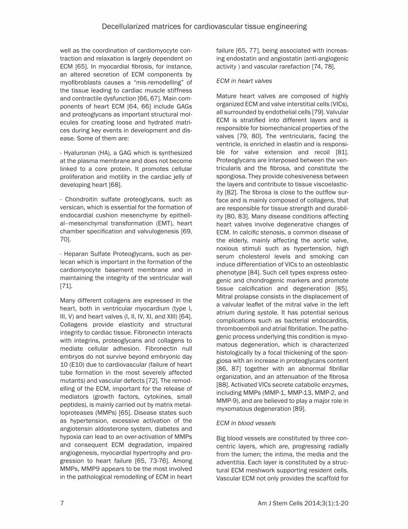
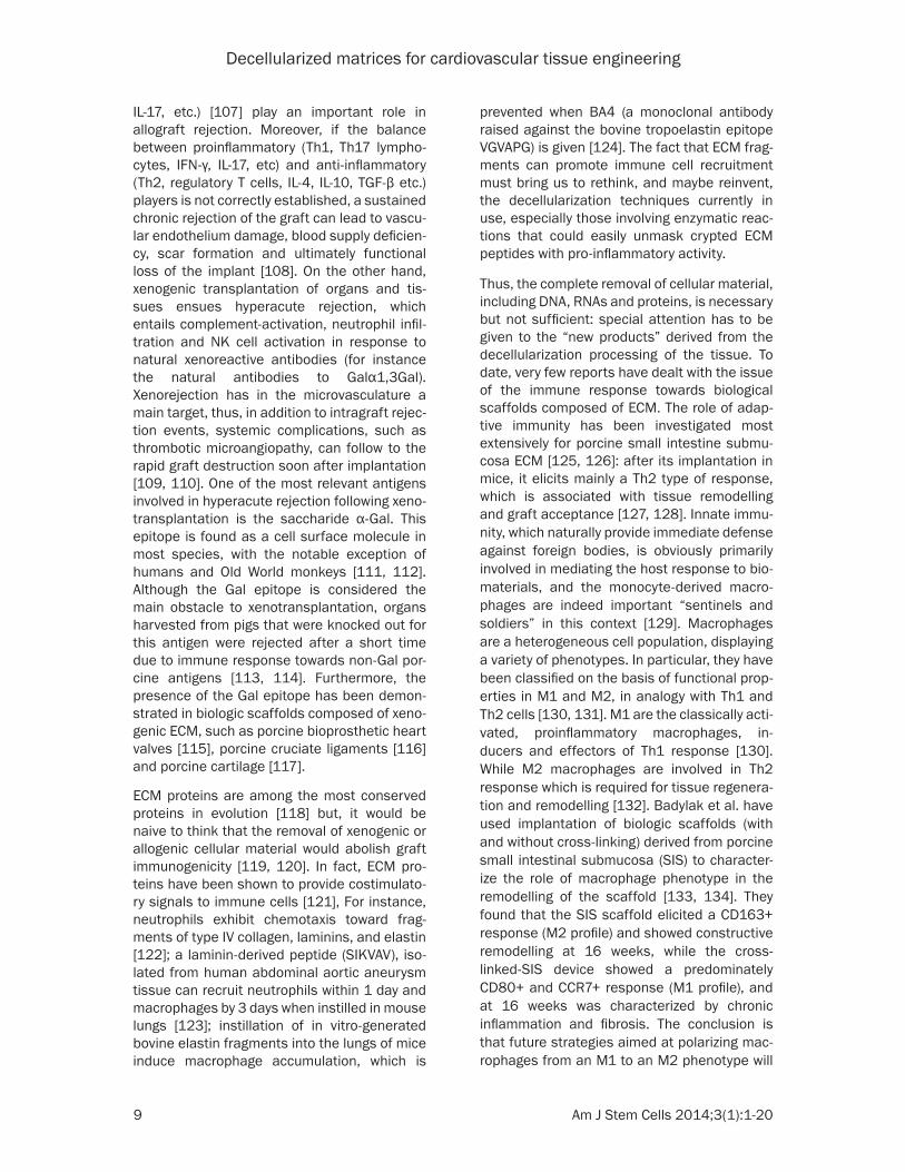
Figure 2. Scheme of different transplantation (TXP) approaches of vessels. Decellularization of the vessel reduces the risk of immune rejection. In vitro cell seeding of the decellularized vessel prior to implantation reduces the risk of thrombus formation and calcifications and induces progenitor recruitment and regeneration in vivo.
Decellularized matrices for cardiovascular tissue engineering
12 Am J Stem Cells 2014;3(1):1-20
inner cell source to promote vascular repair. Tsai et al. developed a mouse model of reste-nosis by grafting a decellularized vessel to the carotid artery. Cells retrieved in the neointimal lesions were endothelial and smooth muscle cells, monocytes, and stem/progenitor cells expressing c-kit, Sca-1 and CD34 [152]. Ex-vivo cultured progenitors displayed the ability to dif-ferentiate into both endothelial and smooth muscle cells. This suggests that the ECM per se is able to recruit vessel progenitors which, if impeded from turning on restenosis, can be a useful source of endothelial and mural cells.
Future perspectives
In future, it will be mandatory to set up decel-lularization techniques that leave an intact ECM and to learn more about ECM biology to exploit native and bioengineered ECM mole-cules that allow a better recruitment of cells in vivo. The concept that ECM degradation can result in products with chemoattractive proper-ties [153] needs to be further developed (Figure 2).
The enhancement of blood perfusion of decel-lularized grafts through the peripheral anasto-mosis could be achieved by providing an imme-diately active angiogenic boost. Many stem cells, including the promising amniotic fluid stem cells, are endowed with a reservoir of sol-uble factors that can exert paracrine effects on capillary ingrowth [154-156]. The in vitro pre-seeding of decellularized ECM can trigger a bet-ter recellularization in vivo. Human embryonic stem cells (ESCs) and human induced pluripo-tent stem cells (iPSCs) can be cultured on decellularized matrices and be reprogrammed into cells capable of angiogenesis and re-endo-thelialization as well as into parenchymal cells with positive implications for cell colonization of big organs, such as the heart [157-159]. However, caution must be used when such undifferentiated cells are used, since the safe-ty profile is not completely investigated yet.
Moreover, the networking between different fields, including but not limited to stem cells, biomaterials, cell and matrix biology, will be the key for a successful application of decellular-ized matrices in the treatment of cardiovascu-lar disease.
Acknowledgements
We thank Dr. John Rhodes, Cardiovascular Research Center, Yale Medical School, for the
critical reading of the manuscript. We have been honored to present in our review some of the unpublished interim results of Humacyte clinical trial, with the kind permission of Prof. Laura Niklason, Yale University. We are grateful to Laura for her terrific input.
Disclosure of conflict of interest
None to declare.
Address correspondence to: Dr. Teodelinda Mira- bella, Cardiovascular Research Center, Yale Medical School, New Haven, CT, 06511, US. E-mail: [email protected]
References
[1] Badylak SF, Freytes DO and Gilbert TW. Extra-cellular matrix as a biological scaffold materi-al: Structure and function. Acta Biomater 2009; 5: 1-13.
[2] Gilbert TW, Sellaro TL and Badylak SF. Decel-lularization of tissues and organs. Biomateri-als 2006; 27: 3675-3683.
[3] McMurray JJ, Adamopoulos S, Anker SD, Auric-chio A, Bohm M, Dickstein K, Falk V, Filippatos G, Fonseca C, Gomez-Sanchez MA, Jaarsma T, Kober L, Lip GY, Maggioni AP, Parkhomenko A, Pieske BM, Popescu BA, Ronnevik PK, Rutten FH, Schwitter J, Seferovic P, Stepinska J, Trin-dade PT, Voors AA, Zannad F, Zeiher A; Task Force for the Diagnosis and Treatment of Acute and Chronic Heart Failure 2012 of the Euro-pean Society of Cardiology; Bax JJ, Baumgart-ner H, Ceconi C, Dean V, Deaton C, Fagard R, Funck-Brentano C, Hasdai D, Hoes A, Kirchhof P, Knuuti J, Kolh P, McDonagh T, Moulin C, Popescu BA, Reiner Z, Sechtem U, Sirnes PA, Tendera M, Torbicki A, Vahanian A, Windecker S, McDonagh T, Sechtem U, Bonet LA, Avraamides P, Ben Lamin HA, Brignole M, Coca A, Cowburn P, Dargie H, Elliott P, Flachskampf FA, Guida GF, Hardman S, Iung B, Merkely B, Mueller C, Nanas JN, Nielsen OW, Orn S, Paris-sis JT, Ponikowski P; ESC Committee for Prac-tice Guidelines. ESC guidelines for the diagno-sis and treatment of acute and chronic heart failure 2012: The Task Force for the Diagnosis and Treatment of Acute and Chronic Heart Fail-ure 2012 of the European Society of Cardiolo-gy. Developed in collaboration with the Heart Failure Association (HFA) of the ESC. Eur J Heart Fail 2012; 14: 803-869.
[4] Go AS, Mozaffarian D, Roger VL, Benjamin EJ, Berry JD, Borden WB, Bravata DM, Dai S, Ford ES, Fox CS, Franco S, Fullerton HJ, Gillespie C, Hailpern SM, Heit JA, Howard VJ, Huffman MD,
Decellularized matrices for cardiovascular tissue engineering
13 Am J Stem Cells 2014;3(1):1-20
Kissela BM, Kittner SJ, Lackland DT, Lichtman JH, Lisabeth LD, Magid D, Marcus GM, Marelli A, Matchar DB, McGuire DK, Mohler ER, Moy CS, Mussolino ME, Nichol G, Paynter NP, Sch-reiner PJ, Sorlie PD, Stein J, Turan TN, Virani SS, Wong ND, Woo D, Turner MB; American Heart Association Statistics Committee and Stroke Statistics Subcommittee. Heart disease and stroke statistics--2013 update: a report from the American Heart Association. Circula-tion 2013; 127: e6-e245.
[5] Banner NR, Bonser RS, Clark AL, Clark S, Cow-burn PJ, Gardner RS, Kalra PR, McDonagh T, Rogers CA, Swan L, Parameshwar J, Thomas HL and Williams SG. UK guidelines for referral and assessment of adults for heart transplan-tation. Heart 2011; 97: 1520-1527.
[6] Mehra MR, Kobashigawa J, Starling R, Russell S, Uber PA, Parameshwar J, Mohacsi P, Augus-tine S, Aaronson K and Barr M. Listing criteria for heart transplantation: International Society for Heart and Lung Transplantation guidelines for the care of cardiac transplant candi-dates--2006. J Heart Lung Transplant 2006; 25: 1024-1042.
[7] Vega JD, Moore J, Murray S, Chen JM, Johnson MR and Dyke DB. Heart transplantation in the United States, 1998-2007. Am J Transplant 2009; 9: 932-941.
[8] Zimmermann WH, Melnychenko I and Eschen-hagen T. Engineered heart tissue for regenera-tion of diseased hearts. Biomaterials 2004; 25: 1639-1647.
[9] Zimmermann WH, Melnychenko I, Wasmeier G, Didie M, Naito H, Nixdorff U, Hess A, Budin-sky L, Brune K, Michaelis B, Dhein S, Schwo-erer A, Ehmke H and Eschenhagen T. Engi-neered heart tissue grafts improve systolic and diastolic function in infarcted rat hearts. Nat Med 2006; 12: 452-458.
[10] Shimizu T, Yamato M, Kikuchi A and Okano T. Cell sheet engineering for myocardial tissue reconstruction. Biomaterials 2003; 24: 2309-2316.
[11] Radisic M, Deen W, Langer R and Vunjak-Nova-kovic G. Mathematical model of oxygen distri-bution in engineered cardiac tissue with paral-lel channel array perfused with culture medium containing oxygen carriers. Am J Physiol Heart Circ Physiol 2005; 288: H1278-1289.
[12] Radisic M, Park H, Chen F, Salazar-Lazzaro JE, Wang Y, Dennis R, Langer R, Freed LE and Vun-jak-Novakovic G. Biomimetic approach to car-diac tissue engineering: oxygen carriers and channeled scaffolds. Tissue Eng 2006; 12: 2077-2091.
[13] Park H, Radisic M, Lim JO, Chang BH and Vun-jak-Novakovic G. A novel composite scaffold for
cardiac tissue engineering. In Vitro Cell Dev Biol Anim 2005; 41: 188-196.
[14] Song JJ and Ott HC. Organ engineering based on decellularized matrix scaffolds. Trends Mol Med 2011; 17: 424-432.
[15] Badylak SF, Lantz GC, Coffey A and Geddes LA. Small intestinal submucosa as a large diame-ter vascular graft in the dog. J Surg Res 1989; 47: 74-80.
[16] Lantz GC, Badylak SF, Coffey AC, Geddes LA and Blevins WE. Small intestinal submucosa as a small-diameter arterial graft in the dog. J Invest Surg 1990; 3: 217-227.
[17] Lantz GC, Badylak SF, Coffey AC, Geddes LA and Sandusky GE. Small intestinal submucosa as a superior vena cava graft in the dog. J Surg Res 1992; 53: 175-181.
[18] Robinson KA, Li J, Mathison M, Redkar A, Cui J, Chronos NA, Matheny RG and Badylak SF. Ex-tracellular matrix scaffold for cardiac repair. Circulation 2005; 112: I135-143.
[19] Ott HC, Matthiesen TS, Goh SK, Black LD, Kren SM, Netoff TI and Taylor DA. Perfusion-decellu-larized matrix: using nature’s platform to engi-neer a bioartificial heart. Nat Med 2008; 14: 213-221.
[20] Soto-Gutierrez A, Wertheim JA, Ott HC and Gil-bert TW. Perspectives on whole-organ assem-bly: moving toward transplantation on demand. J Clin Invest 2012; 122: 3817-3823.
[21] Brinkley DM and Gelfand EV. Valvular heart disease: classic teaching and emerging para-digms. Am J Med 2013; 126: 1035-1042.
[22] Nkomo VT, Gardin JM, Skelton TN, Gottdiener JS, Scott CG and Enriquez-Sarano M. Burden of valvular heart diseases: a population-based study. Lancet 2006; 368: 1005-1011.
[23] Bonow RO, Carabello BA, Chatterjee K, de Leon AC Jr, Faxon DP, Freed MD, Gaasch WH, Lytle BW, Nishimura RA, O’Gara PT, O’Rourke RA, Otto CM, Shah PM, Shanewise JS; Ameri-can College of Cardiology/American Heart As-sociation Task Force on Practice G. 2008 fo-cused update incorporated into the ACC/AHA 2006 guidelines for the management of pa-tients with valvular heart disease: a report of the American College of Cardiology/American Heart Association Task Force on Practice Guidelines (Writing Committee to revise the 1998 guidelines for the management of pa-tients with valvular heart disease). Endorsed by the Society of Cardiovascular Anesthesiolo-gists, Society for Cardiovascular Angiography and Interventions, and Society of Thoracic Sur-geons. J Am Coll Cardiol 2008; 52: e1-142.
[24] Brown JM, O’Brien SM, Wu C, Sikora JA, Griffith BP and Gammie JS. Isolated aortic valve re-placement in North America comprising 108,687 patients in 10 years: changes in risks, valve types, and outcomes in the Society
Decellularized matrices for cardiovascular tissue engineering
14 Am J Stem Cells 2014;3(1):1-20
of Thoracic Surgeons National Database. J Thorac Cardiovasc Surg 2009; 137: 82-90.
[25] Cannegieter SC, Rosendaal FR, Wintzen AR, van der Meer FJ, Vandenbroucke JP and Briet E. Optimal oral anticoagulant therapy in pa-tients with mechanical heart valves. N Engl J Med 1995; 333: 11-17.
[26] Chikwe J, Filsoufi F and Carpentier AF. Pros-thetic valve selection for middle-aged patients with aortic stenosis. Nat Rev Cardiol 2010; 7: 711-719.
[27] Human P and Zilla P. Inflammatory and im-mune processes: the neglected villain of bio-prosthetic degeneration? J Long Term Eff Med Implants 2001; 11: 199-220.
[28] Smedira NG, Blackstone EH, Roselli EE, Laffey CC and Cosgrove DM. Are allografts the bio-logic valve of choice for aortic valve replace-ment in nonelderly patients? Comparison of explantation for structural valve deterioration of allograft and pericardial prostheses. J Tho-rac Cardiovasc Surg 2006; 131: 558-564, e4.
[29] Rippel RA, Ghanbari H and Seifalian AM. Tis-sue-engineered heart valve: future of cardiac surgery. World J Surg 2012; 36: 1581-1591.
[30] Vesely I. Heart valve tissue engineering. Circ Res 2005; 97: 743-755.
[31] Lam MT and Wu JC. Biomaterial applications in cardiovascular tissue repair and regeneration. Expert Rev Cardiovasc Ther 2012; 10: 1039-1049.
[32] Dainese L, Guarino A, Burba I, Esposito G, Pompilio G, Polvani G and Rossini A. Heart valve engineering: decellularized aortic homo-graft seeded with human cardiac stromal cells. J Heart Valve Dis 2012; 21: 125-134.
[33] Cigliano A, Gandaglia A, Lepedda AJ, Zinellu E, Naso F, Gastaldello A, Aguiari P, De Muro P, Gerosa G, Spina M and Formato M. Fine struc-ture of glycosaminoglycans from fresh and de-cellularized porcine cardiac valves and pericar-dium. Biochem Res Int 2012; 2012: 979351.
[34] O’Brien MF, Goldstein S, Walsh S, Black KS, El-kins R and Clarke D. The SynerGraft valve: a new acellular (nonglutaraldehyde-fixed) tissue heart valve for autologous recellularization first experimental studies before clinical im-plantation. Semin Thorac Cardiovasc Surg 1999; 11: 194-200.
[35] Simon P, Kasimir MT, Seebacher G, Weigel G, Ullrich R, Salzer-Muhar U, Rieder E and Wolner E. Early failure of the tissue engineered por-cine heart valve SYNERGRAFT in pediatric pa-tients. Eur J Cardiothorac Surg 2003; 23: 1002-1006; discussion 1006.
[36] Brown JW, Ruzmetov M, Eltayeb O, Rodefeld MD and Turrentine MW. Performance of Syner-Graft decellularized pulmonary homograft in patients undergoing a Ross procedure. Ann
Thorac Surg 2011; 91: 416-422; discussion 422-413.
[37] Perri G, Polito A, Esposito C, Albanese SB, Fran-calanci P, Pongiglione G and Carotti A. Early and late failure of tissue-engineered pulmo-nary valve conduits used for right ventricular outflow tract reconstruction in patients with congenital heart disease. Eur J Cardiothorac Surg 2012; 41: 1320-1325.
[38] Konertz W, Angeli E, Tarusinov G, Christ T, Kroll J, Dohmen PM, Krogmann O, Franzbach B, Pace Napoleone C and Gargiulo G. Right ven-tricular outflow tract reconstruction with decel-lularized porcine xenografts in patients with congenital heart disease. J Heart Valve Dis 2011; 20: 341-347.
[39] Dohmen PM. Clinical results of implanted tis-sue engineered heart valves. HSR Proc Inten-sive Care Cardiovasc Anesth 2012; 4: 225-231.
[40] Hall MJ, DeFrances CJ, Williams SN, Golosins-kiy A and Schwartzman A. National Hospital Discharge Survey: 2007 summary. Natl Health Stat Report 2010; 1-20, 24.
[41] Salacinski HJ, Goldner S, Giudiceandrea A, Hamilton G, Seifalian AM, Edwards A and Car-son RJ. The mechanical behavior of vascular grafts: a review. J Biomater Appl 2001; 15: 241-278.
[42] Liu SQ, Moore MM and Yap C. Prevention of mechanical stretch-induced endothelial and smooth muscle cell injury in experimental vein grafts. J Biomech Eng 2000; 122: 31-38.
[43] Meng X, Mavromatis K and Galis ZS. Mechani-cal stretching of human saphenous vein grafts induces expression and activation of matrix-degrading enzymes associated with vascular tissue injury and repair. Exp Mol Pathol 1999; 66: 227-237.
[44] Fitzpatrick JC, Clark PM and Capaldi FM. Effect of decellularization protocol on the mechanical behavior of porcine descending aorta. Int J Bio-mater 2010; 2010.
[45] Klinkert P, Post PN, Breslau PJ and van Bockel JH. Saphenous vein versus PTFE for above-knee femoropopliteal bypass. A review of the literature. Eur J Vasc Endovasc Surg 2004; 27: 357-362.
[46] Zhang L, Ao Q, Wang A, Lu G, Kong L, Gong Y, Zhao N and Zhang X. A sandwich tubular scaf-fold derived from chitosan for blood vessel tis-sue engineering. J Biomed Mater Res A 2006; 77: 277-284.
[47] McClendon MT and Stupp SI. Tubular hydro-gels of circumferentially aligned nanofibers to encapsulate and orient vascular cells. Bioma-terials 2012; 33: 5713-5722.
[48] Matsumura G, Nitta N, Matsuda S, Sakamoto Y, Isayama N, Yamazaki K and Ikada Y. Long-
Decellularized matrices for cardiovascular tissue engineering
15 Am J Stem Cells 2014;3(1):1-20
term results of cell-free biodegradable scaf-folds for in situ tissue-engineering vasculature: in a canine inferior vena cava model. PLoS One 2012; 7: e35760.
[49] Matsumura G, Isayama N, Matsuda S, Taki K, Sakamoto Y, Ikada Y and Yamazaki K. Long-term results of cell-free biodegradable scaf-folds for in situ tissue engineering of pulmo-nary artery in a canine model. Biomaterials 2013; 34: 6422-6428.
[50] Pankajakshan D and Agrawal DK. Scaffolds in tissue engineering of blood vessels. Can J Physiol Pharmacol 2010; 88: 855-873.
[51] Kakisis JD, Liapis CD, Breuer C and Sumpio BE. Artificial blood vessel: the Holy Grail of pe-ripheral vascular surgery. J Vasc Surg 2005; 41: 349-354.
[52] L’Heureux N, Dusserre N, Konig G, Victor B, Keire P, Wight TN, Chronos NA, Kyles AE, Greg-ory CR, Hoyt G, Robbins RC and McAllister TN. Human tissue-engineered blood vessels for adult arterial revascularization. Nat Med 2006; 12: 361-365.
[53] Poh M, Boyer M, Solan A, Dahl SL, Pedrotty D, Banik SS, McKee JA, Klinger RY, Counter CM and Niklason LE. Blood vessels engineered from human cells. Lancet 2005; 365: 2122-2124.
[54] Kelm JM, Lorber V, Snedeker JG, Schmidt D, Broggini-Tenzer A, Weisstanner M, Odermatt B, Mol A, Zund G and Hoerstrup SP. A novel con-cept for scaffold-free vessel tissue engineer-ing: self-assembly of microtissue building blocks. J Biotechnol 2010; 148: 46-55.
[55] Malone JM, Brendel K, Duhamel RC and Rein-ert RL. Detergent-extracted small-diameter vascular prostheses. J Vasc Surg 1984; 1: 181-191.
[56] Lalka SG, Oelker LM, Malone JM, Duhamel RC, Kevorkian MA, Raper BA, Nixon JC, Etchberger KJ, Dalsing MC, Cikrit DF, et al. Acellular vascu-lar matrix: a natural endothelial cell substrate. Ann Vasc Surg 1989; 3: 108-117.
[57] Zou Y and Zhang Y. Mechanical evaluation of decellularized porcine thoracic aorta. J Surg Res 2012; 175: 359-368.
[58] Schaner PJ, Martin ND, Tulenko TN, Shapiro IM, Tarola NA, Leichter RF, Carabasi RA and Dimuzio PJ. Decellularized vein as a potential scaffold for vascular tissue engineering. J Vasc Surg 2004; 40: 146-153.
[59] Martin ND, Schaner PJ, Tulenko TN, Shapiro IM, Dimatteo CA, Williams TK, Hager ES and DiMuzio PJ. In vivo behavior of decellularized vein allograft. J Surg Res 2005; 129: 17-23.
[60] Teebken OE, Puschmann C, Rohde B, Burgwitz K, Winkler M, Pichlmaier AM, Weidemann J and Haverich A. Human iliac vein replacement with a tissue-engineered graft. Vasa 2009; 38: 60-65.
[61] Nelson CM and Bissell MJ. Of extracellular ma-trix, scaffolds, and signaling: tissue architec-ture regulates development, homeostasis, and cancer. Annu Rev Cell Dev Biol 2006; 22: 287-309.
[62] Eddy AA. Molecular basis of renal fibrosis. Pe-diatr Nephrol 2000; 15: 290-301.
[63] Ghosh AK, Quaggin SE and Vaughan DE. Mo-lecular basis of organ fibrosis: potential thera-peutic approaches. Exp Biol Med (Maywood) 2013; 238: 461-481.
[64] Lockhart M, Wirrig E, Phelps A and Wessels A. Extracellular matrix and heart development. Birth Defects Res A Clin Mol Teratol 2011; 91: 535-550.
[65] Mishra PK, Givvimani S, Chavali V and Tyagi SC. Cardiac matrix: A clue for future therapy. Biochim Biophys Acta 2013; 1832: 2271-2276.
[66] Howard CM and Baudino TA. Dynamic cell-cell and cell-ECM interactions in the heart. J Mol Cell Cardiol 2013; [Epub ahead of print].
[67] Davis J and Molkentin JD. Myofibroblasts: Trust your heart and let fate decide. J Mol Cell Car-diol 2013; [Epub ahead of print].
[68] Toole BP. Hyaluronan in morphogenesis. Se-min Cell Dev Biol 2001; 12: 79-87.
[69] Henderson DJ and Copp AJ. Versican expres-sion is associated with chamber specification, septation, and valvulogenesis in the develop-ing mouse heart. Circ Res 1998; 83: 523-532.
[70] Kern CB, Twal WO, Mjaatvedt CH, Fairey SE, Toole BP, Iruela-Arispe ML and Argraves WS. Proteolytic cleavage of versican during cardiac cushion morphogenesis. Dev Dyn 2006; 235: 2238-2247.
[71] Costell M, Carmona R, Gustafsson E, Gonza-lez-Iriarte M, Fassler R and Munoz-Chapuli R. Hyperplastic conotruncal endocardial cush-ions and transposition of great arteries in per-lecan-null mice. Circ Res 2002; 91: 158-164.
[72] George EL, Georges-Labouesse EN, Patel-King RS, Rayburn H and Hynes RO. Defects in meso-derm, neural tube and vascular development in mouse embryos lacking fibronectin. Devel-opment 1993; 119: 1079-1091.
[73] Moshal KS, Tyagi N, Moss V, Henderson B, Steed M, Ovechkin A, Aru GM and Tyagi SC. Early induction of matrix metalloproteinase-9 transduces signaling in human heart end stage failure. J Cell Mol Med 2005; 9: 704-713.
[74] Givvimani S, Tyagi N, Sen U, Mishra PK, Qip-shidze N, Munjal C, Vacek JC, Abe OA and Tyagi SC. MMP-2/TIMP-2/TIMP-4 versus MMP-9/TIMP-3 in transition from compensatory hyper-trophy and angiogenesis to decompensatory heart failure. Arch Physiol Biochem 2010; 116: 63-72.
Decellularized matrices for cardiovascular tissue engineering
16 Am J Stem Cells 2014;3(1):1-20
[75] Mishra PK, Tyagi N, Sen U, Joshua IG and Tyagi SC. Synergism in hyperhomocysteinemia and diabetes: role of PPAR gamma and tempol. Cardiovasc Diabetol 2010; 9: 49.
[76] Cleutjens JP. The role of matrix metalloprotein-ases in heart disease. Cardiovasc Res 1996; 32: 816-821.
[77] Tyagi SC. Proteinases and myocardial extracel-lular matrix turnover. Mol Cell Biochem 1997; 168: 1-12.
[78] Grant MA and Kalluri R. Structural basis for the functions of endogenous angiogenesis inhibi-tors. Cold Spring Harb Symp Quant Biol 2005; 70: 399-410.
[79] Hinton RB and Yutzey KE. Heart valve structure and function in development and disease. Annu Rev Physiol 2011; 73: 29-46.
[80] Schoen FJ. Evolving concepts of cardiac valve dynamics: the continuum of development, functional structure, pathobiology, and tissue engineering. Circulation 2008; 118: 1864-1880.
[81] Vesely I. The role of elastin in aortic valve me-chanics. J Biomech 1998; 31: 115-123.
[82] Eckert CE, Fan R, Mikulis B, Barron M, Car-ruthers CA, Friebe VM, Vyavahare NR and Sacks MS. On the biomechanical role of gly-cosaminoglycans in the aortic heart valve leaf-let. Acta Biomater 2013; 9: 4653-4660.
[83] Krishnamurthy VK, Opoka AM, Kern CB, Guilak F, Narmoneva DA and Hinton RB. Maladaptive matrix remodeling and regional biomechanical dysfunction in a mouse model of aortic valve disease. Matrix Biol 2012; 31: 197-205.
[84] Mohler ER 3rd, Gannon F, Reynolds C, Zimmer-man R, Keane MG and Kaplan FS. Bone forma-tion and inflammation in cardiac valves. Circu-lation 2001; 103: 1522-1528.
[85] Rajamannan NM, Subramaniam M, Rickard D, Stock SR, Donovan J, Springett M, Orszulak T, Fullerton DA, Tajik AJ, Bonow RO and Spels-berg T. Human aortic valve calcification is as-sociated with an osteoblast phenotype. Circu-lation 2003; 107: 2181-2184.
[86] Grande-Allen KJ, Griffin BP, Ratliff NB, Cos-grove DM and Vesely I. Glycosaminoglycan pro-files of myxomatous mitral leaflets and chor-dae parallel the severity of mechanical alterations. J Am Coll Cardiol 2003; 42: 271-277.
[87] Gupta V, Barzilla JE, Mendez JS, Stephens EH, Lee EL, Collard CD, Laucirica R, Weigel PH and Grande-Allen KJ. Abundance and location of proteoglycans and hyaluronan within normal and myxomatous mitral valves. Cardiovasc Pathol 2009; 18: 191-197.
[88] Tamura K, Fukuda Y, Ishizaki M, Masuda Y, Ya-manaka N and Ferrans VJ. Abnormalities in elastic fibers and other connective-tissue com-
ponents of floppy mitral valve. Am Heart J 1995; 129: 1149-1158.
[89] Rabkin E, Aikawa M, Stone JR, Fukumoto Y, Libby P and Schoen FJ. Activated interstitial myofibroblasts express catabolic enzymes and mediate matrix remodeling in myxomatous heart valves. Circulation 2001; 104: 2525-2532.
[90] Didangelos A, Yin X, Mandal K, Baumert M, Ja-hangiri M and Mayr M. Proteomics character-ization of extracellular space components in the human aorta. Mol Cell Proteomics 2010; 9: 2048-2062.
[91] Paulsson M. Basement membrane proteins: structure, assembly, and cellular interactions. Crit Rev Biochem Mol Biol 1992; 27: 93-127.
[92] Glover C, Ma X, Chen YX, Miller H, Veinot J, La-binaz M and O’Brien E. Human in-stent reste-nosis tissue obtained by means of coronary atherectomy consists of an abundant proteo-glycan matrix with a paucity of cell prolifera-tion. Am Heart J 2002; 144: 702-709.
[93] ER OB, Ma X, Simard T, Pourdjabbar A and Hib-bert B. Pathogenesis of neointima formation following vascular injury. Cardiovasc Hematol Disord Drug Targets 2011; 11: 30-39.
[94] Glover C and O’Brien ER. Pathophysiological insights from studies of retrieved coronary atherectomy tissue. Semin Interv Cardiol 2000; 5: 167-173.
[95] Rosenbloom J, Abrams WR and Mecham R. Ex-tracellular matrix 4: the elastic fiber. FASEB J 1993; 7: 1208-1218.
[96] Arteaga-Solis E, Gayraud B and Ramirez F. Elastic and collagenous networks in vascular diseases. Cell Struct Funct 2000; 25: 69-72.
[97] Morris CA. Genetic aspects of supravalvular aortic stenosis. Curr Opin Cardiol 1998; 13: 214-219.
[98] Gray JR and Davies SJ. A clinical severity grad-ing scale for Marfan syndrome. J Med Genet 1996; 33: 758-759.
[99] Segura AM, Luna RE, Horiba K, Stetler-Steven-son WG, McAllister HA Jr, Willerson JT and Fer-rans VJ. Immunohistochemistry of matrix me-talloproteinases and their inhibitors in thoracic aortic aneurysms and aortic valves of patients with Marfan’s syndrome. Circulation 1998; 98: II331-337; discussion II337-338.
[100] Wagenseil JE and Mecham RP. Vascular extra-cellular matrix and arterial mechanics. Physiol Rev 2009; 89: 957-989.
[101] Beighton P, De Paepe A, Steinmann B, Tsipou-ras P and Wenstrup RJ. Ehlers-Danlos syn-dromes: revised nosology, Villefranche, 1997. Ehlers-Danlos National Foundation (USA) and Ehlers-Danlos Support Group (UK). Am J Med Genet 1998; 77: 31-37.
Decellularized matrices for cardiovascular tissue engineering
17 Am J Stem Cells 2014;3(1):1-20
[102] Coen M, Gabbiani G and Bochaton-Piallat ML. Myofibroblast-mediated adventitial remodel-ing: an underestimated player in arterial pa-thology. Arterioscler Thromb Vasc Biol 2011; 31: 2391-2396.
[103] Shi Y, Pieniek M, Fard A, O’Brien J, Mannion JD and Zalewski A. Adventitial remodeling after coronary arterial injury. Circulation 1996; 93: 340-348.
[104] Bailey MT, Pillarisetti S, Xiao H and Vyavahare NR. Role of elastin in pathologic calcification of xenograft heart valves. J Biomed Mater Res A 2003; 66: 93-102.
[105] Herbst TJ, McCarthy JB, Tsilibary EC and Furcht LT. Differential effects of laminin, intact type IV collagen, and specific domains of type IV col-lagen on endothelial cell adhesion and migra-tion. J Cell Biol 1988; 106: 1365-1373.
[106] Gould DS and Auchincloss H Jr. Direct and indi-rect recognition: the role of MHC antigens in graft rejection. Immunol Today 1999; 20: 77-82.
[107] Sanchez-Fueyo A and Strom TB. Immunologic basis of graft rejection and tolerance following transplantation of liver or other solid organs. Gastroenterology 2011; 140: 51-64.
[108] Seetharam A, Tiriveedhi V and Mohanakumar T. Alloimmunity and autoimmunity in chronic rejection. Curr Opin Organ Transplant 2010; 15: 531-536.
[109] Galvao FH, Soler W, Pompeu E, Waisberg DR, Mello ES, Costa AC, Teodoro W, Velosa AP, Capelozzi VL, Antonangelo L, Catanozi S, Mar-tins A, Malbouisson LM, Cruz RJ Jr, Figueira ER, Filho JA, Chaib E and D’Albuquerque LA. Immu-noglobulin G profile in hyperacute rejection af-ter multivisceral xenotransplantation. Xeno-transplantation 2012; 19: 298-304.
[110] Schuurman HJ, Cheng J and Lam T. Pathology of xenograft rejection: a commentary. Xeno-transplantation 2003; 10: 293-299.
[111] Oriol R, Ye Y, Koren E and Cooper DK. Carbohy-drate antigens of pig tissues reacting with hu-man natural antibodies as potential targets for hyperacute vascular rejection in pig-to-man organ xenotransplantation. Transplantation 1993; 56: 1433-1442.
[112] Cooper DK, Good AH, Koren E, Oriol R, Mal-colm AJ, Ippolito RM, Neethling FA, Ye Y, Ro-mano E and Zuhdi N. Identification of alpha-galactosyl and other carbohydrate epitopes that are bound by human anti-pig antibodies: relevance to discordant xenografting in man. Transpl Immunol 1993; 1: 198-205.
[113] Kuwaki K, Tseng YL, Dor FJ, Shimizu A, Houser SL, Sanderson TM, Lancos CJ, Prabharasuth DD, Cheng J, Moran K, Hisashi Y, Mueller N, Yamada K, Greenstein JL, Hawley RJ, Patience C, Awwad M, Fishman JA, Robson SC, Schuur-
man HJ, Sachs DH and Cooper DK. Heart transplantation in baboons using alpha1,3-ga-lactosyltransferase gene-knockout pigs as do-nors: initial experience. Nat Med 2005; 11: 29-31.
[114] Chen G, Qian H, Starzl T, Sun H, Garcia B, Wang X, Wise Y, Liu Y, Xiang Y, Copeman L, Liu W, Jevnikar A, Wall W, Cooper DK, Murase N, Dai Y, Wang W, Xiong Y, White DJ and Zhong R. Acute rejection is associated with antibodies to non-Gal antigens in baboons using Gal-knock-out pig kidneys. Nat Med 2005; 11: 1295-1298.
[115] Konakci KZ, Bohle B, Blumer R, Hoetzenecker W, Roth G, Moser B, Boltz-Nitulescu G, Gor-litzer M, Klepetko W, Wolner E and Ankersmit HJ. Alpha-Gal on bioprostheses: xenograft im-mune response in cardiac surgery. Eur J Clin Invest 2005; 35: 17-23.
[116] Stone KR, Abdel-Motal UM, Walgenbach AW, Turek TJ and Galili U. Replacement of human anterior cruciate ligaments with pig ligaments: a model for anti-non-gal antibody response in long-term xenotransplantation. Transplanta-tion 2007; 83: 211-219.
[117] Stone KR, Ayala G, Goldstein J, Hurst R, Wal-genbach A and Galili U. Porcine cartilage trans-plants in the cynomolgus monkey. III. Trans-plantation of alpha-galactosidase-treated porcine cartilage. Transplantation 1998; 65: 1577-1583.
[118] Hutter H, Vogel BE, Plenefisch JD, Norris CR, Proenca RB, Spieth J, Guo C, Mastwal S, Zhu X, Scheel J and Hedgecock EM. Conservation and novelty in the evolution of cell adhesion and extracellular matrix genes. Science 2000; 287: 989-994.
[119] Elkins RC, Lane MM, Capps SB, McCue C and Dawson PE. Humoral immune response to al-lograft valve tissue pretreated with an antigen reduction process. Semin Thorac Cardiovasc Surg 2001; 13: 82-86.
[120] Meyer SR, Nagendran J, Desai LS, Rayat GR, Churchill TA, Anderson CC, Rajotte RV, Lakey JR and Ross DB. Decellularization reduces the im-mune response to aortic valve allografts in the rat. J Thorac Cardiovasc Surg 2005; 130: 469-476.
[121] Adair-Kirk TL and Senior RM. Fragments of ex-tracellular matrix as mediators of inflamma-tion. Int J Biochem Cell Biol 2008; 40: 1101-1110.
[122] Senior RM, Hinek A, Griffin GL, Pipoly DJ, Crouch EC and Mecham RP. Neutrophils show chemotaxis to type IV collagen and its 7S do-main and contain a 67 kD type IV collagen binding protein with lectin properties. Am J Respir Cell Mol Biol 1989; 1: 479-487.
Decellularized matrices for cardiovascular tissue engineering
18 Am J Stem Cells 2014;3(1):1-20
[123] Adair-Kirk TL, Atkinson JJ, Broekelmann TJ, Doi M, Tryggvason K, Miner JH, Mecham RP and Senior RM. A site on laminin alpha 5, AQAR-SAASKVKVSMKF, induces inflammatory cell production of matrix metalloproteinase-9 and chemotaxis. J Immunol 2003; 171: 398-406.
[124] Houghton AM, Quintero PA, Perkins DL, Ko-bayashi DK, Kelley DG, Marconcini LA, Me-cham RP, Senior RM and Shapiro SD. Elastin fragments drive disease progression in a mu-rine model of emphysema. J Clin Invest 2006; 116: 753-759.
[125] Allman AJ, McPherson TB, Badylak SF, Merrill LC, Kallakury B, Sheehan C, Raeder RH and Metzger DW. Xenogeneic extracellular matrix grafts elicit a TH2-restricted immune response. Transplantation 2001; 71: 1631-1640.
[126] Allman AJ, McPherson TB, Merrill LC, Badylak SF and Metzger DW. The Th2-restricted im-mune response to xenogeneic small intestinal submucosa does not influence systemic pro-tective immunity to viral and bacterial patho-gens. Tissue Eng 2002; 8: 53-62.
[127] Chen N and Field EH. Enhanced type 2 and di-minished type 1 cytokines in neonatal toler-ance. Transplantation 1995; 59: 933-941.
[128] Chen Y, Chen J, Liu Z, Liang S, Luan X, Long F, Peng Y, Yan L and Gong J. Relationship be-tween TH1/TH2 cytokines and immune toler-ance in liver transplantation in rats. Transplant Proc 2008; 40: 2691-2695.
[129] Fujiwara N and Kobayashi K. Macrophages in inflammation. Curr Drug Targets Inflamm Aller-gy 2005; 4: 281-286.
[130] Mills CD, Kincaid K, Alt JM, Heilman MJ and Hill AM. M-1/M-2 macrophages and the Th1/Th2 paradigm. J Immunol 2000; 164: 6166-6173.
[131] Mantovani A, Sica A and Locati M. Macrophage polarization comes of age. Immunity 2005; 23: 344-346.
[132] Gordon S and Taylor PR. Monocyte and macro-phage heterogeneity. Nat Rev Immunol 2005; 5: 953-964.
[133] Badylak SF and Gilbert TW. Immune response to biologic scaffold materials. Semin Immunol 2008; 20: 109-116.
[134] Badylak SF, Valentin JE, Ravindra AK, McCabe GP and Stewart-Akers AM. Macrophage pheno-type as a determinant of biologic scaffold re-modeling. Tissue Eng Part A 2008; 14: 1835-1842.
[135] Ariganello MB, Simionescu DT, Labow RS and Lee JM. Macrophage differentiation and polar-ization on a decellularized pericardial biomate-rial. Biomaterials 2011; 32: 439-449.
[136] Ariganello MB, Labow RS and Lee JM. In vitro response of monocyte-derived macrophages to a decellularized pericardial biomaterial. J Biomed Mater Res A 2010; 93: 280-288.
[137] Brown BN, Ratner BD, Goodman SB, Amar S and Badylak SF. Macrophage polarization: an opportunity for improved outcomes in biomate-rials and regenerative medicine. Biomaterials 2012; 33: 3792-3802.
[138] Brown BN, Londono R, Tottey S, Zhang L, Kukla KA, Wolf MT, Daly KA, Reing JE and Badylak SF. Macrophage phenotype as a predictor of con-structive remodeling following the implanta-tion of biologically derived surgical mesh mate-rials. Acta Biomater 2012; 8: 978-987.
[139] Martin I, Wendt D and Heberer M. The role of bioreactors in tissue engineering. Trends Bio-technol 2004; 22: 80-86.
[140] Macchiarini P, Jungebluth P, Go T, Asnaghi MA, Rees LE, Cogan TA, Dodson A, Martorell J, Bell-ini S, Parnigotto PP, Dickinson SC, Hollander AP, Mantero S, Conconi MT and Birchall MA. Clinical transplantation of a tissue-engineered airway. Lancet 2008; 372: 2023-2030.
[141] Go T, Jungebluth P, Baiguero S, Asnaghi A, Mar-torell J, Ostertag H, Mantero S, Birchall M, Bad-er A and Macchiarini P. Both epithelial cells and mesenchymal stem cell-derived chondro-cytes contribute to the survival of tissue-engi-neered airway transplants in pigs. J Thorac Cardiovasc Surg 2010; 139: 437-443.
[142] Walles T, Puschmann C, Haverich A and Mertsching H. Acellular scaffold implantation--no alternative to tissue engineering. Int J Artif Organs 2003; 26: 225-234.
[143] Kasimir MT, Weigel G, Sharma J, Rieder E, See-bacher G, Wolner E and Simon P. The decellu-larized porcine heart valve matrix in tissue en-gineering: platelet adhesion and activation. Thromb Haemost 2005; 94: 562-567.
[144] Assmann A, Delfs C, Munakata H, Schiffer F, Horstkotter K, Huynh K, Barth M, Stoldt VR, Ka-miya H, Boeken U, Lichtenberg A and Akhyari P. Acceleration of autologous in vivo recellulariza-tion of decellularized aortic conduits by fibro-nectin surface coating. Biomaterials 2013; 34: 6015-6026.
[145] Lu Q and Rounds S. Focal adhesion kinase and endothelial cell apoptosis. Microvasc Res 2012; 83: 56-63.
[146] Ota T, Sawa Y, Iwai S, Kitajima T, Ueda Y, Cop-pin C, Matsuda H and Okita Y. Fibronectin-he-patocyte growth factor enhances reendotheli-alization in tissue-engineered heart valve. Ann Thorac Surg 2005; 80: 1794-1801.
[147] Huang SD, Liu XH, Bai CG, Lu FL, Yuan Y, Gong DJ and Xu ZY. Synergistic effect of fibronectin and hepatocyte growth factor on stable cell-matrix adhesion, re-endothelialization, and re-constitution in developing tissue-engineered heart valves. Heart Vessels 2007; 22: 116-122.
Decellularized matrices for cardiovascular tissue engineering
19 Am J Stem Cells 2014;3(1):1-20
[148] Zhou M, Liu Z, Wei Z, Liu C, Qiao T, Ran F, Bai Y, Jiang X and Ding Y. Development and valida-tion of small-diameter vascular tissue from a decellularized scaffold coated with heparin and vascular endothelial growth factor. Artif Or-gans 2009; 33: 230-239.
[149] Miller-Kasprzak E and Jagodzinski PP. Endo-thelial progenitor cells as a new agent contrib-uting to vascular repair. Arch Immunol Ther Exp (Warsz) 2007; 55: 247-259.
[150] Rehman J, Li J, Orschell CM and March KL. Pe-ripheral blood “endothelial progenitor cells” are derived from monocyte/macrophages and secrete angiogenic growth factors. Circulation 2003; 107: 1164-1169.
[151] Medina RJ, O’Neill CL, O’Doherty TM, Knott H, Guduric-Fuchs J, Gardiner TA and Stitt AW. My-eloid angiogenic cells act as alternative M2 macrophages and modulate angiogenesis through interleukin-8. Mol Med 2011; 17: 1045-1055.
[152] Tsai TN, Kirton JP, Campagnolo P, Zhang L, Xiao Q, Zhang Z, Wang W, Hu Y and Xu Q. Con-tribution of stem cells to neointimal formation of decellularized vessel grafts in a novel mouse model. Am J Pathol 2012; 181: 362-373.
[153] Reing JE, Zhang L, Myers-Irvin J, Cordero KE, Freytes DO, Heber-Katz E, Bedelbaeva K, McIn-tosh D, Dewilde A, Braunhut SJ and Badylak SF. Degradation products of extracellular ma-trix affect cell migration and proliferation. Tis-sue Eng Part A 2009; 15: 605-614.
[154] Mirabella T, Cilli M, Carlone S, Cancedda R and Gentili C. Amniotic liquid derived stem cells as reservoir of secreted angiogenic factors capa-ble of stimulating neo-arteriogenesis in an ischemic model. Biomaterials 2011; 32: 3689-3699.
[155] Mirabella T, Hartinger J, Lorandi C, Gentili C, van Griensven M and Cancedda R. Proangio-genic soluble factors from amniotic fluid stem cells mediate the recruitment of endothelial progenitors in a model of ischemic fasciocuta-neous flap. Stem Cells Dev 2012; 21: 2179-2188.
[156] Mirabella T, Gentili C, Daga A and Cancedda R. Amniotic fluid stem cells in a bone microenvi-ronment: driving host angiogenic response. Stem Cell Res 2013; 11: 540-551.
[157] Ng SL, Narayanan K, Gao S and Wan AC. Lin-eage restricted progenitors for the repopula-tion of decellularized heart. Biomaterials 2011; 32: 7571-7580.
[158] Lu TY, Lin B, Kim J, Sullivan M, Tobita K, Sala-ma G and Yang L. Repopulation of decellular-ized mouse heart with human induced pluripo-tent stem cell-derived cardiovascular progenitor cells. Nat Commun 2013; 4: 2307.
[159] Margariti A, Winkler B, Karamariti E, Zam-petaki A, Tsai TN, Baban D, Ragoussis J, Huang
Y, Han JD, Zeng L, Hu Y and Xu Q. Direct repro-gramming of fibroblasts into endothelial cells capable of angiogenesis and reendothelializa-tion in tissue-engineered vessels. Proc Natl Acad Sci U S A 2012; 109: 13793-13798.
[160] Mirsadraee S, Wilcox HE, Korossis SA, Kearney JN, Watterson KG, Fisher J and Ingham E. De-velopment and characterization of an acellular human pericardial matrix for tissue engineer-ing. Tissue Eng 2006; 12: 763-773.
[161] Singelyn JM, Sundaramurthy P, Johnson TD, Schup-Magoffin PJ, Hu DP, Faulk DM, Wang J, Mayle KM, Bartels K, Salvatore M, Kinsey AM, Demaria AN, Dib N and Christman KL. Cathe-ter-deliverable hydrogel derived from decellu-larized ventricular extracellular matrix increas-es endogenous cardiomyocytes and preserves cardiac function post-myocardial infarction. J Am Coll Cardiol 2012; 59: 751-763.
[162] Singelyn JM, DeQuach JA, Seif-Naraghi SB, Lit-tlefield RB, Schup-Magoffin PJ and Christman KL. Naturally derived myocardial matrix as an injectable scaffold for cardiac tissue engineer-ing. Biomaterials 2009; 30: 5409-5416.
[163] Seif-Naraghi SB, Singelyn JM, Salvatore MA, Osborn KG, Wang JJ, Sampat U, Kwan OL, Stra-chan GM, Wong J, Schup-Magoffin PJ, Braden RL, Bartels K, DeQuach JA, Preul M, Kinsey AM, DeMaria AN, Dib N and Christman KL. Safety and efficacy of an injectable extracellu-lar matrix hydrogel for treating myocardial in-farction. Sci Transl Med 2013; 5: 173ra125.
[164] Wainwright JM, Czajka CA, Patel UB, Freytes DO, Tobita K, Gilbert TW and Badylak SF. Prep-aration of cardiac extracellular matrix from an intact porcine heart. Tissue Eng Part C Meth-ods 2010; 16: 525-532.
[165] Weymann A, Loganathan S, Takahashi H, Schies C, Claus B, Hirschberg K, Soos P, Kork-maz S, Schmack B, Karck M and Szabo G. De-velopment and evaluation of a perfusion de-cellularization porcine heart model--generation of 3-dimensional myocardial neoscaffolds. Circ J 2011; 75: 852-860.
[166] Akhyari P, Aubin H, Gwanmesia P, Barth M, Hoffmann S, Huelsmann J, Preuss K and Lich-tenberg A. The quest for an optimized protocol for whole-heart decellularization: a compari-son of three popular and a novel decellulariza-tion technique and their diverse effects on cru-cial extracellular matrix qualities. Tissue Eng Part C Methods 2011; 17: 915-926.
[167] Oberwallner B, Brodarac A, Choi YH, Saric T, Anic P, Morawietz L and Stamm C. Preparation of cardiac extracellular matrix scaffolds by de-cellularization of human myocardium. J Biomed Mater Res A 2013; [Epub ahead of print].
[168] Leyh RG, Wilhelmi M, Rebe P, Fischer S, Kofidis T, Haverich A and Mertsching H. In vivo repopu-
Decellularized matrices for cardiovascular tissue engineering
20 Am J Stem Cells 2014;3(1):1-20
lation of xenogeneic and allogeneic acellular valve matrix conduits in the pulmonary circula-tion. Ann Thorac Surg 2003; 75: 1457-1463; discussion 1463.
[169] Grauss RW, Hazekamp MG, Oppenhuizen F, van Munsteren CJ, Gittenberger-de Groot AC and DeRuiter MC. Histological evaluation of decellularised porcine aortic valves: matrix changes due to different decellularisation methods. Eur J Cardiothorac Surg 2005; 27: 566-571.
[170] Erdbrugger W, Konertz W, Dohmen PM, Posner S, Ellerbrok H, Brodde OE, Robenek H, Moder-sohn D, Pruss A, Holinski S, Stein-Konertz M and Pauli G. Decellularized xenogenic heart valves reveal remodeling and growth potential in vivo. Tissue Eng 2006; 12: 2059-2068.
[171] Bloch O, Erdbrugger W, Volker W, Schenk A, Posner S, Konertz W and Dohmen PM. Extra-cellular matrix in deoxycholic acid decellular-ized aortic heart valves. Med Sci Monit 2012; 18: BR487-492.
[172] Rieder E, Kasimir MT, Silberhumer G, See-bacher G, Wolner E, Simon P and Weigel G. Decellularization protocols of porcine heart valves differ importantly in efficiency of cell re-moval and susceptibility of the matrix to recel-lularization with human vascular cells. J Tho-rac Cardiovasc Surg 2004; 127: 399-405.
[173] Naso F, Gandaglia A, Formato M, Cigliano A, Lepedda AJ, Gerosa G and Spina M. Differen-tial distribution of structural components and hydration in aortic and pulmonary heart valve conduits: Impact of detergent-based cell re-moval. Acta Biomater 2010; 6: 4675-4688.
[174] Ota T, Taketani S, Iwai S, Miyagawa S, Furuta M, Hara M, Uchimura E, Okita Y and Sawa Y. Novel method of decellularization of porcine valves using polyethylene glycol and gamma ir-radiation. Ann Thorac Surg 2007; 83: 1501-1507.
[175] Honge JL, Funder JA, Jensen H, Dohmen PM, Konertz WF and Hasenkam JM. Recellulariza-tion of decellularized mitral heart valves in ju-venile pigs. J Heart Valve Dis 2010; 19: 584-592.
[176] Jordan JE, Williams JK, Lee SJ, Raghavan D, Atala A and Yoo JJ. Bioengineered self-seeding heart valves. J Thorac Cardiovasc Surg 2012; 143: 201-208.
[177] Pellegata AF, Asnaghi MA, Stefani I, Maestroni A, Maestroni S, Dominioni T, Zonta S, Zerbini G and Mantero S. Detergent-enzymatic decellu-larization of swine blood vessels: insight on mechanical properties for vascular tissue engi-neering. Biomed Res Int 2013; 2013: 918753.
[178] Mangold S, Schrammel S, Huber G, Niemeyer M, Schmid C, Stangassinger M and Hoenicka M. Evaluation of decellularized human umbili-cal vein (HUV) for vascular tissue engineering - comparison with endothelium-denuded HUV. J Tissue Eng Regen Med 2012; [Epub ahead of print].
[179] Gui L, Muto A, Chan SA, Breuer CK and Nikla-son LE. Development of decellularized human umbilical arteries as small-diameter vascular grafts. Tissue Eng Part A 2009; 15: 2665-2676.
[180] Wilshaw SP, Rooney P, Berry H, Kearney JN, Homer-Vanniasinkam S, Fisher J and Ingham E. Development and characterization of acel-lular allogeneic arterial matrices. Tissue Eng Part A 2012; 18: 471-483.
[181] Dahl SL, Koh J, Prabhakar V and Niklason LE. Decellularized native and engineered arterial scaffolds for transplantation. Cell Transplant 2003; 12: 659-666.
[182] Pellegata AF, Asnaghi MA, Zonta S, Zerbini G and Mantero S. A novel device for the automat-ic decellularization of biological tissues. Int J Artif Organs 2012; 35: 191-198.
[183] Quint C, Kondo Y, Manson RJ, Lawson JH, Dar-dik A and Niklason LE. Decellularized tissue-engineered blood vessel as an arterial conduit. Proc Natl Acad Sci U S A 2011; 108: 9214-9219.
[184] Azhim A, Yamagami K, Muramatsu K, Morimo-to Y and Tanaka M. The use of sonication treat-ment to completely decellularize blood arter-ies: a pilot study. Conf Proc IEEE Eng Med Biol Soc 2011; 2011: 2468-2471.