Embed Size (px)
Citation preview
Review ArticleHepatorenal Syndrome: Aetiology, Diagnosis, and Treatment
G. Low,1,2,3 G. J. M. Alexander,4 and D. J. Lomas1,3
1Department of Radiology, Cambridge University Hospitals NHS Foundation Trust, Cambridge CB2 0QQ, UK2University of Alberta, Edmonton, AB, Canada T6G 2B73University of Cambridge School of Clinical Medicine, Cambridge Biomedical Campus, P.O. Box 218, Cambridge CB2 0QQ, UK4Division of Gastroenterology & Hepatology, Cambridge University Hospitals NHS Foundation Trust, Cambridge CB2 0QQ, UK
Correspondence should be addressed to G. Low; [email protected]
Received 19 October 2014; Accepted 9 December 2014
Academic Editor: Paolo Gionchetti
Copyright © 2015 G. Low et al. This is an open access article distributed under the Creative Commons Attribution License, whichpermits unrestricted use, distribution, and reproduction in any medium, provided the original work is properly cited.
Acute renal impairment is common in patients with chronic liver disease, occurring in approximately 19% of hospitalised patientswith cirrhosis. A variety of types of renal impairment are recognised. The most important of these is the hepatorenal syndrome,a functional renal impairment due to circulatory and neurohormonal abnormalities that underpin cirrhosis. It is one of the mostsevere complications of cirrhosis with survival often measured in weeks to months. A variety of treatment options exist with earlydiagnosis and appropriate treatment providing the best hope for cure. This paper provides a comprehensive and up-to-date reviewof hepatorenal syndrome and lays out the topic according to the following sections: pathophysiology, historical developments,diagnostic criteria and limitations, epidemiology, precipitating factors, predictors, clinical and laboratory findings, prognosis,treatment options, prophylaxis, and conclusion.
1. Introduction
Hepatorenal syndrome (HRS) is a unique form of functionalrenal failure due to diminished renal blood flow,which occurstypically in kidneys that are histologically normal. It is asevere complication of advanced liver disease and character-istically affects patientswith cirrhosis and ascites. Prognosis ispoor with survival commonly measured in weeks to months.Due to the absence of recognised biomarkers, the diagnosisof HRS relies on a combination of clinical and laboratory cri-teria. Several treatment options exist, and early diagnosis andtreatment provide the best hope of survival. In this paper, acomprehensive review of HRS is presented based on currentknowledge. The information is organised according to thefollowing sections: pathophysiology, historical developments,diagnostic criteria and limitations, epidemiology, precipi-tating factors, predictors, clinical and laboratory findings,prognosis, treatment options, prophylaxis, and conclusion.
2. Pathophysiology
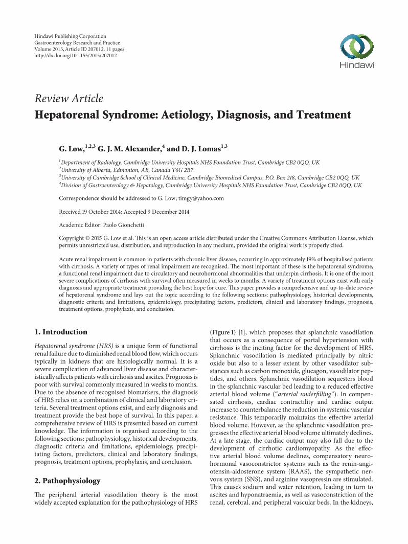
The peripheral arterial vasodilation theory is the mostwidely accepted explanation for the pathophysiology of HRS
(Figure 1) [1], which proposes that splanchnic vasodilationthat occurs as a consequence of portal hypertension withcirrhosis is the inciting factor for the development of HRS.Splanchnic vasodilation is mediated principally by nitricoxide but also to a lesser extent by other vasodilator sub-stances such as carbon monoxide, glucagon, vasodilator pep-tides, and others. Splanchnic vasodilation sequesters bloodin the splanchnic vascular bed leading to a reduced effectivearterial blood volume (“arterial underfilling”). In compen-sated cirrhosis, cardiac contractility and cardiac outputincrease to counterbalance the reduction in systemic vascularresistance. This temporarily maintains the effective arterialblood volume. However, as the splanchnic vasodilation pro-gresses the effective arterial blood volumeultimately declines.At a late stage, the cardiac output may also fall due to thedevelopment of cirrhotic cardiomyopathy. As the effec-tive arterial blood volume declines, compensatory neuro-hormonal vasoconstrictor systems such as the renin-angi-otensin-aldosterone system (RAAS), the sympathetic ner-vous system (SNS), and arginine vasopressin are stimulated.This causes sodium and water retention, leading in turn toascites and hyponatraemia, as well as vasoconstriction of therenal, cerebral, and peripheral vascular beds. In the kidneys,
Hindawi Publishing CorporationGastroenterology Research and PracticeVolume 2015, Article ID 207012, 11 pageshttp://dx.doi.org/10.1155/2015/207012
2 Gastroenterology Research and Practice
Cirrhosis
Portal (sinusoidal) hypertension
Splanchnic/systemicvasodilatation
Decreased effective arterial blood volume
Activation of neurohumoral systems(RAAS, SNS, and ADH)
Sodium and waterretention
Ascites andhyponatremia
Renal vasoconstriction
Decreased renal blood flow
HRS
Increasedcardiac output
High-outputheart failure
Figure 1: A flow diagram outlines the main pathophysiologic mechanisms involved in the development of HRS (reproduced with permissionfrom “JohnWiley and Sons,”Garcia-Tsao et al. [1]). RAAS—renin angiotensin aldosterone system; SNS—sympathetic nervous system;ADH—antidiuretic hormone; HRS—hepatorenal syndrome.
local renal vasodilators such as prostaglandins are initiallyable to counterbalance the effects of the neurohormonal vaso-constrictor systems. Ultimately, this proves inadequate, asrenal vasoconstrictor tone predominates. The end result ofthis process is a severe decline in renal blood flow leadingto reduced glomerular filtration rate (GFR) and the develop-ment of HRS.
3. Historical Developments
The concept of HRS has its origins in important incrementaldiscoveries that extend as far back as the 19th century.Frerichs (1861) and Flint (1863) reported an associationbetween advanced liver disease and a type of renal impair-ment that is characterised by oliguria, the absence of protein-uria, and normal renal histology [2, 3]. By relating the renalimpairment to disturbances in the systemic circulation, theauthors offered the first pathophysiologic interpretation ofHRS. In 1932, Helvig and Schutz introduced the term “aliver and kidney syndrome,” to describe a type of acuterenal impairment that occurred following biliary surgery [4].The definition of HRS evolved with time, and the termbecame synonymous with severe organ dysfunction thatinvolved both the liver and kidneys simultaneously. In 1956,Hecker and Sherlock studied nine patients with advancedliver disease and renal impairment characterised by oliguria,hyponatraemia, low urinary sodium excretion, and absenceof proteinuria [5, 6]. All patients died in hospital andpostmortem findings showed normal renal histology. Bycorrelating these observations with other relevant findings(i.e., low blood pressure, high cardiac output, and highlyoxygenised peripheral venous blood), the authors postulated
that peripheral arterial vasodilation was the key underlyingmechanism for the development of HRS [5, 6]. This theorywas strengthened by the observation that noradrenaline andvolume expansion performed in three patients led to a tran-sient improvement in the renal function. In 1970, Epstein etal. demonstrated the importance of splanchnic and systemicvasodilation together with renal vasoconstriction as a foun-dational concept in the pathophysiology of HRS [7]. In 1972,Vesin described HRS as “functional renal insufficiency incirrhosis” and noted that the disease was often terminal [8].Koppel et al. in 1969 and Iwatsuki et al. in 1973 provided com-pelling evidence for the functional nature ofHRS [9, 10]. Kop-pel et al. reported reversal of the renal dysfunction when kid-neys belonging to six patients with advanced liver disease andHRS were transplanted into recipients with end-stage kidneydisease and normal liver function [9]. Iwatsuki et al. reportedthat liver transplantation in three cirrhotic patients withHRS led to improved liver and renal function within twoweeks of operation [10]. Investigations by Schroeder et al.,Arroyo et al., and Ring-Larsen et al. contributed to the under-standing of the role of neurohormonal vasoconstrictor path-ways (RAAS and SNS) in the genesis of HRS [11–13]. Buildingon advances in the field, Schrier et al. proposed an updatedhypothesis for the pathophysiology of HRS, the peripheralarterial vasodilation theory [14]. In 1978, a consensus con-ference met in Sassari, Italy, to define diagnostic criteriafor HRS [15]. The Sassari criteria did not receive widespreadacceptance and were considered restrictive by many practic-ing physicians [15]. Finally, 1996 marked a watershed, whenthe International Ascites Club (IAC) produced diagnosticcriteria for HRS that were adopted internationally [15].Thesewere later revised in 2007 [16].
Gastroenterology Research and Practice 3
4. Diagnostic Criteria and Their Limitations
Due to the lack of specific biochemical or radiologic markers,the diagnosis of HRS is based on criteria for excluding othercauses of renal impairment that may be found in cirrhosis.The criteria defined by the IAC are listed as follows.
Diagnostic Criteria forHRS (Reproducedwith Permission from“John Wiley and Sons,” Arroyo et al. [15])
Major Criteria(i) Chronic or acute liver disease with advanced hepatic
failure and portal hypertension.(ii) Low GFR as indicated by serum creatinine >
1.5mg/dL or 24 hr creatinine clearance < 40mL/min.(iii) Absence of shock, on-going bacterial infection, and
current or recent treatment with nephrotoxic drugsand absence of gastrointestinal fluid losses (repeatedvomiting or intense diarrhoea) or renal fluid losses(weight loss > 500 g/day for several days in patientswith ascites without peripheral oedema or 1000 g/dayin patients with peripheral oedema).
(iv) No sustained improvement in renal function(decrease in serum creatinine ≤ 1.5mg/dL or increasein creatinine clearance to ≥ 40mL/min) followingdiuretic withdrawal and expansion of plasma volumewith 1.5 L of isotonic saline.
(v) Proteinuria < 500mg/dL and no sonographic evi-dence of obstructive uropathy or parenchymal renaldisease.
Additional Criteria(i) Urine volume < 500mL/day.(ii) Urinary sodium < 10mEq/L.(iii) Urinary osmolality greater than plasma osmolality.(iv) Urine red blood cells < 50 per high power field.(v) Serum sodium < 130mEq/L.
Revised Diagnostic Criteria for HRS (Reproduced with Permis-sion from “BMJ Publishing Group Limited,” Salerno et al. [16])
(i) Cirrhosis with ascites.(ii) Serum creatinine > 133 𝜇mol/L (1.5mg/dL).(iii) No improvement in serum creatinine (decrease to a
level of ≤ 133 𝜇mol/L) after ≥ 2 days with diureticwithdrawal and volume expansion with albumin; therecommended dose of albumin is 1 g/kg of bodyweight/day up to a maximum of 100 g/day.
(iv) Absence of shock.(v) No current or recent treatment with nephrotoxic
drugs.(vi) Absence of parenchymal kidney disease as indicated
by proteinuria> 500mg/day, microscopic haematuria(>50 red blood cells per high power field), and/orabnormal renal ultrasonography.
The revised criteria incorporate several new iterations whichinclude (i) removal of creatinine clearance, (ii) recognitionthat on-going bacterial infection, in the absence of septicshock, no longer excludes a diagnosis of HRS, (iii) preferencefor the choice of albumin rather than saline for plasmaexpansion, and (iv) removal of the minor diagnostic criteria[16].
The IAC classifies HRS into type 1 and type 2. Type1 HRS is a rapid progressive renal impairment defined bydoubling of the serum creatinine to a level > 2.5mg/dL or >226𝜇mol/L in less than two weeks [16]. Type 2 HRS ismoderate renal impairment (serum creatinine > 1.5 and up to2.5mg/dL or > 133 and up to 226𝜇mol/L) with a steady pro-gressive course that evolves over weeks to months [16].Acute deterioration in circulatory, renal, and hepatic functionis characteristic of type 1 HRS, while these abnormalitiesdevelop more gradually in type 2 HRS. Type 1 HRS is oftenassociated with a precipitating factor, while type 2 HRStypically develops de novo in patients with refractory ascites.Rarely, type 2 HRSmay progress into type 1 HRS as a result ofa triggering event. Prognosis is poor ranging frommonths intype 2 HRS to weeks in type 1 HRS.
The IAC diagnostic criteria have several shortcomings.The serum creatinine should be interpreted with caution inpatients with cirrhosis [17–20]. These patients have lowerbaseline serum creatinine than normal due to (i) reducedendogenous creatinine production related to decreased hep-atic synthesis and decreased muscle mass frommalnutrition,(ii) medication related increased tubular secretion of creati-nine, (iii) fluctuations in serum creatinine in patients withcirrhosis and large volume ascites (e.g., following diuretictherapy or paracentesis with volume expansion), and (iv)laboratory based underestimations of serumcreatinine due tointeractions with bilirubin [17–20]. As such, creatinine basedmeasurements run the risk of overestimating renal functionand underestimating the severity of renal impairment, whichraises two important considerations. Firstly, a need exists todevelopmore accurate laboratory and imaging biomarkers ofrenal function. Cystatin C and NGAL (neutrophil gelatinase-associated lipocalin) have been advocated as renal biomark-ers but are expensive and not widely available. As yet, stan-dard imaging tests have proven unreliable for detecting HRSand differentiating it from other types of renal impairment.Secondly, expert consultation and consensus are required todetermine if the serum creatinine threshold (>1.5mg/dL) forHRS should be lowered to allow patients to be diagnosed andtreated while being at an earlier stage [21].
Current IAC criteria do not consider the clinical scenarioof HRS developing in patients with underlying chronic renaldisease.There is an increasing realisation that patients that fitthis category do exist but go unrecognized according to thedefinitions of the existing criteria [21, 22]. Munoz proposesthat patients with HRS superimposed on chronic renaldisease be categorised as having type 3 HRS [22]. Finally, itshould be acknowledged that adherence to the IAC criteriais not always possible. As HRS is a diagnosis of exclusion, thediagnostic pathway can be complex at times, labour intensive,and prone to error. Some patients that do not fulfil the fullIAC diagnostic criteria may be treated as having “presumed”
4 Gastroenterology Research and Practice
HRS, based on the index of clinical suspicion. Three studieshighlight these challenges. Salerno et al., in a study involving253 patients with cirrhosis and renal failure, found that thediagnosis of HRS could only be “presumed” in 36% (𝑛 = 42)as not all the IAC criteria could be met [23]. In 17% (𝑛 = 20),diuretic therapy was tapered but not stopped and in 6% (𝑛 =7) urinalysis could not be performed due to oligo-anuriaor showed red blood cells and/or proteins due to bladdercatheterisation or due to previous parenchymal renal disease,while in 7% (𝑛 = 8) ultrasound showed preexisting renalabnormalities [23]. Interestingly, the authors found no sig-nificant differences in the clinical characteristics, clinical-pathologic scores, or outcomes between presumed cases com-pared with cases that met the full criteria [23]. Servin-Abadet al. performed a retrospective analysis on 140 patients diag-nosed with HRS from 1996 to 2004. The authors found thatonly 41 patients (29.3%) met the IAC criteria [24]. Causes ofmisdiagnosis included parenchymal renal disease (15.2%),acute tubular necrosis (ATN) (35.4%), active sepsis (34.3%),drug induced renal disease (4%), and others (11.1%) [24].Wattet al. found that only 59% of patients with a clinical diagnosisof HRS met IAC criteria [25].
5. Epidemiology
Incidence. In a study in 1993, prior to the introduction ofIAC criteria, Gines et al. reported that HRS had an incidenceof 18% at one year and 39% at five years in patients withcirrhosis and ascites [26]. In a 2010 study utilising the reviseddiagnostic criteria, Montoliu et al. evaluated the incidenceof functional renal impairment in 263 consecutive cirrhoticpatients with ascites [27]. The authors found that 49%of patients developed functional renal impairment duringfollow-up (mean follow-up of 41 months). The annual inci-dence of HRS was 7.6% (𝑛 = 20) (type 1 = 7, type 2 = 13).
Prevalence. The prevalence of HRS (utilising the reviseddiagnostic criteria) in patients with cirrhosis and ascitesranged from 13 to 45.8%. Salerno et al. performed a prospec-tive study of 253 consecutive patients with cirrhosis andrenal impairment admitted to 21 Italian hospitals [23]. Theprevalence ofHRSwas 45.8% (𝑛 = 116) (30% type 1 and 15.8%type 2). A prospective study byMartin-Llahi et al. of 562 con-secutive patients with cirrhosis and renal impairment admit-ted to a single institution found HRS prevalence of 13% [28].A prospective study by Thabut et al. of 100 consecutivepatients with cirrhosis and renal impairment admitted to fiveFrench hospitals found HRS prevalence of 27% [29]. Retro-spective studies suggest thatHRS is present in 17%of cirrhoticpatients admitted to hospital with ascites and in >50% ofpatients dying from end-stage liver failure [30, 31].
Age. Most patients with HRS are in their sixth or seventhdecade. Salerno et al. reported the mean age as 62 ± 1.2 years(type 1 HRS) and 68 ± 1.6 years (type 2 HRS) [23], whileMartin-Llahi et al. reported a mean age of 60 ± 12 years [28].
A pooled analysis by Garcia-Tsao et al. in 509 HRS patientsfrom 14 studies found a mean age of 54 years [1].
Gender. There is a male preponderance, which is reflective ofthe gender balance of the underlying cirrhosis. Salerno et al.reported that 76.3% of type 1 HRS and 70% of type 2 HRSpatientsweremales [23], similar toMartin-Llahi et al. (76.7%)[28] and Garcia-Tsao et al. [1].
Aetiology of Cirrhosis. Salerno et al. identified the aetiologyof cirrhosis as alcohol (type 1 HRS 46.1%; type 2 HRS 55%),viral (type 1 HRS 31.6%; type 2 HRS 40%), alcohol + viral(type 1 HRS 10.5%; type 2 HRS 2.5%), and others (type 1 HRS11.8%; type 2 HRS 2.5%) [23]. According to the pooled anal-ysis by Garcia-Tsao et al., alcohol-related cirrhosis was theunderlying aetiology in 57% (40 to 78%, interquartile range,IQR) [1].
6. Precipitating Factors
HRS develops on the background of advanced liver disease,as highlighted by the high mean Child-Pugh (CP) score of11.2 (11-12; IQR) [1]. Cirrhosis is themost commonunderlyingliver disease but other aetiologies include fulminant hepaticfailure and severe acute alcohol-related hepatitis [32]. Thefrequency of HRS in fulminant hepatic failure and severeacute alcohol-related hepatitis has been reported to be as highas 55% and 30%, respectively [22, 33, 34]. HRS may occurspontaneously (typically in type 2 HRS) or may be triggeredby a precipitating factor (in >70% of cases of type 1 HRS) [35].
The most common precipitating factor is spontaneousbacterial peritonitis (SBP). SBP refers to infection of asciticfluid (typically by enteric Gram-negative bacteria) in theabsence of a specific intra-abdominal source for the sepsis.SBP has a close chronologic and pathologic connection withHRS where it typically precedes its onset. Follo et al. [36]found that SBP precipitated HRS in 28% of cases despiteappropriate treatment and resolution of infection. Renalimpairment was transient in 21 cases (32%), stable in 26 cases(40%), and progressive in 18 cases (28%). SBP may triggerHRS via two postulated mechanisms [35, 37]: (i) releaseof proinflammatory cytokines (interleukin-6 and tumournecrosis factor) and endotoxins leading to increased produc-tion of nitric oxide and other vasodilator substances and (ii)sepsis-induced cardiomyopathy leading to decreased cardiacoutput.
The second most common precipitating factor for HRS islarge volume paracentesis (LVP) without plasma expansion.LVP exacerbates the hyperdynamic circulation in cirrhosis,which leads to progressive systemic vasodilation and arterialunderfilling. Gines et al. found that LVP (4–6 L/day), per-formed without intravenous albumin replacement, precipi-tated HRS in 21% of 53 cases [38]. In contrast, there wereno cases of HRS when LVP was performed with intravenousalbumin replacement. Cardenas et al. found that renal impair-ment occurred in 11% of 175 of cirrhotic patients thatexperienced gastrointestinal bleeding [39]. In these patients,the aetiology was ATN not HRS. Gastrointestinal bleeding
Gastroenterology Research and Practice 5
may induce a systemic inflammatory response associatedwith activation of proinflammatory cytokines that stimulatenitric oxide and other vasodilator substances. Furthermore,gastrointestinal bleeding increases susceptibility to infection,a vicious cycle that may generate further cytokines release,and rebleeding. Certain medications such as nonsteroidalanti-inflammatory drugs (NSAIDs) can precipitate HRS inthose with borderline renal function. Renal vasoconstrictionis initially counterbalanced by increased renal production ofvasodilating prostaglandins (e.g., renal prostaglandin E2 andprostacyclin). NSAIDs can inhibit renal prostaglandin syn-thesis and thus aggravate renal vasoconstriction. Intravascu-lar volume depletion by injudicious diuretic use has also beenconsidered as a potential trigger for HRS; however, evidenceto support this is lacking [32]. Biliary obstruction mayprecipitate HRS due to the action of bile acids and oxidativestress from free radical-induced tissue damage. Bile acids canalter the renal handling of electrolytes and water by blockingthe sodium-hydrogen antiport protein [40]. Oxidative stresspromotes the formation of a variety of vasoconstrictor sub-stances including endothelin-1, cysteinyl leukotrienes, andF2-isoprostanes [40].
7. Predictors of HRS
Several studies have been performed to investigate potentialpredictors for HRS. Gines et al., in a study of 234 patients,identified 16 variables that may be useful as predictors ofHRS on univariate analysis [26]. The variables includedhepatomegaly, oesophageal varices, history of ascites, nutri-tional status, GFR, blood urea nitrogen, serum sodium andpotassium, plasma renin activity, plasma noradrenaline,serum and urinary osmolality, urinary sodium excretion, freewater clearance after a water load, and mean arterial pressure(MAP). However, only three independent variables, absenceof hepatomegaly, high plasma renin activity, and low serumsodium, were found to be predictive of HRS on multivariateanalysis. Montoliu et al., in a study of 263 patients, foundthat older age, high baseline serum creatinine, and a high CPscore were independent predictors for HRS on multivariateanalysis [27]. These variables may reflect the longer durationof the liver disease and the greater severity of the liver andrenal impairment. Platt et al., in a study of 180 patients withnonazotemic liver disease, reported that the resistive index(RI) of the intrarenal arteries on Doppler ultrasound pre-dicted the development of renal dysfunction, including HRS[41]. Renal dysfunction developed in 55% of patients with anelevated RI at baseline (≥0.7), including 6% of patients witha normal RI. HRS developed in 26% of patients with elevatedbaseline RI and 1% of patients with normal baseline RI.Patients that went on to develop HRS had higher baseline RIs(0.77) than patients that developed non-HRS renal impair-ment (0.72, 𝑃 < 0.05) or patients with preserved normalrenal function (0.65, 𝑃 < 0.01). The RI may be regardedas a barometer of the intrarenal vascular tone and thisis elevated in HRS due to increased vasoconstrictor activity.
8. Clinical and Laboratory Findings
HRS does not have specific clinical findings. Its physicalmanifestations broadly reflect the underlying advanced liverdisease, renal impairment, and circulatory abnormalitiespresent. Clinical findings of advanced liver disease includehepatomegaly, ascites, stigmata of portal hypertension (e.g.,gastroesophageal varices, caput medusa, hepatic encephalop-athy, etc.), jaundice, pruritus, coagulopathy, gynaecomastia,finger clubbing, palmar erythema, spider naevi, and con-stitutional disturbances such as weakness, fatigue, anorexia,and poor nutritional status. Patients with type 1 HRS aremore severely affected than patients with type 2 HRS.Acute oliguria is typically present in type 1 HRS, while theurine output shows a more gradual decline in type 2 HRS.Circulatory disturbances include a hyperdynamic circulationand reduced systemic vascular resistance. This may manifestclinically as lowMAP, low jugular venous pressure, tachycar-dia, a bounding pulse, and wide pulse pressure.The followinglaboratory findings are suggestive of HRS [26]: elevatedplasma renin activity, elevated plasma noradrenaline activity,hyponatraemia, hyperkalaemia, elevated blood urea nitro-gen, decreased plasma osmolality, elevated urinary osmolal-ity, and decreased urinary sodium excretion. Serum abnor-malities that reflect the severity of the liver disease includehyperbilirubinemia, hypoalbuminemia, and prolonged pro-thrombin time.
9. Prognosis
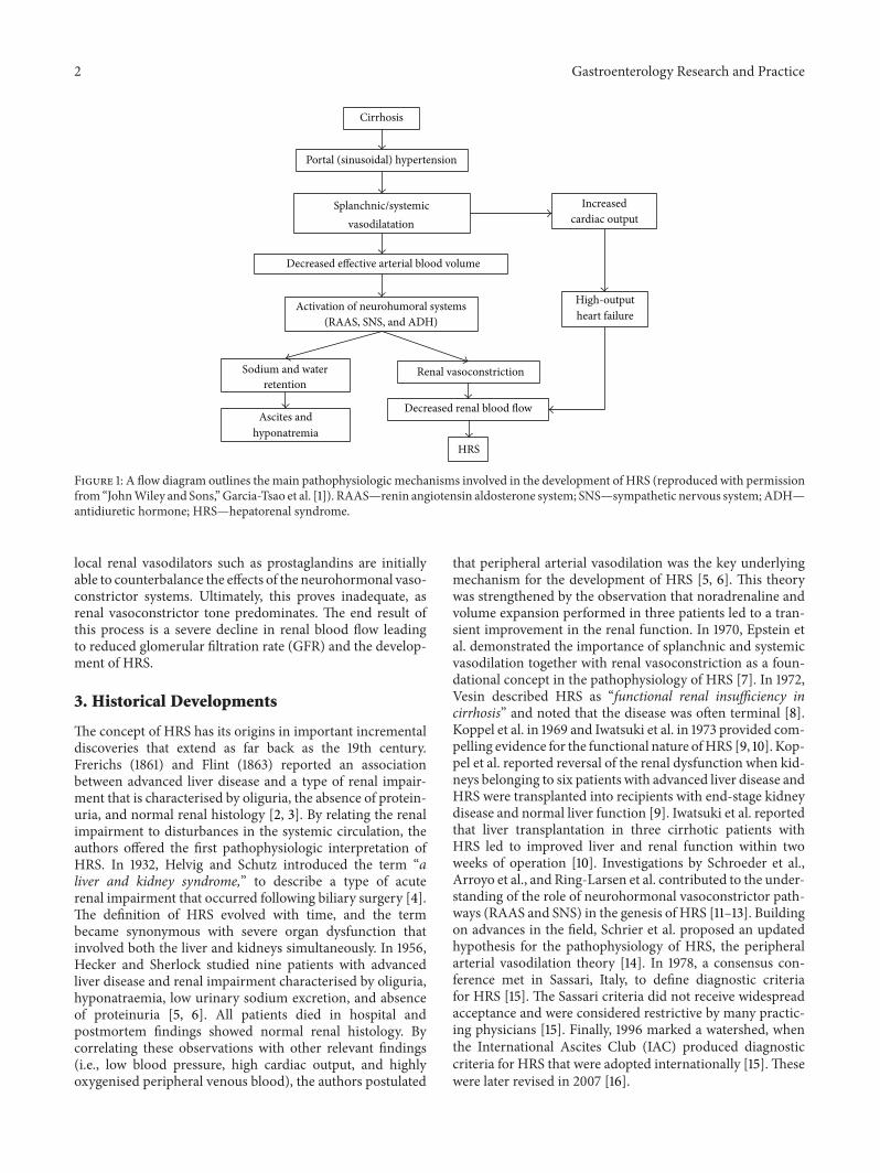
HRS is one of the most lethal complications of cirrhosis(Figure 2). Prognosis is invariably poor ranging frommonthsin type 2 HRS to weeks to months in type 1 HRS [42, 43].Studies have been performed to define clinical parametersthat track survival in HRS. In 2005, Alessandria et al. studied105 patients with cirrhosis and HRS (39% type 1 and 61% type2) [42] and found that HRS type and MELD (model for end-stage liver disease) score were variables that associated inde-pendently with survival onmultivariate analysis. MELD is aninternationally recognised scoring system developed forpatients with advanced liver disease that is a predictor ofthree-month mortality and determines priority listing forliver transplantation [42, 44]. It is calculated according to thefollowing formula: 9.6 ×loge (creatinine mg/dL) + 3.8 ×loge(bilirubin mg/dL) + 11.2 ×loge (international normalisedratio, INR) + 6.4 [42]. Patients with type 1 HRS had a MELDscore ≥ 20 and a median survival of one month [42]. For type2 HRS, the median survival was 11 months for a MELD score< 20 and threemonths for aMELD score≥ 20 [42]. Schepke etal. reported similar findings in a 2006 study that involved 88patients with cirrhosis and renal failure [43], some with HRS(17–39.8% type 1, 22.7% type 2), and non-HRS renal impair-ment. On multivariate analysis, the authors found that type 1HRS and the MELD score were independent variables asso-ciated with survival. The mean MELD score was higher inpatients with HRS than in patients with non-HRS renalimpairment (23.8 versus 18.3, 𝑃 = 0.002) [43]. The estimatedsurvival time was 3.4 months for type 1 HRS, 10.9 months fortype 2 HRS, and 16.1 months for non-HRS renal impairment

6 Gastroenterology Research and Practice
1.0
0.8
0.6
0.4
0.2
0
0 3 6 9 12
Prob
abili
ty
Months
P < 0.01
P < 0.0001 Type 2 HRS
Type 1 HRS
Cirrhosis with asciteswithout HRS
Figure 2: Probability of survival in cirrhotic patients with different types of renal impairment: nonazotemic patients (bold continuous line),type 2 HRS (dotted line), and type 1 HRS (continuous line) (reproduced with permission from “BMJ Publishing Group Limited,” Salernoet al. [16]).
[43]. In 2010, Yang et al. performed a longitudinal assessmentof prognostic factors in 103 patients with HRS (65% type 1and 35% type 2) [45] and revealed that temporal changes (Δ)in four clinical parameters were associated with survivalon time dependent multivariate analysis. These parametersincludedΔMELD score simple, Δ serum creatinine,Δ serumbilirubin, and Δ serum albumin [45]. ΔMELD score simplewas calculated according to the following formula: [3.8 ×log bilirubin (mg/dL)] + 9.6 × log [creatinine (mg/dL) +6.43] [45]. The authors found that ΔMELD score simple wassuperior to baseline and changes inMELD score in predictingprognosis.The authors also found that increasing serum crea-tinine and bilirubin affected survival adversely, while increas-ing serum albumin had a beneficial effect. In a 2012 studyinvolving 68 patients with type 1 HRS, Martinez et al.reported that the aetiology of the liver disease, the serumcreatinine at the time of initiation of treatment, and theurinary sodiumwere useful prognostic factors [46].The aeti-ologies of the liver disease included (in decreasing order ofsurvival) autoimmune hepatitis, cardiac, idiopathic, viral,viral + alcohol, alcohol, and neoplasia.The authors also foundthat higher serum creatinine on admission and a urinarysodium < 5mEq/L were associated negatively with survival.
10. Treatment Options
Vasoconstrictor Therapy. This is the primary medical treat-ment for type 1 HRS. These drugs function by causing vaso-constriction of the grossly dilated splanchnic vascular bedresulting in increased systemic vascular return and increasedMAP, which in turn suppresses the RAAS and SNS andimproves renal perfusion. Albumin augments the potency ofvasoconstrictor drugs by improving cardiac function andincreasing the effective arterial blood volume. The AcuteDialysis Quality Initiative (ADQI) work group recommendsthe use of vasoconstrictor drugs combined with plasmaexpansion with albumin, as first line treatment for type 1HRS [47]. Terlipressin is the vasoconstrictor drug of choicein Europe. It is a vasopressin analogue that acts on the V1
vasopressin receptors in vascular smooth muscle cells. Theresults of randomised controlled trials and meta-analysissuggest that the combination of terlipressin and albumin iseffective in improving renal function in 40 to 50% of patientswith type 1 HRS [48–50]. Patients with type 2 HRS mayalso benefit. Response to treatment is characterised by adecrease in the serum creatinine and an increase in the urinevolume, serum sodium, and MAP. Median response timeto treatment is 14 days [48]. Predictors of response includepretreatment bilirubin < 10mg/dL and an increase of MAP ≥5mmHg at day three of therapy [50–52]. Following with-drawal of treatment, HRS recurs in 20%, although retreat-ment is generally effective [16]. Terlipressin may cause organand peripheral ischaemia and is contraindicated in patientswith cardiovascular disease, cerebrovascular disease, andperipheral vascular disease. Significant complications arereported in 10 to 12% [16, 48–50]. It is unlikely that vaso-constrictor drugs improve survival beyond the short term.A multicentre randomised controlled trial comparing terli-pressin and albumin to albumin alone in 46 patients withHRS showed improved renal function in the former group(43.5% versus 8.7%, 𝑃 = 0.017), but no survival advantage ineither group at three months (27% versus 19%, 𝑃 = 0.7) [53].A second multicentre randomised controlled trial in 56patients with HRS comparing terlipressin to placebo andalbumin found similar survival for both groups at 180 days(42.9% versus 37.5%, 𝑃 = 0.8) [54]. Other vasoconstrictordrugs used in HRS include noradrenaline, midodrine, andoctreotide. These have lower costs than terlipressin and,unlike terlipressin, are licensed for clinical use in NorthAmerica. Noradrenaline and midodrine are 𝛼-adrenergicagonists, which act on the 𝛼
1-adrenergic receptors in vascular
smooth muscle cells. Octreotide is a long acting somatostatinanalogue that inhibits glucagon and other vasodilator pep-tides. Only limited information is available about the efficacyof these alternative drugs.
Transjugular Intrahepatic Portosystemic Shunt (TIPS). TIPSinvolves the insertion of an intrahepatic stent that connects
Gastroenterology Research and Practice 7
the portal vein to the hepatic vein. This shunts portalblood into the systemic circulation, which reduces the portalpressure and increases the systemic venous return. In turn,this treats the arterial underfilling and the overactivity of theRAAS and SNS. Unfortunately, most patients with HRS areineligible for TIPS due to contraindications (e.g., INR > 2,serum bilirubin > 5mg/dL, CP score > 11, and cardiopul-monary disease). Furthermore, TIPS can aggravate the liverfailure and precipitate encephalopathy.
Brensing et al. assessed the outcome following TIPS in 41nontransplantHRSpatients [55]. 31 patients receivedTIPS (14patients, type 1, and 17 patients, type 2), and tenwere excludeddue to advanced liver disease. Renal function showed agradual nonnormalized improvement within two weeks afterTIPS. Overall survival following TIPS was 81% at threemonths, 71% at six months, 48% at 12 months, and 35% at 18months. In contrast, seven of the ten non-TIPS patients diedwithin three months. The authors concluded that TIPS couldprovide a survival benefit in well-selected HRS patients.Testino et al. performed TIPS in 18 type 2 HRS patients withrefractory ascites that were awaiting liver transplantation[56]. The study found that TIPS improved the renal functionand led to complete resolution of ascites in eight patients andpartial resolution in ten patients. The authors suggested thatTIPS may be used as a bridge to transplantation by preparingpatients for surgery with improved renal function. Guevaraet al. assessed the effects of TIPS on renal function and thevasoactive systems [57] and found improved renal function tononnormalized levels in six of seven patients with type 1HRS.The serum creatinine fell from 5 ± 0.8 to 1.8± 0.4mg/dL by 30days. Reduced activity of the RAAS and SNS was reflected bya reduction in serum renin, aldosterone, and norepinephrinelevels. Testino et al. performed TIPS in nine patients withtype 1 HRS and acute alcohol-related hepatitis [58], whichresulted in a nonnormalized improvement in renal func-tion (decreased serum creatinine and blood urea nitrogenand increased urine volumes). The serum creatinine fellfrom5.2± 0.9mg/dL to 1.6± 0.6mg/dL by 30 days.Wong et al.evaluated the combination of medical therapy (midodrine,octreotide, and albumin) and TIPS in type 1 HRS [59];serum creatinine decreased to < 135 𝜇mol/L for at least threedays following medical therapy in ten patients. Half of theresponders underwent TIPS and showed resolution of asciteswith normalisation of renal function (GFR 96 ± 20mL/min)and serum renin and aldosterone by 12 months.
The ADQI work group recommends that (i) TIPS shouldnot be used as the first line treatment for type 1 HRS due toinsufficient data and (ii) TIPS may be used in patients withtype 2 HRS and refractory ascites [47].
Extracorporeal Support Systems. Renal replacement therapy(RRT) may be used to treat specific complications of renalimpairment such as metabolic acidosis, hyperkalaemia, vol-ume overload, and uraemic symptoms. It may have a rolein patients who are unresponsive to vasoconstrictor drugsand where TIPS is contraindicated. In some circumstances,it may provide a bridge to liver transplantation. Side effectsinclude hypotension, coagulopathy, and infection. Continu-ous venovenous haemofiltration is preferred to intermittent
haemodialysis in unstable patients and those at risk ofdeveloping raised intracranial pressure. The ADQI workgroup recommends that RRT should be avoided in type 1HRSpatients unless there is an acute reversible component or anintention to pursue transplantation [47].
Molecular adsorbent recirculating system (MARS) is amodified dialysis technique for extracting albumin boundand water-soluble substances from the blood. This removesvasodilator substances such as nitric oxide, tumour necrosisfactor, and cytokines, which are implicated in the pathogene-sis of HRS. Mitzner et al. performed a randomised controlledstudy in type 1 HRS patients undergoing either MARS (eightpatients) or standard treatment (five patients) [60] and founda significant reduction in the serum creatinine and bilirubinand a significant increase in the serum sodiumandprothrom-bin activity. The MARS group showed improved short-termsurvival comparedwith the control group (survival was 37.5%at seven days and 25% at 30 days forMARS versus 0% at sevendays for the control group). In a separate study involvingeight subjects, Mitzner et al. found that MARS improvedmultiorgan function in type 1 HRS patients [61]. Wong et al.performed a study involving six type 1 HRS patients withrefractory ascites that had failed vasoconstrictor treatment[62] and found thatMARSwas not associatedwith significantimprovements in theGFR, neurohormonal levels, or systemichaemodynamics. In a study of 32 patients, Lavayssiere et al.found that only 28% of type 1 HRS patients showed completerenal recovery after 28 days [63]. More recently, a largerandomised controlled trial (RELIEF) involving 19 Europeancentres comparing MARS with standard therapy in patientswith acute-on-chronic liver failure [64] reported that whileMARS provided temporary organ benefit (liver, kidney, andbrain), it did not improve overall survival.
Prometheus is an extracorporeal technique involvingfractional plasma separation and adsorptionwith haemodial-ysis for removing water-soluble and albumin bound sub-stances. Rifai et al. found that Prometheus treatment in tenHRS patients led to improved serum creatinine, urea, biliru-bin, bile acids, and ammonia concentrations [65]. Given thelack of a definitive survival benefit and high costs, the ADQIwork group suggests that extracorporeal support systemsshould be limited to research protocols.
Liver Transplantation. Liver transplantation is the definitivetreatment for HRS [66–72]. The five-year survival for HRS is60% for patients that underwent liver transplantation com-pared with 0% for patients that did not undergo liver trans-plantation [66]. The use of the MELD scoring system, whichallocates liver grafts according to the “sickest first” policy, hasincreased the proportion of HRS patients that have receiveda liver transplant. In addition to orthotopic liver transplan-tation (OLT) [40], some centres have also performed livingdonor liver transplantation (LDLT). Advantages of LDLTinclude increasing the donor pool, the possibility of plannedsurgery, shorter ischaemic duration, and younger donor age[73]. A potential pitfall is that the smaller graft may be insuf-ficient to sustain hepatic function [68]. A report by Lee et al.involving 71 HRS patients (48OLT and 23 LDLT) found com-parable outcomes for both techniques [73]. The three-yearsurvival was 85.3% for LDLT compared with 60.9% for OLT.
8 Gastroenterology Research and Practice
Patients with HRS have worse posttransplant outcomesthan patients without HRS. This includes reduced short- andlong-term survival, increased risk of bleeding and infection,and longer hospital stay [66, 67]. The five-year survival com-paring patients with HRS that have received a liver transplantto patients without HRS that have received a liver transplantis 60% versus 68% (𝑃 < 0.03) [66, 67]. The pretransplantrenal function is a major predictor of outcomes followingtransplantation, with less favourable outcomes for patientswith pretransplant renal dysfunction [66, 72]. Restuccia et al.suggested that reversal of the renal dysfunction prior to trans-plantation improves posttransplant outcomes [68].The studyfound that HRS patients treated with vasopressin analoguesbefore transplantation had similar outcomes to controls withnormal renal function [68]. Both groups had comparablesurvival (three-year survival 100% in HRS-treated groupversus 83% in controls,𝑃 = 0.15), frequency of posttransplantrenal dysfunction, and length of hospital stay.
Between 58 and 94% of patients with HRS demonstraterecovery of renal function after liver transplantation [74–76].Factors associated with a failure of renal recovery include thetime interval between onset of HRS and transplantation (≥4–6 weeks), dialysis for ≥ 8 weeks, and a serum creatinineof ≥ 2mg/dL [74–77]. Such patients may benefit from simul-taneous liver and kidney transplantation. The ADQI workgroup recommends that liver transplantation alone should beperformed if the duration of type 1 HRS is < 4 weeks, whilesimultaneous liver and kidney transplantation should beperformed if there is a risk that renal recovery will not occur[47].
11. Prophylaxis
Prophylactic treatment may be beneficial in reducing the riskof developing HRS. Sort et al. performed a randomised con-trolled trial in cirrhotic patients with SBP [78]. 126 patientswere randomised to one of two treatment arms, albumin pluscefotaxime (antibiotic) or cefotaxime alone. The study foundthat patients that received albumin plus cefotaxime had alower incidence of developing renal impairment than patientsthat received cefotaxime alone (10% versus 33%, 𝑃 < 0.01).In-hospital and three-month mortality rates were signifi-cantly lower in the albumin plus cefotaxime group comparedwith the cefotaxime group (10% versus 29%, 𝑃 = 0.01,and 22% versus 41%, 𝑃 = 0.03). Pentoxifylline is a phospho-diesterase inhibitor with beneficial effects on renal function.Tyagi et al. performed a randomised controlled trial involving70 patients with cirrhosis, ascites, and a baseline normal renalfunction [79]. Patients were randomised into two treatmentarms, pentoxifylline or placebo. Baseline, one-month, three-month and sixth-month laboratory and clinical parameterswere assessed. HRS developed in two patients in the pen-toxifylline group (2.9%) compared with ten patients in theplacebo group (14.3%). The six-month mortality was one intwo HRS patients in the pentoxifylline group compared withthree in ten HRS patients in the placebo group.
12. Conclusion
Over the last century, much has been learnt about the patho-physiology, clinical behaviour, and natural history of HRS.Standardised diagnostic criteria have been developed andimplemented worldwide, allowing for more uniform diag-nosis and consistent reporting of the disease. Limitations inthe diagnostic criteria exist, but as yet, no reliable diagnosticmarker exists for HRS. Future directions should include thedevelopment of an accurate diagnostic test for HRS. This isimportant as an earlier diagnosis and thus treatment is likelyto improve survival. Several treatment options exist, but, atpresent, only liver transplantation offers a genuine hope forcure and longevity.
Ethical Approval
This is a literature review. It did not involve any investigationson patients and therefore informed consent was not required.No patient information was included.
Conflict of Interests
The authors declare that there is no conflict of interestsregarding the publication of this paper.
Acknowledgment
The work was supported by grant funding by the NationalInstitute of Health Research (UK) Cambridge BiomedicalResearch Centre (NIHR CBRC)
References
[1] G. Garcia-Tsao, C. R. Parikh, and A. Viola, “Acute kidney injuryin cirrhosis,” Hepatology, vol. 48, no. 6, pp. 2064–2077, 2008.
[2] V. Arroyo and R. Bataller, “Historical notes on ascites in cirrho-sis,” in Ascites and Renal Dysfunction in Liver Disease: Patho-genesis, Diagnosis and Treatment, V. Arroyo, P. Gines, J. Rodes,R. W. Schrier, and M. A. Malden, Eds., pp. 3–13, BlackwellScience, 1999.
[3] A. Flint, “Clinical report of hydro-peritoneum, based on anal-ysis of forty-six cases,” The American Journal of the MedicalSciences, vol. 45, no. 90, pp. 306–339, 1863.
[4] F. Helvig and C. Schutz, “A liver and kidney syndrome: clinical,pathological and experimental studies,” The Journal of Surgery,Gynecology and Obstetrics, vol. 55, pp. 570–582, 1932.
[5] R. Hecker and S. Sherlock, “Electrolyte and circulatory changesin terminal liver failure,”The Lancet, vol. 268, no. 6953, pp. 1121–1125, 1956.
[6] V. Arroyo, M. Guevara, and P. Gines, “Hepatorenal syndromein cirrhosis: pathogenesis and treatment,”Gastroenterology, vol.122, no. 6, pp. 1658–1676, 2002.
[7] M. Epstein, D. P. Berk, N. K. Hollenberg et al., “Renal failure inthe patient with cirrhosis. The role of active vasoconstriction,”The American Journal of Medicine, vol. 49, no. 2, pp. 175–185,1970.
[8] P. Vesin, “Functional renal insufficiency in cirrhotics. Course.Mechanism. Treatment,” Archives Francaises des Maladies del”Appareil Digestif, vol. 61, no. 12, pp. 775–786, 1972.
Gastroenterology Research and Practice 9
[9] M. H. Koppel, J. W. Coburn, M. M. Mims, H. Goldstein, J. D.Boyle, and M. E. Rubini, “Transplantation of cadaveric kidneysfrom patients with hepatorenal syndrome. Evidence for thefunctionalnature of renal failure in advanced liver disease,”TheNew England Journal ofMedicine, vol. 280, no. 25, pp. 1367–1371,1969.
[10] S. Iwatsuki,M.M. Popovtzer, J. L. Corman et al., “Recovery from“hepatorenal syndrome” after orthotopic liver transplantation,”The New England Journal of Medicine, vol. 289, no. 22, pp. 1155–1159, 1973.
[11] E. T. Schroeder, R. H. Eich, H. Smulyan, A. B. Gould, and G. J.Gabuzda, “Plasma renin level in hepatic cirrhosis. Relation tofunctional renal failure,”The American Journal of Medicine, vol.49, no. 2, pp. 186–191, 1970.
[12] V. Arroyo, R. Planas, J. Gaya et al., “Sympathetic nervousactivity, renin-angiotensin system and renal excretion ofprostaglandin E
2in cirrhosis. Relationship to functional renal
failure and sodium and water excretion,” European Journal ofClinical Investigation, vol. 13, no. 3, pp. 271–278, 1983.
[13] H.Ring-Larsen, B.Hesse, J.H.Henriksen, andN. J. Christensen,“Sympathetic nervous activity and renal and systemic hemo-dynamics in cirrhosis: plasma norepinephrine concentration,hepatic extraction, and renal relase,” Hepatology, vol. 2, no. 3,pp. 304–310, 1982.
[14] R. W. Schrier, V. Arroyo, M. Bernardi, M. Epstein, J. H. Henrik-sen, and J. Rodes, “Peripheral arterial vasodilation hypothesis: aproposal for the initiation of renal sodium and water retentionin cirrhosis,” Hepatology, vol. 8, no. 5, pp. 1151–1157, 1988.
[15] V. Arroyo, P. Gines, A. L. Gerbes et al., “Definition and diag-nostic criteria of refractory ascites and hepatorenal syndromein cirrhosis,” Hepatology, vol. 23, no. 1, pp. 164–176, 1996.
[16] F. Salerno, A. Gerbes, P. Gines, F. Wong, and V. Arroyo,“Diagnosis, prevention and treatment of hepatorenal syndromein cirrhosis,” Gut, vol. 56, no. 9, pp. 1310–1318, 2007.
[17] C. Francoz,D.Glotz, R.Moreau, and F.Durand, “The evaluationof renal function and disease in patients with cirrhosis,” Journalof Hepatology, vol. 52, no. 4, pp. 605–613, 2010.
[18] D. S. Sherman, D. N. Fish, and I. Teitelbaum, “Assessing renalfunction in cirrhotic patients: problems and pitfalls,” AmericanJournal of Kidney Diseases, vol. 41, no. 2, pp. 269–278, 2003.
[19] M. A. Papadakis and A. I. Arieff, “Unpredictability of clinicalevaluation of renal function in cirrhosis. Prospective study,”TheAmerican Journal of Medicine, vol. 82, no. 5, pp. 945–952, 1987.
[20] L. Caregaro, F. Menon, P. Angeli et al., “Limitations of serumcreatinine level and creatinine clearance as filtration markers incirrhosis,” Archives of Internal Medicine, vol. 154, no. 2, pp. 201–205, 1994.
[21] F. Wong, M. K. Nadim, J. A. Kellum et al., “Working Party pro-posal for a revised classification system of renal dysfunction inpatients with cirrhosis,” Gut, vol. 60, no. 5, pp. 702–709, 2011.
[22] S. J. Munoz, “The hepatorenal syndrome,” Medical Clinics ofNorth America, vol. 92, no. 4, pp. 813–837, 2008.
[23] F. Salerno, M. Cazzaniga, M. Merli et al., “Diagnosis, treatmentand survival of patients with hepatorenal syndrome: a survey ondaily medical practice,” Journal of Hepatology, vol. 55, no. 6, pp.1241–1248, 2011.
[24] L. R. A. Servin-Abad, G. Contreras, V. Caban, L. Arosemena,and A. Arcila, “Retrospective analysis of 140 patients labelled ashepatorenal syndrome in a referral center,” Hepatology, vol. 42,p. 543, 2005.
[25] K. Watt, J. Uhanova, and G. Y. Minuk, “Hepatorenal syndrome:diagnostic accuracy, clinical features, and outcome in a tertiarycare center,”American Journal of Gastroenterology, vol. 97, no. 8,pp. 2046–2050, 2002.
[26] A. Gines, A. Escorsell, P. Gines et al., “Incidence, predictivefactors, and prognosis of the hepatorenal syndrome in cirrhosiswith ascites,”Gastroenterology, vol. 105, no. 1, pp. 229–236, 1993.
[27] S. Montoliu, B. Balleste, R. Planas et al., “Incidence and prog-nosis of different types of functional renal failure in cirrhoticpatients with ascites,” Clinical Gastroenterology and Hepatology,vol. 8, pp. 616–622, 2010.
[28] M. Martin-Llahi, M. Guevara, A. Torre et al., “Prognosticimportance of the cause of renal failure in patients withcirrhosis,” Gastroenterology, vol. 140, no. 2, pp. 488–496, 2011.
[29] D. Thabut, J. Massard, A. Gangloff et al., “Model for end-stageliver disease score and systemic inflammatory response aremajor prognostic factors in patients with cirrhosis and acutefunctional renal failure,” Hepatology, vol. 46, no. 6, pp. 1872–1882, 2007.
[30] L. Dagher and K.Moore, “The hepatorenal syndrome,”Gut, vol.49, no. 5, pp. 729–737, 2001.
[31] V. Arroyo, P. Gines, V. Jimenez et al., “Renal dysfunction incirrhosis,” in Oxford Textbook of Clinical Hepatology, J. Bircher,J.-P. Benhamou, andN.McIntyre, Eds., OxfordUniversity Press,Oxford, UK, 1999.
[32] P. Gines, M. Guevara, V. Arroyo, and J. Rodes, “Hepatorenalsyndrome,”The Lancet, vol. 362, no. 9398, pp. 1819–1827, 2003.
[33] K.Moore, “Renal failure in acute liver failure,” European Journalof Gastroenterology and Hepatology, vol. 11, no. 9, pp. 967–975,1999.
[34] S. Verma, K. Ajudia, M. Mendler, and A. Redeker, “Prevalenceof septic events, type 1 hepatorenal syndrome, and mortality insevere alcoholic hepatitis and utility of discriminant functionand MELD score in predicting these adverse events,” DigestiveDiseases and Sciences, vol. 51, no. 9, pp. 1637–1643, 2006.
[35] H. M. Wadei, M. L. Mai, N. Ahsan, and T. A. Gonwa, “Hepa-torenal syndrome: pathophysiology and management,” ClinicalJournal of the American Society of Nephrology, vol. 1, no. 5, pp.1066–1079, 2006.
[36] A. Follo, J. M. Llovet, M. Navasa et al., “Renal impairment afterspontaneous bacterial peritonitis in cirrhosis: incidence, clinicalcourse, predictive factors and prognosis,” Hepatology, vol. 20,no. 6, pp. 1495–1501, 1994.
[37] M. Navasa, A. Follo, X. Filella et al., “Tumor necrosis factor andinterleukin-6 in spontaneous bacterial peritonitis in cirrhosis:relationship with the development of renal impairment andmortality,” Hepatology, vol. 27, no. 5, pp. 1227–1232, 1998.
[38] P. Gines, L. Tito, V. Arroyo et al., “Randomized comparativestudy of therapeutic paracentesis with and without intravenousalbumin in cirrhosis,” Gastroenterology, vol. 94, no. 6, pp. 1493–1502, 1988.
[39] A. Cardenas, P. Gines, J. Uriz et al., “Renal failure after uppergastrointestinal bleeding in cirrhosis: Incidence, clinical course,predictive factors, and short-term prognosis,” Hepatology, vol.34, no. 4 I, pp. 671–676, 2001.
[40] A. Bomzon, S. Holt, and K. Moore, “Bile acids, oxidativestress, and renal function in biliary obstruction,” Seminars inNephrology, vol. 17, no. 6, pp. 549–562, 1997.
[41] J. F. Platt, J. H. Ellis, J. M. Rubin, R. M.Merion, andM. R. Lucey,“Renal duplex Doppler ultrasonography: a noninvasive pre-dictor of kidney dysfunction and hepatorenal failure in liverdisease,” Hepatology, vol. 20, no. 2, pp. 362–369, 1994.
10 Gastroenterology Research and Practice
[42] C. Alessandria, O. Ozdogan, M. Guevara et al., “MELD scoreand clinical type predict prognosis in hepatorenal syndrome:relevance to liver transplantation,”Hepatology, vol. 41, no. 6, pp.1282–1289, 2005.
[43] M. Schepke, B. Appenrodt, J. Heller, J. Zielinski, and T. Sauer-bruch, “Prognostic factors for patientswith cirrhosis and kidneydysfunction in the era of MELD: results of a prospective study,”Liver International, vol. 26, no. 7, pp. 834–839, 2006.
[44] P. S. Kamath and W. R. Kim, “The model for end-stage liverdisease (MELD),” Hepatology, vol. 45, no. 3, pp. 797–805, 2007.
[45] Y.-W. Yang, C.-H.Wu, R.-H.Hu et al., “Longitudinal assessmentof prognostic factors for patients with hepatorenal syndrome ina tertiary center,”Hepatology International, vol. 4, no. 2, pp. 507–510, 2010.
[46] M. O. Martinez, H. Sayles, R. Vivekanandan, S. D’Souza, andM. C. Florescu, “Hepatorenal syndrome: are we missing someprognostic factors?” Digestive Diseases and Sciences, vol. 57, no.1, pp. 210–214, 2012.
[47] M. K. Nadim, J. A. Kellum, A. Davenport et al., “Hepatorenalsyndrome: the 8 th international consensus conference of theAcute Dialysis Quality Initiative (ADQI) Group,” Critical Care,vol. 16, no. 1, article R23, 2012.
[48] P. Gines, P. Angeli, K. Lenz et al., “EASL clinical practiceguidelines on the management of ascites, spontaneous bacterialperitonitis, and hepatorenal syndrome in cirrhosis,” Journal ofHepatology, vol. 53, no. 3, pp. 397–417, 2010.
[49] P. Gines and R. W. Schrier, “Renal failure in cirrhosis,”The NewEngland Journal of Medicine, vol. 361, no. 13, pp. 1279–1290,2009.
[50] R. Moreau and D. Lebrec, “The use of vasoconstrictors inpatientswith cirrhosis: type 1HRS and beyond,”Hepatology, vol.43, no. 3, pp. 385–394, 2006.
[51] T. D. Boyer, A. J. Sanyal, G. Garcia-Tsao et al., “Predictors ofresponse to terlipressin plus albumin in hepatorenal syndrome(HRS) type 1: relationship of serum creatinine to hemodynam-ics,” Journal of Hepatology, vol. 55, no. 2, pp. 315–321, 2011.
[52] A. Nazar, G. H. Pereira, M. Guevara et al., “Predictors ofresponse to therapy with terlipressin and albumin in patientswith cirrhosis and type 1 hepatorenal syndrome,” Hepatology,vol. 51, no. 1, pp. 219–226, 2010.
[53] M. Martın-Llahı, M.-N. Pepin, M. Guevara et al., “Terlipressinand albumin vs albumin in patients with cirrhosis and hepa-torenal syndrome: a randomized study,” Gastroenterology, vol.134, no. 5, pp. 1352–1359, 2008.
[54] A. J. Sanyal, T. Boyer, G. Garcia-Tsao et al., “A randomized,prospective, double-blind, placebo-controlled trial of terli-pressin for type 1 hepatorenal syndrome,” Gastroenterology, vol.134, no. 5, pp. 1360–1368, 2008.
[55] K. A. Brensing, H. Schild, T. Sauerbruch et al., “Long term out-come after transjugular intrahepatic portosystemic stent-shuntin non-transplant cirrhotics with hepatorenal syndrome: aphase II study,” Gut, vol. 47, no. 2, pp. 288–295, 2000.
[56] G. Testino, C. Ferro, A. Sumberaz et al., “Type-2 hepatorenalsyndrome and refractory ascites: role of transjugular intra-hepatic portosystemic stent-shunt in eighteen patients withadvanced cirrhosis awaiting orthotopic liver transplantation,”Hepato-Gastroenterology, vol. 50, no. 54, pp. 1753–1755, 2003.
[57] M. Guevara, P. Gines, J. C. Bandi et al., “Transjugular intrahep-atic portosystemic shunt in hepatorenal syndrome: effects onrenal function and vasoactive systems,” Hepatology, vol. 28, no.2, pp. 416–422, 1998.
[58] G. Testino, S. Leone, C. Ferro, and P. Borro, “Severe acute alco-holic hepatitis and hepatorenal syndrome: role of transjugularintrahepatic portosystemic stent shunt,” Journal ofMedicine andLife, vol. 5, no. 2, pp. 203–205, 2012.
[59] F. Wong, L. Pantea, and K. Sniderman, “Midodrine, octreotide,albumin, and TIPS in selected patients with cirrhosis and type1 hepatorenal syndrome,” Hepatology, vol. 40, no. 1, pp. 55–64,2004.
[60] S. R.Mitzner, J. Stange, S. Klammt et al., “Improvement of hepa-torenal syndrome with extracorporeal albumin dialysis MARS:results of a prospective, randomized, controlled clinical trial,”Liver Transplantation, vol. 6, no. 3, pp. 277–286, 2000.
[61] S. R. Mitzner, S. Klammt, P. Peszynski et al., “Improvement ofmultiple organ functions in hepatorenal syndrome duringalbumin dialysis with the molecular adsorbent recirculatingsystem,”Therapeutic Apheresis, vol. 5, no. 5, pp. 417–422, 2001.
[62] F. Wong, N. Raina, and R. Richardson, “Molecular adsorbentrecirculating system is ineffective in the management of type1 hepatorenal syndrome in patients with cirrhosis with asciteswho have failed vasoconstrictor treatment,” Gut, vol. 59, no. 3,pp. 381–386, 2010.
[63] L. Lavayssiere, S. Kallab, I. Cardeau-Desangles et al., “Impact ofmolecular adsorbent recirculating system on renal recovery intype-1 hepatorenal syndromepatientswith chronic liver failure,”Journal of Gastroenterology and Hepatology (Australia), vol. 28,no. 6, pp. 1019–1024, 2013.
[64] R. Banares, F. Nevens, F. S. Larsen et al., “Extracorporeal albu-min dialysis with the molecular adsorbent recirculating systemin acute-on-chronic liver failure: the RELIEF trial,”Hepatology,vol. 57, no. 3, pp. 1153–1162, 2013.
[65] K. Rifai, T. Ernst, U. Kretschmer et al., “The Prometheus devicefor extracorporeal support of combined liver and renal failure,”Blood Purification, vol. 23, no. 4, pp. 298–302, 2005.
[66] T. A. Gonwa, G. B. Klintmalm, M. Levy, L. S. Jennings, R. M.Goldstein, and B. S. Husberg, “Impact of pretransplant renalfunction on survival after liver transplantation,” Transplanta-tion, vol. 59, no. 3, pp. 361–365, 1995.
[67] T. A. Gonwa, C. A. Morris, R. M. Goldstein, B. S. Husberg, andG. B. Klintmalm, “Long-term survival and renal function fol-lowing liver transplantation in patients with and without hep-atorenal syndrome—experience in 300 patients,” Transplanta-tion, vol. 51, no. 2, pp. 428–430, 1991.
[68] T. Restuccia, R. Ortega, M. Guevara et al., “Effects of treatmentof hepatorenal syndrome before transplantation on posttrans-plantation outcome. A case-control study,” Journal of Hepatol-ogy, vol. 40, no. 1, pp. 140–146, 2004.
[69] P. Seu, A. H. Wilkinson, A. Shaked, and R. W. Busuttil, “Thehepatorenal syndrome in liver transplant recipients.,” AmericanSurgeon, vol. 57, no. 12, pp. 806–809, 1991.
[70] V. Cuervas-Mons, I. Millan, J. S. Gavaler, T. E. Starzl, and D. H.VanThiel, “Prognostic value of preoperatively obtained clinicaland laboratory data in predicting survival following orthotopicliver transplantation,” Hepatology, vol. 6, no. 5, pp. 922–927,1986.
[71] A. Rimola, J. S. Gavaler, R. R. Schade, S. El-Lankany, T. E. Starzl,and D. H. Van Thiel, “Effects of renal impairment on livertransplantation,” Gastroenterology, vol. 93, no. 1, pp. 148–156,1987.
[72] S. Nair, S. Verma, and P. J.Thuluvath, “Pretransplant renal func-tion predicts survival in patients undergoing orthotopic livertransplantation,” Hepatology, vol. 35, no. 5, pp. 1179–1185, 2002.
Gastroenterology Research and Practice 11
[73] J. P. Lee, H. Y. Kwon, J. I. Park et al., “Clinical outcomes ofpatients with hepatorenal syndrome after living donor livertransplantation,” Liver Transplantation, vol. 18, no. 10, pp. 1237–1244, 2012.
[74] X. Xu, Q. Ling, M. Zhang et al., “Outcome of patients with hep-atorenal syndrome type 1 after liver transplantation: Hangzhouexperience,” Transplantation, vol. 87, no. 10, pp. 1514–1519, 2009.
[75] P. E. Marik, K. Wood, and T. E. Starzl, “The course of type 1hepato-renal syndrome post liver transplantation,” NephrologyDialysis Transplantation, vol. 21, no. 2, pp. 478–482, 2006.
[76] P. Angeli and P. Gines, “Hepatorenal syndrome, MELD scoreand liver transplantation: an evolving issue with relevant impli-cations for clinical practice,” Journal of Hepatology, vol. 57, no.5, pp. 1135–1140, 2012.
[77] J. D. Eason, T. A. Gonwa, C. L. Davis, R. S. Sung, D. Gerber,and R. D. Bloom, “Proceedings of consensus conference onsimultaneous liver kidney transplantation (SLK),”TheAmericanJournal of Transplantation, vol. 8, no. 11, pp. 2243–2251, 2008.
[78] P. Sort, M. Navasa, V. Arroyo et al., “Effect of intravenousalbumin on renal impairment andmortality in patients with cir-rhosis and spontaneous bacterial peritonitis,”The New EnglandJournal of Medicine, vol. 341, no. 6, pp. 403–409, 1999.
[79] P. Tyagi, P. Sharma, B. C. Sharma, A. S. Puri, A. Kumar, andS. K. Sarin, “Prevention of hepatorenal syndrome in patientswith cirrhosis and ascites: a pilot randomized control trialbetween pentoxifylline and placebo,” European Journal of Gas-troenterology and Hepatology, vol. 23, no. 3, pp. 210–217, 2011.

Submit your manuscripts athttp://www.hindawi.com
Stem CellsInternational
Hindawi Publishing Corporationhttp://www.hindawi.com Volume 2014
Hindawi Publishing Corporationhttp://www.hindawi.com Volume 2014
MEDIATORSINFLAMMATION
of
Hindawi Publishing Corporationhttp://www.hindawi.com Volume 2014
Behavioural Neurology
EndocrinologyInternational Journal of
Hindawi Publishing Corporationhttp://www.hindawi.com Volume 2014
Hindawi Publishing Corporationhttp://www.hindawi.com Volume 2014
Disease Markers
Hindawi Publishing Corporationhttp://www.hindawi.com Volume 2014
BioMed Research International
OncologyJournal of
Hindawi Publishing Corporationhttp://www.hindawi.com Volume 2014
Hindawi Publishing Corporationhttp://www.hindawi.com Volume 2014
Oxidative Medicine and Cellular Longevity
Hindawi Publishing Corporationhttp://www.hindawi.com Volume 2014
PPAR Research
The Scientific World JournalHindawi Publishing Corporation http://www.hindawi.com Volume 2014
Immunology ResearchHindawi Publishing Corporationhttp://www.hindawi.com Volume 2014
Journal of
ObesityJournal of
Hindawi Publishing Corporationhttp://www.hindawi.com Volume 2014
Hindawi Publishing Corporationhttp://www.hindawi.com Volume 2014
Computational and Mathematical Methods in Medicine
OphthalmologyJournal of
Hindawi Publishing Corporationhttp://www.hindawi.com Volume 2014
Diabetes ResearchJournal of
Hindawi Publishing Corporationhttp://www.hindawi.com Volume 2014
Hindawi Publishing Corporationhttp://www.hindawi.com Volume 2014
Research and TreatmentAIDS
Hindawi Publishing Corporationhttp://www.hindawi.com Volume 2014
Gastroenterology Research and Practice
Hindawi Publishing Corporationhttp://www.hindawi.com Volume 2014
Parkinson’s Disease
Evidence-Based Complementary and Alternative Medicine
Volume 2014Hindawi Publishing Corporationhttp://www.hindawi.com