Embed Size (px)
Citation preview
Review ArticlePrecursor Lesions for Sporadic Pancreatic Cancer:PanIN, IPMN, and MCN
M. Distler,1 D. Aust,2 J. Weitz,1 C. Pilarsky,1 and Robert Grützmann1
1 Department of Visceral, Thoracic, and Vascular Surgery, University Hospital Carl Gustav Carus Dresden, TU Dresden,Fetscher Street 74, 01307 Dresden, Germany
2 Institute for Pathology, University Hospital Carl Gustav Carus Dresden, TU Dresden, Fetscher Street 74, 01307 Dresden, Germany
Correspondence should be addressed to Robert Grutzmann; [email protected]
Received 18 November 2013; Revised 8 February 2014; Accepted 10 February 2014; Published 24 March 2014
Academic Editor: Sonshin Takao
Copyright © 2014 M. Distler et al. This is an open access article distributed under the Creative Commons Attribution License,which permits unrestricted use, distribution, and reproduction in any medium, provided the original work is properly cited.
Pancreatic cancer is still a dismal disease. The high mortality rate is mainly caused by the lack of highly sensitive and specificdiagnostic tools, and most of the patients are diagnosed in an advanced and incurable stage. Knowledge about precursor lesionsfor pancreatic cancer has grown significantly over the last decade, and nowadays we know that mainly three lesions (PanIN, andIPMN, MCN) are responsible for the development of pancreatic cancer. The early detection of these lesions is still challenging butprovides the chance to cure patients before they might get an invasive pancreatic carcinoma. This paper focuses on PanIN, IPMN,and MCN lesions and reviews the current level of knowledge and clinical measures.
1. Introduction
Pancreatic cancer is the fourth leading cause of cancer deathin the USA [1]. The high mortality rate is mainly caused bythe lack of highly sensitive and specific tools to detect thedisease in an early stage, and therefore most of the patientsare diagnosed in advanced tumor stages. Currently, surgicalresection is the only curative treatment option.However, onlya small number of patients (30–40%) presentwith a resectabletumor at the time of diagnosis. The overall five-year survivalafter pancreatic head resection for cancer has been reportedto range between 10 and 25% [2, 3]. The five-year survivalrate can be significantly improved for patients with pancreaticcancer when surgery is possible and patients additionallyreceive adjuvant therapy [4, 5]. However, there are patientsthat relapse shortly after the surgery and, therefore, haveonly a limited life span even after complete resection of thetumor.
Current knowledge about pancreatic carcinogenesis pos-tulates—analogous to other carcinomas—a stepwise progres-sion from intraepithelial neoplasia to invasive cancer [6, 7].Studies of resected pancreata from patients with a family his-tory of pancreatic cancer or from patients with ductal
adenocarcinoma of the pancreas (PDAC) demonstratedthat many patients have multifocal, microscopic pancreaticintraepithelial neoplasms (PanINs) surrounding the tumorand in the remainder of the pancreas [8–11]. Furthermore,cystic lesions of the pancreas like intraductal papillary muci-nous neoplasms (IPMNs) or mucinous cystic neoplasms(MCNs) are well known as precursor lesions for pancreaticcancer (Figure 1) [12–15]. Early detection and treatment ofthe abovementioned precursor lesions could probably savepatients from advancing to invasive pancreatic cancer. In thisreview, a summary of the most important precursor lesionsfor pancreatic cancer, that is, PanIN, IPMN, and MCN, isgiven.
2. Pancreatic IntraepithelialNeoplasia (PanIN)
The most important and well-known precursor of a PDACis pancreatic intraepithelial neoplasia (PanIN). For severaldecades, this noninvasive ductal lesion was described usingmultiple terminologies. Hruban et al. first presented thenowadays generally accepted “PanIN scheme” to classifythese lesions in 2001 (Figure 2) [16, 17].
Hindawi Publishing CorporationBioMed Research InternationalVolume 2014, Article ID 474905, 11 pageshttp://dx.doi.org/10.1155/2014/474905

2 BioMed Research International
IPMN
dysplasia
IPMN
IPMN intermediate
dysplasia
IPMN high-grade dysplasia
MCN
MCNlow-gradelow-gradedysplasia
MCNintermediate
dysplasia
MCNhigh-grade dysplasia
PanIN-1Alesion
PanIN-1Blesion
PanIN-2lesion
PanIN-3lesion
Invasive pancreatic carcinoma
Normal pancreatic (duct) tissue
Figure 1: Model of three distinct morphological pathways to invasive pancreatic carcinoma.
MUC5AC
P16/CDKN2
MUC1MUC1
Normalpancreatic tissue
PanIN-1Alesion
PanIN-1Blesion
PanIN-2lesion
PanIN-3lesion
Invasivecarcinoma
Telomere shortening, KRAS
TP53, SMAD4/DPC4
CpG island hypermethylation
Figure 2: Compendium of molecular changes during the PanIN-progression model. Adapted from [6, 18].
2.1. Histology. A PanIN is a microscopic (usually <5mm)flat or papillary lesion arising in the small intralobularpancreatic ducts [17]. These lesions are characteristicallyasymptomatic. PanINs are composed of columnar to cuboidalcells with varying amounts of mucin and varying degrees ofcytological and architectural atypia [15]. They are classifiedinto three grades: PanIN-1A (flat) and PanIN-1B (papillary)are low-grade lesions with minimal cytological and architec-tural atypia (Figure 3). PanIN-2 lesions (intermediate-gradePanIN) show mild to moderate cytological and architecturalatypia (e.g., nuclear pleomorphism, nuclear crowding, andnuclear hyperchromasia) with frequent papillae (Figure 4)[6, 15]. High-grade PanINs (PanIN-3) are characterized bysevere cytological and architectural atypia. PanIN-3 is alsoreferred to as “carcinoma in situ.” All PanINs are noninvasivelesions that do not trespass the basement membrane [16, 17].
PanIN-3 lesions usually have a papillary morphology butcan also present with a flat or cribriform pattern (Figure 4).In addition, there are some rare variants of PanINs (e.g.,intestinal type, foam gland type, and oncocytic type) that donot seem to have any specific clinical significance [15].
Formal pancreatic carcinogenesis is thought to progressfrom low-grade to high-grade PanIN and then to invasivecancer; this histological progression is paralleled by theaccumulation of genetic changes (Figure 2).
2.2. Immunohistochemical Characteristics. The immunohis-tochemical characteristics of PanINs vary with the grade ofdysplasia. In particular, the apomucins MUC1, MUC2, andMUC5AC are frequently overexpressed in epithelial cancersof the gastrointestinal tract [14, 19]. MUC1 is responsible forthe surveillance of lumen formation and is typically expressed
BioMed Research International 3
(a) (b)
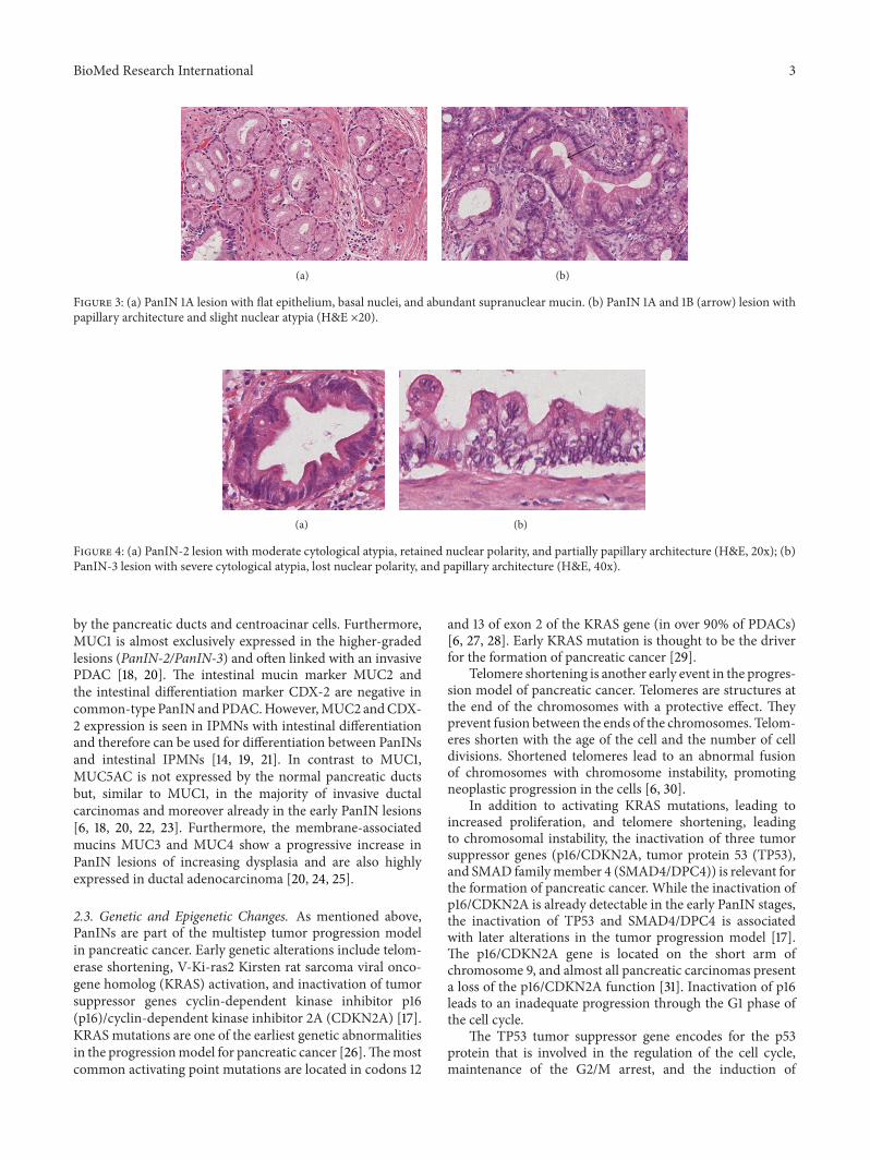
Figure 3: (a) PanIN 1A lesion with flat epithelium, basal nuclei, and abundant supranuclear mucin. (b) PanIN 1A and 1B (arrow) lesion withpapillary architecture and slight nuclear atypia (H&E ×20).
(a) (b)
Figure 4: (a) PanIN-2 lesion with moderate cytological atypia, retained nuclear polarity, and partially papillary architecture (H&E, 20x); (b)PanIN-3 lesion with severe cytological atypia, lost nuclear polarity, and papillary architecture (H&E, 40x).
by the pancreatic ducts and centroacinar cells. Furthermore,MUC1 is almost exclusively expressed in the higher-gradedlesions (PanIN-2/PanIN-3) and often linked with an invasivePDAC [18, 20]. The intestinal mucin marker MUC2 andthe intestinal differentiation marker CDX-2 are negative incommon-type PanIN andPDAC.However,MUC2 andCDX-2 expression is seen in IPMNs with intestinal differentiationand therefore can be used for differentiation between PanINsand intestinal IPMNs [14, 19, 21]. In contrast to MUC1,MUC5AC is not expressed by the normal pancreatic ductsbut, similar to MUC1, in the majority of invasive ductalcarcinomas and moreover already in the early PanIN lesions[6, 18, 20, 22, 23]. Furthermore, the membrane-associatedmucins MUC3 and MUC4 show a progressive increase inPanIN lesions of increasing dysplasia and are also highlyexpressed in ductal adenocarcinoma [20, 24, 25].
2.3. Genetic and Epigenetic Changes. As mentioned above,PanINs are part of the multistep tumor progression modelin pancreatic cancer. Early genetic alterations include telom-erase shortening, V-Ki-ras2 Kirsten rat sarcoma viral onco-gene homolog (KRAS) activation, and inactivation of tumorsuppressor genes cyclin-dependent kinase inhibitor p16(p16)/cyclin-dependent kinase inhibitor 2A (CDKN2A) [17].KRAS mutations are one of the earliest genetic abnormalitiesin the progressionmodel for pancreatic cancer [26].Themostcommon activating point mutations are located in codons 12
and 13 of exon 2 of the KRAS gene (in over 90% of PDACs)[6, 27, 28]. Early KRAS mutation is thought to be the driverfor the formation of pancreatic cancer [29].
Telomere shortening is another early event in the progres-sion model of pancreatic cancer. Telomeres are structures atthe end of the chromosomes with a protective effect. Theyprevent fusion between the ends of the chromosomes. Telom-eres shorten with the age of the cell and the number of celldivisions. Shortened telomeres lead to an abnormal fusionof chromosomes with chromosome instability, promotingneoplastic progression in the cells [6, 30].
In addition to activating KRAS mutations, leading toincreased proliferation, and telomere shortening, leadingto chromosomal instability, the inactivation of three tumorsuppressor genes (p16/CDKN2A, tumor protein 53 (TP53),and SMAD familymember 4 (SMAD4/DPC4)) is relevant forthe formation of pancreatic cancer. While the inactivation ofp16/CDKN2A is already detectable in the early PanIN stages,the inactivation of TP53 and SMAD4/DPC4 is associatedwith later alterations in the tumor progression model [17].The p16/CDKN2A gene is located on the short arm ofchromosome 9, and almost all pancreatic carcinomas presenta loss of the p16/CDKN2A function [31]. Inactivation of p16leads to an inadequate progression through the G1 phase ofthe cell cycle.
The TP53 tumor suppressor gene encodes for the p53protein that is involved in the regulation of the cell cycle,maintenance of the G2/M arrest, and the induction of
4 BioMed Research International
apoptosis. Loss of p53 causes deregulation in cell death andcell division [18, 32].
The gene DPC4 (located on chromosome 18q) encodesfor the SMAD4 protein that plays an important role insignaling through the transforming growth factor type 𝛽(TGF-𝛽) pathway. SMAD4 has a growth-controlling effect byregulating the expression of specific genes [33, 34].Therefore,loss of SMAD4 leads to a decreased growth inhibition andthereby supports the growth of cancer cells.
Epigenetic changes predominantly occur throughmethy-lation of CpG islands, which are located in the promoterregions of genes leading to gene silencing [35]. Numerousstudies have demonstrated hypermethylation of several genesin patientswith pancreatic cancer [36, 37]. Amicroarray anal-ysis by Sato et al. showed that aberrant CpG island hyperme-thylation begins in early stages of PanINs, and its prevalenceprogressively increases during neoplastic progression [38].Furthermore, the detection of aberrantlymethylated genes bymethylation-specific PCR in the pancreatic juice might be aninteresting diagnostic tool for the future [39].
2.4. Clinical Relevance of PanIN. Asmentioned above, PanINlesions are precursor lesions in the stepwise progressionfrom intraepithelial to invasive pancreatic neoplasia. Thismorphological progression is paralleled by an accumulationof genetic changes in which activating KRAS mutations arethought to be the driving force (Figure 2). Early detection ofPanINs would present an opportunity to cure patients beforethey develop invasive pancreatic cancer, but unfortunatelyPanINs are not yet detectable by cross-sectional imagingor endoscopic ultrasound (EUS). Molecular markers in thepancreatic juice may help to solve this dilemma. The mostappropriate approach to the pancreatic resection marginwhen it harbors a PanIN lesion (detected by intraoperativefrozen section) is, however, not clear. In particular, noconsensus has been achieved on how to handle PanIN-3lesions in the resection margin [40]. We would recommendan additional resection in cases with PanIN-3 lesions (in theresectionmargin), whereas an additional resection in PanIN-1 and -2 lesions might not be necessary.
3. Intraductal Papillary MucinousNeoplasm (IPMN)
IPMNs belong to the heterogeneous group of cystic pancre-atic lesions with increasing incidence in recent years [41–43].These cystic lesions were initially reported in the 1990s, andthe term IPMN was established in the 2000 classification ofthe WHO [44]. IPMNs are tumors of the duct epithelium.Papillary epithelial proliferation and mucin production leadto cystic dilatation of involved ducts [43]. IPMNs havebeen proven to be invasive carcinoma precursors, and thusprogression models from noninvasive intraductal tumors viaborderline lesions to invasive carcinoma have been developed(Figure 1) [45–47].
3.1. Histology and Immunohistochemistry. Morphologically,IPMNs are subdivided into the main (MD-IPMN) andbranch duct types (BD-IPMN) according to their site of
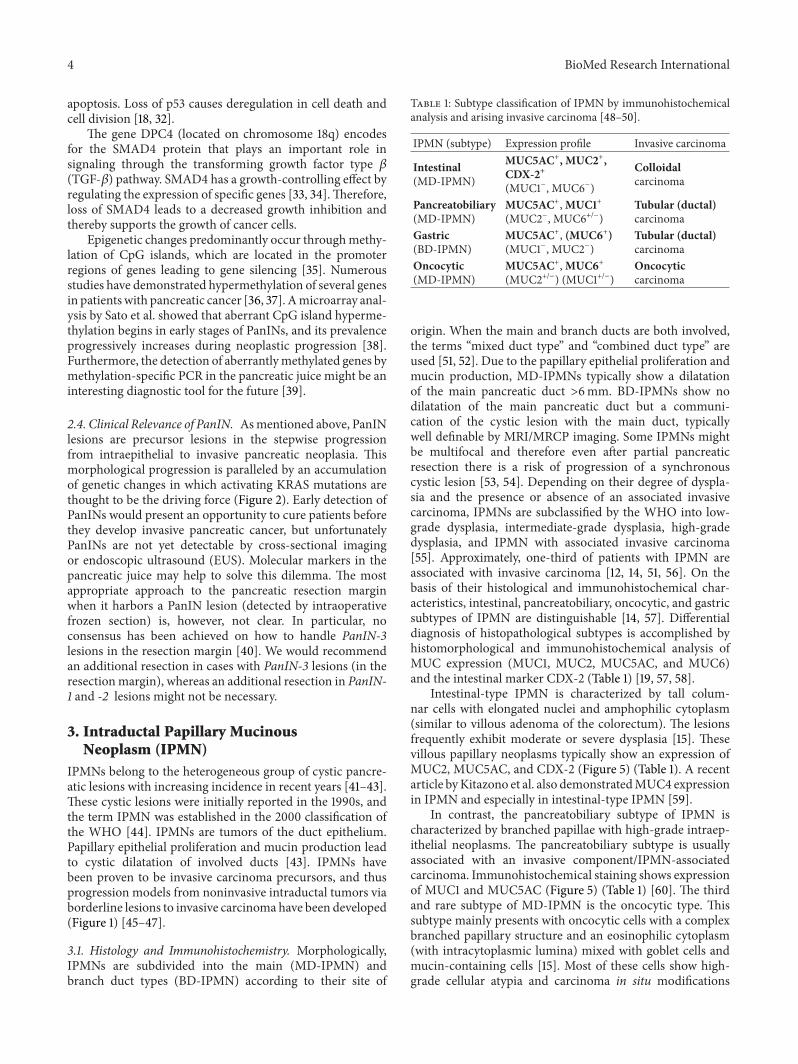
Table 1: Subtype classification of IPMN by immunohistochemicalanalysis and arising invasive carcinoma [48–50].
IPMN (subtype) Expression profile Invasive carcinoma
Intestinal(MD-IPMN)
MUC5AC+, MUC2+,CDX-2+(MUC1−, MUC6−)
Colloidalcarcinoma
Pancreatobiliary(MD-IPMN)
MUC5AC+,MUC1+(MUC2−, MUC6+/−)
Tubular (ductal)carcinoma
Gastric(BD-IPMN)
MUC5AC+, (MUC6+)(MUC1−, MUC2−)
Tubular (ductal)carcinoma
Oncocytic(MD-IPMN)
MUC5AC+,MUC6+(MUC2+/−) (MUC1+/−)
Oncocyticcarcinoma
origin. When the main and branch ducts are both involved,the terms “mixed duct type” and “combined duct type” areused [51, 52]. Due to the papillary epithelial proliferation andmucin production, MD-IPMNs typically show a dilatationof the main pancreatic duct >6mm. BD-IPMNs show nodilatation of the main pancreatic duct but a communi-cation of the cystic lesion with the main duct, typicallywell definable by MRI/MRCP imaging. Some IPMNs mightbe multifocal and therefore even after partial pancreaticresection there is a risk of progression of a synchronouscystic lesion [53, 54]. Depending on their degree of dyspla-sia and the presence or absence of an associated invasivecarcinoma, IPMNs are subclassified by the WHO into low-grade dysplasia, intermediate-grade dysplasia, high-gradedysplasia, and IPMN with associated invasive carcinoma[55]. Approximately, one-third of patients with IPMN areassociated with invasive carcinoma [12, 14, 51, 56]. On thebasis of their histological and immunohistochemical char-acteristics, intestinal, pancreatobiliary, oncocytic, and gastricsubtypes of IPMN are distinguishable [14, 57]. Differentialdiagnosis of histopathological subtypes is accomplished byhistomorphological and immunohistochemical analysis ofMUC expression (MUC1, MUC2, MUC5AC, and MUC6)and the intestinal marker CDX-2 (Table 1) [19, 57, 58].
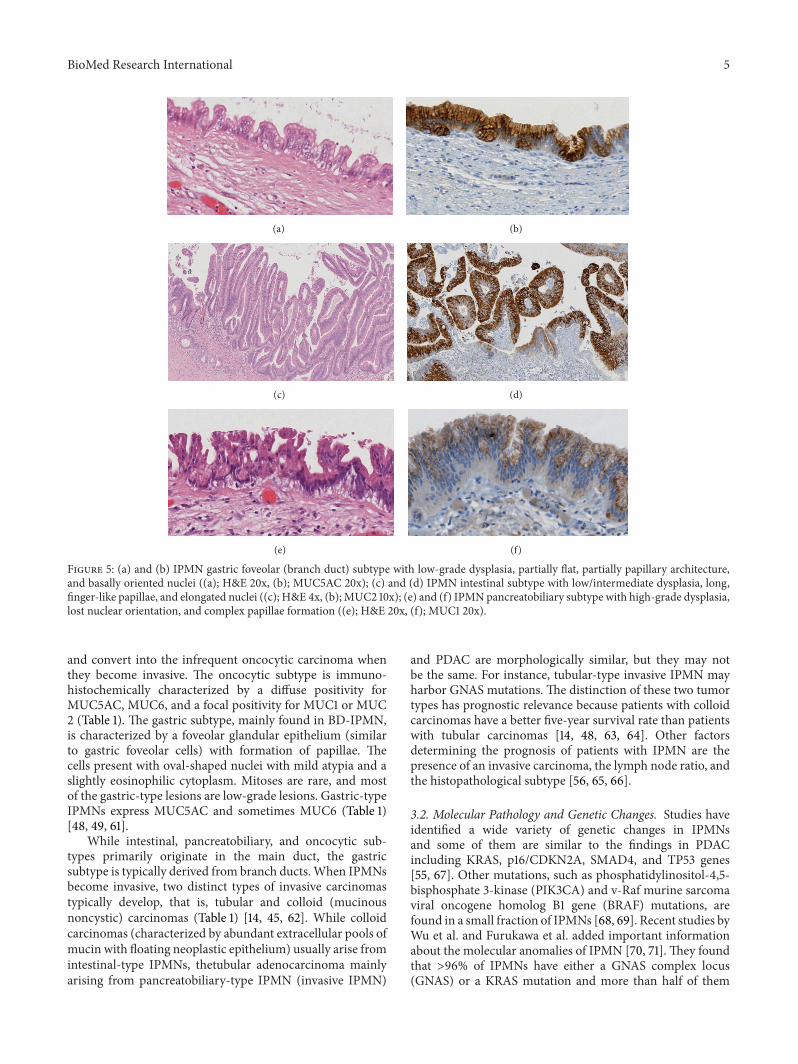
Intestinal-type IPMN is characterized by tall colum-nar cells with elongated nuclei and amphophilic cytoplasm(similar to villous adenoma of the colorectum). The lesionsfrequently exhibit moderate or severe dysplasia [15]. Thesevillous papillary neoplasms typically show an expression ofMUC2, MUC5AC, and CDX-2 (Figure 5) (Table 1). A recentarticle byKitazono et al. also demonstratedMUC4 expressionin IPMN and especially in intestinal-type IPMN [59].
In contrast, the pancreatobiliary subtype of IPMN ischaracterized by branched papillae with high-grade intraep-ithelial neoplasms. The pancreatobiliary subtype is usuallyassociated with an invasive component/IPMN-associatedcarcinoma. Immunohistochemical staining shows expressionof MUC1 and MUC5AC (Figure 5) (Table 1) [60]. The thirdand rare subtype of MD-IPMN is the oncocytic type. Thissubtype mainly presents with oncocytic cells with a complexbranched papillary structure and an eosinophilic cytoplasm(with intracytoplasmic lumina) mixed with goblet cells andmucin-containing cells [15]. Most of these cells show high-grade cellular atypia and carcinoma in situ modifications
BioMed Research International 5
(a) (b)
(c) (d)
(e) (f)
Figure 5: (a) and (b) IPMN gastric foveolar (branch duct) subtype with low-grade dysplasia, partially flat, partially papillary architecture,and basally oriented nuclei ((a); H&E 20x, (b); MUC5AC 20x); (c) and (d) IPMN intestinal subtype with low/intermediate dysplasia, long,finger-like papillae, and elongated nuclei ((c); H&E 4x, (b); MUC2 10x); (e) and (f) IPMNpancreatobiliary subtype with high-grade dysplasia,lost nuclear orientation, and complex papillae formation ((e); H&E 20x, (f); MUC1 20x).
and convert into the infrequent oncocytic carcinoma whenthey become invasive. The oncocytic subtype is immuno-histochemically characterized by a diffuse positivity forMUC5AC, MUC6, and a focal positivity for MUC1 or MUC2 (Table 1). The gastric subtype, mainly found in BD-IPMN,is characterized by a foveolar glandular epithelium (similarto gastric foveolar cells) with formation of papillae. Thecells present with oval-shaped nuclei with mild atypia and aslightly eosinophilic cytoplasm. Mitoses are rare, and mostof the gastric-type lesions are low-grade lesions. Gastric-typeIPMNs express MUC5AC and sometimes MUC6 (Table 1)[48, 49, 61].
While intestinal, pancreatobiliary, and oncocytic sub-types primarily originate in the main duct, the gastricsubtype is typically derived from branch ducts.When IPMNsbecome invasive, two distinct types of invasive carcinomastypically develop, that is, tubular and colloid (mucinousnoncystic) carcinomas (Table 1) [14, 45, 62]. While colloidcarcinomas (characterized by abundant extracellular pools ofmucin with floating neoplastic epithelium) usually arise fromintestinal-type IPMNs, thetubular adenocarcinoma mainlyarising from pancreatobiliary-type IPMN (invasive IPMN)
and PDAC are morphologically similar, but they may notbe the same. For instance, tubular-type invasive IPMN mayharbor GNAS mutations. The distinction of these two tumortypes has prognostic relevance because patients with colloidcarcinomas have a better five-year survival rate than patientswith tubular carcinomas [14, 48, 63, 64]. Other factorsdetermining the prognosis of patients with IPMN are thepresence of an invasive carcinoma, the lymph node ratio, andthe histopathological subtype [56, 65, 66].
3.2. Molecular Pathology and Genetic Changes. Studies haveidentified a wide variety of genetic changes in IPMNsand some of them are similar to the findings in PDACincluding KRAS, p16/CDKN2A, SMAD4, and TP53 genes[55, 67]. Other mutations, such as phosphatidylinositol-4,5-bisphosphate 3-kinase (PIK3CA) and v-Raf murine sarcomaviral oncogene homolog B1 gene (BRAF) mutations, arefound in a small fraction of IPMNs [68, 69]. Recent studies byWu et al. and Furukawa et al. added important informationabout the molecular anomalies of IPMN [70, 71]. They foundthat >96% of IPMNs have either a GNAS complex locus(GNAS) or a KRAS mutation and more than half of them
6 BioMed Research International
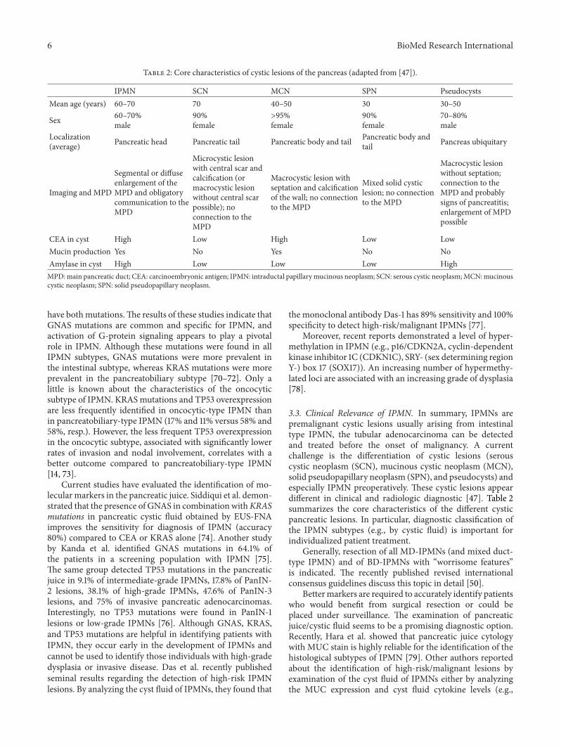
Table 2: Core characteristics of cystic lesions of the pancreas (adapted from [47]).
IPMN SCN MCN SPN PseudocystsMean age (years) 60–70 70 40–50 30 30–50
Sex 60–70%male
90%female
>95%female
90%female
70–80%male
Localization(average) Pancreatic head Pancreatic tail Pancreatic body and tail Pancreatic body and
tail Pancreas ubiquitary
Imaging and MPD
Segmental or diffuseenlargement of theMPD and obligatorycommunication to theMPD
Microcystic lesionwith central scar andcalcification (ormacrocystic lesionwithout central scarpossible); noconnection to theMPD
Macrocystic lesion withseptation and calcificationof the wall; no connectionto the MPD
Mixed solid cysticlesion; no connectionto the MPD
Macrocystic lesionwithout septation;connection to theMPD and probablysigns of pancreatitis;enlargement of MPDpossible
CEA in cyst High Low High Low LowMucin production Yes No Yes No NoAmylase in cyst High Low Low Low HighMPD:main pancreatic duct; CEA: carcinoembryonic antigen; IPMN: intraductal papillarymucinous neoplasm; SCN: serous cystic neoplasm;MCN:mucinouscystic neoplasm; SPN: solid pseudopapillary neoplasm.
have bothmutations.The results of these studies indicate thatGNAS mutations are common and specific for IPMN, andactivation of G-protein signaling appears to play a pivotalrole in IPMN. Although these mutations were found in allIPMN subtypes, GNAS mutations were more prevalent inthe intestinal subtype, whereas KRAS mutations were moreprevalent in the pancreatobiliary subtype [70–72]. Only alittle is known about the characteristics of the oncocyticsubtype of IPMN. KRASmutations and TP53 overexpressionare less frequently identified in oncocytic-type IPMN thanin pancreatobiliary-type IPMN (17% and 11% versus 58% and58%, resp.). However, the less frequent TP53 overexpressionin the oncocytic subtype, associated with significantly lowerrates of invasion and nodal involvement, correlates with abetter outcome compared to pancreatobiliary-type IPMN[14, 73].
Current studies have evaluated the identification of mo-lecularmarkers in the pancreatic juice. Siddiqui et al. demon-strated that the presence of GNAS in combination withKRASmutations in pancreatic cystic fluid obtained by EUS-FNAimproves the sensitivity for diagnosis of IPMN (accuracy80%) compared to CEA or KRAS alone [74]. Another studyby Kanda et al. identified GNAS mutations in 64.1% ofthe patients in a screening population with IPMN [75].The same group detected TP53 mutations in the pancreaticjuice in 9.1% of intermediate-grade IPMNs, 17.8% of PanIN-2 lesions, 38.1% of high-grade IPMNs, 47.6% of PanIN-3lesions, and 75% of invasive pancreatic adenocarcinomas.Interestingly, no TP53 mutations were found in PanIN-1lesions or low-grade IPMNs [76]. Although GNAS, KRAS,and TP53 mutations are helpful in identifying patients withIPMN, they occur early in the development of IPMNs andcannot be used to identify those individuals with high-gradedysplasia or invasive disease. Das et al. recently publishedseminal results regarding the detection of high-risk IPMNlesions. By analyzing the cyst fluid of IPMNs, they found that
the monoclonal antibody Das-1 has 89% sensitivity and 100%specificity to detect high-risk/malignant IPMNs [77].
Moreover, recent reports demonstrated a level of hyper-methylation in IPMN (e.g., p16/CDKN2A, cyclin-dependentkinase inhibitor 1C (CDKN1C), SRY- (sex determining regionY-) box 17 (SOX17)). An increasing number of hypermethy-lated loci are associated with an increasing grade of dysplasia[78].
3.3. Clinical Relevance of IPMN. In summary, IPMNs arepremalignant cystic lesions usually arising from intestinaltype IPMN, the tubular adenocarcinoma can be detectedand treated before the onset of malignancy. A currentchallenge is the differentiation of cystic lesions (serouscystic neoplasm (SCN), mucinous cystic neoplasm (MCN),solid pseudopapillary neoplasm (SPN), and pseudocysts) andespecially IPMN preoperatively. These cystic lesions appeardifferent in clinical and radiologic diagnostic [47]. Table 2summarizes the core characteristics of the different cysticpancreatic lesions. In particular, diagnostic classification ofthe IPMN subtypes (e.g., by cystic fluid) is important forindividualized patient treatment.
Generally, resection of all MD-IPMNs (and mixed duct-type IPMN) and of BD-IPMNs with “worrisome features”is indicated. The recently published revised internationalconsensus guidelines discuss this topic in detail [50].
Bettermarkers are required to accurately identify patientswho would benefit from surgical resection or could beplaced under surveillance. The examination of pancreaticjuice/cystic fluid seems to be a promising diagnostic option.Recently, Hara et al. showed that pancreatic juice cytologywith MUC stain is highly reliable for the identification of thehistological subtypes of IPMN [79]. Other authors reportedabout the identification of high-risk/malignant lesions byexamination of the cyst fluid of IPMNs either by analyzingthe MUC expression and cyst fluid cytokine levels (e.g.,
BioMed Research International 7
(a) (b)
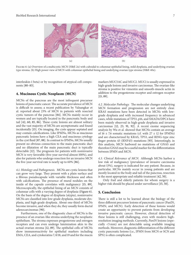
Figure 6: (a) Overview of a multicystic MCN (H&E 2x) with cuboidal to columnar epithelial lining, mild dysplasia, and underlying ovariantype stroma. (b) High power view of MCN with columnar epithelial lining and underlying ovarian type stroma (H&E 40x).
interleukin-1 beta) or by recognition of atypical cell compo-nents [80–83].
4. Mucinous Cystic Neoplasm (MCN)
MCNs of the pancreas are the most infrequent precursorlesions of pancreatic cancer.The accurate prevalence ofMCNis difficult to assess; a recent publication by Valsangkar etal. reported about 23% of MCN in patients with resectedcystic tumors of the pancreas [84]. MCNs mainly occur inwomen and are typically located in the pancreatic body andtail [42, 60, 85, 86]. These cystic lesions are almost solitaryand the vast majority of MCNs are asymptomatic and foundincidentally [15]. On imaging, the cysts appear septated andmay contain calcifications. Like IPMNs, MCNs as mucinouspancreatic lesions have a high CEA and mucinous cytologyin the cyst fluid [87, 88]. In contrast to IPMNs, MCNs usuallypresent no obvious connection to the main pancreatic ductand no dilatation of the main pancreatic duct is typicallydetectable [12]. The prognosis for patients with noninvasiveMCN is very favorable (five-year survival almost 100%), andalso for patients who undergo resection for an invasive MCNthe five-year survival rate is nearly up to 60% [86].
4.1. Histology and Pathogenesis. MCNs are cystic lesions thatcan grow very large. They present with a plain surface anda fibrous pseudocapsule with variable thickness and oftenwith calcifications. The presence of mural nodules on theinside of the capsule correlates with malignancy [15, 89].Microscopically, the epithelial lining of an MCN consists ofcolumnar cells with a varying degree of dysplasia (Figure 6).On the basis of the degree of dysplasia (atypia), noninvasiveMCNs are classified into low-grade dysplasia, moderate dys-plasia, and high-grade dysplasia. About one-third of MCNsbecome invasive, and when they do, they usually form ductaladenocarcinomas [90].
Furthermore, one of the diagnostic clues of MCNs is thepresence of an ovarian-like stroma underlying the neoplasticepithelium. The stroma expresses progesterone and estrogenreceptors and can even undergo luteinization akin to theactual ovarian stroma [12, 89]. The epithelial cells of MCNsshow immunoreactivity for epithelial markers includingEMA, CEA, and cytokeratins 7, 8/18, and 19 and for themucin
markersMUC5AC andMUC2.MUC1 is usually expressed inhigh-grade lesions and invasive carcinomas.The ovarian-likestroma is positive for vimentin and smooth-muscle actin inaddition to the progesterone receptor and estrogen receptor[15, 89].
4.2. Molecular Pathology. Themolecular changes underlyingMCN formation and progression are not entirely clear.KRAS mutations have been detected in MCNs with low-grade dysplasia and with increased frequency in advancedcases, while mutations of TP53, p16, and SMAD4/DPC4 havebeen mainly observed in high-grade dysplasia and invasivecarcinomas [12, 23, 91, 92]. A recent exome sequencinganalysis by Wu et al. showed that MCNs contain an averageof 16 ± 7.6 somatic mutations (cf. with 27 ± 12 for IPMNs)and are characterized by KRAS, TP53, and RING-type zincfinger protein 43 (RNF43) mutations [93]. Interestingly, inthis analysis, MCN harbored no mutations of GNAS andthereforeGNASmay be a usefulmarker for the differentiationbetween IPMN and MCN.
4.3. Clinical Relevance of MCN. Although MCNs harbor alow risk of malignancy (prevalence of invasive carcinomaabout 13%), surgery is indicated for any patient. Because, inparticular, MCNs mainly occur in young patients and aremostly located in the body and tail of the pancreas, resectionis the most appropriate and reliable treatment [42, 50].
Only frail and elderly patients for whom surgery is ahigher risk should be placed under surveillance [15, 50].
5. Conclusion
There is still a lot to be learned about the biology of thethree different precursor lesions of pancreatic cancer (PanIN,IPMN, and MCN). Early detection of these lesions wouldcreate an opportunity to prevent patients from developinginvasive pancreatic cancer. However, clinical detection ofthese lesions is still challenging, even with modern high-resolution imaging methods. Currently, PanIN lesions (espe-cially <5mm) are not detectable using available imagingmethods.Moreover, diagnostic differentiation of the differentcystic pancreatic lesions (i.e., IPMN from MCN or SCN andSPN) is often complicated.
8 BioMed Research International
From this comes the need for reliable biomarkers to detectand differentiate precursor lesions for pancreatic cancer.Analyzing genetic alterations in pancreatic juice/cystic fluidmay be a diagnostic option for the future, as preliminarystudies have already demonstrated. Prospective studies tovalidate these markers are needed to put them into clinicalpractice.
Abbreviations
PanIN: Pancreatic intraepithelial neoplasiaPDAC: Pancreatic ductal adenocarcinomaIPMN: Intraductal papillary mucinous neoplasmMCN: Mucinous cystic neoplasmMD-IPMN: Main duct intraductal papillary mucinous
neoplasmBD-IPMN: Branch duct intraductal papillary
mucinous neoplasmSPN: Solid pseudopapillary neoplasmSCN: Serous cystic neoplasm.
Conflict of Interests
The authors declare that there is no conflict of interestsregarding the publication of this paper.
Acknowledgment
The authors acknowledge the service of “Proof-Reading-Service.com” (Hertfordshire, UK) for English language edit-ing of the paper.
References
[1] R. Siegel, D.Naishadham, andA. Jemal, “Cancer statistics, 2012,”CA Cancer Journal for Clinicians, vol. 62, no. 1, pp. 10–29, 2012.
[2] J. M. Winter, J. L. Cameron, K. A. Campbell et al., “1423Pancreaticoduodenectomies for pancreatic cancer: a single-institution experience,” Journal of Gastrointestinal Surgery , vol.10, no. 9, pp. 1199–1210, 2006.
[3] M. Distler, F. Ruckert, M. Hunger et al., “Evaluation of survivalin patients after pancreatic head resection for ductal adenocar-cinoma,” BMC Surgery, vol. 13, article 12, 2013.
[4] H. Oettle, S. Post, P. Neuhaus et al., “Adjuvant chemother-apy with gemcitabine vs observation in patients undergoingcurative-intent resection of pancreatic cancer: a randomizedcontrolled trial,” The Journal of the American Medical Associa-tion, vol. 297, no. 3, pp. 267–277, 2007.
[5] J. P. Neoptolemos, D. D. Stocken, C. Tudur Smith et al.,“Adjuvant 5-fluorouracil and folinic acid vs observation forpancreatic cancer: composite data from the ESPAC-1 and -3(v1)trials,” British Journal of Cancer, vol. 100, no. 2, pp. 246–250,2009.
[6] J.-B.M. Koorstra, G. Feldmann, N.Habbe, andA.Maitra, “Mor-phogenesis of pancreatic cancer: role of pancreatic intraepithe-lial neoplasia (PanINs),” Langenbeck’s Archives of Surgery, vol.393, no. 4, pp. 561–570, 2008.
[7] D. J. Brat, K. D. Lillemoe, C. J. Yeo, P. B. Warfield, and R. H.Hruban, “Progression of pancreatic intraductal neoplasias to
infiltrating adenocarcinoma of the pancreas,” American Journalof Surgical Pathology, vol. 22, no. 2, pp. 163–169, 1998.
[8] A. L. Cubilla and P. J. Fitzgerald, “Morphological lesions asso-ciated with human primary invasive nonendocrine pancreascancer,” Cancer Research, vol. 36, no. 7, pp. 2690–2698, 1976.
[9] A. Andea, F. Sarkar, and V. N. Adsay, “Clinicopathologicalcorrelates of pancreatic intraepithelial neoplasia: a comparativeanalysis of 82 cases with and 152 cases without pancreatic ductaladenocarcinoma,” Modern Pathology, vol. 16, no. 10, pp. 996–1006, 2003.
[10] K. Brune, T. Abe, M. Canto et al., “Multifocal neoplastic pre-cursor lesions associated with lobular atrophy of the pancreasin patients having a strong family history of pancreatic cancer,”American Journal of Surgical Pathology, vol. 30, no. 9, pp. 1067–1076, 2006.
[11] C. Shi, A. P. Klein, M. Goggins et al., “Increased prevalence ofprecursor lesions in familial pancreatic cancer patients,”ClinicalCancer Research, vol. 15, no. 24, pp. 7737–7743, 2009.
[12] H.Matthaei, R. D. Schulick, R. H. Hruban, and A.Maitra, “Cys-tic precursors to invasive pancreatic cancer,” Nature ReviewsGastroenterology and Hepatology, vol. 8, no. 3, pp. 141–150, 2011.
[13] H. Uehara, A. Nakaizumi, O. Ishikawa et al., “Development ofductal carcinoma of the pancreas during follow-up of branchduct intraductal papillary mucinous neoplasm of the pancreas,”Gut, vol. 57, no. 11, pp. 1561–1565, 2008.
[14] M. Distler, S. Kersting, M. Niedergethmann et al., “Pathohis-tological subtype predicts survival in patients with intraductalpapillary mucinous neoplasm (IPMN) of the pancreas,” Annalsof Surgery, vol. 258, no. 2, pp. 324–330, 2013.
[15] G. Zamboni, K. Hirabayashi, P. Castelli, and A. M. Lennon,“Precancerous lesions of the pancreas,”Best Practice&Research.Clinical Gastroenterology, vol. 27, no. 2, pp. 299–322, 2013.
[16] R. H. Hruban, N. V. Adsay, J. Albores-Saavedra et al., “Pancre-atic intraepithelial neoplasia: a new nomenclature and classifi-cation system for pancreatic duct lesions,” American Journal ofSurgical Pathology, vol. 25, no. 5, pp. 579–586, 2001.
[17] R. H. Hruban, K. Takaori, D. S. Klimstra et al., “An illustratedconsensus on the classification of pancreatic intraepithelial neo-plasia and intraductal papillary mucinous neoplasms,” Amer-ican Journal of Surgical Pathology, vol. 28, no. 8, pp. 977–987,2004.
[18] A. Maitra, N. V. Adsay, P. Argani et al., “Multicomponentanalysis of the pancreatic adenocarcinoma progression modelusing a pancreatic intraepithelial neoplasia tissue microarray,”Modern Pathology, vol. 16, no. 9, pp. 902–912, 2003.
[19] J. Luttges, G. Zamboni, D. Longnecker, and G. Kloppel, “Theimmunohistochemical mucin expression pattern distinguishesdifferent types of intraductal papillary mucinous neoplasms ofthe pancreas and determines their relationship to mucinousnoncystic carcinoma and ductal adenocarcinoma,” AmericanJournal of Surgical Pathology, vol. 25, no. 7, pp. 942–948, 2001.
[20] K. Nagata, M. Horinouchi, M. Saitou et al., “Mucin expressionprofile in pancreatic cancer and the precursor lesions,” Journalof Hepato-Biliary-Pancreatic Surgery, vol. 14, no. 3, pp. 243–254,2007.
[21] N. V. Adsay, K. Merati, A. Andea et al., “The dichotomy in thepreinvasive neoplasia to invasive carcinoma sequence in thepancreas: differential expression of MUC1 and MUC2 supportsthe existence of two separate pathways of carcinogenesis,”Modern Pathology, vol. 15, no. 10, pp. 1087–1095, 2002.
[22] G. E. Kim, H. I. Bae, H. U. Park et al., “Aberrant expressionof MUC5AC and MUC6 gastric mucins and sialyl Tn antigen
BioMed Research International 9
in intraepithelial neoplasms of the pancreas,” Gastroenterology,vol. 123, no. 4, pp. 1052–1060, 2002.
[23] S. Yonezawa, M. Higashi, N. Yamada, and M. Goto, “Precursorlesions of pancreatic cancer,”Gut, vol. 2, no. 3, pp. 137–154, 2008.
[24] H.-U. Park, J.-W. Kim, G. E. Kim et al., “Aberrant expressionof MUC3 and MUC4 membrane-associated mucins and sialylLe(x) antigen in pancreatic intraepithelial neoplasia,” Pancreas,vol. 26, no. 3, pp. e48–e54, 2003.
[25] M. J. Swartz, S. K. Batra, G. C. Varshney et al., “MUC4expression increases progressively in pancreatic intraepithelialneoplasia,” American Journal of Clinical Pathology, vol. 117, no.5, pp. 791–796, 2002.
[26] M. Lohr, G. Kloppel, P. Maisonneuve, A. B. Lowenfels, andJ. Luttges, “Frequency of K-ras mutations in pancreatic intra-ductal neoplasias associated with pancreatic ductal adenocarci-noma and chronic pancreatitis: a meta-analysis,”Neoplasia, vol.7, no. 1, pp. 17–23, 2005.
[27] R. H. Hruban, A. D. M. van Mansfeld, G. J. A. Offerhauset al., “K-ras oncogene activation in adenocarcinoma of thehuman pancreas: a study of 82 carcinomas using a combinationof mutant-enriched polymerase chain reaction analysis andallele-specific oligonucleotide hybridization,” American Journalof Pathology, vol. 143, no. 2, pp. 545–554, 1993.
[28] C. Almoguera, D. Shibata, K. Forrester, J. Martin, N. Arnheim,and M. Perucho, “Most human carcinomas of the exocrinepancreas contain mutant c-K-ras genes,” Cell, vol. 53, no. 4, pp.549–554, 1988.
[29] S. R. Hingorani, E. F. Petricoin III, A. Maitra et al., “Preinvasiveand invasive ductal pancreatic cancer and its early detection inthe mouse,” Cancer Cell, vol. 4, no. 6, pp. 437–450, 2003.
[30] A. Maitra, S. E. Kern, and R. H. Hruban, “Molecular patho-genesis of pancreatic cancer,” Best Practice & Research. ClinicalGastroenterology, vol. 20, no. 2, pp. 211–226, 2006.
[31] C. Caldas, S. A. Hahn, L. T. da Costa et al., “Frequent somaticmutations and homozygous deletions of the p16 (MTS1) gene inpancreatic adenocarcinoma,” Nature Genetics, vol. 8, no. 1, pp.27–32, 1994.
[32] S. E. Kern, “p53: tumor suppression through control of the cellcycle,” Gastroenterology, vol. 106, no. 6, pp. 1708–1711, 1994.
[33] J. Massague, S. W. Blain, and R. S. Lo, “TGFbeta signaling ingrowth control, cancer, and heritable disorders,” Cell, vol. 103,no. 2, pp. 295–309, 2000.
[34] M. Schutte, R. H. Hruban, L. Hedrick et al., “DPC4 gene invarious tumor types,” Cancer Research, vol. 56, no. 11, pp. 2527–2530, 1996.
[35] S. J. Clark, “Action at a distance: epigenetic silencing of largechromosomal regions in carcinogenesis,” Human MolecularGenetics, vol. 16, no. 1, pp. R88–R95, 2007.
[36] A. C. Tan, A. Jimeno, S. H. Lin et al., “Characterizing DNAmethylation patterns in pancreatic cancer genome,” MolecularOncology, vol. 3, no. 5-6, pp. 425–438, 2009.
[37] M. G. House, J. G. Herman, M. Z. Guo et al., “Aberranthypermethylation of tumor suppressor genes in pancreaticendocrine neoplasms,” Annals of Surgery, vol. 238, no. 3, pp.423–432, 2003.
[38] N. Sato, N. Fukushima, R. H. Hruban, and M. Goggins,“CpG island methylation profile of pancreatic intraepithelialneoplasia,”Modern Pathology, vol. 21, no. 3, pp. 238–244, 2008.
[39] M. Goggins, “Identifying molecular markers for the earlydetection of pancreatic neoplasia,” Seminars in Oncology, vol.34, no. 4, pp. 303–310, 2007.
[40] H. Matthaei, S.-M. Hong, S. C. Mayo et al., “Presence of pan-creatic intraepithelial neoplasia in the pancreatic transectionmargin does not influence outcome in patients with R0 resectedpancreatic cancer,” Annals of Surgical Oncology, vol. 18, no. 12,pp. 3493–3499, 2011.
[41] M. Kosmahl, U. Pauser, K. Peters et al., “Cystic neoplasms of thepancreas and tumor-like lesions with cystic features: a reviewof 418 cases and a classification proposal,” Virchows Archiv, vol.445, no. 2, pp. 168–178, 2004.
[42] R. Grutzmann and H. D. Saeger, “Cystic tumors of the pan-creas,” Chirurg, vol. 81, no. 8, pp. 755–769, 2010.
[43] W. R. Brugge, G. Y. Lauwers, D. Sahani, C. Fernandez-DelCastillo, andA. L.Warshaw, “Cystic neoplasms of the pancreas,”The New England Journal of Medicine, vol. 351, no. 12, pp. 1218–1226, 2004.
[44] G. Kloppel, E. Solcia, D. S. Longnecker et al.,Histological Typingof Tumours of the Exocrine Pancreas:WorldHealth OrganizationInternational Histological Classification of Tumours, Springer,New York, NY, USA, 2nd edition, 1998.
[45] N. V. Adsay, K. Merati, O. Basturk et al., “Pathologically andbiologically distinct types of epithelium in intraductal papillarymucinous neoplasms: delineation of an “intestinal” pathway ofcarcinogenesis in the pancreas,” American Journal of SurgicalPathology, vol. 28, no. 7, pp. 839–848, 2004.
[46] N. V. Adsay, K. Merati, H. Nassar et al., “Pathogenesis of colloid(puremucinous) carcinoma of exocrine organs: coupling of gel-forming mucin (MUC2) production with altered cell polarityand abnormal cell-stroma interaction may be the key factor inthe morphogenesis and indolent behavior of colloid carcinomain the breast and pancreas,” American Journal of SurgicalPathology, vol. 27, no. 5, pp. 571–578, 2003.
[47] R. Grutzmann, M. Niedergethmann, C. Pilarsky, G. Kloppel,andH.D. Saeger, “Intraductal papillarymucinous tumors of thepancreas: biology, diagnosis, and treatment,”Oncologist, vol. 15,no. 12, pp. 1294–1309, 2010.
[48] T. Furukawa, T. Hatori, I. Fujita et al., “Prognostic relevanceof morphological types of intraductal papillary mucinous neo-plasms of the pancreas,” Gut, vol. 60, no. 4, pp. 509–516, 2011.
[49] O. Basturk, S. Khayyata, D. S. Klimstra et al., “Preferentialexpression of MUC6 in oncocytic and pancreatobiliary types ofintraductal papillary neoplasms highlights a pyloropancreaticpathway, distinct from the intestinal pathway, in pancreaticcarcinogenesis,”American Journal of Surgical Pathology, vol. 34,no. 3, pp. 364–370, 2010.
[50] M. Tanaka, C. F.-D. Castillo, V. Adsay et al., “Internationalconsensus guidelines 2012 for the management of IPMN andMCN of the pancreas,” Pancreatology, vol. 12, pp. 183–197, 2012.
[51] K. Nagai, R. Doi, A. Kida et al., “Intraductal papillary mucinousneoplasms of the pancreas: clinicopathologic characteristicsand long-term follow-up after resection,” World Journal ofSurgery, vol. 32, no. 2, pp. 271–280, 2008.
[52] R. Grutzmann, S. Post, H. D. Saeger, and M. Niedergethmann,“Intraductal Papillary Mucinous Neoplasia (IPMN) of thepancreas: its diagnosis, treatment, and prognosis,” DeutschesArzteblatt International, vol. 108, no. 46, pp. 788–794, 2011.
[53] R. Salvia, S. Partelli, S. Crippa et al., “Intraductal papillarymuci-nous neoplasms of the pancreas with multifocal involvement ofbranch ducts,” American Journal of Surgery, vol. 198, no. 5, pp.709–714, 2009.
10 BioMed Research International
[54] S. T. Chari, D. Yadav, T. C. Smyrk et al., “Study of recurrenceafter surgical resection of intraductal papillary mucinous neo-plasm of the pancreas,” Gastroenterology, vol. 123, no. 5, pp.1500–1507, 2002.
[55] F. T. Bosman, F. Carneiro, R. H. Hruban, and N. Theise,Classification of Tumours of the Digestive System, WHO, Lyon,France, 2010.
[56] M. Niedergethmann, R. Grutzmann, R. Hildenbrand et al.,“Outcome of invasive and noninvasive intraductal papillary-mucinous neoplasms of the pancreas (IPMN): a 10-year experi-ence,” World Journal of Surgery, vol. 32, no. 10, pp. 2253–2260,2008.
[57] T. Furukawa, G. Kloppel, N. Volkan Adsay et al., “Classificationof types of intraductal papillary-mucinous neoplasm of thepancreas: a consensus study,” Virchows Archiv, vol. 447, no. 5,pp. 794–799, 2005.
[58] S. Yonezawa, M. Horinouchi, M. Osako et al., “Gene expressionof gastric typemucin (MUC5AC) in pancreatic tumors: its rela-tionship with the biological behavior of the tumor,” PathologyInternational, vol. 49, no. 1, pp. 45–54, 1999.
[59] I. Kitazono, M. Higashi, S. Kitamoto et al., “Expression ofMUC4 mucin is observed mainly in the intestinal type ofintraductal papillary mucinous neoplasm of the pancreas,”Pancreas, vol. 42, no. 7, pp. 1120–1128, 2013.
[60] M. Distler, T. Welsch, D. Aust, J. Weitz, and R. Grutzmann,“Intraductal papillary mucinous neoplasm of the pancreas(IPMN)—standards and new aspects,” Zentralbl Chir. In press.
[61] H. Yamaguchi, Y. Kuboki, T. Hatori et al., “The discrete natureand distinguishing molecular features of pancreatic intraductaltubulopapillary neoplasms and intraductal papillary mucinousneoplasms of the gastric type, pyloric gland variant,”The Journalof Pathology, vol. 231, no. 3, pp. 335–341, 2013.
[62] Y. Sadakari, K. Ohuchida, K. Nakata et al., “Invasive carcinomaderived from the nonintestinal type intraductal papillary muci-nous neoplasm of the pancreas has a poorer prognosis than thatderived from the intestinal type,” Surgery, vol. 147, no. 6, pp. 812–817, 2010.
[63] G. A. Poultsides, S. Reddy, J. L. Cameron et al., “Histopathologicbasis for the favorable survival after resection of intraductalpapillary mucinous neoplasm-associated invasive adenocarci-noma of the pancreas,” Annals of Surgery, vol. 251, no. 3, pp.470–476, 2010.
[64] M. Mino-Kenudson, C. Fernandez-del Castillo, Y. Baba et al.,“Prognosis of invasive intraductal papillarymucinous neoplasmdepends on histological and precursor epithelial subtypes,”Gut,vol. 60, no. 12, pp. 1712–1720, 2011.
[65] Y.Murakami, K. Uemura, T. Sudo et al., “Postoperative adjuvantchemotherapy improves survival after surgical resection forpancreatic carcinoma,” Journal of Gastrointestinal Surgery, vol.12, no. 3, pp. 534–541, 2008.
[66] J. Kim, K. T. Jang, S. Mo Park et al., “Prognostic relevance ofpathologic subtypes and minimal invasion in intraductal papil-larymucinous neoplasms of the pancreas,” Tumour Biology, vol.32, no. 3, pp. 535–542, 2011.
[67] S. Fritz, C. Fernandez-del Castillo, M. Mino-Kenudson et al.,“Global genomic analysis of intraductal papillary mucinousneoplasms of the pancreas reveals significant molecular differ-ences compared to ductal adenocarcinoma,” Annals of Surgery,vol. 249, no. 3, pp. 440–447, 2009.
[68] F. Schonleben, W. Qiu, K. C. Bruckman et al., “BRAF andKRAS gene mutations in intraductal papillary mucinous neo-plasm/carcinoma (IPMN/IPMC) of the pancreas,” Cancer Let-ters, vol. 249, no. 2, pp. 242–248, 2007.
[69] D. Mohri, Y. Asaoka, H. Ijichi et al., “Different subtypes ofintraductal papillary mucinous neoplasm in the pancreas havedistinct pathways to pancreatic cancer progression,” Journal ofGastroenterology, vol. 47, no. 2, pp. 203–213, 2012.
[70] J.Wu,H.Matthaei, A.Maitra et al., “RecurrentGNASmutationsdefine an unexpected pathway for pancreatic cyst development,”Science Translational Medicine, vol. 3, no. 92, article 92ra66,2011.
[71] T. Furukawa, Y. Kuboki, E. Tanji et al., “Whole-exome sequenc-ing uncovers frequent GNASmutations in intraductal papillarymucinous neoplasms of the pancreas,” Scientific Reports, vol. 1,article 161, 2011.
[72] M. Dal Molin, H. Matthaei, J. Wu et al., “Clinicopathologicalcorrelates of activatingGNASmutations in intraductal papillarymucinous neoplasm (IPMN) of the pancreas,”Annals of SurgicalOncology, vol. 20, no. 12, pp. 3802–3808, 2013.
[73] H. D. Xiao, H. Yamaguchi, D. Dias-Santagata et al., “Molecularcharacteristics and biological behaviours of the oncocytic andpancreatobiliary subtypes of intraductal papillary mucinousneoplasms,” Journal of Pathology, vol. 224, no. 4, pp. 508–516,2011.
[74] A. A. Siddiqui, T. E. Kowalski, R. Kedika et al., “EUS-guidedpancreatic fluid aspiration for DNA analysis of KRAS andGNASmutations for the evaluation of pancreatic cystic neopla-sia: a pilot study,” Gastrointestinal Endoscopy, vol. 77, no. 4, pp.669–670, 2013.
[75] M. Kanda, S. Knight, M. Topazian et al., “Mutant GNASdetected in duodenal collections of secretin-stimulated pan-creatic juice indicates the presence or emergence of pancreaticcysts,” Gut, vol. 62, no. 7, pp. 1024–1033, 2013.
[76] M. Kanda, Y. Sadakari, M. Borges et al., “Mutant TP53 in duo-denal samples of pancreatic juice from patients with pancreaticcancer or high-grade dysplasia,” Clinical Gastroenterology andHepatology, vol. 11, no. 6, pp. 719.e5–730.e5, 2013.
[77] K. K. Das, H. Xiao, X. Geng et al., “mAb Das-1 is specificfor high-risk and malignant intraductal papillary mucinousneoplasm (IPMN),” Gut, 2013.
[78] S. M. Hong, N. Omura, A. Vincent et al., “Genome-wide CpGisland profiling of intraductal papillary mucinous neoplasms ofthe pancreas,” Clinical Cancer Research, vol. 18, no. 3, pp. 700–712, 2012.
[79] T. Hara, D. Ikebe, A. Odaka et al., “Preoperative histologicalsubtype classification of intraductal papillary mucinous neo-plasms (IPMN) by pancreatic juice cytology with MUC stain,”Annals of Surgery, vol. 257, no. 6, pp. 1103–1111, 2013.
[80] M. B. Pitman, B. A. Centeno, E. S. Daglilar, W. R. Brugge,and M. Mino-Kenudson, “Cytological criteria of high-gradeepithelial atypia in the cyst fluid of pancreatic intraductalpapillary mucinous neoplasms,” Cancer Cytopathology, vol. 122,no. 1, pp. 40–47, 2014.
[81] A. V. Maker, N. Katabi, M. Gonen et al., “Pancreatic cystfluid and serum mucin levels predict dysplasia in intraductalpapillary mucinous neoplasms of the pancreas,” Annals ofSurgical Oncology, vol. 18, no. 1, pp. 199–206, 2011.
[82] A. V. Maker, N. Katabi, L.-X. Qin et al., “Cyst fluid interleukin-1𝛽 (IL1𝛽) levels predict the risk of carcinoma in intraductalpapillary mucinous neoplasms of the pancreas,” Clinical CancerResearch, vol. 17, no. 6, pp. 1502–1508, 2011.
BioMed Research International 11
[83] M. B. Pitman, P. J. Michaels, V. Deshpande,W. R. Brugge, and B.C. Bounds, “Cytological and cyst fluid analysis of small (≤3 cm)branch duct intraductal papillary mucinous neoplasms addsvalue to patient management decisions,” Pancreatology, vol. 8,no. 3, pp. 277–284, 2008.
[84] N. P. Valsangkar, V. Morales-Oyarvide, S. P. Thayer et al., “851resected cystic tumors of the pancreas: a 33-year experience atthe Massachusetts General Hospital,” Surgery, vol. 152, no. 3,supplement 1, pp. S4–S12, 2012.
[85] K. Yamao, A. Yanagisawa, K. Takahashi et al., “Clinicopatholog-ical features and prognosis of mucinous cystic neoplasm withovarian-type stroma: a multi-institutional study of the Japanpancreas society,” Pancreas, vol. 40, no. 1, pp. 67–71, 2011.
[86] S. Crippa, R. Salvia, A. L. Warshaw et al., “Mucinous cysticneoplasm of the pancreas is not an aggressive entity: lessonsfrom 163 resected patients,” Annals of Surgery, vol. 247, no. 4,pp. 571–579, 2008.
[87] S. Cizginer, B. Turner, A. R. Bilge, C. Karaca, M. B. Pitman,and W. R. Brugge, “Cyst fluid carcinoembryonic antigen is anaccurate diagnostic marker of pancreatic mucinous cysts,” Pan-creas, vol. 40, no. 7, pp. 1024–1028, 2011, Erratum in “Cyst fluidcarcinoembryonic antigen is an accurate diagnostic marker ofpancreatic mucinous cysts”, Pancreas, vol. 42, no. 4, pp. 728,2013.
[88] W. G.-U. Park, R. Mascarenhas, M. Palaez-Luna et al., “Diag-nostic performance of cyst fluid carcinoembryonic antigen andamylase in histologically confirmed pancreatic cysts,” Pancreas,vol. 40, no. 1, pp. 42–45, 2011.
[89] G. Zamboni, A. Scarpa, G. Bogina et al., “Mucinous cystictumors of the pancreas: clinicopathological features, prognosis,and relationship to other mucinous cystic tumors,” AmericanJournal of Surgical Pathology, vol. 23, no. 4, pp. 410–422, 1999.
[90] M. L. Baker, E. S. Seeley, R. Pai et al., “Invasive mucinouscystic neoplasms of the pancreas,” Experimental and MolecularPathology, vol. 93, no. 3, pp. 345–349, 2012.
[91] R. E. Jimenez, A. L. Warshaw, K. Z’graggen et al., “Sequentialaccumulation of K-ras mutations and p53 overexpression inthe progression of pancreatic mucinous cystic neoplasms tomalignancy,”Annals of Surgery, vol. 230, no. 4, pp. 501–511, 1999.
[92] C. A. Iacobuzio-Donahue, R. E. Wilentz, P. Argani et al., “Dpc4protein in mucinous cystic neoplasms of the pancreas: frequentloss of expression in invasive carcinomas suggests a role ingenetic progression,” American Journal of Surgical Pathology,vol. 24, no. 11, pp. 1544–1548, 2000.
[93] J. Wu, Y. Jiao, M. Dal Molin et al., “Whole-exome sequencing ofneoplastic cysts of the pancreas reveals recurrent mutations incomponents of ubiquitin-dependent pathways,” Proceedings ofthe National Academy of Sciences of the United States of America,vol. 108, no. 52, pp. 21188–21193, 2011.

Submit your manuscripts athttp://www.hindawi.com
Stem CellsInternational
Hindawi Publishing Corporationhttp://www.hindawi.com Volume 2014
Hindawi Publishing Corporationhttp://www.hindawi.com Volume 2014
MEDIATORSINFLAMMATION
of
Hindawi Publishing Corporationhttp://www.hindawi.com Volume 2014
Behavioural Neurology
EndocrinologyInternational Journal of
Hindawi Publishing Corporationhttp://www.hindawi.com Volume 2014
Hindawi Publishing Corporationhttp://www.hindawi.com Volume 2014
Disease Markers
Hindawi Publishing Corporationhttp://www.hindawi.com Volume 2014
BioMed Research International
OncologyJournal of
Hindawi Publishing Corporationhttp://www.hindawi.com Volume 2014
Hindawi Publishing Corporationhttp://www.hindawi.com Volume 2014
Oxidative Medicine and Cellular Longevity
Hindawi Publishing Corporationhttp://www.hindawi.com Volume 2014
PPAR Research
The Scientific World JournalHindawi Publishing Corporation http://www.hindawi.com Volume 2014
Immunology ResearchHindawi Publishing Corporationhttp://www.hindawi.com Volume 2014
Journal of
ObesityJournal of
Hindawi Publishing Corporationhttp://www.hindawi.com Volume 2014
Hindawi Publishing Corporationhttp://www.hindawi.com Volume 2014
Computational and Mathematical Methods in Medicine
OphthalmologyJournal of
Hindawi Publishing Corporationhttp://www.hindawi.com Volume 2014
Diabetes ResearchJournal of
Hindawi Publishing Corporationhttp://www.hindawi.com Volume 2014
Hindawi Publishing Corporationhttp://www.hindawi.com Volume 2014
Research and TreatmentAIDS
Hindawi Publishing Corporationhttp://www.hindawi.com Volume 2014
Gastroenterology Research and Practice
Hindawi Publishing Corporationhttp://www.hindawi.com Volume 2014
Parkinson’s Disease
Evidence-Based Complementary and Alternative Medicine
Volume 2014Hindawi Publishing Corporationhttp://www.hindawi.com