Embed Size (px)
Citation preview
Review ArticleProstate Cancer Radiation Therapy:What Do Clinicians Have to Know?
Ben G. L. Vanneste,1 Evert J. Van Limbergen,1 Emile N. van Lin,1
Joep G. H. van Roermund,2 and Philippe Lambin1
1Department of Radiation Oncology (MAASTRO Clinic), GROW School for Oncology and Developmental Biology,Maastricht University Medical Centre, Maastricht, Netherlands2Department of Urology, Maastricht University Medical Centre, Maastricht, Netherlands
Correspondence should be addressed to Ben G. L. Vanneste; [email protected]
Received 31 August 2016; Revised 18 October 2016; Accepted 31 October 2016
Academic Editor: Seyed Behzad Jazayeri
Copyright © 2016 Ben G. L. Vanneste et al. This is an open access article distributed under the Creative Commons AttributionLicense, which permits unrestricted use, distribution, and reproduction in any medium, provided the original work is properlycited.
Radiotherapy (RT) for prostate cancer (PC) has steadily evolved over the last decades, with improving biochemical disease-freesurvival. Recently population based research also revealed an association between overall survival and doses ≥ 75.6 Gray (Gy) inmen with intermediate- and high-risk PC. Examples of improved RT techniques are image-guided RT, intensity-modulated RT,volumetric modulated arc therapy, and stereotactic ablative body RT, which could facilitate further dose escalation. Brachytherapyis an internal form of RT that also developed substantially. New devices such as rectum spacers and balloons have been developed tospare rectal structures. Newer techniques like protons and carbon ions have the intrinsic characteristics maximising the dose on thetumour while minimising the effect on the surrounding healthy tissue, but clinical data are needed for confirmation in randomisedphase III trials. Furthermore, it provides an overview of an important discussion issue in PC treatment between urologists andradiation oncologists: the comparison between radical prostatectomy and RT. Current literature reveals that all possible treatmentmodalities have the same cure rate, but a different toxicity pattern. We recommend proposing the possible different treatmentmodalities with their own advantages and side-effects to the individual patient. Clinicians and patients should make treatmentdecisions together (shared decision-making) while using patient decision aids.
1. Introduction
Prostate cancer (PC) is the most common cancer amongmales in the Western world, with more than 1.11 millionnew cases diagnosed in 2012 and 307,000 deaths [1, 2]. Thelifetime risk of developing PC is 1 in 8 [3]. It is expectedthat the incidence will substantially increase in the comingdecades due to the aging population, which makes it a hugehealth care problem. The total economic costs of PC inEurope are estimated to exceed €8.43 billion [4]. One of thebiggest challenges in the 21st century will be to offer the bestindividualised treatment at reasonable costs.
External-beam radiotherapy (EBRT) and brachytherapy(BT) are potentially curative therapies for PC. RT has under-gone tremendous improvements in the last decades. Doseescalation in prostate EBRT leads to improved locoregional
control, biochemical disease-free survival (bDFS), distantmetastasis-free survival, PC specific mortality, and evenoverall survival in intermediate- and high-risk PC [5–11].However, dose escalation is limited by toxicity of surroundinghealthy tissues, and therefore improved tumour control isexpected to come at the cost of higher toxicity, greatlyimpacting patients’ quality of life [12–14]. However, doseescalation is possible due to advances in different RT tech-niques, sophisticated computer-based treatment planning,and/or development of extra devices, avoiding increased dosedelivery to the surrounding healthy tissue.Thepurpose of thisarticle is to provide insight into the enormous improvementsin RT techniques to practicing clinicians and primary caredoctors and to develop a greater comfort level when referringpatients to a radiation oncologist. Furthermore, it providesan overview of an important discussion issue concerning
Hindawi Publishing CorporationBioMed Research InternationalVolume 2016, Article ID 6829875, 14 pageshttp://dx.doi.org/10.1155/2016/6829875
2 BioMed Research International
EBRT procedure
Preparatory phasecreating a RT plan
Delivery phasedelivering RT plan to a patient
Fiducials:marking the prostate
Images:MRI to optimize delineation of the prostate
CT required for dose calculations
Dose calculation:3D-CRT ≥ IMRT ≥ VMAT
Decision support system:need for rectum spacer or not?Yes No?
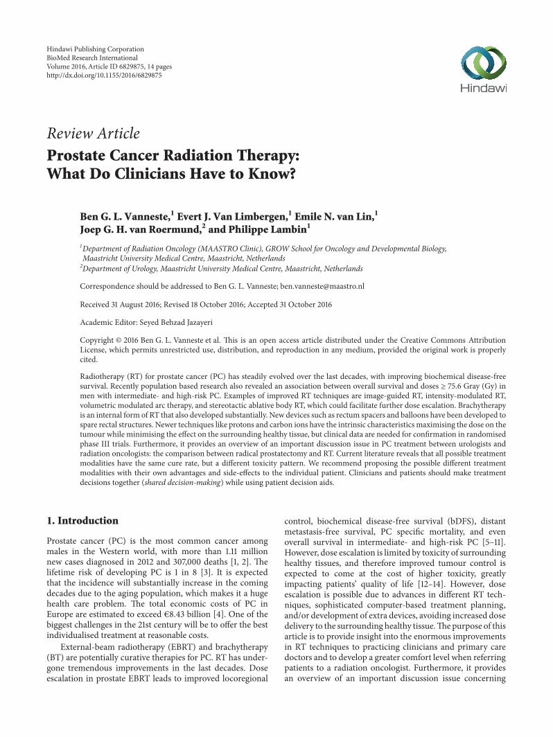
Figure 1: Overview of an EBRT procedure.
RT from a clinician’s perspective: the comparison betweenoperation and RT.
2. Overview of External BeamRadiation Treatments
In EBRT a dose of ionising radiation is generated by anexternal X-ray source. In the past this was a cobalt-60source machine, but nowadays a high-tech tele-therapy unitis used for this purpose [15, 16]. Linear accelerators are thesource of electronic induced irradiation. The radiation beamleaves the linear accelerator by a gantry. Different optionsof machines are commercially available: a traditional linearaccelerator where the gantry can rotate around the patient(Arc therapy). Other possibilities are tomotherapy (=helicaltherapy) where the radiation dose is delivered slice-by-slice[17], or cyberknife (=a robotic radiosurgery system) wherethe location of the prostate is identified during treatment andactive corrections are made for movements of the prostateduring treatment delivery [18]. Evolving radiation techniquesas protons and carbon ions are also introduced and arediscussed below. Over the last 20 years the methods ofdelivering a dose of ionising radiation to a target area havechanged incrementally.
An EBRT procedure consists of 2 main parts (Figure 1).First, in a preparatory phase an RT plan needs to be
created. This process is referred to as RT planning. Secondly,the linear accelerator requires delivering this plan to a patientin an appropriate fashion: the RT dose delivery.
In the preparatory phase, images of the patient areacquired. On these scans the clinical target area is delineatedto which the radiotherapy dose is prescribed. In the 90sthis area was delineated on conventional planar 2D X-rays,on which the target area (the prostate and seminal vesicles)could only be assumed. Later, CT based planning wasintroduced [19]. On the latter the target areas are visualised
and can be delineated directly leading to up to one-thirdless geographical miss of the target [20]. Another advantageof CT based planning was that also critical structures likerectal wall and bladder around the target could be visualisedand subsequently spared from radiation, by avoiding theX-ray beams to pass through them. Currently an MRI isbeing integrated more broadly into the planning process.MRI allows us to delineate the prostate more precisely fromthe pelvic diaphragm, and the base of the prostate canbe differentiated more precisely from the seminal vesicles[21, 22]. An additional MRI changes the delineation of theclinical target volume in 18% to 20% of cases compared toCT based planning [23, 24]. Moreover, tumour extensionin and outside of the prostate and invasion in the seminalvesicles are better visible on MRI and therefore more oftenincluded in the target volumes [24, 25]. Chang and colleaguesreported significant volume changes with MRI delineation:extracapsular extension was significantly more incorporatedinto target volumes with the addition of MRI (40%) incomparison with CT (32%). The seminal vesicles are alsomore often included: 18% versus 3%, respectively. In addition,CT scans overestimate prostate volume by 10% to 45% [21, 22,26–32]. Furthermore, anMRI revealed an important decreaseof the interobserver delineation variation, especially at theprostatic apex [33].We expect that a correct delineation of thetarget volume will result in better treatment outcome, withless toxicity, but until now this is not proven yet.
In addition to improved radiotherapy planning, develop-ments were introduced to verify correct dose delivery duringthe whole course of RT over the several fractions deliveredaccording to the radiotherapy plan. In earlier times patientswere positioned on a linear accelerator using surrogate refer-ence points: external reference points like skin lines or tattoopoints or using bony landmarks visualised by conventionalplain X-ray photographs taken on the linear accelerator.However, as it is known that the prostate and the seminal
BioMed Research International 3
3DCRT IMRT VMAT
PSPT IMPT BT
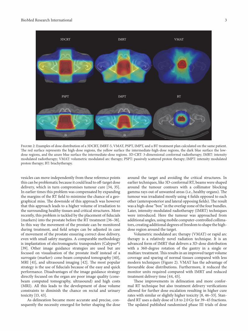
Figure 2: Examples of dose distribution of a 3DCRT, IMRT-5, VMAT, PSPT, IMPT, and a BT treatment plan calculated on the same patient.The red surface represents the high-dose regions, the yellow surface the intermediate-high-dose regions, the dark blue surface the low-dose regions, and the azure blue surface the intermediate-dose regions. 3D-CRT: 3-dimensional conformal radiotherapy; IMRT: intensitymodulated radiotherapy; VMAT: volumetric modulated arc therapy; PSPT: passively scattered proton therapy; IMPT: intensity modulatedproton therapy; BT: brachytherapy.
vesicles can move independently from these reference pointsthis can be problematic because it could lead to off-target dosedelivery, which in turn compromises tumour cure [34, 35].In earlier times this problem was compensated by expandingthe margins of the RT field to minimise the chance of a geo-graphical miss. The downside of this approach was howeverthat this approach leads to a higher volume of irradiation tothe surrounding healthy tissues and critical structures. Morerecently, this problem is tackled by the placement of fiducials(markers) into the prostate before the RT treatment [36–38].In this way the movement of the prostate can be monitoredduring treatment, and field setups can be adjusted in caseof movement of the prostate ensuring correct dose delivery,even with small safety margins. A comparable methodologyis implantation of electromagnetic transponders (Calypso�)[39]. Other image guidance strategies are used but arefocused on visualisation of the prostate itself instead of asurrogate (marker): cone-beam computed tomography [40],MRI [41], and ultrasound imaging [42]. The most popularstrategy is the use of fiducials because of the easy and quickperformance. Disadvantages of the image guidance strategydirectly focused on the organ are poor image quality (cone-beam computed tomography, ultrasound) and high costs(MRI). All this leads to the development of dose volumeconstraints to diminish the chance on rectal and urinarytoxicity [13, 43].
As delineation became more accurate and precise, con-sequently the necessity emerged for better shaping the dose
around the target and avoiding the critical structures. Inearlier techniques, like 3D-conformal RT, beams were shapedaround the tumour contours with a collimator blockinggamma rays out of unwanted areas (i.e., healthy organs). Thetumour was irradiated mostly using 4 fields opposed to eachother (anteroposterior and lateral opposing fields).The resultwas a high-dose “box” in the overlap zone of the four bundles.Later, intensity-modulated radiotherapy (IMRT) techniqueswere introduced. Here the tumour was approached fromadditional angles, usingmobile computer-controlled collima-tors, creating additional degrees of freedom to shape the high-dose region around the target.
Volumetric modulated arc therapy (VMAT) or rapid arctherapy is a relatively novel radiation technique. It is anadvanced form of IMRT that delivers a 3D-dose distributionwith a 360-degree rotation of the gantry in a single ormultiarc treatment.This results in an improved target volumecoverage and sparing of normal tissues compared with lessmodern techniques (Figure 2). VMAT has the advantage offavourable dose distributions. Furthermore, it reduced themonitor units required compared with IMRT and reducedtreatment delivery time [44, 45].
These improvements in delineation and more confor-mal RT technique but also treatment delivery verificationsallowed for further dose escalation resulting in higher curerates with similar or slightly higher toxicity [8, 46–53]. Stan-dard RT uses a daily dose of 1.8 to 2.0Gy for 39–45 fractions.The updated published randomised phase III trials of dose
4 BioMed Research International
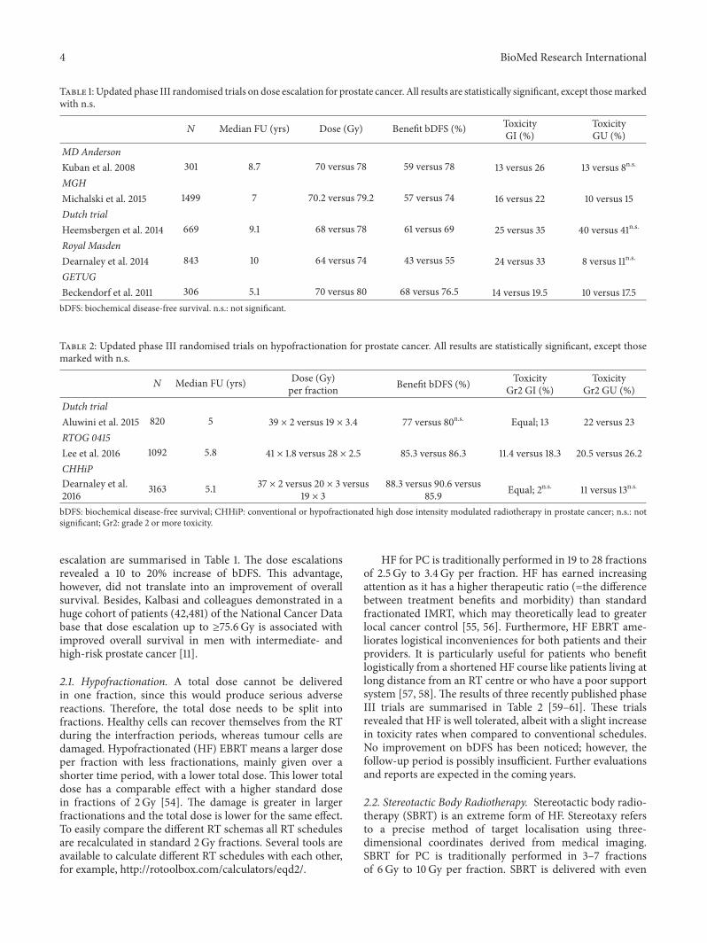
Table 1: Updated phase III randomised trials on dose escalation for prostate cancer. All results are statistically significant, except thosemarkedwith n.s.
𝑁 Median FU (yrs) Dose (Gy) Benefit bDFS (%) ToxicityGI (%)
ToxicityGU (%)
MD AndersonKuban et al. 2008 301 8.7 70 versus 78 59 versus 78 13 versus 26 13 versus 8n.s.
MGHMichalski et al. 2015 1499 7 70.2 versus 79.2 57 versus 74 16 versus 22 10 versus 15Dutch trialHeemsbergen et al. 2014 669 9.1 68 versus 78 61 versus 69 25 versus 35 40 versus 41n.s.
Royal MasdenDearnaley et al. 2014 843 10 64 versus 74 43 versus 55 24 versus 33 8 versus 11n.s.
GETUGBeckendorf et al. 2011 306 5.1 70 versus 80 68 versus 76.5 14 versus 19.5 10 versus 17.5bDFS: biochemical disease-free survival. n.s.: not significant.
Table 2: Updated phase III randomised trials on hypofractionation for prostate cancer. All results are statistically significant, except thosemarked with n.s.
𝑁 Median FU (yrs) Dose (Gy)per fraction Benefit bDFS (%) Toxicity
Gr2 GI (%)Toxicity
Gr2 GU (%)Dutch trialAluwini et al. 2015 820 5 39 × 2 versus 19 × 3.4 77 versus 80n.s. Equal; 13 22 versus 23RTOG 0415Lee et al. 2016 1092 5.8 41 × 1.8 versus 28 × 2.5 85.3 versus 86.3 11.4 versus 18.3 20.5 versus 26.2CHHiPDearnaley et al.2016 3163 5.1 37 × 2 versus 20 × 3 versus
19 × 388.3 versus 90.6 versus
85.9 Equal; 2n.s. 11 versus 13n.s.
bDFS: biochemical disease-free survival; CHHiP: conventional or hypofractionated high dose intensity modulated radiotherapy in prostate cancer; n.s.: notsignificant; Gr2: grade 2 or more toxicity.
escalation are summarised in Table 1. The dose escalationsrevealed a 10 to 20% increase of bDFS. This advantage,however, did not translate into an improvement of overallsurvival. Besides, Kalbasi and colleagues demonstrated in ahuge cohort of patients (42,481) of the National Cancer Database that dose escalation up to ≥75.6Gy is associated withimproved overall survival in men with intermediate- andhigh-risk prostate cancer [11].
2.1. Hypofractionation. A total dose cannot be deliveredin one fraction, since this would produce serious adversereactions. Therefore, the total dose needs to be split intofractions. Healthy cells can recover themselves from the RTduring the interfraction periods, whereas tumour cells aredamaged. Hypofractionated (HF) EBRT means a larger doseper fraction with less fractionations, mainly given over ashorter time period, with a lower total dose. This lower totaldose has a comparable effect with a higher standard dosein fractions of 2Gy [54]. The damage is greater in largerfractionations and the total dose is lower for the same effect.To easily compare the different RT schemas all RT schedulesare recalculated in standard 2Gy fractions. Several tools areavailable to calculate different RT schedules with each other,for example, http://rotoolbox.com/calculators/eqd2/.
HF for PC is traditionally performed in 19 to 28 fractionsof 2.5 Gy to 3.4Gy per fraction. HF has earned increasingattention as it has a higher therapeutic ratio (=the differencebetween treatment benefits and morbidity) than standardfractionated IMRT, which may theoretically lead to greaterlocal cancer control [55, 56]. Furthermore, HF EBRT ame-liorates logistical inconveniences for both patients and theirproviders. It is particularly useful for patients who benefitlogistically from a shortened HF course like patients living atlong distance from an RT centre or who have a poor supportsystem [57, 58]. The results of three recently published phaseIII trials are summarised in Table 2 [59–61]. These trialsrevealed that HF is well tolerated, albeit with a slight increasein toxicity rates when compared to conventional schedules.No improvement on bDFS has been noticed; however, thefollow-up period is possibly insufficient. Further evaluationsand reports are expected in the coming years.
2.2. Stereotactic Body Radiotherapy. Stereotactic body radio-therapy (SBRT) is an extreme form of HF. Stereotaxy refersto a precise method of target localisation using three-dimensional coordinates derived from medical imaging.SBRT for PC is traditionally performed in 3–7 fractionsof 6Gy to 10Gy per fraction. SBRT is delivered with even
BioMed Research International 5
higher than standard precision procedures, for example, acustomised body pillow formed by vacuum suction [62]. Justlike in conventional EBRT there is an evolution with moredose guidance and higher precision (see above).The availableliterature consists mainly of several nonrandomised phase IItrials. Recently, a largemulti-institutional trial of 1100 patientswas reported. Separate prospective phase 2 protocols oflocalised PCpatients fromdifferent institutes treated between2003 and 2011 were pooled for analysis [63]. With a medianfollow-up of 36 months, the five-year bDFS rate was 93%.As this series mostly consisted of low- and intermediate-risk patients and follow-up is still limited, this treatment isonly recommended for selected low- and intermediate-riskpatients with localised PC. That the acute urogenital toxicityseemed higher than conventional EBRT [64] might pose adisadvantage. On the other hand, low late urinary and rectaltoxicities after median follow-up of three years were reported[65]. Data from published prostate SBRT trials have shownlate grade 3 GI and GU toxicities within the 3%. However,this data is preliminary and prospective randomised phase IIItrials and additional follow-up are required to further clarifythe relative differences between both treatment modalities.
3. Brachytherapy
BT is an internal RT, where radiation comes from animplanted source, such as seeds or capsules. BT permits anextreme dose escalation far exceeding other RT modalities.Furthermore, no extra treatment margin is necessary forset-up errors. In general, two types of BT are clinicallyused: low-dose rate (LDR) and high-dose rate (HDR). InLDR radioactive sources are permanently implanted in theprostate, whereas at HDR temporary needles are placed inthe prostate in which a radioactive source irradiates theprostate temporarily. Both modalities can be used either asa monotherapy or as a boost with EBRT. Monotherapies aregenerally used for low- and intermediate-risk PC, whereascombined therapy usually is used for intermediate- and high-risk PC [66].The logistics are themain advantage of LDR: youcan implant it with small shields, whereas HDR is applied in aspecialised shielded room for radioprotection issue. LDR hasthe disadvantage that some extensions are difficult to cover,for example, seminal vesicle extension and extra capsularextension, which can be adequately covered by HDR.
3.1. Low-Dose Rate. Permanent seed implantation involvesinjecting approximately 50–125 radioactive seeds into theprostate depending on the volume [67]. General or spinalanaesthesia is required. The seed implantation is performedunder TRUS guidance via the transperineal approach, withthe patient placed in dorsal lithotomy position. LDR isaccomplished in an outpatient single visit setting. Individual(loose) seeds or stranded seeds (seeds linked together indissolvable suture material) are used in LDR [66]. Strandedseeds minimise seed migration and improve dose delivery[68, 69].The planned RT dose is emitted over several monthswith an average dose rate of 0.1 Gy/h, depending on thespecific isotope [70]. Iodine-125 (I-125) and palladium-103(Pd-103) are mostly used. Pd-103 has a higher dose rate and
is more frequently used in the United States.The prescriptiondose varies from 145Gy for I-125 to 120Gy for Pd-103.The BTalone is an option for patientswith low- and intermediate-riskdisease when there are only limited features, such as a serumPSA between 10 and 20 ng/mL or small volumeGleason score7 [68, 70].
Grimm et al. conducted a comprehensive literaturereview to identify over 18,000 papers involving treatmentof localised PC published during 2000–2010 [71]. Selectioncriteria were made based on the following criteria: medianfollow-up of at least five years (which is still short for PC);patient stratification into pretreatment risk; both clinicaland pathological staging; accepted standard definitions forPSA failure; minimum patients number for each risk groupwhich was accepted as 100 for low- and intermediate- and50 for high-risk group; and results published in peer-reviewjournals only. All the study outcomeswere calculated for eachrisk group and suggested that BT alone, particularly seedimplant, provides superior bDFS in low-risk patients. For theintermediate-risk group, combinationRT (EBRT+BT) seemsto be equal to BT alone. For high-risk patients combinationRT with or without androgen deprivation therapy seemsto be superior. Furthermore, in a recently reported ran-domised trial (ASCENDE-RT, NCT00175396), a LDR boostwas demonstrated to be much more effective than an EBRTboost in high-risk prostate cancer patients: a 9-year BRFSof 83% versus 63% [72]. However, these results should beinterpreted with some caution because this is only publishedin an abstract form: no mention of image guidance or qualityassurance is made, yet. Toxicity rates are also not clearlymentioned in this abstract. Although these results encouragechoosing BT as an element of management, it should beremembered that selection bias may play a main role.
3.2. High-Dose Rate. With HDR BT, transperineal cathetersare first inserted in the prostate under general or spinalanaesthesia. The hollow catheters are connected to an HDR“afterloader” with an isotope, mostly iridium-192 (Ir-192).The dose rate is at least 12Gy/h. The afterloader machineloads the hollow catheters while the BT team is outsidethe shielded room for radioprotection issues. This machinepushes a wire connected to the radioactive source into eachof the different catheters, one by one under computer-control,utilising stop positions and dwell times according to the plan.After treatment, the afterloader withdraws the sources. Afterthe BT treatment the catheters are removed. No radioactiveseeds are left in the body.
HDR is often used in a combination therapy with EBRT.Outcomes are superior to those achieved with EBRT alone[73–77]. One phase III trial is reported by Mount VernonHospital where they compared EBRT (55Gy, 20x) with EBRT(37.5 Gy, 13x) and HDR boost (17Gy, 2x) [73]. Hoskin et al.demonstrated a 7-year BRFS rate of 75% compared with 61%,respectively, with similar incidence of severe late urinary andrectal morbidity. An ongoing randomised trial (PROBACH,NTR3897) will further evaluate the value of HDR as a boosttherapy in intermediate- and high-risk PC.
Another older phase III trial is reported by Sathya andcolleagues [78]. They proved that the combination of HDR
6 BioMed Research International
Dos
e
Depth
Tumour
Protons
Photons
Ideal curve
Bragg peak
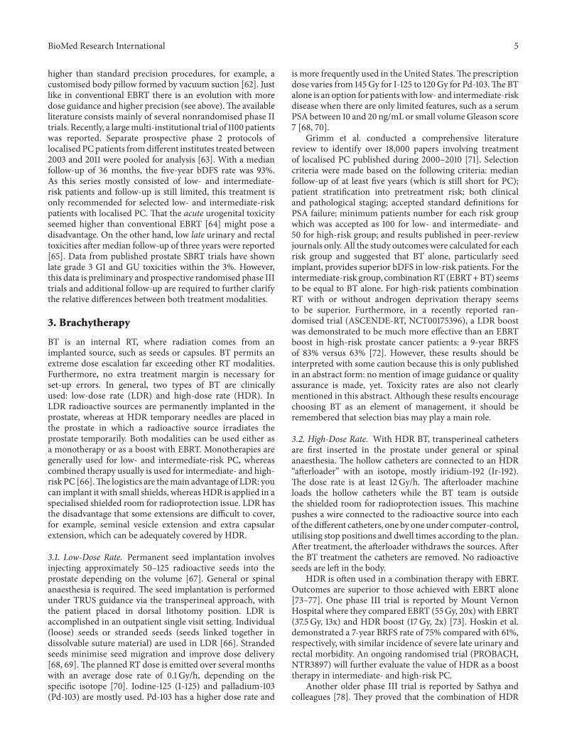
Figure 3: The Bragg peak demonstrating the plots energy loss ofionising radiation during its travel through the body. Maximumenergy deposition at the target area (tumour) without energy lossafter the target (healthy organs).
plus EBRT was superior to EBRT alone for a 5 years BRFS of71% compared with 39%. This is logic when comparing thetotal dose schedules to the prostate: the combination therapywas superior with 75 to 80Gy (comparable with nowadaysEBRT schedules) in comparison with EBRT only where thegiven dose was inferior with 66Gy and with 2 cm safetymargins.
Although the interest in monotherapy HDR is growing,no phase III trials are conducted. Several nonrandomisedseries are reported on the results of monotherapy HDR inmultiple and in single fractions, which are promising.
4. New Techniques: Proton Therapy,Carbon Ion
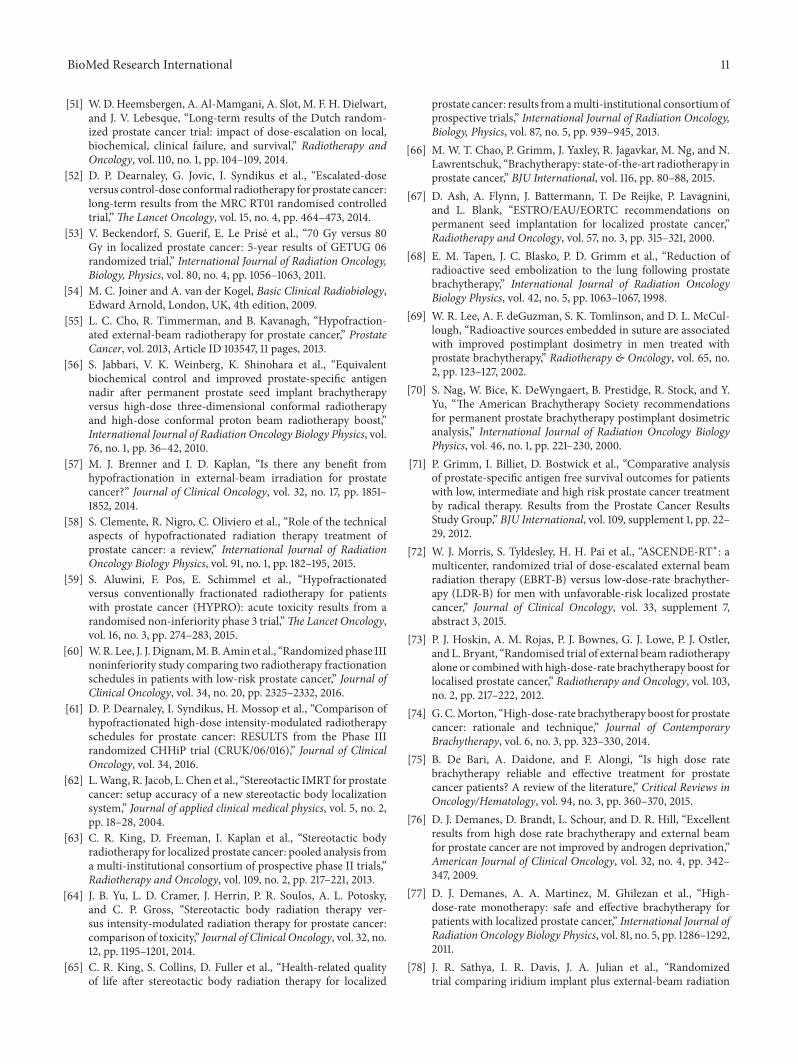
Newer RT techniques which utilise heavy particles such asprotons and carbon ions have a potential dosimetric benefitof the so-called “Bragg” peak (Figure 3). This means thatthe maximum dose delivery occurs immediately before theparticles come to rest. This means that the maximum effecton the tumour can be determined while minimising theimpact on the surrounding healthy tissue. These approachesare currently in development [79–81].
Zietman et al. published the only randomised seriescurrently available, comparing a high- to a low-proton boost,resulting in a significant increase in bDFS in the high-dosearm [8].
Carbon ions seem more efficient than protons which canbe explained by the fact that carbon ion beams are twice tothree times more effective than protons or photons [82, 83].Habl and colleagues published an HF schedule using eithercarbon ions or protons resulting in comparable acute toxic-ities [84]. Long-term outcome data on these treatments are
not yet available. However, until now, no evidence is shownto support the use of protons in preference to conventional RTfor patients with prostate cancer; neither technique had beenshown to give improved results over the others with respectto disease control or toxicity [85].
An ongoing multi-institutional phase III-randomisedtrial (PARTIQoL, NCT01617161) evaluates the value of pro-tons in low- and intermediate-risk PC in comparison withIMRT. This trial will probably shed light on the additionalvalue of protons in comparison with conventional IMRT forPC. In any event, we believe the future lies in multifactorialdecision support systems calculating for each individualpatient the outcome and the cost-effectiveness of the varioustreatments [86, 87].
5. New Devices: Balloon/Spacer
Another way to reduce toxicity is to physically create somespace between the healthy organ (rectum) and the targetedarea (prostate). As ionising radiation decreases by the inversesquare law, even a few millimetres of increased separationcan lead to sparing the healthy organ for high doses of radia-tion.
To spare rectal structures several spacer devices aredeveloped [88].These can be divided into endorectal balloonsand relatively novel rectum spacers. Endorectal balloons areplaced into the rectum for each daily treatment. Althoughthe ventral anorectal wall is pushed towards the prostate,the distance from the posterior rectal wall to the prostate isincreased with an overall effect proved to be beneficial in RT[89].
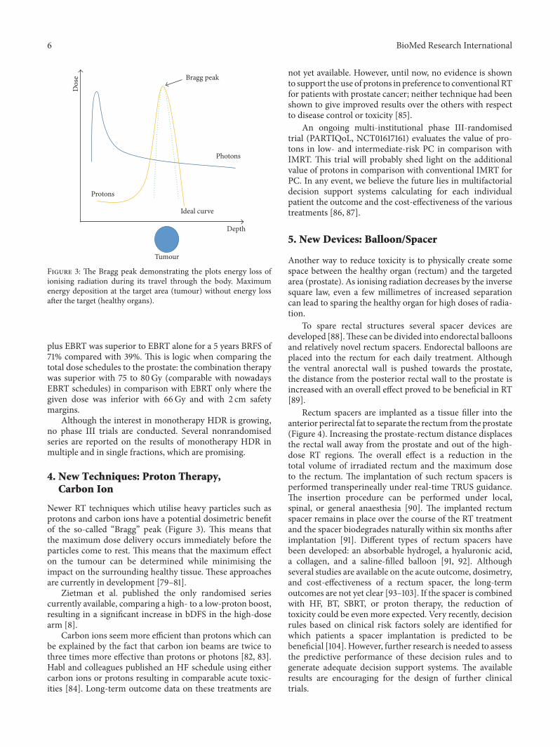
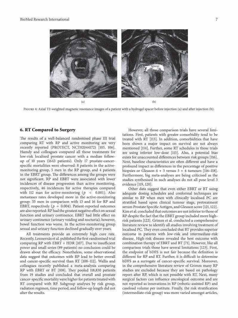
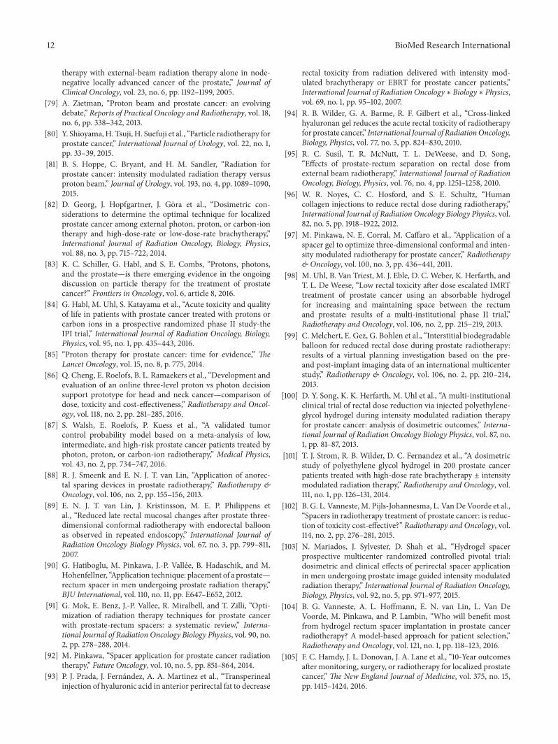
Rectum spacers are implanted as a tissue filler into theanterior perirectal fat to separate the rectum from the prostate(Figure 4). Increasing the prostate-rectum distance displacesthe rectal wall away from the prostate and out of the high-dose RT regions. The overall effect is a reduction in thetotal volume of irradiated rectum and the maximum doseto the rectum. The implantation of such rectum spacers isperformed transperineally under real-time TRUS guidance.The insertion procedure can be performed under local,spinal, or general anaesthesia [90]. The implanted rectumspacer remains in place over the course of the RT treatmentand the spacer biodegrades naturally within six months afterimplantation [91]. Different types of rectum spacers havebeen developed: an absorbable hydrogel, a hyaluronic acid,a collagen, and a saline-filled balloon [91, 92]. Althoughseveral studies are available on the acute outcome, dosimetry,and cost-effectiveness of a rectum spacer, the long-termoutcomes are not yet clear [93–103]. If the spacer is combinedwith HF, BT, SBRT, or proton therapy, the reduction oftoxicity could be even more expected. Very recently, decisionrules based on clinical risk factors solely are identified forwhich patients a spacer implantation is predicted to bebeneficial [104]. However, further research is needed to assessthe predictive performance of these decision rules and togenerate adequate decision support systems. The availableresults are encouraging for the design of further clinicaltrials.
BioMed Research International 7
Prostate
Rectum
(a)
Spacer
(b)
Figure 4: Axial T2-weighted magnetic resonance images of a patient with a hydrogel spacer before injection (a) and after injection (b).
6. RT Compared to Surgery
The results of a well-balanced randomised phase III trialcomparing RT with RP and active monitoring are veryrecently reported (PROTECT, NCT02044172) [105, 106].Hamdy and colleagues compared all those treatments forlow-risk localised prostate cancer with a median follow-up of 10 years (1643 patients). Only 17 prostate-cancer-specific mortalities were observed: 8 patients in the active-monitoring group, 5 men in the RP group, and 4 patientsin the EBRT group. The differences among the groups werenot significant. RP and EBRT were associated with lowerincidences of disease progression than active monitoring,respectively, 46 incidences for active therapies comparedwith 112 man for active-monitoring (𝑝 < 0.001). Alsometastases rates developed more in the active-monitoringgroup: 33 men in comparison with 13 and 16 for RP andEBRT, respectively (𝑝 = 0.004). Patient-reported outcomesare also reported: RP had the greatest negative effect on sexualfunction and urinary continence. EBRT had little effect onurinary continence (urinary voiding and nocturia); however,bowel function was worse. In the active-monitoring groupsexual and urinary function declined gradually over years.
All treatments provide an extremely high cure rate.Recently, Lennernas et al. published the first randomised trialcomparing RP with EBRT + HDR [107]. Due to insufficientpower and small series (89 patients) no conclusion could bedrawn about the efficacy. Nonetheless, some observationaldata suggest that outcomes with RP lead to better overalland cancer-specific survival than RT [108–112]. Wallis andcolleagues recently published a meta-analysis comparingRP with EBRT or BT [108]. They pooled 118,830 patientsfrom 19 studies and concluded that overall and prostatecancer-specificmortalitywere higher for patients treatedwithRT compared with RP. Subgroup analyses by risk group,radiation regimen, time period, and follow-up length did notalter the results.
However, all those comparison trials have several limi-tations. First, patients with greater comorbidity tend to betreated with RT [113]. In addition, comorbidities that havebeen shown a major impact on survival are not alwaysmentioned [114]. Further, some RT schedules in those trialsare using inferior low-dose [115]. Also, a potential biasexists for unaccounted differences between risk groups [116].Next, baseline characteristics are often different and have aprofound impact as differences in the percentage of positivebiopsies or Gleason 4 + 3 versus 3 + 4 tumours [116–118].Furthermore, big meta-analyses are being criticised as thestudies synthesised in such analyses do not all pose level 3evidence [119, 120].
Other data suggest that even either EBRT or BT usingadequate dosing schedules and conformal techniques aresimilar to RP when men with clinically localised PC arestratified based upon clinical tumour stage, pretreatmentserumProstate Specific Antigen, andGleason score [121, 122].Kim et al. concluded that outcomes are not inferior to those ofRP despite the fact that the EBRT group included more high-risk patients [122]. Grimm et al. conducted a comprehensiveliterature review to identify all studies involving treatment oflocalised PC.They even concluded that BT provides superioroutcome in patients with low-risk and intermediate-riskdisease. High-risk disease revealed the best outcome withcombination therapy of EBRT and BT [71]. However, like allcomparison trials those have several limitations [123]. First,the endpoint of bDFS is not fair because the definition isdifferent for RP and RT. Further, it is difficult to determinebDFS as a surrogate of cancer-specific survival. Moreover,in the comprehensive literature review of Grimm many RPstudies are excluded because they are based on pathologyreport after RP, which is not possible with RT. Next, manysurgical factors can influence oncological outcome and arenot reported as innovations in RP (robotic-assisted RP) andcaseload volume per institute. Finally, the risk stratification(intermediate-risk group) was more varied amongst articles,
8 BioMed Research International
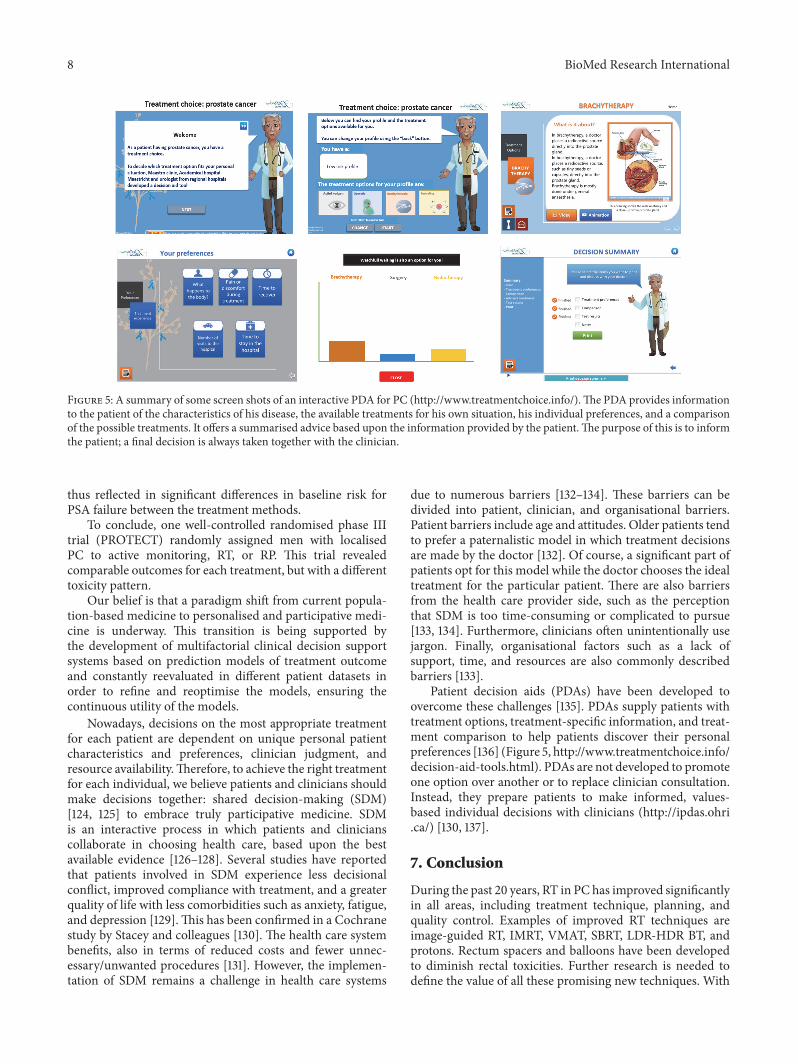
Figure 5: A summary of some screen shots of an interactive PDA for PC (http://www.treatmentchoice.info/).The PDA provides informationto the patient of the characteristics of his disease, the available treatments for his own situation, his individual preferences, and a comparisonof the possible treatments. It offers a summarised advice based upon the information provided by the patient.The purpose of this is to informthe patient; a final decision is always taken together with the clinician.
thus reflected in significant differences in baseline risk forPSA failure between the treatment methods.
To conclude, one well-controlled randomised phase IIItrial (PROTECT) randomly assigned men with localisedPC to active monitoring, RT, or RP. This trial revealedcomparable outcomes for each treatment, but with a differenttoxicity pattern.
Our belief is that a paradigm shift from current popula-tion-based medicine to personalised and participative medi-cine is underway. This transition is being supported bythe development of multifactorial clinical decision supportsystems based on prediction models of treatment outcomeand constantly reevaluated in different patient datasets inorder to refine and reoptimise the models, ensuring thecontinuous utility of the models.
Nowadays, decisions on the most appropriate treatmentfor each patient are dependent on unique personal patientcharacteristics and preferences, clinician judgment, andresource availability.Therefore, to achieve the right treatmentfor each individual, we believe patients and clinicians shouldmake decisions together: shared decision-making (SDM)[124, 125] to embrace truly participative medicine. SDMis an interactive process in which patients and clinicianscollaborate in choosing health care, based upon the bestavailable evidence [126–128]. Several studies have reportedthat patients involved in SDM experience less decisionalconflict, improved compliance with treatment, and a greaterquality of life with less comorbidities such as anxiety, fatigue,and depression [129].This has been confirmed in a Cochranestudy by Stacey and colleagues [130]. The health care systembenefits, also in terms of reduced costs and fewer unnec-essary/unwanted procedures [131]. However, the implemen-tation of SDM remains a challenge in health care systems
due to numerous barriers [132–134]. These barriers can bedivided into patient, clinician, and organisational barriers.Patient barriers include age and attitudes. Older patients tendto prefer a paternalistic model in which treatment decisionsare made by the doctor [132]. Of course, a significant part ofpatients opt for this model while the doctor chooses the idealtreatment for the particular patient. There are also barriersfrom the health care provider side, such as the perceptionthat SDM is too time-consuming or complicated to pursue[133, 134]. Furthermore, clinicians often unintentionally usejargon. Finally, organisational factors such as a lack ofsupport, time, and resources are also commonly describedbarriers [133].
Patient decision aids (PDAs) have been developed toovercome these challenges [135]. PDAs supply patients withtreatment options, treatment-specific information, and treat-ment comparison to help patients discover their personalpreferences [136] (Figure 5, http://www.treatmentchoice.info/decision-aid-tools.html). PDAs are not developed to promoteone option over another or to replace clinician consultation.Instead, they prepare patients to make informed, values-based individual decisions with clinicians (http://ipdas.ohri.ca/) [130, 137].
7. Conclusion
During the past 20 years, RT in PC has improved significantlyin all areas, including treatment technique, planning, andquality control. Examples of improved RT techniques areimage-guided RT, IMRT, VMAT, SBRT, LDR-HDR BT, andprotons. Rectum spacers and balloons have been developedto diminish rectal toxicities. Further research is needed todefine the value of all these promising new techniques. With
BioMed Research International 9
those technical implementations the long-term bDFS areimproved. We recommend dose escalation up to ≥75.6Gy(calculated as standard fractionations of 2Gy). Doses up to75.6Gy is associated with improved overall survival in menwith intermediate- and high-risk prostate cancer. HF is anattractive therapeutic option, and the randomised phase IIItrials revealed a slight increase of toxicity rates in comparisonto conventional schedules.
An important discussion issue between urologistsand radiation oncologists is summarised: the comparisonbetween RP and RT. The results of a well-balanced random-ised phase III trial comparing RT with RP and activemonitoring are very recently reported. The outcomes of RPand RT are similar, but they differ significantly in termsof the side-effects. We recommend proposing differenttreatment modalities to the individual patient characteristicsand preferences. For each individual, we recommend thatclinicians and patients should make decisions together,shared decision-making, while using patient decision aids.
Competing Interests
The authors declare that there is no conflict of interestsregarding the publication of this paper.
References
[1] J. Ferlay, E. Steliarova-Foucher, J. Lortet-Tieulent et al., “Cancerincidence and mortality patterns in Europe: estimates for 40countries in 2012,” European Journal of Cancer, vol. 49, no. 6,pp. 1374–1403, 2013.
[2] J. Ferlay, I. Soerjomataram, R. Dikshit et al., “Cancer incidenceand mortality worldwide: sources, methods and major patternsin GLOBOCAN 2012,” International Journal of Cancer, vol. 136,no. 5, pp. E359–E386, 2015.
[3] “Lifetime risk was calculated by the Statistical InformationTeam at Cancer Research UK,” 2012, http://www.cancer-researchuk.org/health-professional/cancer-statistics/statistics-by-cancer-type/prostate-cancer.
[4] A. Heidenreich, P. J. Bastian, J. Bellmunt et al., “EAU guidelineson prostate cancer. Part 1: screening, diagnosis, and local treat-ment with curative intent—update 2013,” EuropeanUrology, vol.65, no. 1, pp. 124–137, 2014.
[5] S. T. H. Peeters, W. D. Heemsbergen, P. C. M. Koper et al.,“Dose-response in radiotherapy for localized prostate cancer:results of the Dutch multicenter randomized phase III trialcomparing 68Gy of radiotherapywith 78Gy,” Journal of ClinicalOncology, vol. 24, no. 13, pp. 1990–1996, 2006.
[6] D. P. Dearnaley, M. R. Sydes, J. D. Graham et al., “Escalated-dose versus standard-dose conformal radiotherapy in prostatecancer: first results from the MRC RT01 randomised controlledtrial,”The Lancet Oncology, vol. 8, no. 6, pp. 475–487, 2007.
[7] G. A. Viani, E. J. Stefano, and S. L. Afonso, “Higher-than-conventional radiation doses in localized prostate cancer treat-ment: a meta-analysis of randomized, controlled Trials,” Inter-national Journal of Radiation Oncology Biology Physics, vol. 74,no. 5, pp. 1405–1418, 2009.
[8] A. L. Zietman, K. Bae, J. D. Slater et al., “Randomized trial com-paring conventional-dose with high-dose conformal radiation
therapy in early-stage adenocarcinoma of the prostate: long-term results fromProtonRadiationOncologyGroup/AmericanCollege of Radiology 95-09,” Journal of Clinical Oncology, vol.28, no. 7, pp. 1106–1111, 2010.
[9] M. J. Zelefsky, Z. Fuks, M. Hunt et al., “High dose radia-tion delivered by intensity modulated conformal radiotherapyimproves the outcome of localized prostate cancer,” Journal ofUrology, vol. 166, no. 3, pp. 876–881, 2001.
[10] M. M. Kim, K. E. Hoffman, L. B. Levy et al., “Improvement inprostate cancer survival over time: a 20-year analysis,” CancerJournal, vol. 18, no. 1, pp. 1–8, 2012.
[11] A. Kalbasi, J. Li, A. T. Berman et al., “Dose-escalated irradiationand overall survival inmenwith nonmetastatic prostate cancer,”JAMA Oncology, vol. 1, no. 7, pp. 897–906, 2015.
[12] A. S. Glass, J. E. Cowan, M. J. Fuldeore et al., “Patient demo-graphics, quality of life, and disease features of men with newlydiagnosed prostate cancer: trends in the PSA era,” Urology, vol.82, no. 1, pp. 60–65, 2013.
[13] V. Fonteyne, P. Ost, F. Vanpachtenbeke et al., “Rectal toxicityafter intensity modulated radiotherapy for prostate cancer:which rectal dose volume constraints should we use?” Radio-therapy and Oncology, vol. 113, no. 3, pp. 398–403, 2014.
[14] B. G. L. Vanneste, L. Van De Voorde, R. J. de Ridder, E. J. VanLimbergen, P. Lambin, and E. N. van Lin, “Chronic radiationproctitis: tricks to prevent and treat,” International Journal ofColorectal Disease, vol. 30, no. 10, pp. 1293–1303, 2015.
[15] F. W. George, C. E. Carlton Jr., R. F. Dykhuizen, and J. R.Dillon, “Cobalt-60 telecurietherapy in the definitive treatmentof carcinoma of ther prostate: a preluminary report,” Journal ofUrology, vol. 93, pp. 102–109, 1965.
[16] A. Dal Pra and L. Souhami, “Prostate cancer radiation therapy:a physician’s perspective,”PhysicaMedica, vol. 32, no. 3, pp. 438–445, 2016.
[17] S. Scobioala, C. Kittel, N. Wissmann et al., “A treatmentplanning study comparing tomotherapy, volumetric modulatedarc therapy, Sliding Window and proton therapy for low-riskprostate carcinoma,” Radiation Oncology, vol. 11, no. 1, 2016.
[18] W. Kilby, J. R. Dooley, G. Kuduvalli, S. Sayeh, and C. R. MaurerJr., “The CyberKnife� robotic radiosurgery system in 2010,”Technology in Cancer Research and Treatment, vol. 9, no. 5, pp.433–452, 2010.
[19] C. A. Perez and L.W. Brady, Chapter 51 Prostate, J. B. Lippincott& Co, Philadelphia, Pa, USA, 2nd edition, 1992.
[20] N. N. Low, S. Vijayakumar, I. Rosenberg et al., “Beam’s eye viewbased prostate treatment planning: is it useful?” InternationalJournal of Radiation Oncology, Biology, Physics, vol. 19, no. 3, pp.759–768, 1990.
[21] M.Milosevic, S. Voruganti, R. Blend et al., “Magnetic resonanceimaging (MRI) for localization of the prostatic apex: com-parison to computed tomography (CT) and urethrography,”Radiotherapy and Oncology, vol. 47, no. 3, pp. 277–284, 1998.
[22] C. Rasch, I. Barillot, P. Remeijer, A. Touw,M.VanHerk, and J. V.Lebesque, “Definition of the prostate in CT and MRI: A Multi-observer Study,” International Journal of Radiation OncologyBiology Physics, vol. 43, no. 1, pp. 57–66, 1999.
[23] P. J. Horsley, N. J. Aherne, G. V. Edwards et al., “Planningmagnetic resonance imaging for prostate cancer intensity-modulated radiation therapy: impact on target volumes, radio-therapy dose and androgen deprivation administration,” Asia-Pacific Journal of Clinical Oncology, vol. 11, no. 1, pp. 15–21, 2015.
10 BioMed Research International
[24] J. H. Chang, D. Lim Joon, B. T. Nguyen et al., “MRI scans signif-icantly change target coverage decisions in radical radiotherapyfor prostate cancer,” Journal of Medical Imaging and RadiationOncology, vol. 58, no. 2, pp. 237–243, 2014.
[25] G. M. Villeirs and G. O. De Meerleer, “Magnetic resonanceimaging (MRI) anatomy of the prostate and application of MRIin radiotherapy planning,” European Journal of Radiology, vol.63, no. 3, pp. 361–368, 2007.
[26] M. Roach III, P. Faillace-Akazawa, C. Malfatti, J. Holland,and H. Hricak, “Prostate volumes defined by magnetic reso-nance imaging and computerized tomographic scans for three-dimensional conformal radiotherapy,” International Journal ofRadiation Oncology Biology Physics, vol. 35, no. 5, pp. 1011–1018,1996.
[27] M. Debois, R. Oyen, F. Maes et al., “The contribution of mag-netic resonance imaging to the three-dimensional treatmentplanning of localized prostate cancer,” International Journal ofRadiation Oncology Biology Physics, vol. 45, no. 4, pp. 857–865,1999.
[28] G. L. Sannazzari, R. Ragona, M. G. Ruo Redda, F. R. Giglioli, G.Isolato, and A. Guarneri, “CT-MRI image fusion for delineationof volumes in three-dimensional conformal radiation therapyin the treatment of localized prostate cancer,” British Journal ofRadiology, vol. 75, no. 895, pp. 603–607, 2002.
[29] W. L. Smith, C. Lewis, G. Bauman et al., “Prostate volumecontouring: a 3D analysis of segmentation using 3DTRUS, CT,and MR,” International Journal of Radiation Oncology BiologyPhysics, vol. 67, no. 4, pp. 1238–1247, 2007.
[30] B. Hentschel, W. Oehler, D. Strauß, A. Ulrich, and A. Malich,“Definition of the CTV prostate in CT and MRI by usingCT-MRI image fusion in IMRT planning for prostate cancer,”Strahlentherapie und Onkologie, vol. 187, no. 3, pp. 183–190, 2011.
[31] L. Sander, N. C. Langkilde, M. Holmberg, and J. Carl, “MRItarget delineation may reduce long-term toxicity after prostateradiotherapy,” Acta Oncologica, vol. 53, no. 6, pp. 809–814, 2014.
[32] T. Seppala, H. Visapaa, J. Collan et al., “Converting from CT-to MRI-only-based target definition in radiotherapy of local-ized prostate cancer: a comparison between two modalities,”Strahlentherapie und Onkologie, vol. 191, no. 11, pp. 862–868,2015.
[33] G. M. Villeirs, K. Van Vaerenbergh, L. Vakaet et al., “Interob-server delineation variation using CT versus combined CT +MRI in intensity-modulated radiotherapy for prostate cancer,”Strahlentherapie und Onkologie, vol. 181, no. 7, pp. 424–430,2005.
[34] J. Liang, Q.Wu, and D. Yan, “The role of seminal vesicle motionin target margin assessment for online image-guided radio-therapy for prostate cancer,” International Journal of RadiationOncology Biology Physics, vol. 73, no. 3, pp. 935–943, 2009.
[35] E.-J. Rijkhorst, A. Lakeman, J. Nijkamp et al., “Strategies foronline organ motion correction for intensity-modulated radio-therapy of prostate cancer: prostate, rectum, and bladder doseeffects,” International Journal of Radiation Oncology BiologyPhysics, vol. 75, no. 4, pp. 1254–1260, 2009.
[36] J. F. Langenhuijsen, E. N. J. T. van Lin, L. A. Kiemeney et al.,“Ultrasound-guided transrectal implantation of gold markersfor prostate localization during external beam radiotherapy:complication rate and risk factors,” International Journal ofRadiation Oncology Biology Physics, vol. 69, no. 3, pp. 671–676,2007.
[37] Z. S. Fawaz, M. Yassa, D. H. Nguyen, and P. Vavassis, “Fiducialmarker implantation in prostate radiation therapy: complica-tion rates and technique,” Cancer/Radiotherapie, vol. 18, no. 8,pp. 736–739, 2014.
[38] J. Sveistrup, P. M. af Rosenschold, J. O. Deasy et al., “Improve-ment in toxicity in high risk prostate cancer patients treatedwith image-guided intensity-modulated radiotherapy com-pared to 3D conformal radiotherapy without daily imageguidance,” Radiation Oncology, vol. 9, no. article 44, 2014.
[39] R. D. Foster, T. D. Solberg, H. S. Li et al., “Comparison oftransabdominal ultrasound and electromagnetic transpondersfor prostate localization,” Journal of Applied Clinical MedicalPhysics, vol. 11, no. 1, p. 2924, 2010.
[40] M. Oldham, D. Letourneau, L. Watt et al., “Cone-beam-CTguided radiation therapy: a model for on-line application,”Radiotherapy & Oncology, vol. 75, no. 3, pp. 271.e1–271.e8, 2005.
[41] B. W. Raaymakers, J. J. W. Lagendijk, J. Overweg et al.,“Integrating a 1.5 TMRI scanner with a 6MV accelerator: proofof concept,” Physics in Medicine and Biology, vol. 54, no. 12, pp.N229–N237, 2009.
[42] A. Y. C. Fung, K. M. Ayyangar, D. Djajaputra, R. M. Nehru,and C. A. Enke, “Ultrasound-based guidance of intensity-modulated radiation therapy,”Medical Dosimetry, vol. 31, no. 1,pp. 20–29, 2006.
[43] V. Carillo, C. Cozzarini, T. Rancati et al., “Relationships betweenbladder dose-volume/surface histograms and acute urinarytoxicity after radiotherapy for prostate cancer,” Radiotherapyand Oncology, vol. 111, no. 1, pp. 100–105, 2014.
[44] E. A. Mellon, K. Javedan, T. J. Strom et al., “A dosimetriccomparison of volumetric modulated arc therapy with step-and-shoot intensity modulated radiation therapy for prostatecancer,” Practical Radiation Oncology, vol. 5, no. 1, pp. 11–15,2015.
[45] D. Palma, E. Vollans, K. James et al., “Volumetric modulatedarc therapy for delivery of prostate radiotherapy: comparisonwith intensity-modulated radiotherapy and three-dimensionalconformal radiotherapy,” International Journal of RadiationOncology Biology Physics, vol. 72, no. 4, pp. 996–1001, 2008.
[46] M. J. Zelefsky, E. J. Levin, M. Hunt et al., “Incidence of laterectal and urinary toxicities after three-dimensional confor-mal radiotherapy and intensity-modulated radiotherapy forlocalized prostate cancer,” International Journal of RadiationOncology Biology Physics, vol. 70, no. 4, pp. 1124–1129, 2008.
[47] A. Pollack, G. K. Zagars, G. Starkschall et al., “Prostate cancerradiation dose response: results of theM. D. Anderson phase IIIrandomized trial,” International Journal of Radiation OncologyBiology Physics, vol. 53, no. 5, pp. 1097–1105, 2002.
[48] D. A. Kuban, S. L. Tucker, L. Dong et al., “Long-term resultsof the M. D. anderson randomized dose-escalation trial forprostate cancer,” International Journal of Radiation OncologyBiology Physics, vol. 70, no. 1, pp. 67–74, 2008.
[49] J. M. Michalski, J. Moughan, and J. Purdy, “A randomized trialof 79.2Gy versus 70.2Gy radiation therapy (RT) for localizedprostate cancer,” Journal of Clinical Oncology, vol. 33, supple-ment 7, abstract 4, 2015.
[50] A. Al-Mamgani, W. L. J. van Putten, W. D. Heemsbergen et al.,“Update of Dutch multicenter dose-escalation trial of radio-therapy for localized prostate cancer,” International Journal ofRadiation Oncology Biology Physics, vol. 72, no. 4, pp. 980–988,2008.
BioMed Research International 11
[51] W. D. Heemsbergen, A. Al-Mamgani, A. Slot, M. F. H. Dielwart,and J. V. Lebesque, “Long-term results of the Dutch random-ized prostate cancer trial: impact of dose-escalation on local,biochemical, clinical failure, and survival,” Radiotherapy andOncology, vol. 110, no. 1, pp. 104–109, 2014.
[52] D. P. Dearnaley, G. Jovic, I. Syndikus et al., “Escalated-doseversus control-dose conformal radiotherapy for prostate cancer:long-term results from the MRC RT01 randomised controlledtrial,”The Lancet Oncology, vol. 15, no. 4, pp. 464–473, 2014.
[53] V. Beckendorf, S. Guerif, E. Le Prise et al., “70 Gy versus 80Gy in localized prostate cancer: 5-year results of GETUG 06randomized trial,” International Journal of Radiation Oncology,Biology, Physics, vol. 80, no. 4, pp. 1056–1063, 2011.
[54] M. C. Joiner and A. van der Kogel, Basic Clinical Radiobiology,Edward Arnold, London, UK, 4th edition, 2009.
[55] L. C. Cho, R. Timmerman, and B. Kavanagh, “Hypofraction-ated external-beam radiotherapy for prostate cancer,” ProstateCancer, vol. 2013, Article ID 103547, 11 pages, 2013.
[56] S. Jabbari, V. K. Weinberg, K. Shinohara et al., “Equivalentbiochemical control and improved prostate-specific antigennadir after permanent prostate seed implant brachytherapyversus high-dose three-dimensional conformal radiotherapyand high-dose conformal proton beam radiotherapy boost,”International Journal of Radiation Oncology Biology Physics, vol.76, no. 1, pp. 36–42, 2010.
[57] M. J. Brenner and I. D. Kaplan, “Is there any benefit fromhypofractionation in external-beam irradiation for prostatecancer?” Journal of Clinical Oncology, vol. 32, no. 17, pp. 1851–1852, 2014.
[58] S. Clemente, R. Nigro, C. Oliviero et al., “Role of the technicalaspects of hypofractionated radiation therapy treatment ofprostate cancer: a review,” International Journal of RadiationOncology Biology Physics, vol. 91, no. 1, pp. 182–195, 2015.
[59] S. Aluwini, F. Pos, E. Schimmel et al., “Hypofractionatedversus conventionally fractionated radiotherapy for patientswith prostate cancer (HYPRO): acute toxicity results from arandomised non-inferiority phase 3 trial,”The Lancet Oncology,vol. 16, no. 3, pp. 274–283, 2015.
[60] W.R. Lee, J. J. Dignam,M. B.Amin et al., “Randomized phase IIInoninferiority study comparing two radiotherapy fractionationschedules in patients with low-risk prostate cancer,” Journal ofClinical Oncology, vol. 34, no. 20, pp. 2325–2332, 2016.
[61] D. P. Dearnaley, I. Syndikus, H. Mossop et al., “Comparison ofhypofractionated high-dose intensity-modulated radiotherapyschedules for prostate cancer: RESULTS from the Phase IIIrandomized CHHiP trial (CRUK/06/016),” Journal of ClinicalOncology, vol. 34, 2016.
[62] L.Wang, R. Jacob, L. Chen et al., “Stereotactic IMRT for prostatecancer: setup accuracy of a new stereotactic body localizationsystem,” Journal of applied clinical medical physics, vol. 5, no. 2,pp. 18–28, 2004.
[63] C. R. King, D. Freeman, I. Kaplan et al., “Stereotactic bodyradiotherapy for localized prostate cancer: pooled analysis froma multi-institutional consortium of prospective phase II trials,”Radiotherapy and Oncology, vol. 109, no. 2, pp. 217–221, 2013.
[64] J. B. Yu, L. D. Cramer, J. Herrin, P. R. Soulos, A. L. Potosky,and C. P. Gross, “Stereotactic body radiation therapy ver-sus intensity-modulated radiation therapy for prostate cancer:comparison of toxicity,” Journal of Clinical Oncology, vol. 32, no.12, pp. 1195–1201, 2014.
[65] C. R. King, S. Collins, D. Fuller et al., “Health-related qualityof life after stereotactic body radiation therapy for localized
prostate cancer: results from amulti-institutional consortiumofprospective trials,” International Journal of Radiation Oncology,Biology, Physics, vol. 87, no. 5, pp. 939–945, 2013.
[66] M. W. T. Chao, P. Grimm, J. Yaxley, R. Jagavkar, M. Ng, and N.Lawrentschuk, “Brachytherapy: state-of-the-art radiotherapy inprostate cancer,” BJU International, vol. 116, pp. 80–88, 2015.
[67] D. Ash, A. Flynn, J. Battermann, T. De Reijke, P. Lavagnini,and L. Blank, “ESTRO/EAU/EORTC recommendations onpermanent seed implantation for localized prostate cancer,”Radiotherapy and Oncology, vol. 57, no. 3, pp. 315–321, 2000.
[68] E. M. Tapen, J. C. Blasko, P. D. Grimm et al., “Reduction ofradioactive seed embolization to the lung following prostatebrachytherapy,” International Journal of Radiation OncologyBiology Physics, vol. 42, no. 5, pp. 1063–1067, 1998.
[69] W. R. Lee, A. F. deGuzman, S. K. Tomlinson, and D. L. McCul-lough, “Radioactive sources embedded in suture are associatedwith improved postimplant dosimetry in men treated withprostate brachytherapy,” Radiotherapy & Oncology, vol. 65, no.2, pp. 123–127, 2002.
[70] S. Nag, W. Bice, K. DeWyngaert, B. Prestidge, R. Stock, and Y.Yu, “The American Brachytherapy Society recommendationsfor permanent prostate brachytherapy postimplant dosimetricanalysis,” International Journal of Radiation Oncology BiologyPhysics, vol. 46, no. 1, pp. 221–230, 2000.
[71] P. Grimm, I. Billiet, D. Bostwick et al., “Comparative analysisof prostate-specific antigen free survival outcomes for patientswith low, intermediate and high risk prostate cancer treatmentby radical therapy. Results from the Prostate Cancer ResultsStudy Group,” BJU International, vol. 109, supplement 1, pp. 22–29, 2012.
[72] W. J. Morris, S. Tyldesley, H. H. Pai et al., “ASCENDE-RT∗: amulticenter, randomized trial of dose-escalated external beamradiation therapy (EBRT-B) versus low-dose-rate brachyther-apy (LDR-B) for men with unfavorable-risk localized prostatecancer,” Journal of Clinical Oncology, vol. 33, supplement 7,abstract 3, 2015.
[73] P. J. Hoskin, A. M. Rojas, P. J. Bownes, G. J. Lowe, P. J. Ostler,and L. Bryant, “Randomised trial of external beam radiotherapyalone or combinedwith high-dose-rate brachytherapy boost forlocalised prostate cancer,” Radiotherapy and Oncology, vol. 103,no. 2, pp. 217–222, 2012.
[74] G.C.Morton, “High-dose-rate brachytherapy boost for prostatecancer: rationale and technique,” Journal of ContemporaryBrachytherapy, vol. 6, no. 3, pp. 323–330, 2014.
[75] B. De Bari, A. Daidone, and F. Alongi, “Is high dose ratebrachytherapy reliable and effective treatment for prostatecancer patients? A review of the literature,” Critical Reviews inOncology/Hematology, vol. 94, no. 3, pp. 360–370, 2015.
[76] D. J. Demanes, D. Brandt, L. Schour, and D. R. Hill, “Excellentresults from high dose rate brachytherapy and external beamfor prostate cancer are not improved by androgen deprivation,”American Journal of Clinical Oncology, vol. 32, no. 4, pp. 342–347, 2009.
[77] D. J. Demanes, A. A. Martinez, M. Ghilezan et al., “High-dose-rate monotherapy: safe and effective brachytherapy forpatients with localized prostate cancer,” International Journal ofRadiation Oncology Biology Physics, vol. 81, no. 5, pp. 1286–1292,2011.
[78] J. R. Sathya, I. R. Davis, J. A. Julian et al., “Randomizedtrial comparing iridium implant plus external-beam radiation
12 BioMed Research International
therapy with external-beam radiation therapy alone in node-negative locally advanced cancer of the prostate,” Journal ofClinical Oncology, vol. 23, no. 6, pp. 1192–1199, 2005.
[79] A. Zietman, “Proton beam and prostate cancer: an evolvingdebate,” Reports of Practical Oncology and Radiotherapy, vol. 18,no. 6, pp. 338–342, 2013.
[80] Y. Shioyama,H. Tsuji, H. Suefuji et al., “Particle radiotherapy forprostate cancer,” International Journal of Urology, vol. 22, no. 1,pp. 33–39, 2015.
[81] B. S. Hoppe, C. Bryant, and H. M. Sandler, “Radiation forprostate cancer: intensity modulated radiation therapy versusproton beam,” Journal of Urology, vol. 193, no. 4, pp. 1089–1090,2015.
[82] D. Georg, J. Hopfgartner, J. Gora et al., “Dosimetric con-siderations to determine the optimal technique for localizedprostate cancer among external photon, proton, or carbon-iontherapy and high-dose-rate or low-dose-rate brachytherapy,”International Journal of Radiation Oncology, Biology, Physics,vol. 88, no. 3, pp. 715–722, 2014.
[83] K. C. Schiller, G. Habl, and S. E. Combs, “Protons, photons,and the prostate—is there emerging evidence in the ongoingdiscussion on particle therapy for the treatment of prostatecancer?” Frontiers in Oncology, vol. 6, article 8, 2016.
[84] G. Habl, M. Uhl, S. Katayama et al., “Acute toxicity and qualityof life in patients with prostate cancer treated with protons orcarbon ions in a prospective randomized phase II study-theIPI trial,” International Journal of Radiation Oncology, Biology,Physics, vol. 95, no. 1, pp. 435–443, 2016.
[85] “Proton therapy for prostate cancer: time for evidence,” TheLancet Oncology, vol. 15, no. 8, p. 775, 2014.
[86] Q. Cheng, E. Roelofs, B. L. Ramaekers et al., “Development andevaluation of an online three-level proton vs photon decisionsupport prototype for head and neck cancer—comparison ofdose, toxicity and cost-effectiveness,” Radiotherapy and Oncol-ogy, vol. 118, no. 2, pp. 281–285, 2016.
[87] S. Walsh, E. Roelofs, P. Kuess et al., “A validated tumorcontrol probability model based on a meta-analysis of low,intermediate, and high-risk prostate cancer patients treated byphoton, proton, or carbon-ion radiotherapy,” Medical Physics,vol. 43, no. 2, pp. 734–747, 2016.
[88] R. J. Smeenk and E. N. J. T. van Lin, “Application of anorec-tal sparing devices in prostate radiotherapy,” Radiotherapy &Oncology, vol. 106, no. 2, pp. 155–156, 2013.
[89] E. N. J. T. van Lin, J. Kristinsson, M. E. P. Philippens etal., “Reduced late rectal mucosal changes after prostate three-dimensional conformal radiotherapy with endorectal balloonas observed in repeated endoscopy,” International Journal ofRadiation Oncology Biology Physics, vol. 67, no. 3, pp. 799–811,2007.
[90] G. Hatiboglu, M. Pinkawa, J.-P. Vallee, B. Hadaschik, and M.Hohenfellner, “Application technique: placement of a prostate—rectum spacer in men undergoing prostate radiation therapy,”BJU International, vol. 110, no. 11, pp. E647–E652, 2012.
[91] G. Mok, E. Benz, J.-P. Vallee, R. Miralbell, and T. Zilli, “Opti-mization of radiation therapy techniques for prostate cancerwith prostate-rectum spacers: a systematic review,” Interna-tional Journal of Radiation Oncology Biology Physics, vol. 90, no.2, pp. 278–288, 2014.
[92] M. Pinkawa, “Spacer application for prostate cancer radiationtherapy,” Future Oncology, vol. 10, no. 5, pp. 851–864, 2014.
[93] P. J. Prada, J. Fernandez, A. A. Martinez et al., “Transperinealinjection of hyaluronic acid in anterior perirectal fat to decrease
rectal toxicity from radiation delivered with intensity mod-ulated brachytherapy or EBRT for prostate cancer patients,”International Journal of Radiation Oncology ∗ Biology ∗ Physics,vol. 69, no. 1, pp. 95–102, 2007.
[94] R. B. Wilder, G. A. Barme, R. F. Gilbert et al., “Cross-linkedhyaluronan gel reduces the acute rectal toxicity of radiotherapyfor prostate cancer,” International Journal of RadiationOncology,Biology, Physics, vol. 77, no. 3, pp. 824–830, 2010.
[95] R. C. Susil, T. R. McNutt, T. L. DeWeese, and D. Song,“Effects of prostate-rectum separation on rectal dose fromexternal beam radiotherapy,” International Journal of RadiationOncology, Biology, Physics, vol. 76, no. 4, pp. 1251–1258, 2010.
[96] W. R. Noyes, C. C. Hosford, and S. E. Schultz, “Humancollagen injections to reduce rectal dose during radiotherapy,”International Journal of Radiation Oncology Biology Physics, vol.82, no. 5, pp. 1918–1922, 2012.
[97] M. Pinkawa, N. E. Corral, M. Caffaro et al., “Application of aspacer gel to optimize three-dimensional conformal and inten-sity modulated radiotherapy for prostate cancer,” Radiotherapy& Oncology, vol. 100, no. 3, pp. 436–441, 2011.
[98] M. Uhl, B. Van Triest, M. J. Eble, D. C. Weber, K. Herfarth, andT. L. De Weese, “Low rectal toxicity after dose escalated IMRTtreatment of prostate cancer using an absorbable hydrogelfor increasing and maintaining space between the rectumand prostate: results of a multi-institutional phase II trial,”Radiotherapy and Oncology, vol. 106, no. 2, pp. 215–219, 2013.
[99] C. Melchert, E. Gez, G. Bohlen et al., “Interstitial biodegradableballoon for reduced rectal dose during prostate radiotherapy:results of a virtual planning investigation based on the pre-and post-implant imaging data of an international multicenterstudy,” Radiotherapy & Oncology, vol. 106, no. 2, pp. 210–214,2013.
[100] D. Y. Song, K. K. Herfarth, M. Uhl et al., “A multi-institutionalclinical trial of rectal dose reduction via injected polyethylene-glycol hydrogel during intensity modulated radiation therapyfor prostate cancer: analysis of dosimetric outcomes,” Interna-tional Journal of Radiation Oncology Biology Physics, vol. 87, no.1, pp. 81–87, 2013.
[101] T. J. Strom, R. B. Wilder, D. C. Fernandez et al., “A dosimetricstudy of polyethylene glycol hydrogel in 200 prostate cancerpatients treated with high-dose rate brachytherapy ± intensitymodulated radiation therapy,” Radiotherapy and Oncology, vol.111, no. 1, pp. 126–131, 2014.
[102] B. G. L. Vanneste, M. Pijls-Johannesma, L. VanDeVoorde et al.,“Spacers in radiotherapy treatment of prostate cancer: is reduc-tion of toxicity cost-effective?” Radiotherapy and Oncology, vol.114, no. 2, pp. 276–281, 2015.
[103] N. Mariados, J. Sylvester, D. Shah et al., “Hydrogel spacerprospective multicenter randomized controlled pivotal trial:dosimetric and clinical effects of perirectal spacer applicationin men undergoing prostate image guided intensity modulatedradiation therapy,” International Journal of Radiation Oncology,Biology, Physics, vol. 92, no. 5, pp. 971–977, 2015.
[104] B. G. Vanneste, A. L. Hoffmann, E. N. van Lin, L. Van DeVoorde, M. Pinkawa, and P. Lambin, “Who will benefit mostfrom hydrogel rectum spacer implantation in prostate cancerradiotherapy? A model-based approach for patient selection,”Radiotherapy and Oncology, vol. 121, no. 1, pp. 118–123, 2016.
[105] F. C. Hamdy, J. L. Donovan, J. A. Lane et al., “10-Year outcomesafter monitoring, surgery, or radiotherapy for localized prostatecancer,” The New England Journal of Medicine, vol. 375, no. 15,pp. 1415–1424, 2016.

BioMed Research International 13
[106] J. L. Donovan, F. C. Hamdy, J. A. Lane et al., “Patient-reported outcomes after monitoring, surgery, or radiotherapyfor prostate cancer,” New England Journal of Medicine, vol. 375,no. 15, pp. 1425–1437, 2016.
[107] B. Lennernas, K. Majumder, J.-E. Damber et al., “Radicalprostatectomy versus high-dose irradiation in localized/locallyadvanced prostate cancer: a swedish multicenter randomizedtrial with patient-reported outcomes,” Acta Oncologica, vol. 54,no. 6, pp. 875–881, 2015.
[108] C. J. D. Wallis, R. Saskin, R. Choo et al., “Surgery versus radio-therapy for clinically-localized prostate cancer: a systematicreview and meta-analysis,” European Urology, vol. 70, no. 1, pp.21–30, 2016.
[109] P. Sooriakumaran, T. Nyberg, O. Akre et al., “Comparativeeffectiveness of radical prostatectomy and radiotherapy inprostate cancer: observational study of mortality outcomes,”British Medical Journal, vol. 348, Article ID g1502, 2014.
[110] F. Petrelli, I. Vavassori, A. Coinu, K. Borgonovo, E. Sarti, andS. Barni, “Radical prostatectomy or radiotherapy in high-riskprostate cancer: a systematic review and metaanalysis,” ClinicalGenitourinary Cancer, vol. 12, no. 4, pp. 215–224, 2014.
[111] K. Akakura, H. Suzuki, T. Ichikawa et al., “A randomized trialcomparing radical prostatectomy plus endocrine therapy versusexternal beam radiotherapy plus endocrine therapy for locallyadvanced prostate cancer: results at median follow-up of 102months,” Japanese Journal of Clinical Oncology, vol. 36, no. 12,pp. 789–793, 2006.
[112] J. Y. Lee, K. S. Cho, J. K. Kwon et al., “A competing risk analysisof cancer-specific mortality of initial treatment with radicalprostatectomy versus radiation therapy in clinically localizedhigh-risk prostate cancer,” Annals of Surgical Oncology, vol. 21,no. 12, pp. 4026–4033, 2014.
[113] S. Houterman, M. L. G. Janssen-Heijnen, A. J. M. Hendrikx,H. A. Van Den Berg, and J. W. W. Coebergh, “Impact ofcomorbidity on treatment and prognosis of prostate cancerpatients: a population-based study,” Critical Reviews in Oncol-ogy/Hematology, vol. 58, no. 1, pp. 60–67, 2006.
[114] M. Roach III and K. Thomas, “Overview of randomized con-trolled treatment trials for clinically localized prostate cancer:implications for active surveillance and the United Statespreventative task force report on screening?” Journal of theNational Cancer Institute.Monographs, vol. 2012, no. 45, pp. 221–229, 2012.
[115] A. Rane, “Surgery or radiotherapy for prostate cancer?” BritishMedical Journal, vol. 348, Article ID g1580, 2014.
[116] M. Roach III, “Radical prostatectomy v radiation: only a ran-domised trial can provide the answer,” British Medical Journal,vol. 348, Article ID g2266, 2014.
[117] S. H. Giordano, Y.-F. Kuo, Z. Duan, G. N. Hortobagyi, J.Freeman, and J. S. Goodwin, “Limits of observational data indetermining outcomes from cancer therapy,” Cancer, vol. 112,no. 11, pp. 2456–2466, 2008.
[118] J. B. Eifler, E. B. Humphreys, M. Agro, A. W. Partin, B. J. Trock,and M. Han, “Causes of death after radical prostatectomy at alarge tertiary center,”The Journal of Urology, vol. 188, no. 3, pp.798–801, 2012.
[119] C. J. Wallis, R. Saskin, R. Choo et al., “Surgery versus radiother-apy for clinically-localized prostate cancer: a systematic reviewand meta-analysis,” European Urology, vol. 70, no. 1, pp. 21–30,2016, European Urology, vol. 70, no. 1, pp. e15-6, 2016.
[120] C. J. Wallis, R. Saskin, R. Choo et al., “Surgery versus radiother-apy for clinically-localized prostate cancer: a systematic review
and meta-analysis,” European Urology, vol. 70, no. 1, pp. 21–30,2016.
[121] R. F. Wolff, S. Ryder, A. Bossi et al., “A systematic reviewof randomised controlled trials of radiotherapy for localisedprostate cancer,” European Journal of Cancer, vol. 51, no. 16, pp.2345–2367, 2015.
[122] Y.-J. Kim, K. H. Cho, H. R. Pyo et al., “Radical prostatectomyversus external beam radiotherapy for localized prostate can-cer: comparison of treatment outcomes,” Strahlentherapie undOnkologie, vol. 191, no. 4, pp. 321–329, 2015.
[123] P. Sooriakumaran, M. Spahn, and P. Wiklund, “Apples andoranges: comparison of treatment methods for prostate cancerusing biochemical recurrence as an endpoint,” BJU Interna-tional, vol. 110, no. 4, pp. 477–478, 2012.
[124] P. Lambin, J. Zindler, B. G. Vanneste et al., “Decision supportsystems for personalized and participative radiation oncology,”Advanced Drug Delivery Reviews, 2016.
[125] P. Lambin, J. Zindler, B. Vanneste et al., “Modern clinicalresearch: how rapid learning health care and cohort multiplerandomised clinical trials complement traditional evidencebased medicine,” Acta Oncologica, vol. 54, no. 9, pp. 1289–1300,2015.
[126] F. Legare, D. Stacey, N. Briere et al., “A conceptual frameworkfor interprofessional shared decision making in home care:protocol for a feasibility study,” BMC Health Services Research,vol. 11, article no. 23, 2011.
[127] G. Elwyn, A. O’Connor, D. Stacey et al., “International PatientDecision Aids Standards (IPDAS) Collaboration. Developinga quality criteria framework for patient decision aids: onlineinternational Delphi consensus process,” British Medical Jour-nal, vol. 333, no. 7565, p. 417, 2006.
[128] G. Elwyn, S. Laitner, A. Coulter, E. Walker, P. Watson, and R.Thomson, “Implementing shared decision making in the NHS,”BMJ, vol. 341, Article ID c5146, 2010.
[129] J. D. Tariman,D. L. Berry, B. Cochrane, A.Doorenbos, andA.D.Schepp, “Preferred and actual participation roles during healthcare decision making in persons with cancer: a systematicreview,” Annals of Oncology, vol. 21, no. 6, pp. 1145–1151, 2009.
[130] D. Stacey, F. Legare, N. F. Col et al., “Decision aids for peoplefacing health treatment or screening decisions,” The CochraneDatabase of Systematic Reviews, no. 1, Article ID CD001431,2014.
[131] E.O. Lee andE. J. Emanuel, “Shared decisionmaking to improvecare and reduce costs,” The New England Journal of Medicine,vol. 368, no. 1, pp. 6–8, 2013.
[132] H. L. Kane, M. T. Halpern, L. B. Squiers, K. A. Treiman,and L. A. McCormack, “Implementing and evaluating shareddecision making in oncology practice,” CA: A Cancer Journalfor Clinicians, vol. 64, no. 6, pp. 377–388, 2014.
[133] F. Legare, S. Ratte, K. Gravel, and I. D. Graham, “Barriers andfacilitators to implementing shared decision-making in clinicalpractice: update of a systematic review of health professionals’perceptions,” Patient Education and Counseling, vol. 73, no. 3,pp. 526–535, 2008.
[134] K. Gravel, F. Legare, and I. D. Graham, “Barriers and facilitatorsto implementing shared decision-making in clinical practice: asystematic review of health professionals’ perceptions,” Imple-mentation Science, vol. 1, article 16, 2006.
[135] A. M. O’Connor, A. Rostom, V. Fiset et al., “Decision aidsfor patients facing health treatment or screening decisions:systematic review,” BritishMedical Journal, vol. 319, no. 7212, pp.731–734, 1999.

14 BioMed Research International
[136] J. D. Harrison, L. Masya, P. Butow et al., “Implementing patientdecision support tools: moving beyond academia?” PatientEducation and Counseling, vol. 76, no. 1, pp. 120–125, 2009.
[137] D. Stacey, F. Legare, A. Lyddiatt et al., “Translating evidence tofacilitate shared decision making: development and usability ofa consult decision aid prototype,” The Patient, vol. 9, no. 6, pp.571–582, 2016.

Submit your manuscripts athttp://www.hindawi.com
Stem CellsInternational
Hindawi Publishing Corporationhttp://www.hindawi.com Volume 2014
Hindawi Publishing Corporationhttp://www.hindawi.com Volume 2014
MEDIATORSINFLAMMATION
of
Hindawi Publishing Corporationhttp://www.hindawi.com Volume 2014
Behavioural Neurology
EndocrinologyInternational Journal of
Hindawi Publishing Corporationhttp://www.hindawi.com Volume 2014
Hindawi Publishing Corporationhttp://www.hindawi.com Volume 2014
Disease Markers
Hindawi Publishing Corporationhttp://www.hindawi.com Volume 2014
BioMed Research International
OncologyJournal of
Hindawi Publishing Corporationhttp://www.hindawi.com Volume 2014
Hindawi Publishing Corporationhttp://www.hindawi.com Volume 2014
Oxidative Medicine and Cellular Longevity
Hindawi Publishing Corporationhttp://www.hindawi.com Volume 2014
PPAR Research
The Scientific World JournalHindawi Publishing Corporation http://www.hindawi.com Volume 2014
Immunology ResearchHindawi Publishing Corporationhttp://www.hindawi.com Volume 2014
Journal of
ObesityJournal of
Hindawi Publishing Corporationhttp://www.hindawi.com Volume 2014
Hindawi Publishing Corporationhttp://www.hindawi.com Volume 2014
Computational and Mathematical Methods in Medicine
OphthalmologyJournal of
Hindawi Publishing Corporationhttp://www.hindawi.com Volume 2014
Diabetes ResearchJournal of
Hindawi Publishing Corporationhttp://www.hindawi.com Volume 2014
Hindawi Publishing Corporationhttp://www.hindawi.com Volume 2014
Research and TreatmentAIDS
Hindawi Publishing Corporationhttp://www.hindawi.com Volume 2014
Gastroenterology Research and Practice
Hindawi Publishing Corporationhttp://www.hindawi.com Volume 2014
Parkinson’s Disease
Evidence-Based Complementary and Alternative Medicine
Volume 2014Hindawi Publishing Corporationhttp://www.hindawi.com