Embed Size (px)
Citation preview
APRIL 2014RCCLREVIEW OF CORNEA & CONTACT LENSES
Supplement to
Can these specialty lenses go
mainstream? Yes, experts say.
Here’s how.
Unlock Your Scleral Potential
Earn 1 CE credit — Treating the Tear Film: Nutrition is Essential
10 Dos and Don’ts of
Scleral Contact Lens
fitting techniques
Simplify Your Coding
for specialty lenses
SPECIAL ISSUE:
fitting new patients
in Scleral Lenses
Scleral Lens Design 101
Landing zone Lessons
001_rcl0414_cover.indd 1 4/4/14 10:00 AM

BLINK-ACTIVATEDMOISTURIZING AGENTS
MOISTURIZING AGENTSPVA, PEG and HPMC help deliver comfort from insertion to the end of the day.Visit myalcon.com
THIS IS WHY patients canenjoy refreshing comfort with every blink—no matter what the day may bring.
DAILIES® AquaComfort Plus® contact lenses are the only daily disposable contact lenses with blink-activated moisture,* which helps result in a stable tear film with refreshing comfort throughout the day—every day.1
PERFORMANCE DRIVEN BY SCIENCE™
*Based on DAILIES® AquaComfort Plus® sphere contact lenses.
Reference: 1. Wolffsohn J, Hunt O, Chowdhury A. Objective clinical performance of ‘comfort-enhanced’ daily disposable soft contact lenses. Cont Lens Anterior Eye. 2010;33(2):88-92.
See product instructions for complete wear, care, and safety information.
© 2013 Novartis 11/13 DAF14004JAD
TEAR FILM STABILITYTear fi lm stability helps support clear vision.
BLINK-ACTIVATED MOISTUREMoisture is released with every blink, which helps result in a stable tear fi lm.
RCCL0414_Alcon Dailies.indd 1 3/27/14 2:57 PM

contentsReview of Cornea & Contact Lenses | April 2014
/ReviewofCorneaAndContactLenses #rcclmag
departments
10 Putting Scleral LensesFront and CenterCommonly reserved only for patients with irregular corneas, sclerals can be an excellent option even for patients with ‘normal’ ones.By Melissa Barnett, OD, and Brooke Messer, OD
14 Managing the CurveSuccessfully fi tting scleral lenses may seem like a complex task. But understanding a few basic principles of lens curvature will make it simpler than you initially thought. By Christine W. Sindt, OD
24CE — Treating the Tear Film: Nutrition is EssentialAn often forgotten element of ocular surface health is a balanced diet that includes essential fatty acids. By Jeff rey Anshel, OD
News Review4How Safe is DALK?;
CLs Trump IOLs for Aphakic Infants
My Perspective 6Mixed Signals
By Joseph P. Shovlin, OD
Lens Care Insights 7 A Crack in the Surface
By Christine W. Sindt, OD
32 The GP Expert
Dust Off Those Diagnostic Lenses
By Stephanie L. Woo, OD
Derail Dropouts8 Stick the Landing
By Mile Brujic, OD, and Jason R. Miller, OD, MBA
Out of the Box
“You Don’t Know My Staff”
By Gary Gerber, OD
Are Solutions Actually the Problem?Pharma Science & Practice30 FQs: The Secret of Our Success
By Elyse L. Chaglasian, OD, and Tammy P. Than, OD, MS
features
34
REVIEW OF CORNEA & CONTACT LENSES | APRIL 2014 3
18 10 Dos and Don’ts of Scleral LensesFitting scleral lenses can be a tricky process, but proper preparation can ensuresuccess when fi tting this unique modality.By Stephanie L. Woo, OD
20Simplify Your Specialized CodingBuilding a practice that focuses on fi tting specialty lenses can be both rewarding and challenging. Don’t let coding issues prevent success.By John Rumpakis, OD, MBA
003_rccl0414_TOC.indd 3 4/3/14 2:23 PM
N ewly developed corneal transplant techniques may result in poorer patient outcomes
than older methods, according to a recent study published in the Febru-ary 2014 Ophthalmology.
Researchers collected data from the Australian Corneal Graft Reg-istry, established in 1985 to record the outcomes of corneal transplants performed nationally, and com-pared both graft survival and visual outcomes between patients who had penetrating keratoplasty, endokera-toplasty and deep anterior lamellar keratoplasty (DALK).
The data collected in the obser-vational, prospective cohort study included 13,920 penetrating kera-toplasties, 2,287 endokeratoplasties and 858 DALKs performed between January 1996 and February 2013.
According to the study, both graft survival (p<0.001) and visual outcome (p<0.001) were statistically better in patients who had penetrat-ing keratoplasties than in those who received DALK.
Additionally, patients with Fuchs’ dystrophy who received penetrating keratoplasties experienced better graft survival (p<0.001) and visual outcomes (p<0.001) than those who had endokeratoplasties. Penetrating keratoplasties also achieved better graft survivability than endokerato-plasties in patients with pseudopha-kic bullous keratopathy (p<0.001), but endokeratoplasties actually achieved better visual outcomes than penetrating keratoplasties in these same patients (p<0.001).
“This study’s conclusion that lamellar corneal surgery achieves
inferior outcomes as compared to the traditional full-thickness penetrating keratoplasty is based on life expectancy of the transplant and visual outcomes,” says Eric Donnen-feld, MD, president of the American Society of Cataract and Refractive Surgery. But he doesn’t believe these parameters tell the whole story.
“Penetrating keratoplasty is as-sociated with a mean of fi ve diopters of astigmatism and anisometropia,” Dr. Donnenfeld says, “a risk of severe visual loss due to trauma approaching 1% and a dramatically higher incidence of glaucoma.” Ad-ditionally, the study makes no men-tion of how the patients achieved their reported vision (i.e., via GP lenses or spectacles).
While the results suggest that graft survival in DALK and endokerato-plasty procedures is worse than that of penetrating keratoplasties with the same indications over the same timeframe, the researchers also note that more lamellar procedures are being performed every year.
“I agree that an interface as-sociated with lamellar surgery will reduce best-corrected visual acuity by zero to two lines of vision,” says Dr. Donnenfeld. He also notes that graft expectancy may be reduced, but the benefi ts of DALK make it a very reliable procedure overall.
“The dramatic increase in safety, speed of visual rehabilita-tion, reduced astigmatism and ease of repeat surgery make lamellar surgery the procedure of choice for most patients who have an option,” Dr. Donnenfeld concludes.
Coster DJ, Lowe MT, Keane MC, Williams KA. A comparison of lamellar and penetrating keratoplasty outcomes: a registry study. Ophthalmology. 2014 Feb 1. [Epub ahead of print]
News Review
How Safe is DALK?
4 REVIEW OF CORNEA & CONTACT LENSES | APRIL 2014
IN BRIEF
• The use of an aspirating specu-lum on patients with dry eye may actually aggravate and exacer-bate the condition, according to a recent prospective study, pub-lished in Cornea, that examined 58 eyes of 58 patients who under-went cataract surgery. Half the eyes were treated with an aspirat-ing speculum and the other half with a non-aspirating speculum. The aspirating speculum group demonstrated significant aggrava-tion by conjunctival staining on day one postoperatively, while no such staining was evident in the non-aspirating group. Additionally, the aspirating group exhibited an increased conjunctivochalasis grade from baseline to post-op day one (25 aspirating eyes vs. 13 non-aspirating eyes).
Moon H, et al. Short-term influence of aspirating speculum use on dry eye after cataract surgery. Cornea. 2014 March. 373-375.
• Bausch + Lomb recently launched a new hydrogen per-oxide cleaning and disinfecting solution, PeroxiClear 3%, which it says can provide up to 20 hours of moisture. Additionally, while providing the same disinfecting capabilities of traditional peroxide solutions, the formula neutralizes in only four hours, according to the company. PeroxiClear will hit shelves in April and May 2014.
• Alcon has added toric and multifocal designs to its Dailies AquaComfort Plus line of one-day contact lenses. The new Dailies AquaComfort Plus Toric lenses come in a number of cylinder powers, including -0.75, -1.25 and -1.75 diopters, and offer 10 total axes. The Dailies AquaComfort Plus Multifocal lenses offer dis-tance powers ranging from +6.00 to -10.00 and three levels of add power for both advanced and emerging presbyopes.
004_rccl0414_news.indd 4 4/3/14 10:40 AM
U sing contact lenses to correct aphakia in infants follow-ing cataract surgery may provide better
results than IOL implementa-tion, says a study published in the March 6 JAMA Ophthalmology.
The Infant Aphakia Treatment Study, a randomized clinical trial conducted at 12 different sites, com-pared IOLs to contact lenses in 114 infants with unilateral congenital cataracts.
The infants were between one and six months old when cataract surgery was performed. Through random assignment, half were given IOLs while the other half received contact lenses to treat aphakia following surgery. Each child was assessed in a follow-up visit approxi-mately four years following surgery (age 4.5).
No signifi cant differences in medi-an visual acuity were found between the two groups, but the children who received IOLs experienced sig-nifi cantly more adverse events than those who were left aphakic. The study also found that more subjects in the IOL group received at least one additional intraocular surgical procedure than those in the contact lens group (contact lens, 21%; IOL, 72%; p<0.001).
The researchers concluded that IOL implantation should be re-served for infants when the cost and handling of contact lenses would prove to be such an insurmountable obstacle that the end result would be signifi cant periods of uncor-rected aphakia.
Lambert SR, et al. Comparison of contact lens and intraocular lens correction of monocular aphakia during infancy: a randomized clinical trial of hotv optotype acuity at age 4.5 years and clinical findings at age 5 years. JAMA Ophthalmol. 2014 Mar 6. [Epub ahead of print]
REVIEW OF CORNEA & CONTACT LENSES | APRIL 2014 5
CLs Trump IOLs for Aphakic Infants
JOBSON PROFESSIONAL PUBLICATIONS GROUP11 Campus Blvd., Suite 100Newtown Square, PA 19073Telephone (610) 492-1000Fax (610) 492-1049
Editorial inquiries (610) 492-1003Advertising inquiries (610) 492-1011E-mail [email protected]
EDITORIAL STAFFEDITOR-IN-CHIEFJack Persico [email protected]
ASSOCIATE EDITORFrank Auletto [email protected]
CLINICAL EDITORJoseph P. Shovlin, OD, [email protected]
EXECUTIVE EDITORArthur B. Epstein, OD, [email protected]
ASSOCIATE CLINICAL EDITORChristine W. Sindt, OD, [email protected]
CONSULTING EDITORMilton M. Hom, OD, [email protected]
SENIOR ART/PRODUCTION DIRECTORJoe Morris [email protected]
GRAPHIC DESIGNERMatt Egger [email protected]
AD PRODUCTION MANAGERScott Tobin [email protected]
BUSINESS STAFFVICE PRESIDENT OPERATIONSCasey Foster [email protected]
SALES MANAGER, NORTHEAST, MID ATLANTIC, OHIOJames Henne [email protected]
SALES MANAGER, SOUTHEAST, WEST Michele Barrett [email protected]
EDITORIAL BOARDMark B. Abelson, MDJames V. Aquavella, MDEdward S. Bennett, ODAaron Bronner, ODBrian Chou, ODS. Barry Eiden, ODGary Gerber, ODSusan Gromacki, ODBrien Holden, PhDBruce Koffler, MDJeffrey Charles Krohn, ODKenneth A. Lebow, ODKelly Nichols, ODRobert Ryan, ODJack Schaeffer, ODKirk Smick, ODBarry Weissman, OD
REVIEW BOARDKenneth Daniels, ODDesmond Fonn, Dip Optom M OptomRobert M. Grohe, ODPatricia Keech, ODJerry Legerton , ODCharles B. Slonim, MDMary Jo Stiegemeier, ODLoretta B. Szczotka, ODMichael A. Ward, FCLSABarry M. Weiner, OD
Advertiser IndexAlcon Laboratories ...................... Cover 2, Cover 4, Page 13Bausch + Lomb .............................................................Page 23CooperVision ................................................................ Cover 3Menicon ........................................................................ Page 29
RCCLREVIEW OF CORNEA & CONTACT LENSES
Preserved prednisolone acetate suspension 1% is more toxic to the cornea than non-preserved methylprednisolone sodium succinate 1%, according to a study published in the April edition of Cornea. Thirty-eight eyes received non-preserved methylprednisolone 1% hourly, and 34 eyes were treated with preserved prednisolone 1% followed by a two-week tapering regimen. Both groups demonstrated similar inflammation reso-lution, but subjects using non-preserved methylprednisolone 1% showed significantly lower corneal fluorescein staining score (p<0.001) and reported milder subjective ocular discomfort than those using preserved prednisolone.
Preservatives Linked to Discomfort
004_rccl0414_news.indd 5 4/3/14 10:41 AM
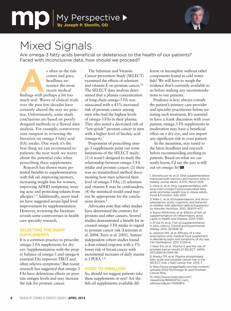
A s often as the tide comes and goes, headlines an-nounce the most recent medical
fi ndings with perhaps a bit too much zeal. Waves of clinical trials over the past few decades have certainly altered the way we prac-tice. Unfortunately, some study conclusions are based on poorly designed methods or a fl awed data analysis. For example, controversy runs rampant in reviewing the literature on omega-3 fatty acid (FA) intake. One week it’s the best thing we can recommend to patients; the next week we worry about the potential risks when prescribing these supplements.
Research has shown many po-tential benefi ts to supplementation with fi sh oil: improving memory, increasing weight loss for women, improving ADHD symptoms, treat-ing acne and protecting infants from allergies.1-5 Additionally, many stud-ies have suggested serum lipid level improvement by supplementation. However, reviewing the literature reveals some controversies in health care specialty research.
SELECTING THE RIGHT SUPPLEMENTSIt is a common practice to prescribe omega-3 FA supplements for dry eye. Supplementation with the prop-er balance of omega-3 and omega-6 essential FAs improves TBUT and often relieves symptoms.6 But recent research has suggested that omega-3 FAs have deleterious effects on pros-tate antigen levels and may increase the risk for prostate cancer.
The Selenium and Vitamin Cancer prevention Study (SELECT) examined the effects of selenium and vitamin E on prostate cancer.7,8
The SELECT data analysis deter-mined that a plasma concentration of long-chain omega-3 FA was associated with a 43% increased risk of prostate cancer among men who had the highest levels of omega-3 FAs in their plasma. They also noted a decreased risk of “low-grade” prostate cancer in men with a higher level of linoleic acid (omega-6).7
Proponents of prescribing ome-ga-3 supplements point out some limitations of the SELECT study: (1) it wasn’t designed to study the relationship between omega-3 FA intake and prostate cancer; (2) there was no standardized method docu-menting how men achieved their level of omega-3 FAs; (3) selenium and vitamin E may be confounders; (4) the statistical model used may not be appropriate for the conclu-sions drawn.9
Advocates note that other studies have determined the contrary for prostate and other cancers. Several studies demonstrated a benefi t for in-creased omega-3 FA intake in regard to prostate cancer risk (Lietzman et al. 2004, Terry et al. 2001). Sixteen independent cohort studies found a dose-related response with a 5% lower risk of breast cancer with incremental increases of daily marine n-3 PUFA.9,10
HARD TO SWALLOWSo, should we suggest patients take these supplements or not? Are the fi sh oil supplements available dif-
ferent or incomplete without other components found in cold water fi sh? We will have to weigh the evidence that’s currently available to us before making any recommenda-tions to our patients.
Prudence is key; always consult the patient’s primary care provider and specialty practitioner before ini-tiating such treatment. It’s essential to have a frank discussion with your patient; omega-3 FA supplements in moderation may have a benefi cial effect on a dry eye, and not impart any signifi cant risk to your patient.
In the meantime, stay tuned to the latest headlines and research before recommending anything to patients. Based on what we cur-rently know, I’d say the jury is still out on omega-3s! RCCL
1. Stonehouse W, et al. DHA supplementation improved both memory and reaction time in healthy young adults. AJCN. March 2013.2. Irene A, et al. Prior supplementation with long chain omega-3 polyunsaturated fatty acids promotes weight loss in obese adults. Food & Function. Feb 2013.3. Milte C, et al. Eicosapentaenoic and doco-sahexaenoic acids, cognition, and behavior in children with attention-defi cit/hyperactiv-ity disorder. Nutrition. 2012; 28:670–677. 4. Burns-Whitmore, et al. Effects of fi sh oil supplementation on infl ammatory acne. Lipids in Health and Disease. 2012; 11:1655. D’Vaz N, et al. Fish oil supplementation in early infancy. Clinical and Experimental Allergy. 2012; 42:1206–16. 6. Jackson MA, et al. Effi cacy of a new prescription-only medical food supplement in alleviating signs and symptoms of dry eye. Clin Ophthalmol. 2011; 5:1201-6.7. Klein EA, et al. Vitamin E and the risk of prostate cancer results of SELECT. JAMA. 2011;306(14):1549-56.8. Brasky TM, et al. Plasma phospholipid fatty acids and prostate cancer risk in the SELECT trial. J Natl Cancer Inst. 2013; 7.9. http://www.omega3galil.com/wp-content/uploads/2013/10/Omega-3s-and-Prostate-Cancer-Risk. 10. http://www.medscape.com/viewarticle/808402?src=wnl_editspecol&uac=142918FK.
Mixed SignalsAre omega-3 fatty acids benefi cial or deleterious to the health of our patients? Faced with inconclusive data, how should we proceed?
6 REVIEW OF CORNEA & CONTACT LENSES | APRIL 2014
My Perspective By Joseph P. Shovlin, OD
006_rccl0414_mp.indd 6 4/3/14 10:14 AM
REVIEW OF CORNEA & CONTACT LENSES | APRIL 2014 7
There are many things patients do—things they will never admit to—that lead to the destruction of their GP
lenses. Clearly, in their eyes, the fault must be some manufacturing defect. Or maybe it’s a faulty lens? But one thing is for sure: it is not something the patient has done! After all, these are the same lenses they have worn for 30 years, and this is the fi rst time this problem has ever occurred. These patients believe it is your responsibility to honor a warrantee (be it real or imagined) and exchange the lens.
Fortunately, there are a number of early signs that indicate improp-er lens care. Being well aware of these behaviors can help to prevent costly remakes.
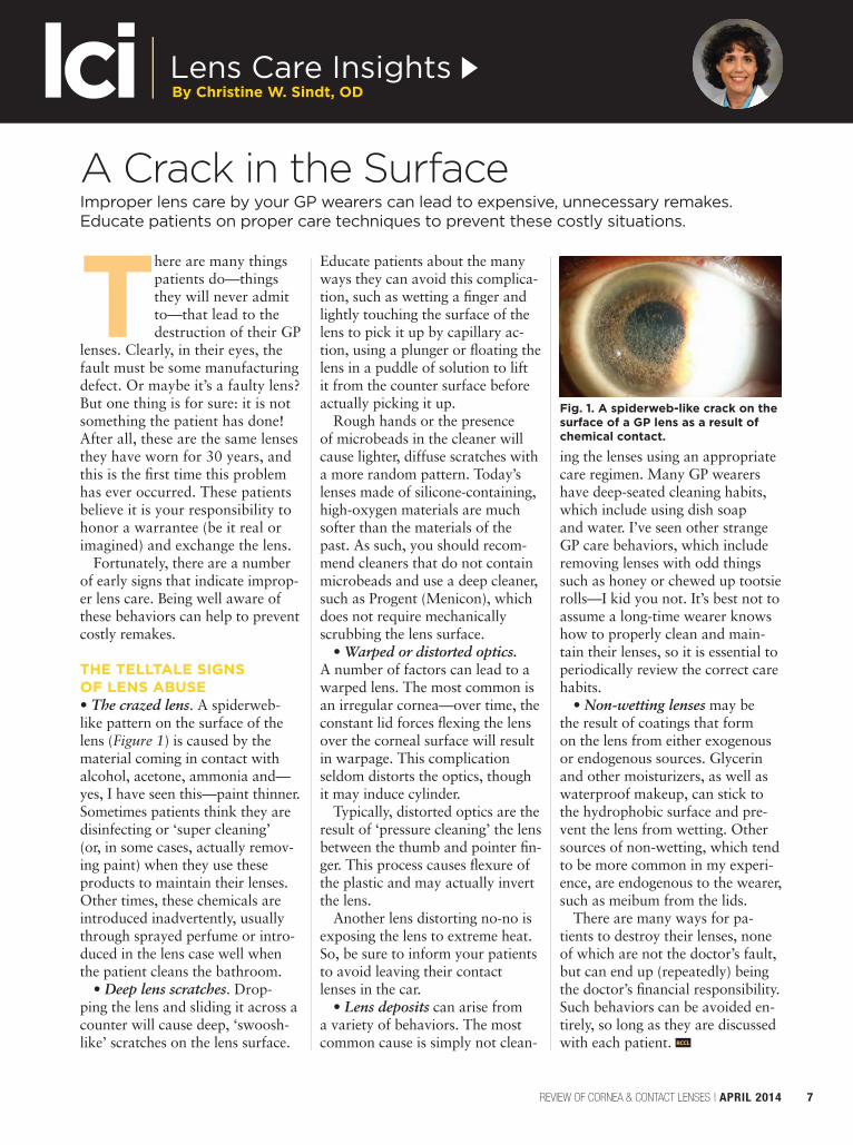
THE TELLTALE SIGNS OF LENS ABUSE• The crazed lens. A spiderweb-like pattern on the surface of the lens (Figure 1) is caused by the material coming in contact with alcohol, acetone, ammonia and—yes, I have seen this—paint thinner. Sometimes patients think they are disinfecting or ‘super cleaning’ (or, in some cases, actually remov-ing paint) when they use these products to maintain their lenses. Other times, these chemicals are introduced inadvertently, usually through sprayed perfume or intro-duced in the lens case well when the patient cleans the bathroom.
• Deep lens scratches. Drop-ping the lens and sliding it across a counter will cause deep, ‘swoosh-like’ scratches on the lens surface.
Educate patients about the many ways they can avoid this complica-tion, such as wetting a fi nger and lightly touching the surface of the lens to pick it up by capillary ac-tion, using a plunger or fl oating the lens in a puddle of solution to lift it from the counter surface before actually picking it up.
Rough hands or the presence of microbeads in the cleaner will cause lighter, diffuse scratches with a more random pattern. Today’s lenses made of silicone-containing, high-oxygen materials are much softer than the materials of the past. As such, you should recom-mend cleaners that do not contain microbeads and use a deep cleaner, such as Progent (Menicon), which does not require mechanically scrubbing the lens surface.
• Warped or distorted optics.A number of factors can lead to a warped lens. The most common is an irregular cornea—over time, the constant lid forces fl exing the lens over the corneal surface will result in warpage. This complication seldom distorts the optics, though it may induce cylinder.
Typically, distorted optics are the result of ‘pressure cleaning’ the lens between the thumb and pointer fi n-ger. This process causes fl exure of the plastic and may actually invert the lens.
Another lens distorting no-no is exposing the lens to extreme heat. So, be sure to inform your patients to avoid leaving their contact lenses in the car.
• Lens deposits can arise from a variety of behaviors. The most common cause is simply not clean-
ing the lenses using an appropriate care regimen. Many GP wearers have deep-seated cleaning habits, which include using dish soap and water. I’ve seen other strange GP care behaviors, which include removing lenses with odd things such as honey or chewed up tootsie rolls—I kid you not. It’s best not to assume a long-time wearer knows how to properly clean and main-tain their lenses, so it is essential to periodically review the correct care habits.
• Non-wetting lenses may be the result of coatings that form on the lens from either exogenous or endogenous sources. Glycerin and other moisturizers, as well as waterproof makeup, can stick to the hydrophobic surface and pre-vent the lens from wetting. Other sources of non-wetting, which tend to be more common in my experi-ence, are endogenous to the wearer, such as meibum from the lids.
There are many ways for pa-tients to destroy their lenses, none of which are not the doctor’s fault, but can end up (repeatedly) being the doctor’s fi nancial responsibility. Such behaviors can be avoided en-tirely, so long as they are discussed with each patient. RCCL
A Crack in the SurfaceImproper lens care by your GP wearers can lead to expensive, unnecessary remakes. Educate patients on proper care techniques to prevent these costly situations.
Fig. 1. A spiderweb-like crack on the surface of a GP lens as a result of chemical contact.
By Christine W. Sindt, ODLens Care Insights
007_rccl0414_lenscare.indd 7 4/3/14 10:15 AM
Scleral lenses have al-lowed a number of our irregular cornea patients to see the world in a manner that
may have previously been impos-sible. Because these lenses vault the cornea, they create a wholly new refractive surface—freeing the clinician from the constraints of the patient’s corneal anatomy. Scleral lenses also provide a stable visual experience; unlike tradition-al small-diameter GP lenses, they do not move with each blink.
We have used sclerals on a num-ber of normal corneas and have prevented patients from dropping out of lens wear. Patients with high levels of ametropia (specifi cally those with high levels of astigma-tism), who are dissatisfi ed with the stability of their vision, have done quite well with this design. We have also used this modality to allow patients to re-enter lens wear following years of discontinuation.
But, as with any lens choice, we must consider the disadvantages of the modality along with its advantages so that we can ex-ercise caution when prescribing these lenses. Because this lens is meant to clear the cornea entirely, an ideal fi t must provide both adequate central and limbal clear-ance. As a result, the lens should rest solely on the conjunctiva and underlying sclera via its scleral landing zone. It is thus critical to respect the interaction between the lens and the conjunctiva—that may very well be what makes the difference between a successful and an unsuccessful fi t.
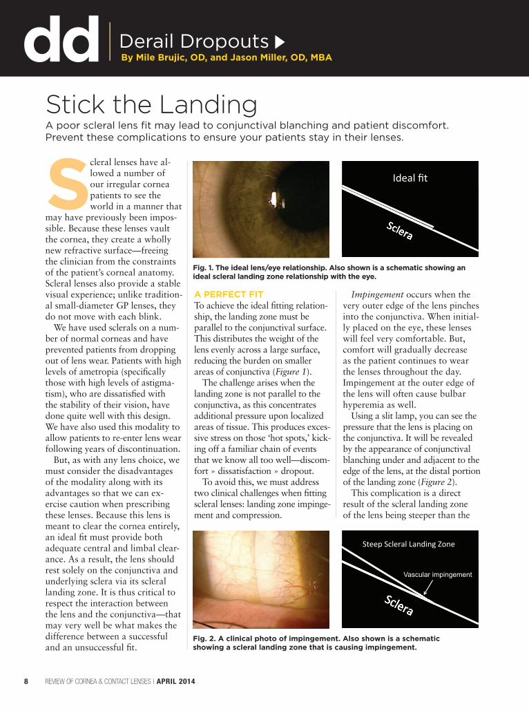
A PERFECT FITTo achieve the ideal fi tting relation-ship, the landing zone must be parallel to the conjunctival surface. This distributes the weight of the lens evenly across a large surface, reducing the burden on smaller areas of conjunctiva (Figure 1).
The challenge arises when the landing zone is not parallel to the conjunctiva, as this concentrates additional pressure upon localized areas of tissue. This produces exces-sive stress on those ‘hot spots,’ kick-ing off a familiar chain of events that we know all too well—discom-fort » dissatisfaction » dropout.
To avoid this, we must address two clinical challenges when fi tting scleral lenses: landing zone impinge-ment and compression.
Impingement occurs when the very outer edge of the lens pinches into the conjunctiva. When initial-ly placed on the eye, these lenses will feel very comfortable. But, comfort will gradually decrease as the patient continues to wear the lenses throughout the day. Impingement at the outer edge of the lens will often cause bulbar hyperemia as well.
Using a slit lamp, you can see the pressure that the lens is placing on the conjunctiva. It will be revealed by the appearance of conjunctival blanching under and adjacent to the edge of the lens, at the distal portion of the landing zone (Figure 2).
This complication is a direct result of the scleral landing zone of the lens being steeper than the
Stick the LandingA poor scleral lens fi t may lead to conjunctival blanching and patient discomfort. Prevent these complications to ensure your patients stay in their lenses.
By Mile Brujic, OD, and Jason Miller, OD, MBADerail Dropouts
8 REVIEW OF CORNEA & CONTACT LENSES | APRIL 2014
Fig. 1. The ideal lens/eye relationship. Also shown is a schematic showing an ideal scleral landing zone relationship with the eye.
Vascular impingement
Fig. 2. A clinical photo of impingement. Also shown is a schematic showing a scleral landing zone that is causing impingement.
008_rccl0414_DD.indd 8 4/3/14 10:15 AM
Go Further—Without Leaving Home
Expand your clinical skills and catch up on your CE
requirements, all from the
comfort of your own home.
Review offers nearly 100 hours of COPE-approved continuing
education — right now! It’s just a click away. Our extensive
library of exams runs the gamut from
keratoconus to fundus autofluorescence, and everything in between.
www.revoptom.com/continuing_education
scleral profi le. To resolve this par-ticular challenge, fl atten the scleral landing zone. This will allow the lens to more evenly distribute its weight over the conjunctiva, which in turn reduces the localized pres-sure that causes the conjunctival blanching beneath the edge of the lens.
Because of the obvious pressure on the vascular supply in the un-derlying conjunctiva, it is important to resolve this fi tting challenge as soon as it’s seen. Additionally, these patients typically experience signifi -cantly higher amounts of debris and
clouding under the lens than normal and, as such, require more frequent removal and rinsing of the lens.
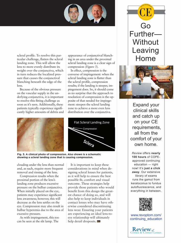
Compression results when the proximal portion of the lens’s landing zone produces excessive pressure on the bulbar conjunctiva. When initially placed on the eye, patients may experience signifi cant lens awareness; however, this will decrease as the lens settles on the eye. Compression may also result in bulbar hyperemia due to the area of excessive pressure.
As with impingement, this too can be seen at the slit lamp. The
appearance of conjunctival blanch-ing in an area under the proximal scleral landing zone is a clear sign of compression (Figure 3).
In effect, compression is the converse of impingement: when the scleral landing zone is fl atter than the scleral profi le, compression results; if the landing is steeper, im-pingement does. So, it should come as no surprise that the approach to resolution of compression is the op-posite of that needed for impinge-ment: steepen the scleral landing zone to achieve a more even lens distribution over the conjunctiva.
It is important to keep these considerations in mind when de-signing scleral lenses for patients, as it will help to ensure the best possible fi t, comfort and visual outcome. These strategies help provide those patients who would benefi t from this design the great-est chance of doing so, and will also help to keep individuals in contact lenses who may have oth-erwise considered discontinuing lens wear. Ensuring your patients are experiencing an ideal lens-to-eye relationship will ultimately help derail dropouts. RCCL
Compression
Fig. 3. A clinical photo of compression. Also shown is a schematic showing a scleral landing zone that is causing compression.
008_rccl0414_DD.indd 9 4/3/14 10:15 AM

Dr. Barnett is a Principal Optom-
etrist at the UC Davis Medical
Center. She specializes in
anterior segment disease and
specialty contact lenses.
Dr. Messer practices at the Cor-
nea and Contact Lens Institute
of Minnesota. She specializes in
fi tting specialty contact lenses.
ABOUT THE AUTHORS
10 REVIEW OF CORNEA & CONTACT LENSES | APRIL 2014
Clinicians have been fi tting scleral contact lenses for over a cen-tury; indeed, this de-sign marked our fi rst
opportunity to correct refractive error without spectacle lenses. Their virtues in vision correction and promotion of ocular surface healing have long been known, but sev-eral hurdles made the mainstream viability of scleral contact lenses short lived. The earliest sclerals were made of fragile and potentially harmful glass, which was then suc-ceeded by PMMA—a material that offered poor oxygen transmissibil-ity, leading to the discontinuation of lens wear due to corneal hypoxia. Because these materials required the lenses to be handmade, they were impossible to replicate in the instance of breakage or loss.
Fortunately, the modality has improved in recent years; we now have hyper-oxygen permeable materials, computer-driven lathes and diagnostic imaging to guide fi ts—making scleral lenses more comfortable and easier to fi t than many other options. There is nearly three decades of documentation and hundreds of scientifi c studies proving the success and benefi ts of wearing scleral contact lenses.
Thanks to the recent advances in scleral lens designs, patients with irregular corneas and ocular surface disease are now enjoying healthy lens wear and great vision with
similar initial comfort to that of a soft contact lens.
However, the rapid advancement of corneal RGP and hydrogel soft lenses (and their silicone hydrogel successors) over the same period rel-egated sclerals to specialty practices serving niche patient populations. We would argue that this is short-sighted and does a disservice to many patients who might be better served with a scleral design.
REFOCUSING A MINDSETOver the past three decades, the use of (highly oxygen permeable) scleral lenses for a number of ocular conditions has increased dramati-cally, and the applications for them continue to grow. Ocular surface disease was the fi rst recognized use for scleral lenses, and remains the only indication currently recognized by Medicare for coverage as a pros-thetic device. Patients with corneal ectasia represent a rapidly growing group of scleral lens wearers. Addi-tionally, scleral lenses are restoring vision to patients with neurotrophic and exposure keratopathies, reduc-ing the need for tarsorrhaphies and corneal transplants.
With each passing year, laborato-ries continue to improve their lens designs to encompass more visual performance needs. For example, scleral lens technology develop-ments made in just the last decade include front surface toric lenses for residual astigmatism and toric
peripheral curves for eyes with greater than average scleral toric-ity. A number of laboratories also offer reverse geometry design lenses and multifocal scleral lenses. There is even technology to precisely customize a lens to the specifi c shape of an individual’s irregular globe. These options allow us to offer customized care for our entire patient population—improving vision, comfort and overall quality of life for habitual and new lens wearers alike.
It’s not just the technology and designs of scleral lenses that continue to evolve, however. We’re now beginning to use more sophis-ticated fi tting techniques to expand the pool of patients who can be considered candidates for scleral contact lenses. For example, scleral lenses aren’t only for patients with irregular corneas; patients with normal corneas are now considered great candidates—especially when their visual needs exceed the typical parameters of soft lenses.
Scleral lens wear in patients who do not have irregular corneas is
PUTTING SCLERAL LENSES
Commonly reserved only for patients with irregular corneas, sclerals can be an excellent option even for patients with ‘normal’ ones.
SPECIAL ISSUE: unlock your scleral potential
010_rccl0414F1_F2.indd 10 4/3/14 2:03 PM
becoming so popular that labora-tories are designing scleral lenses made specifi cally to vault normal corneas. Patients with high astigma-tism who desire a multifocal option are no longer restricted to corneal gas permeable lenses—even though, in certain cases (e.g., a patient with ocular surface disease, such as dry eye or refractive error), these designs remain a great option for lens wear.
Adding patients with normal eyes to the scleral lens-wearing popula-tion has signaled a major shift in philosophy from years past—and it’s continuing to gain popularity. As habitual contact lens wear-ers reach their forties and contact lens dropout becomes more likely, sclerals can offer us another way to keep patients wearing lenses despite the onset of presbyopia and ocular surface disease that may otherwise be a deterrent.
Overall lens diameter is another aspect of scleral lenses that has seen a signifi cant change. The theory be-hind lens diameter selection remains the same: the more sagittal depth needed to vault the corneal apex, the larger the lens diameter should be. What has changed, though, is the range of diameters available for scleral lens designs. Now, diameters come as small as 14.3mm, while still maintaining a full corneal and limbal vault, rather than using a corneoscleral fi tting technique.
While this design is not appropri-ate for all irregular corneas, it has
been very successful with normal corneas, and a vault of as little as 100 microns is considered accept-able in many cases. These smaller-diameter lenses are less intimidating
for some patients and clinicians, easier for patients to handle and, in some cases, may be a less expensive option. In these smaller-diameter lenses, the limbal-scleral curvature is mostly regular and has less inter-patient variability near the cornea than the peripheral sclera. As with all scleral designs, limbal clear-ance must be maintained during lens wear to preserve the health of limbal stem cells.
A fi nal change in fi tting technique worth noting is the decline in use of fenestrations—a lens alteration used very selectively now, on a case-by-case basis. We have known for 30 years that fenestrating lenses does not increase oxygen transmission or improve tear exchange in any mean-ingful way. Fenestrations should only be used for easier removal, due to
the suction effect. In very rare cases, fenestration may be used to release CO2 from beneath the lens; however, this is not always successful.
To make the fi tting process smoother for practitioners, scleral fi tting sets now offer expand-ed parameters to expedite the process. In addi-tion to numerous base curves, many fi tting sets include lenses with differ-ent overall diam-eters, changes to
the peripheral curve lifts, different curvatures to contour the limbus and even different specifi cations in sagittal depth.
NEW PATIENTS, NEW VISIONWith all these advances in scleral lens parameters and technology, nearly every patient in your offi ce becomes a scleral lens candidate. The most receptive are likely those who experience frustration with their current soft contact lenses—usually due to the modality failing to adequately meet their vision needs. It’s up to the practitioner to ensure every patient is satisfi ed with their lenses, and it is acceptable to ask your patient whether or not they are satisfi ed with their quality of vision. Patients often believe their current lenses are the best option
REVIEW OF CORNEA & CONTACT LENSES | APRIL 2014 11
COMPANYAcculensAcculensArt OpticalAdvanced Vision TechnologiesBlanchardBlanchardDakota SciencesEssilorMetro OpticsLens DynamicsTruformValley Contax
LENSMaxim PlusComfort SL PlusSo2Clear ProgressiveAVTMSDOne FitSo2Clear ProgressiveJupiter PlusSo2Clear ProgressiveDyna Semi-ScleralDigiform MultifocalStable Near
Table 1. Multifocal Scleral Lenses
FRONT AND CENTER
By Melissa Barnett, OD, and Brooke Messer, OD
010_rccl0414F1_F2.indd 11 4/3/14 2:04 PM
available, so they rarely bother to ask questions regarding new products in the contact lens world. Patients who typically fi t this mind-set include those with high astig-matism, residual refractive error post-LASIK surgery or presbyopia.
Those who have high astigma-tism are especially pleased with scleral lenses, as they can rotate without having an effect on vision. If the astigmatic topography shows limbus-to-limbus toricity, it may extend onto the sclera and produce some lens fl exure. As a practitioner, you’ll see with-the-rule astigmatism on retinoscopy and the patient may comment on vision fl uctuation or reduced acuity with a spherical over-refraction. A spherocylindrical over-refraction and keratometry or to-pography over the lens will provide some insight on how much fl exure is occurring. Typically, increasing the center thickness of the lens by anywhere from 0.05mm to 0.20mm is our fi rst adjustment, but decreas-ing the overall diameter is another option that may reduce fl exure. In our experience, a fi nal option when attempting to decrease fl exure is to add toric peripheral curves to the lenses in an effort a better contour to the patient’s sclera.
Post-refractive surgery patients have altered corneal geometry and an elevated likelihood of dry eye; they may struggle with vision issues related to the stability of the fi t. The oblate-shaped cornea no longer fi ts the prolate toric soft lenses, which causes patients to become frustrated with lens movement and rota-tion. Scleral lenses are an excellent option for these patients because the tear lens corrects for corneal astigmatism and helps with dry eye. Occasionally, to maintain an even tear fi lm from limbus to limbus, a reverse geometry lens is needed for an oblate cornea. Many post-LASIK patients want to return to contact
lenses when presbyopia becomes a problem. Fortunately, there are a number of well-designed multifocal scleral lens options on the market today. Table 1 lists many examples.
Many well-designed multifocal scleral lenses are on the market to-day. These lenses are simultaneous vision designs, as these lenses move minimally on the eye. Most designs are center-near, with the exception of the AVT scleral multifocal and Jupiter Plus lenses, which are a center-distance design. The Jupiter Plus lens offers an intermediate-near add, but not full-near add.
MAKING THE SOFT-TO-SCLERAL REFITIf, following this review, you’re ea-ger to fi nd opportunities for fi tting new patients in scleral lenses, start by considering those with normal corneas who wear soft lenses for astigmatism or presbyopia who complain of fl uctuating vision or poor night vision. To success-fully convert our soft lens-wearing patients, we must refi t them quickly when problems arise and achieve better results than soft torics or multifocals while maintaining good lens comfort.
First, assess their current frustra-tion level as well as their desire to change modalities and undergo a new fi tting procedure. Once the patient has agreed to try new lenses, use revelant anatomical data—their topographies, corneal diameter mea-surement, palpebral fi ssure widths—to help select an appropriate lens design. We’re all aware that scleral lenses provide better initial comfort when compared to smaller diameter corneal GP lenses, but we need to be sure we select an appropriate design to ensure a good fi t in our soft contact lens wearers. In doing so, we provide an excellent fi rst experience, ensuring the patient remains excited about the vision potential offered by
this new lens design. The following tips can help with
the education and fi tting process: • Lens diameter selection. The
overall diameter of the scleral lens is the fi rst decision you’ll have to make. This factor is the primary reason scleral lenses provide more comfort than corneal gas permeable lenses. Diameter is also important because it determines the sagittal depth of the lens. Many patients believe their soft lenses are more comfortable than GP contacts due to the biocompatibility of the mate-rial, but we know that it’s actually because they have a larger overall diameter and experience less lid interaction and lens movement.
That is an important point to communicate to patients. The larger lenses sit behind both lids, which reduces the interaction between the eyelid and the lens, and promotes excellent comfort and lens stability. Once patients understand this con-cept, the notion of wearing “hard” lenses is not nearly as intimidating.
When selecting the diameter, do so based on patient factors such as corneal diameter, the sagittal depth required to vault any corneal ir-regularity or ocular surface disease, scleral factors (e.g., pinguecula, conjunctival chalasis or toricity) and your personal experience with various lens designs. If the cornea is 12mm or more in overall diameter, you may want to begin with a lens that is at least 14.5mm to 15mm, so it can appropriately and comfort-ably vault both the central cornea and limbus.
An average palpebral fi ssure width is about 10mm. If a patient has an average sized cornea with a small to average palpebral fi s-sure width, lenses that are 14mm to 15mm in overall diameter will perform well, and the patient will appreciate how easily the lens can
12 REVIEW OF CORNEA & CONTACT LENSES | APRIL 2014
PUTTING SCLERAL LENSES FRONT AND CENTER
(Continued on page 17)
010_rccl0414F1_F2.indd 12 4/3/14 2:15 PM
Recommend CLEAR CARE® Solution and learn more at MYALCON.COM
The scientifically proven formula of CLEAR CARE® Solution deeply cleans, then neutralizes, to create a gentle saline similar to natural tears. The result is pure comfort and is why CLEAR CARE® has the most loyal patients of any lens care brand.2
^Trademarks are the property of their respective owners.
References: 1. A market research study conducted amongst 107 US contact lens wearers representative of CLEAR CARE® purchasers in the United States, 2007. 2. Based on third party industry report 52 weeks ending 12/29/12; Alcon data on fi le. 3. Alcon data on fi le, 2009. 4. SOFTWEAR™ Saline package insert. 5. Paugh, Jerry R, et al. Ocular response to hydrogen peroxide. American Journal of Optometry & Physiological Optics: 1988; 65:2,91–98.
© 2014 Novartis 02/14 CCS14004ADi
THIS IS WHY 4 out of 5 patients agree their lenses feel like new.1
SOFTWEARTM Saline2
CLEAR CARE® Solution1
RESIDUAL H2O2 IN PARTS PER MILLION (PPM)
OCULAR AWARENESS THRESHOLD3
2050 40 60 80 100
Range of Residual H2O2 on Lens:
The Science Behind a Pristine, Clean Lens:
PERFORMANCE DRIVEN BY SCIENCE™
Triple-Action Cleaning• Patented formula deeply cleans• Carries away dirt & debris• Pluronic^ 17R4 lifts away protein
Pristine, Clean Lens• Less residual H2O2
3-5
• Irritant-free comfort• No added preservatives
Pluronic 17R4
RCCL0414_Alcon Clear Care.indd 1 3/27/14 2:49 PM
14 REVIEW OF CORNEA & CONTACT LENSES | APRIL 2014
Dr. Sindt is the Associate Clinical Editor of
Review of Cornea & Contact Lenses, and
of Review of Optometry.
She is also the Director,
Contact Lens Service
and an associate
professor of clinical
ophthalmology and
visual sciences at the
University of Iowa.
ABOUT THE AUTHOR
14 REVIEW OF CORNEA & CONTACT LENSES | APRIL 2014
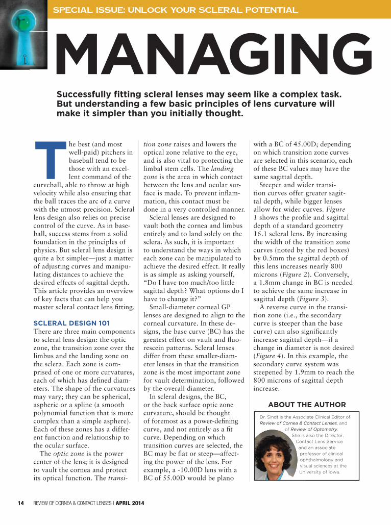
The best (and most well-paid) pitchers in baseball tend to be those with an excel-lent command of the
curveball, able to throw at high velocity while also ensuring that the ball traces the arc of a curve with the utmost precision. Scleral lens design also relies on precise control of the curve. As in base-ball, success stems from a solid foundation in the principles of physics. But scleral lens design is quite a bit simpler—just a matter of adjusting curves and manipu-lating distances to achieve the desired effects of sagittal depth. This article provides an overview of key facts that can help you master scleral contact lens fi tting.
SCLERAL DESIGN 101There are three main components to scleral lens design: the optic zone, the transition zone over the limbus and the landing zone on the sclera. Each zone is com-prised of one or more curvatures, each of which has defi ned diam-eters. The shape of the curvatures may vary; they can be spherical, aspheric or a spline (a smooth polynomial function that is more complex than a simple asphere). Each of these zones has a differ-ent function and relationship to the ocular surface.
The optic zone is the power center of the lens; it is designed to vault the cornea and protect its optical function. The transi-
tion zone raises and lowers the optical zone relative to the eye, and is also vital to protecting the limbal stem cells. The landing zone is the area in which contact between the lens and ocular sur-face is made. To prevent infl am-mation, this contact must be done in a very controlled manner.
Scleral lenses are designed to vault both the cornea and limbus entirely and to land solely on the sclera. As such, it is important to understand the ways in which each zone can be manipulated to achieve the desired effect. It really is as simple as asking yourself, “Do I have too much/too little sagittal depth? What options do I have to change it?”
Small-diameter corneal GP lenses are designed to align to the corneal curvature. In these de-signs, the base curve (BC) has the greatest effect on vault and fl uo-rescein patterns. Scleral lenses differ from these smaller-diam-eter lenses in that the transition zone is the most important zone for vault determination, followed by the overall diameter.
In scleral designs, the BC, or the back surface optic zone curvature, should be thought of foremost as a power-defi ning curve, and not entirely as a fi t curve. Depending on which transition curves are selected, the BC may be fl at or steep—affect-ing the power of the lens. For example, a -10.00D lens with a BC of 55.00D would be plano
with a BC of 45.00D; depending on which transition zone curves are selected in this scenario, each of these BC values may have the same sagittal depth.
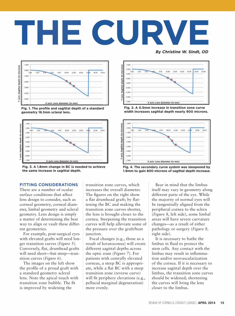
Steeper and wider transi-tion curves offer greater sagit-tal depth, while bigger lenses allow for wider curves. Figure 1 shows the profi le and sagittal depth of a standard geometry 16.1 scleral lens. By increasing the width of the transition zone curves (noted by the red boxes) by 0.5mm the sagittal depth of this lens increases nearly 800 microns (Figure 2). Conversely, a 1.8mm change in BC is needed to achieve the same increase in sagittal depth (Figure 3).
A reverse curve in the transi-tion zone (i.e., the secondary curve is steeper than the base curve) can also signifi cantly increase sagittal depth—if a change in diameter is not desired (Figure 4). In this example, the secondary curve system was steepened by 1.9mm to reach the 800 microns of sagittal depth increase.
Successfully fi tting scleral lenses may seem like a complex task. But understanding a few basic principles of lens curvature will make it simpler than you initially thought.
MANAGING SPECIAL ISSUE: unlock your scleral potential
010_rccl0414F1_F2.indd 14 4/3/14 2:04 PM
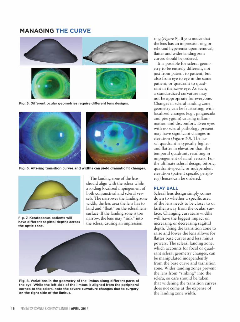
FITTING CONSIDERATIONSThere are a number of ocular surface conditions that affect lens design to consider, such as corneal geometry, corneal diam-eter, limbal geometry and scleral geometry. Lens design is simply a matter of determining the best way to align or vault these differ-ent geometries.
For example, post-surgical eyes with elevated grafts will need lon-ger transition curves (Figure 5). Conversely, fl at, drumhead grafts will need short—but steep—tran-sition curves (Figure 6).
The images on the left show the profi le of a proud graft with a standard geometry scleral lens. Note the apical touch with transition zone bubble. The fi t is improved by widening the
transition zone curves, which increases the overall diameter. The fi gures on the right show a fl at drumhead graft; by fl at-tening the BC and making the transition zone curves shorter, the lens is brought closer to the cornea. Steepening the transition curves will help alleviate some of the pressure over the graft/host junction.
Focal changes (e.g., those as a result of keratoconus) will create different sagittal depths across the optic zone (Figure 7). For patients with centrally elevated corneas, a steep BC is appropri-ate, while a fl at BC with a steep transition zone (reverse curve) will fi t periphery elevations (e.g., pellucid marginal degeneration) more evenly.
Bear in mind that the limbus itself may vary in geometry along different parts of the eye. While the majority of normal eyes will be tangentially aligned from the peripheral cornea to the sclera (Figure 8, left side), some limbal areas will have severe curvature changes—as a result of either pathology or surgery (Figure 8,right side).
It is necessary to bathe the limbus in fl uid to protect the stem cells. Any contact with the limbus may result in infl amma-tion and/or neovascularization of the cornea. If it is necessary to increase sagittal depth over the limbus, the transition zone curves should be widened; shortening the curves will bring the lens closer to the limbus.
REVIEW OF CORNEA & CONTACT LENSES | APRIL 2014 15
Fig. 3. A 1.8mm change in BC is needed to achieve the same increase in sagittal depth.
X axis: Lens diameter (in mm)
Y a
xis:
Sag
itta
l dep
th (
in m
icro
ns)
Fig. 4. The secondary curve system was steepened by 1.9mm to gain 800 microns of sagittal depth increase.
X axis: Lens diameter (in mm)
Y a
xis:
Sag
itta
l dep
th (
in m
icro
ns)
Fig. 2. A 0.5mm increase in transition zone curve width increases sagittal depth nearly 900 microns.
X axis: Lens diameter (in mm)
Y a
xis:
Sag
itta
l dep
th (
in m
icro
ns)
Fig. 1. The profi le and sagittal depth of a standard geometry 16.1mm scleral lens.
X axis: Lens diameter (in mm)
Y a
xis:
Sag
itta
l dep
th (
in m
icro
ns)
THE CURVEBy Christine W. Sindt, OD
010_rccl0414F1_F2.indd 15 4/3/14 2:04 PM
MANAGING THE CURVE
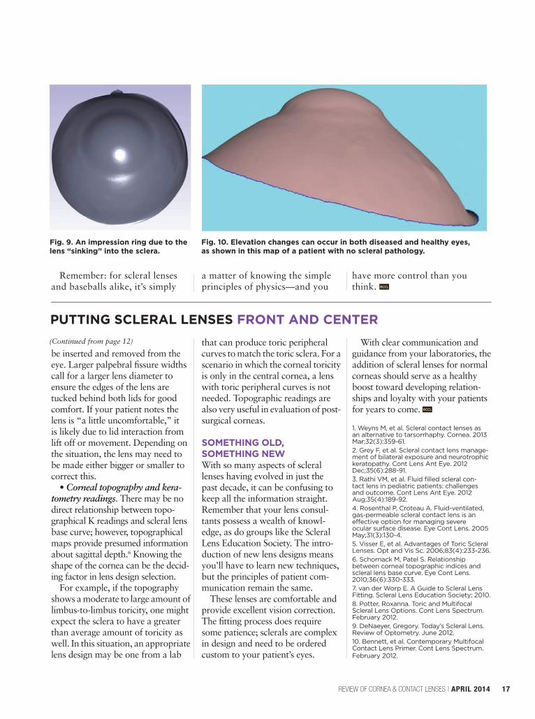
The landing zone of the lens should align with the sclera while avoiding localized impingement of both conjunctival and scleral ves-sels. The narrower the landing zone width, the less area the lens has to land and “fl oat” on the scleral lens surface. If the landing zone is too narrow, the lens may “sink” into the sclera, causing an impression
ring (Figure 9). If you notice that the lens has an impression ring or rebound hyperemia upon removal, fl atter and wider landing zone curves should be ordered.
It is possible for scleral geom-etry to be entirely different, not just from patient to patient, but also from eye to eye in the same patient, or quadrant to quad-rant in the same eye. As such, a standardized curvature may not be appropriate for everyone. Changes in scleral landing zone geometry can be frustrating, with localized changes (e.g., pinguecula and pterygium) causing infl am-mation and discomfort. Even eyes with no scleral pathology present may have signifi cant changes in elevation (Figure 10). The na-sal quadrant is typically higher and fl atter in elevation than the temporal quadrant, resulting in impingement of nasal vessels. For the ultimate scleral design, bitoric, quadrant-specifi c or independent elevation (patient specifi c periph-ery) lenses can be ordered.
PLAY BALLScleral lens design simply comes down to whether a specifi c area of the lens needs to be closer to or farther away from the ocular sur-face. Changing curvature widths will have the biggest impact on increasing or decreasing sagittal depth. Using the transition zone to raise and lower the lens allows for fl atter base curves and less minus powers. The scleral landing zone, which accounts for focal or quad-rant scleral geometry changes, can be manipulated independently from the base curve and transition zone. Wider landing zones prevent the lens from “sinking” into the sclera, so care should be taken that widening the transition curves does not come at the expense of the landing zone width.
16 REVIEW OF CORNEA & CONTACT LENSES | APRIL 2014
Fig. 5. Diff erent ocular geometries require diff erent lens designs.
Fig. 6. Altering transition curves and widths can yield dramatic fi t changes.
Fig. 8. Variations in the geometry of the limbus along diff erent parts of the eye. While the left side of the limbus is aligned from the peripheral cornea to the sclera, note the severe curvature changes due to surgery on the right side of the limbus.
Fig. 7. Keratoconus patients will have diff erent sagittal depths across the optic zone.
010_rccl0414F1_F2.indd 16 4/3/14 2:04 PM
be inserted and removed from the eye. Larger palpebral fi ssure widths call for a larger lens diameter to ensure the edges of the lens are tucked behind both lids for good comfort. If your patient notes the lens is “a little uncomfortable,” it is likely due to lid interaction from lift off or movement. Depending on the situation, the lens may need to be made either bigger or smaller to correct this.
• Corneal topography and kera-tometry readings. There may be no direct relationship between topo-graphical K readings and scleral lens base curve; however, topographical maps provide presumed information about sagittal depth.6 Knowing the shape of the cornea can be the decid-ing factor in lens design selection.
For example, if the topography shows a moderate to large amount of limbus-to-limbus toricity, one might expect the sclera to have a greater than average amount of toricity as well. In this situation, an appropriate lens design may be one from a lab
that can produce toric peripheral curves to match the toric sclera. For a scenario in which the corneal toricity is only in the central cornea, a lens with toric peripheral curves is not needed. Topographic readings are also very useful in evaluation of post-surgical corneas.
SOMETHING OLD, SOMETHING NEWWith so many aspects of scleral lenses having evolved in just the past decade, it can be confusing to keep all the information straight. Remember that your lens consul-tants possess a wealth of knowl-edge, as do groups like the Scleral Lens Education Society. The intro-duction of new lens designs means you’ll have to learn new techniques, but the principles of patient com-munication remain the same.
These lenses are comfortable and provide excellent vision correction. The fi tting process does require some patience; sclerals are complex in design and need to be ordered custom to your patient’s eyes.
With clear communication and guidance from your laboratories, the addition of scleral lenses for normal corneas should serve as a healthy boost toward developing relation-ships and loyalty with your patients for years to come. RCCL
1. Weyns M, et al. Scleral contact lenses as an alternative to tarsorrhaphy. Cornea. 2013 Mar;32(3):359-61.2. Grey F, et al. Scleral contact lens manage-ment of bilateral exposure and neurotrophic keratopathy. Cont Lens Ant Eye. 2012 Dec;35(6):288-91. 3. Rathi VM, et al. Fluid fi lled scleral con-tact lens in pediatric patients: challenges and outcome. Cont Lens Ant Eye. 2012 Aug;35(4):189-92.4. Rosenthal P, Croteau A. Fluid-ventilated, gas-permeable scleral contact lens is an effective option for managing severe ocular surface disease. Eye Cont Lens. 2005 May;31(3):130-4.5. Visser E, et al. Advantages of Toric Scleral Lenses. Opt and Vis Sc. 2006;83(4):233-236.6. Schornack M, Patel S. Relationship between corneal topographic indices and scleral lens base curve. Eye Cont Lens. 2010;36(6):330-333.7. van der Worp E. A Guide to Scleral Lens Fitting. Scleral Lens Education Society; 2010. 8. Potter, Roxanna. Toric and Multifocal Scleral Lens Options. Cont Lens Spectrum. February 2012.9. DeNaeyer, Gregory. Today’s Scleral Lens. Review of Optometry. June 2012. 10. Bennett, et al. Contemporary Multifocal Contact Lens Primer. Cont Lens Spectrum. February 2012.
Remember: for scleral lenses and baseballs alike, it’s simply
a matter of knowing the simple principles of physics—and you
have more control than you think. RCCL
PUTTING SCLERAL LENSES FRONT AND CENTER
REVIEW OF CORNEA & CONTACT LENSES | APRIL 2014 17
Fig. 9. An impression ring due to the lens “sinking” into the sclera.
Fig. 10. Elevation changes can occur in both diseased and healthy eyes, as shown in this map of a patient with no scleral pathology.
(Continued from page 12)
010_rccl0414F1_F2.indd 17 4/3/14 2:05 PM

ABOUT THE AUTHOR
18 REVIEW OF CORNEA & CONTACT LENSES | APRIL 2014
The use of scleral con-tact lenses has grown exponentially since 2006.1 Over the past decade, clinicans have
started to consider scleral lens de-signs as an option for all contact lens patients. Improved materials and technology for fi tting and manufacturing have helped create the renaissance this modality is currently experiencing.
Scleral lenses offer some dis-tinct advantages, including superb comfort, stability of fi t and excellent vision.2 Despite these advantages, some diffi culties can lead to dissat-isfaction in your scleral lens wearers. This article will highlight 10 impor-tant “do’s and don’ts” of scleral lenses.
1.DON’T FORGET TO EXPLAIN THE ENTIRE
FITTING PROCESS.When a patient comes in for a consultation, referral or compre-hensive exam, and scleral lenses are found to be the best option for that particular patient, discussing the entire fi tting process is critical to their success with scleral lenses. Be sure to inform the patient of just how long each appointment will be, and discuss the schedule of a typical scleral lens patient.
For example, a common schedule may begin with a one-hour diag-nostic fi tting, followed by a 30-min-ute dispensing visit, which is then followed by a one-hour training session on insertion and removal techniques. Finally, inform patients
that there will be additional follow-up visits after the initial fi tting.
Patients new to contact lens wear, and those who have never worn specialty lenses, need to be informed of the complex fi tting and follow-up care involved. Detailing this during the fi rst visit eliminates potential patient frustration, and will defi ne their expectations for the scleral lens fi tting process appropriately.
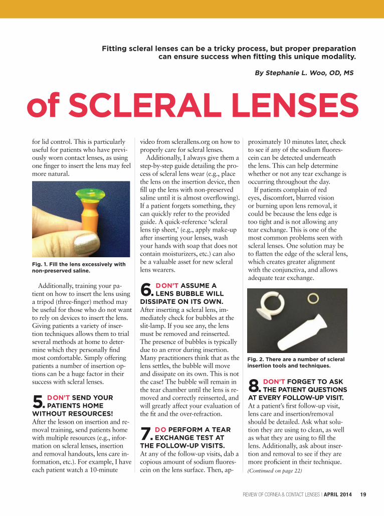
2.DO FILL THE LENS BOWL EXCESSIVELY.
Because most scleral lens fi ttings involve patients who are new to the modality, it is inevitable that there will be some insertion diffi culties. This is especially true of patients who have never previously worn any type of contact lenses. Scleral lenses are inserted much differently than conventional soft contact lenses or corneal gas permeable lenses. The patient must tuck their chin into their chest and look down with their nose pointed to the fl oor. While this may sound strange to most patients, there is a legitimate reason for this: the liquid that fi lls the scleral lens needs to remain in the bowl.
Filling the lens excessively with non-preserved saline will help prevent insertion bubbles in the case of excessive eye movement, blinking and other errors (Figure 1). As some of the fl uid will undoubtedly be spilled during the insertion process, excessively fi lling the lens bowl ensures there will be enough liquid left to still yield an appropriate fi t.
3.DON’T LET GO OF THE INSERTION DEVICE FIRST!
In one of the more common “rookie mistakes,” patients will hold their eyelids open, insert the lens, remove their insertion device (e.g., plunger, fi ngers, plastic ring, etc.) and then release their eyelids. Fortunately, this mistake is also one of the easiest to remedy. After the lens is inserted on the eye, inform patients to release their eyelids fi rst, and then release the insertion device. Releasing the eyelids fi rst allows the lens to become trapped under the lids and onto the eye.
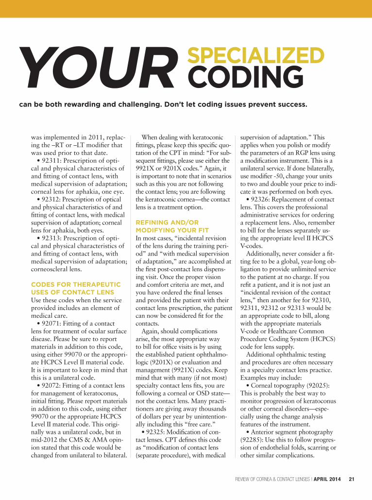
4.DO INFORM YOUR PATIENTS OF SEVERAL
WAYS TO INSERT THE LENS.During the insertion and removal training, be sure to thoroughly explain and demonstrate several insertion techniques (Figure 2). For instance, the large plunger may work well for some patients, but many others will fi nd that a scleral ring/orthodontic band/O-ring offers a greater amount of control. Using these devices allows the lens to rest on one fi nger, freeing the others
Dr. Woo graduated from the Southern Cali-
fornia College of Optometry and completed a
Cornea/Contact Lens residency at the Univer-
sity of Missouri - St. Louis. She
is a Fellow of the American
Academy of Optometry
and a Fellow of the Scleral
Lens Education Society.
She currently practices
at Havasu Eye Center in
Lake Havasu, AZ.
SPECIAL ISSUE: unlock your scleral potential
DOs andDON'Ts10
018_rccl0414F3_F4.indd 18 4/3/14 1:10 PM
for lid control. This is particularly useful for patients who have previ-ously worn contact lenses, as using one fi nger to insert the lens may feel more natural.
Additionally, training your pa-tient on how to insert the lens using a tripod (three-fi nger) method may be useful for those who do not want to rely on devices to insert the lens. Giving patients a variety of inser-tion techniques allows them to trial several methods at home to deter-mine which they personally fi nd most comfortable. Simply offering patients a number of insertion op-tions can be a huge factor in their success with scleral lenses.
5.DON’T SEND YOUR PATIENTS HOME
WITHOUT RESOURCES!After the lesson on insertion and re-moval training, send patients home with multiple resources (e.g., infor-mation on scleral lenses, insertion and removal handouts, lens care in-formation, etc.). For example, I have each patient watch a 10-minute
video from sclerallens.org on how to properly care for scleral lenses.
Additionally, I always give them a step-by-step guide detailing the pro-cess of scleral lens wear (e.g., place the lens on the insertion device, then fi ll up the lens with non-preserved saline until it is almost overfl owing). If a patient forgets something, they can quickly refer to the provided guide. A quick-reference ‘scleral lens tip sheet,’ (e.g., apply make-up after inserting your lenses, wash your hands with soap that does not contain moisturizers, etc.) can also be a valuable asset for new scleral lens wearers.
6.DON’T ASSUME A LENS BUBBLE WILL
DISSIPATE ON ITS OWN.After inserting a scleral lens, im-mediately check for bubbles at the slit-lamp. If you see any, the lens must be removed and reinserted. The presence of bubbles is typically due to an error during insertion. Many practitioners think that as the lens settles, the bubble will move and dissipate on its own. This is not the case! The bubble will remain in the tear chamber until the lens is re-moved and correctly reinserted, and will greatly affect your evaluation of the fi t and the over-refraction.
7.DO PERFORM A TEAR EXCHANGE TEST AT
THE FOLLOW-UP VISITS.At any of the follow-up visits, dab a copious amount of sodium fl uores-cein on the lens surface. Then, ap-
proximately 10 minutes later, check to see if any of the sodium fl uores-cein can be detected underneath the lens. This can help determine whether or not any tear exchange is occurring throughout the day.
If patients complain of red eyes, discomfort, blurred vision or burning upon lens removal, it could be because the lens edge is too tight and is not allowing any tear exchange. This is one of the most common problems seen with scleral lenses. One solution may be to fl atten the edge of the scleral lens, which creates greater alignment with the conjunctiva, and allows adequate tear exchange.
8.DON’T FORGET TO ASK THE PATIENT QUESTIONS
AT EVERY FOLLOW-UP VISIT.At a patient’s fi rst follow-up visit, lens care and insertion/removal should be detailed. Ask what solu-tion they are using to clean, as well as what they are using to fi ll the lens. Additionally, ask about inser-tion and removal to see if they are more profi cient in their technique.
Fitting scleral lenses can be a tricky process, but proper preparation can ensure success when fi tting this unique modality.
REVIEW OF CORNEA & CONTACT LENSES | APRIL 2014 19
Fig. 1. Fill the lens excessively with non-preserved saline.
Fig. 2. There are a number of scleral insertion tools and techniques.
(Continued on page 22)
of SCLERAL LENSESBy Stephanie L. Woo, OD, MS
018_rccl0414F3_F4.indd 19 4/3/14 1:22 PM

Dr. Rumpakis is currently President and CEO
of Practice Resource Management, Inc., a fi rm
that specializes in providing a full
array of consulting, appraisal
and management services
for health care profession-
als and industry partners.
He is also the Clinical
Coding Editor for Review of Optometry.
ABOUT THE AUTHOR
Building a practice that focuses on fi tting specialty lenses
20 REVIEW OF CORNEA & CONTACT LENSES | APRIL 2014
I f you’d like to insulate yourself from the commod-ity-based world of dispos-able contact lens margin erosion, a specialty contact
lens practice is one effective way to do so. Specialty lenses may not get as much attention as other practice-building opportunities, but I believe this area of optometric practice has tremendous potential for growth. Yet it’s also a hotbed of frustration for many practitioners—especially given the confusion and apprehension surrounding medical coding and reimbursement.
Generally, when a practitioner considers fi tting a patient in a specialty contact lens, it is typically predicated on the presence of a spe-cifi c medical condition or refractive complication caused by an existing corneal condition. Ocular surface disease (OSD) is an emerging area of both wellness and medical care within many practices today. Build-ing a specialty contact lens practice can be both extraordinarily satisfy-ing and challenging. So, my goal is a simple one: to make coding for and getting reimbursed for specialty contact lenses the easiest part of your very specialized practice.
BACK TO BASICSBefore getting into specifi c codes, it’s necessary to review some basics. In order to know how to properly document your medical record and to use the correct Current Proce-dural Terminology (CPT) code to describe the appropriate services
provided, it is important to under-stand and keep up to date with the current defi nitions of the contact lens fi tting codes as described in the CPT. Just because you are fi tting contact lenses doesn’t mean that you can forget or ignore the funda-mental concepts of medical neces-sity, or the requirements of the chief complaint in your medical record.
As mentioned earlier, because you will often be working with a disease process or a medical condi-tion, you must be very familiar with the requirements for the appropriate use of the 920XX and 992XX codes for your offi ce visits that get coded in addition to your contact lens services.
The CPT has the following to say about contact lens fi tting: The prescription of contact lens includes specifi cation of optical and physical characteristics (such as power, size, curvature, fl exibility, gas permeability). It is not a part of the general ophthalmological ser-vices. The fi tting of contact lenses includes instruction and training of the wearer and incidental revi-sion of the lens during the training period. Supply of materials may be reported as part of the service of fi tting, or may be reported sepa-rately using the appropriate supply codes. Follow-up of successfully fi tted extended wear lenses is re-ported as part of a general oph-thalmological service (92012).
Now that we’ve got the basics
covered, let’s dive into the handful of CPT codes that cover the fi tting of contact lenses.
TRADITIONAL CONTACT LENS FITTING CODES1
These are the bread-and-butter codes used for routine contact lens services.
• 92310: Contact lens fi tting. This is defi ned by CPT as the “pre-scription of optical and physical characteristics of and fi tting of a contact lens, with medical supervi-sion of adaptation; corneal lens, both eyes, except for aphakia.” A 92310 should be charged for the fi tting of contact lenses, and also encompasses services up to the point at which you would issue a contact lens prescription. This code does not include contact lens follow-up care after the lenses have been dispensed.
This code is charged every visit in which a new lens is placed on a patient’s eye, or when the fi t is altered. Incidental revisions, such as power changes without altering the fi t, are not billed as a new fi t-ting. Keep in mind that the modi-fi er -52 should be used if fi tting only one eye; this is a change that
SIMPLIFYSPECIAL ISSUE: unlock your scleral potential
BY JOHN RUMPAKIS, OD, MBA
018_rccl0414F3_F4.indd 20 4/3/14 1:22 PM
was implemented in 2011, replac-ing the –RT or –LT modifi er that was used prior to that date.
• 92311: Prescription of opti-cal and physical characteristics of and fi tting of contact lens, with medical supervision of adaptation; corneal lens for aphakia, one eye.
• 92312: Prescription of optical and physical characteristics of and fi tting of contact lens, with medical supervision of adaptation; corneal lens for aphakia, both eyes.
• 92313: Prescription of opti-cal and physical characteristics of and fi tting of contact lens, with medical supervision of adaptation; corneoscleral lens.
CODES FOR THERAPEUTIC USES OF CONTACT LENS Use these codes when the service provided includes an element of medical care.
• 92071: Fitting of a contact lens for treatment of ocular surface disease. Please be sure to report materials in addition to this code, using either 99070 or the appropri-ate HCPCS Level II material code. It is important to keep in mind that this is a unilateral code.
• 92072: Fitting of a contact lens for management of keratoconus, initial fi tting. Please report materials in addition to this code, using either 99070 or the appropriate HCPCS Level II material code. This origi-nally was a unilateral code, but in mid-2012 the CMS & AMA opin-ion stated that this code would be changed from unilateral to bilateral.
When dealing with keratoconic fi ttings, please keep this specifi c quo-tation of the CPT in mind: “For sub-sequent fi ttings, please use either the 9921X or 9201X codes.” Again, it is important to note that in scenarios such as this you are not following the contact lens; you are following the keratoconic cornea—the contact lens is a treatment option.
REFINING AND/OR MODIFYING YOUR FITIn most cases, “incidental revision of the lens during the training peri-od” and “with medical supervision of adaptation,” are accomplished at the fi rst post-contact lens dispens-ing visit. Once the proper vision and comfort criteria are met, and you have ordered the fi nal lenses and provided the patient with their contact lens prescription, the patient can now be considered fi t for the contacts.
Again, should complications arise, the most appropriate way to bill for offi ce visits is by using the established patient ophthalmo-logic (9201X) or evaluation and management (9921X) codes. Keep mind that with many (if not most) specialty contact lens fi ts, you are following a corneal or OSD state—not the contact lens. Many practi-tioners are giving away thousands of dollars per year by unintention-ally including this “free care.”
• 92325: Modifi cation of con-tact lenses. CPT defi nes this code as “modifi cation of contact lens (separate procedure), with medical
supervision of adaptation.” This applies when you polish or modify the parameters of an RGP lens using a modifi cation instrument. This is a unilateral service. If done bilaterally, use modifi er -50, change your units to two and double your price to indi-cate it was performed on both eyes.
• 92326: Replacement of contact lens. This covers the professional administrative services for ordering a replacement lens. Also, remember to bill for the lenses separately us-ing the appropriate level II HCPCS V-codes.
Additionally, never consider a fi t-ting fee to be a global, year-long ob-ligation to provide unlimited service to the patient at no charge. If you refi t a patient, and it is not just an “incidental revision of the contact lens,” then another fee for 92310, 92311, 92312 or 92313 would be an appropriate code to bill, along with the appropriate materials V-code or Healthcare Common Procedure Coding System (HCPCS) code for lens supply.
Additional ophthalmic testing and procedures are often necessary in a specialty contact lens practice. Examples may include:
• Corneal topography (92025): This is probably the best way to monitor progression of keratoconus or other corneal disorders—espe-cially using the change analysis features of the instrument.
• Anterior segment photography (92285): Use this to follow progres-sion of endothelial folds, scarring or other similar complications.
REVIEW OF CORNEA & CONTACT LENSES | APRIL 2014 21
YOUR SPECIALIZED CODING
can be both rewarding and challenging. Don’t let coding issues prevent success.
018_rccl0414F3_F4.indd 21 4/3/14 1:23 PM
Also ask about quality of vision and comfort. For example, if a patient complains of blurred vision and discomfort upon insertion that progresses as the day goes on, it is likely due to the presence of a bub-ble. Explaining the need to reinsert the lenses can help the patient to understand why they were having a problem in the fi rst place, and offer-ing a solution will give them more confi dence in the product.
9.DON’T “SET PATIENTS FREE” AFTER THEIR
ONE-WEEK FOLLOW-UP.Scleral lens patients need to be moni-tored more closely than conventional soft contact lens wearers. This is be-cause problems such as lens fogging and tight lens syndrome can arise in scleral lens wearers. If a patient is doing well at their one-week follow-
up visit, it would be advisable to see them in about one month, and then three months after that. At each visit, check the lens surface for scratches and build up, then check their tear chamber for clouding before adding sodium fl uorescein. Remove the lens-es and check their cornea for staining, neovascularization and edema. Regu-larly checking for complications will help detect issues sooner, and give the patient the perception that their lenses are truly a custom product.
10.DO DEVELOP A CONTRACT FOR
SPECIALTY LENS PATIENTS.Many insurance policies will not reimburse properly for a scleral lens fi tting or for the lenses themselves. In such cases, I would advise you to create a scleral lens contract. Be sure to highlight the costs associated with both the fi tting and the lenses.
Also, it is important to include a description of what the fi tting fee covers (e.g., one-hour diagnostic fi tting, one-hour dispense, etc.). The contract should include a section detailing what happens if the lenses don't work for any reason; does the patient get any sort of refund? For example, I keep the entire fi tting fee, but I will refund the patient the cost of the lenses—minus shipping and restocking fees. When a contract is written, read and signed, both par-ties know exactly what to expect.
Fitting scleral lenses can be dif-fi cult for both patients and prac-titioners.These 10 simple tips can help simplify the process and ensure scleral lens success. RCCL
1. Nichols, J. Annual contact lens report. Contact Lens Spectrum. Jan 2012.
2. Visser ES, et al. Modern scleral lenses part I. Clinical features. Eye and Contact Lens. 2007;33:13-20.
• Endothelial photography and cell count (92286): This is great for following degenerative changes to the endothelial cell layer resulting in therapeutic decisions. Be sure to pay close attention to National Cover-age Determination (NCD 80.8) for additional rules and regulations regarding use of this code.
• Pachymetry (76514): This de-scribes the determination of corneal thickness by ultrasound. Use this to monitor progressive thinning of the corneal apex. Keep in mind that the once-a-lifetime limitation associated with this code only applies with respect to the diagnosis of glaucoma and corneal use, and is dependent upon the medical necessity that you establish in the medical record.
“DOES MY INSURANCE COVER THIS?”Insurance benefi ts for specialty contact lenses vary greatly. Some
managed vision care plans have benefi ts that their members can purchase that will cover specialty lenses; however, not all policies or benefi ts are the same. Remember to consult your local carriers’ medical policies as well as your provider agreement for specifi cs on specialty contact lens coverage. Be mindful of patients who haven’t purchased coverage for medically necessary contact lenses—you can’t “create” coverage if it simply doesn’t exist.
So, if there is an exclusion for all contact lenses—or no coverage at all—tell the patient at the initial visit what the total charges will be. Explain to the patient exactly what the fee does and does not cover, including lens exchanges and offi ce visits. Once you’ve discussed it with the patient, use an Advanced Benefi -ciary Notice (ABN) form to properly document this disclosure. An ABN is required for Medicare claims (and
is accepted by other carriers), and is the best method to inform patients of suspected out of pocket costs.
When completing the ABN form, use the appropriate modifi er -GA, -GX, -GY, or -GZ, appended to the CPT or HCPCS code. This will indi-cate to the carrier that the patient has received appropriate disclosure of personal fi nancial responsibility and has attested to that with their signature on the ABN form.
Building a specialty contact lens practice can be a very rewarding endeavor. Employing all the cut-ting-edge technology available to us, and our expertise in both optics and the ocular surface, optometry can serve our patients very well by providing the best patient out-comes while keeping our practices safe and profi table. RCCL
1. Current Procedural Terminology (CPT) 2014. American Medical Association, p. 539.
22 REVIEW OF CORNEA & CONTACT LENSES | APRIL 2014
10 DO’S AND DON’TS OF SCLERAL LENSES
SIMPLIFY YOUR SPECIALIZED CODING
(Continued from page 19)
018_rccl0414F3_F4.indd 22 4/3/14 1:23 PM

Boston® Materials
Finally, you have reached your destination.
For keratoconus and other corneal irregularities, Boston is pleased to offer a variety of GP lens fitting solutions. For example, our high Dk GP materials, including Boston XO® and Boston XO2
®, are available in a variety of large diameters. Boston XO2 , with a hyper-Dk of 141, is an ideal scleral lens material that provides healthy GP lens wear along with comfort and stability. Our authorized Boston manufacturers are pleased to offer a variety of contact lens designs to fit your most challenging patients.
Now, for irregular corneas, your GP lens options are all in one place.Visit fit-boston.com for details.
© 2013 Bausch & Lomb Incorporated. ®/™ are trademarks of Bausch & Lomb Incorporated. HL6237
RO0613_BL Boston.indd 1 5/21/13 3:30 PM

1 CE Credit
TREATING THE
TEAR FILM
D ry eye syndrome (DES) goes by many names—dys-functional tear syndrome, chronic
dry eye, ocular surface disease and keratoconjunctivitis sicca to name a few—but the approach to manage-ment rarely varies. Considering the most recent research into the infl ammatory component of this disorder, most authorities are now calling this condition dry eye disease (DED). For many years, the logical treatment has been to supplement the tear fi lm with additional lubri-cation. But as the science behind tear production has advanced, we now realize that it takes more than simply “adding moisture” to resolve this problem (Figure 1).
A number of new DED treatment methods have emerged, such as enhanced artifi cial tears (e.g. stabi-lizers, mucin enhancers, thickeners, etc., to make them last longer in the eye), punctal plugs, lipid layer im-provements, tear quality enhance-ments and epithelial surface treat-ments. In just the past few years, oral supplementation has given practitioners a new approach to improving, and perhaps resolving, this condition. This article will look
at the facts and fi ction surrounding the oral treatment of DED, as well as its effects on patient comfort.
ESSENTIAL FATTY ACIDS: ‘GOOD’ FATS FOR OCULAR HEALTHFatty acids (FAs) are important sources of fuel for our bodies be-cause their metabolism yields large quantities of adenosine triphosphate (ATP). A number of different cell types can use either glucose or fatty acids for this purpose. In particular, heart and skeletal muscle prefer fatty acids. FAs are comprised of carboxylic acids that have a long tail, or a chain (Figure 2), which can either be saturated or unsaturated. They have an even number chain of carbon atoms, from four to 28, and can be derived from triglycerides or phospholipids. When not attached to other molecules, they are known as “free” fatty acids.
Fatty acids are involved in pro-ducing essential life energy in our bodies from food substances and then moving that energy through-out our systems. They govern growth, vitality and mental state. They connect oxygen, electron transport and energy in the process of oxidation.
Some of these FAs are considered “essential,” as they are a neces-sary part of our diet. Essential FAs (EFAs) are also important in oxygen transfer, hemoglobin production and control of nutrients through cell membranes. They have the abil-ity to markedly shorten recovery time from fatigue. EFAs are integral in preventing damage from hard fats, as they are “anti-sticky” and tend to disperse them.1
EFAs play a part in almost every function of our body. This discus-sion will address two essential fatty acids: omega-6 and omega-3.
Omega-6 fatty acids are the most plentiful in our diet. They are found in most everything we eat that con-tains fat, including meat, most seed and vegetable oils, dairy products and eggs. Omega-3 fatty acids are
Dr. Anshel is in private practice in
Carlsbad, Calif. He is the president of
the Ocular Nutrition Society and is a
fellow in the American Academy of
Optometry. Additionally, Dr. Anshel
serves as a contributing editor
to Review of Optometry. He
lectures nationally to eye
care providers on nutrition
and nutritional genomics
topics.
ABOUT THE AUTHOR
24 REVIEW OF CORNEA & CONTACT LENSES | APRIL 2014
Nutrition is EssentialAn often forgotten element of ocular surface health is a balanced diet
that includes essential fatty acids.
By Jeff rey Anshel, OD
(COPE Approval Pending)
024_rccl0414_F5CE.indd 24 4/3/14 3:13 PM
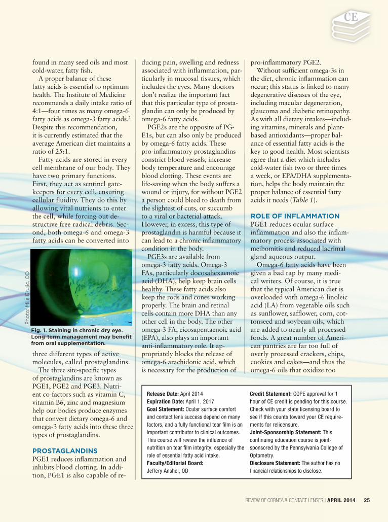
found in many seed oils and most cold-water, fatty fi sh.
A proper balance of these fatty acids is essential to optimum health. The Institute of Medicine recommends a daily intake ratio of 4:1—four times as many omega-6 fatty acids as omega-3 fatty acids.2
Despite this recommendation, it is currently estimated that the average American diet maintains a ratio of 25:1.
Fatty acids are stored in every cell membrane of our body. They have two primary functions. First, they act as sentinel gate-keepers for every cell, ensuring cellular fl uidity. They do this by allowing vital nutrients to enter the cell, while forcing out de-structive free radical debris. Sec-ond, both omega-6 and omega-3 fatty acids can be converted into
three different types of active molecules, called prostaglandins.
The three site-specifi c types of prostaglandins are known as PGE1, PGE2 and PGE3. Nutri-ent co-factors such as vitamin C, vitamin B6, zinc and magnesium help our bodies produce enzymes that convert dietary omega-6 and omega-3 fatty acids into these three types of prostaglandins.
PROSTAGLANDINSPGE1 reduces infl ammation and inhibits blood clotting. In addi-tion, PGE1 is also capable of re-
ducing pain, swelling and redness associated with infl ammation, par-ticularly in mucosal tissues, which includes the eyes. Many doctors don’t realize the important fact that this particular type of prosta-glandin can only be produced by omega-6 fatty acids.
PGE2s are the opposite of PG-E1s, but can also only be produced by omega-6 fatty acids. These pro-infl ammatory prostaglandins constrict blood vessels, increase body temperature and encourage blood clotting. These events are life-saving when the body suffers a wound or injury, for without PGE2 a person could bleed to death from the slightest of cuts, or succumb to a viral or bacterial attack. However, in excess, this type of prostaglandin is harmful because it can lead to a chronic infl ammatory condition in the body.
PGE3s are available from omega-3 fatty acids. Omega-3 FAs, particularly docosahexaenoic acid (DHA), help keep brain cells healthy. These fatty acids also keep the rods and cones working properly. The brain and retinal cells contain more DHA than any other cell in the body. The other omega-3 FA, eicosapentaenoic acid (EPA), also plays an important anti-infl ammatory role. It ap-propriately blocks the release of omega-6 arachidonic acid, which is necessary for the production of
pro-infl ammatory PGE2. Without suffi cient omega-3s in
the diet, chronic infl ammation can occur; this status is linked to many degenerative diseases of the eye, including macular degeneration, glaucoma and diabetic retinopathy. As with all dietary intakes—includ-ing vitamins, minerals and plant-based antioxidants—proper bal-ance of essential fatty acids is the key to good health. Most scientists agree that a diet which includes cold-water fi sh two or three times a week, or EPA/DHA supplementa-tion, helps the body maintain the proper balance of essential fatty acids it needs (Table 1).
ROLE OF INFLAMMATIONPGE1 reduces ocular surface infl ammation and also the infl am-matory process associated with meibomitis and reduced lacrimal gland aqueous output.
Omega-6 fatty acids have been given a bad rap by many medi-cal writers. Of course, it is true that the typical American diet is overloaded with omega-6 linoleic acid (LA) from vegetable oils such as sunfl ower, saffl ower, corn, cot-tonseed and soybean oils, which are added to nearly all processed foods. A great number of Ameri-can pantries are far too full of overly processed crackers, chips, cookies and cakes—and thus the omega-6 oils that oxidize too
Release Date: April 2014Expiration Date: April 1, 2017Goal Statement: Ocular surface comfort and contact lens success depend on many factors, and a fully functional tear film is an important contributor to clinical outcomes. This course will review the influence of nutrition on tear film integrity, especially the role of essential fatty acid intake.Faculty/Editorial Board: Jeffery Anshel, OD
Credit Statement: COPE approval for 1 hour of CE credit is pending for this course. Check with your state licensing board to see if this counts toward your CE require-ments for relicensure.Joint-Sponsorship Statement: This continuing education course is joint-sponsored by the Pennsylvania College of Optometry.Disclosure Statement: The author has no financial relationships to disclose.
REVIEW OF CORNEA & CONTACT LENSES | APRIL 2014 25
Fig. 1. Staining in chronic dry eye. Long-term management may benefi t from oral supplementation.
Ph
oto
: M
ile B
rujic, O
D
024_rccl0414_F5CE.indd 25 4/3/14 10:50 AM
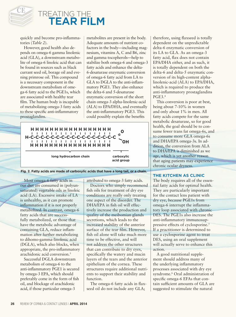
quickly and become pro-infl amma-tories (Table 2).
However, good health also de-pends on omega-6 gamma linolenic acid (GLA), a downstream metabo-lite of omega-6 linoleic acid that can be found in sources such as black currant seed oil, borage oil and eve-ning primrose oil. This compound is a necessary component in the downstream metabolism of ome-ga-6 fatty acid to the PGE1s, which are associated with healthy tear fi lm. The human body is incapable of metabolizing omega-3 fatty acids to these specifi c anti-infl ammatory prostaglandins.
Most omega-6 fatty acids in our diet are consumed in (polyun-saturated) vegetable oils as linoleic acid (LA). Excessive intake of LA is unhealthy, as it can promote infl ammation if it is not properly metabolized. In contrast, omega-6 fatty acids that are success-fully metabolized, or those that have the metabolic advantage of containing GLA, reduce infl am-mation after further metabolizing to dihomo-gamma-linolenic acid (DGLA), which also blocks, when appropriate, the pro-infl ammatory arachidonic acid conversion.3
Successful DGLA downstream metabolism of omega-6 to the anti-infl ammatory PGE1 is secured by omega-3 EPA, which should preferably come in the form of fi sh oil, and blockage of arachidonic acid, if those particular omega-3
metabolites are present in the body. Adequate amounts of nutrient co-factors in the body—including mag-nesium, vitamins A, C and B6, zinc and gamma tocopherols—help to stabilize both omega-6 and omega-3 fatty acids and enhance the delta-6-desaturase enzymatic conversion of omega-6 fatty acid from LA to GLA to DGLA to the anti-infl am-matory PGE1. They also enhance the delta-6 and 5-desaturase enzymatic conversion of the short chain omega-3 alpha-linolenic-acid (ALA) to EPA/DHA, and eventually the anti-infl ammatory PGE3. This could possibly explain the benefi ts
attributed to omega-3 fatty acids.Doctors who simply recommend
fi sh oils for treatment of dry eye conditions are really only treating one aspect of the disorder. The DHA/EPA in fi sh oil will effec-tively increase the production and quality of the meibomian glands secretions, which leads to the increased stability of the anterior surface of the tear fi lm. However, fi sh oil alone will take much more time to be effective, and will not address the other structures that can contribute to dry eyes, specifi cally the watery and mucin layers of the tears and the anterior epithelium of the cornea. These structures require additional nutri-ents to support their stability and function.
The omega-6 fatty acids in fl ax-seed oil do not include any GLA;
therefore, using fl axseed is totally dependent on the unpredictable delta-6 enzymatic conversion of its LA to GLA. As an omega-3 fatty acid, fl ax does not contain EPA/DHA either, and as such, it is totally dependent on both the delta-6 and delta-5 enzymatic con-version of its high-content alpha-linolenic-acid (ALA) to EPA/DHA, which is required to produce the anti-infl ammatory prostaglandins PGE3.4
This conversion is poor at best, being about 7-10% in women and only about 1% in men. All fatty acids compete for the same metabolic desaturase, so for good health, the goal should be to con-sume fewer trans fat omega-6s, and to consume more GLA omega-6s and DHA/EPA omega-3s. In ad-dition, the conversion from ALA to DHA/EPA is diminished as we age, which is yet another reason that aging patients may experience chronic ocular dryness.
THE KITCHEN AS CLINICThe body requires all of the essen-tial fatty acids for optimal health. They are particularly important for the contact lens patient with dry eye, because PGE1s from omega-6 interrupt the infl amma-tory loop associated with chronic DES. The PGE1s also increase the anti-infl ammatory immunosup-pressive effects of cyclosporine.5
If a practitioner is determined to use a cyclosporine agent to treat DES, using an oral supplement will actually serve to enhance this action.
A good nutritional supple-ment should address many of the underlying infl ammatory processes associated with dry eye syndrome.6 Oral administration of specifi c omega-6 EFAs that con-tain suffi cient amounts of GLA are suggested to stimulate the natural
26 REVIEW OF CORNEA & CONTACT LENSES | APRIL 2014
TREATING THE
TEAR FILM
Fig. 2. Fatty acids are made of carboxylic acids that have a long tail, or a chain.
024_rccl0414_F5CE.indd 26 4/3/14 10:18 AM
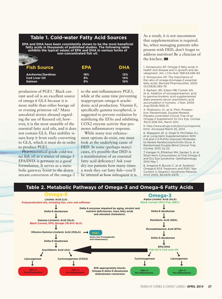
production of PGE1.7 Black cur-rant seed oil is an excellent source of omega-6 GLA because it is more stable than either borage oil or evening primrose oil.8 Many anecdotal stories abound regard-ing the use of fl axseed oil; how-ever, it is the most unstable of the essential fatty acid oils, and it does not contain GLA. Flax stability is-sues keep it from easily converting to GLA, which it must do in order to produce PGE1.
Pharmaceutical grade cold-wa-ter fi sh oil as a source of omega-3 EPA/DHA is germane to a good formulation. It serves as a meta-bolic gateway boost to the down-stream conversion of the omega-3
to the anti-infl ammatory PGE3, while at the same time preventing inappropriate omega-6 arachi-donic acid production. Vitamin E, specifi cally gamma tocopherol, is suggested to prevent oxidation by stabilizing the EFAs and inhibiting COX2 enzyme activity that pro-motes infl ammatory response.
While many tear enhance-ment options do exist, one must look at the underlying cause of DED. In some (perhaps many) cases, it’s possible that DED is a manifestation of an essential fatty acid defi ciency! Ask your dry eye patients how many times a week they eat fatty fi sh—you’ll be amazed at how infrequent it is.
As a result, it is not uncommon that supplementation is required. So, when managing patients who present with DED, don’t forget to address nutrition! Be a clinician of the kitchen. RCCL
1. Simopoulos AP. Omega-3 fatty acids in health and disease and in growth and de-velopment. Am J Clin Nutr 1991;54:438–63. 2. Simopoulos AP. The importance of the ratio of omega-6/omega-3 essential fatty acids. Biomed Pharmacother. 2002 Oct;56(8):365-79.3. Barham JB1, Edens MB, Fonteh AN, et al. Addition of eicosapentaenoic acid to gamma-linolenic acid-supplemented diets prevents serum arachidonic acid accumulation in humans. J Nutr. 2000 Aug;130(8):1925-31.4. Wojtowicz JC, et al. Pilot, Prospec-tive, Randomized, Double-Masked, Placebo-controlled Clinical Trial of an Omega-3 Supplement for Dry Eye. Cornea. 30(3):308-314, March 2011.5. http://www.drugs.com/pro/cyclosporine.html- Accessed March 25, 20146. Sheppard JD Jr, Singh R, McClellan AJ, et al. Long-term Supplementation With n-6 and n-3 PUFAs Improves Moderate-to-Severe Keratoconjunctivitis Sicca: A Randomized Double-Blind Clinical Trial. Cornea. 2013 Jul 23.7. Kangari H, Eftekhari MH, Sardari S, et al. Short-term Consumption of Oral Omega-3 and Dry Eye Syndrome. Ophthalmology. 2013 May 1.8. Aragona P, Bucolo C, et al. Systemic Omega-6 EFA Treatment and PGE1 Tear Content in Sjögren’s Syndrome Patients. IOVS 2005; 46:4474-4479.
REVIEW OF CORNEA & CONTACT LENSES | APRIL 2014 27
Fish Source EPA DHA Anchovies/Sardines 18% 12% Cod Liver Oil 9% 14% Salmon 9% 10%
EPA and DHA have been consistently shown to be the most benefi cial fatty acids in thousands of published studies. The following table
exhibits the typical values of EPA and DHA in various forms of non-concentrated fi sh oil.
Table 1. Cold-water Fatty Acid Sources
Fish So
Delta 6 enzymes impaired by aging, alcohol and nutrient defi ciencies, trans fatty acids
and elevated cholesterol.
EPA appropriately blocks Omega-6 delta-5-desaturase
downstream conversion
Omega-6Linoleic Acid (LA)
Polyunsaturated oils, including fl ax, corn and saffl ower
Delta-6 desaturase
Gamma-Linolenic Acid (GLA)Black Currant, EPO, Borage (18-24% GLA)
Dihomo-Gamma-Linolenic Acid (DGLA)
Delta-5-desaturase
Arachidonic Acid (AA)
Lipoxygenase Cyclooxygenase (COX2)
Series One Prostaglandin Anti-infl ammatory
PGE1
Omega-3Alpha-Linoleic Acid (ALA)
Black Currant (15%) Flax (85%)
Delta-6 desaturase
Steridonic Acid (SDA)
Eicosatetiaenoic Acid (ETA)
Delta-5-desaturase
EPA/DHA Fish Oil & Cod Liver Oil
Cyclooxygenase Lipoxygenase
PGE—3 LBT—5 Pro-infl ammatory Pro-infl ammatory
Table 2. Metabolic Pathways of Omega-3 and Omega-6 Fatty Acids
PGE—2 Pro-infl ammatoryLBT—4
Pro-infl ammatory
024_rccl0414_F5CE.indd 27 4/3/14 3:14 PM

Examination Answer Sheet Valid for credit through April 1, 2017
This exam can be taken online at www.reviewofcontactlenses.com. Upon passing the exam, you can view your results immediately. You can also view your test history at any time from the website.
Treating the Tear Film: Nutrition is Essential
Directions: Select one answer for each question in the exam and completely darken the appropriate circle. A minimum score of 70% is required to earn credit.
Mail to: Jobson - Optometric CE, PO Box 488, Canal Street Station, New York, NY 10013
Payment: Remit $20 with this exam. Make check payable to Jobson Medical Information LLC.
COPE approval for 1 hour of CE credit is pending for this course.
This course is joint-sponsored by the Pennsylvania College of Optometry
There is an eight-to-10 week processing time for this exam.
1. A B C D 1 = Excellent 2 = Very Good 3 = Good 4 = Fair 5 = Poor
2. A B C D Rate the effectiveness of how well the activity: 3. A B C D
4. A B C D 11. Met the goal statement: 1 2 3 4 5
5. A B C D 12. Related to your practice needs: 1 2 3 4 5
6. A B C D 13. Will help you improve patient care: 1 2 3 4 5
7. A B C D 14. Avoided commercial bias/influence: 1 2 3 4 5
8. A B C D 15. How would you rate the overall 9. A B C D quality of the material presented? 1 2 3 4 5
10. A B C D 16. Your knowledge of the subject was increased: Greatly Somewhat Little 17. The difficulty of the course was: Complex Appropriate Basic How long did it take to complete this course? Comments on this course:
Suggested topics for future CE articles:
Please retain a copy for your records. Please print clearly.
You must choose and complete one of the following three identifier types:
1 SS # - -
Last 4 digits of your SS # and date of birth State Code and License #: (Example: NY12345678)
2 - 3
First Name
Last Name
The following is your: Home Address Business Address
Business Name
Address
City State
ZIP
Telephone # - -
Fax # - -
By submitting this answer sheet, I certify that I have read the lesson in its entirety and completed the self-assessment exam personally based on the material presented. I have not obtained the answers to this exam by any fraudulent or improper means.
Signature Date
Lesson 109885 RO-RCCL-0414
1. Which of the following is not a source of GLA? a. Black currant seed oil b. Evening primrose oil c. Flax seed oil d. Borage oil
2. Fatty acids that are required to be a part of the diet are termed:
a. Required fatty acids b. Essential fatty acids c. Endogenous fatty acids d. Exogenous fatty acids
3. Which nutrients are important co-factors in Omega EFA conversion?
a. Vitamins C and B6 b. Zinc c. Magnesium d. All of the above
4. Which of these nutrients produce prostaglandin E1? a. Alpha-linolenic acid b. Dihomo-Gamma-linolenic acid c. Eicosapentaenoic acid d. Docosahexaenoic acid
5. Prostaglandin E1 is most responsible for: a. Anti-inflammatory mucins b. Pro-inflammatory cytokines c. Pro-inflammatory mucins d. Pro-inflammatory lipoxgenase
6. The fatty acid that directly leads to pro-inflammatory prostaglandin E2 is:
a. Docosahexaenoic acid b. Eicosapentaenoic acid c. Linoleic acid d. Arachidonic acid
7. The conversion of alpha-linoleic acid to EPA/DHA is: a. About 100% in adults b. About 10% in men c. About 10% in women d. About 1% in women
8. The best balance of omega-6 to omega-3 essential fatty acids is:
a. 10:1 b. 15:1 c. 75:1 d. 4:1
9. DHA is most abundant in which tissues? a. Joints b. Brain and retina c. Collagen d. Skin
10. Flax seed oils can be problematic in treating dry eye because:
a. They are highly unstable b. The contain too much omega-3 c. The contain too much omega-6 d. They come from a vegetable source
CE TEST
28 REVIEW OF CORNEA & CONTACT LENSES | APRIL 2014
TREATING THE
TEAR FILM
024_rccl0414_F5CE.indd 28 4/3/14 10:18 AM
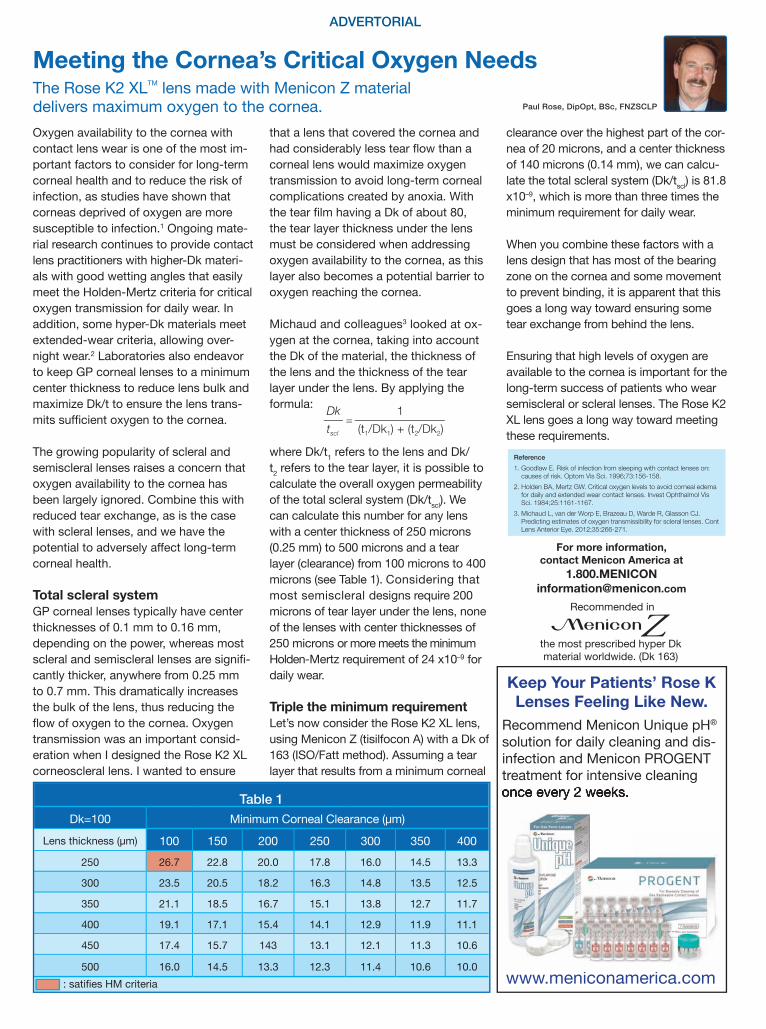
Meeting the Cornea’s Critical Oxygen NeedsThe Rose K2 XLTM lens made with Menicon Z material delivers maximum oxygen to the cornea.Oxygen availability to the cornea with contact lens wear is one of the most im-portant factors to consider for long-term corneal health and to reduce the risk of infection, as studies have shown that corneas deprived of oxygen are more susceptible to infection.1 Ongoing mate-rial research continues to provide contact lens practitioners with higher-Dk materi-als with good wetting angles that easily meet the Holden-Mertz criteria for critical oxygen transmission for daily wear. In addition, some hyper-Dk materials meet extended-wear criteria, allowing over-night wear.2 Laboratories also endeavor to keep GP corneal lenses to a minimum center thickness to reduce lens bulk and maximize Dk/t to ensure the lens trans-mits suf cient oxygen to the cornea.
The growing popularity of scleral and semiscleral lenses raises a concern that oxygen availability to the cornea has been largely ignored. Combine this with reduced tear exchange, as is the case with scleral lenses, and we have the potential to adversely affect long-term corneal health.
Total scleral systemGP corneal lenses typically have center thicknesses of 0.1 mm to 0.16 mm, depending on the power, whereas most scleral and semiscleral lenses are signi -cantly thicker, anywhere from 0.25 mm to 0.7 mm. This dramatically increases the bulk of the lens, thus reducing the ow of oxygen to the cornea. Oxygen transmission was an important consid-eration when I designed the Rose K2 XL corneoscleral lens. I wanted to ensure
that a lens that covered the cornea and had considerably less tear ow than a corneal lens would maximize oxygen transmission to avoid long-term corneal complications created by anoxia. With the tear lm having a Dk of about 0, the tear layer thickness under the lens must be considered when addressing oxygen availability to the cornea, as this layer also becomes a potential barrier to oxygen reaching the cornea.
Michaud and colleagues3 looked at ox-ygen at the cornea, taking into account the Dk of the material, the thickness of the lens and the thickness of the tear layer under the lens. By applying the formula:
where Dk/t1 refers to the lens and Dk/t2 refers to the tear layer, it is possible to calculate the overall oxygen permeability of the total scleral system (Dk/tscl). We can calculate this number for any lens with a center thickness of 250 microns (0.25 mm) to 500 microns and a tear layer (clearance) from 100 microns to 400 microns (see Table 1). Considering that most semiscleral designs require 200 microns of tear layer under the lens, none of the lenses with center thicknesses of 250 microns or more meets the minimum Holden-Mertz requirement of 24 x10–9 for daily wear.
Triple the minimum requirementLet’s now consider the Rose K2 XL lens, using Menicon Z (tisilfocon A) with a Dk of 163 (ISO/Fatt method). Assuming a tear layer that results from a minimum corneal
clearance over the highest part of the cor-nea of 20 microns, and a center thickness of 140 microns (0.14 mm), we can calcu-late the total scleral system (Dk/tscl) is 1. x10–9, which is more than three times the minimum requirement for daily wear.
When you combine these factors with a lens design that has most of the bearing zone on the cornea and some movement to prevent binding, it is apparent that this goes a long way toward ensuring some tear exchange from behind the lens.
Ensuring that high levels of oxygen are available to the cornea is important for the long-term success of patients who wear semiscleral or scleral lenses. The Rose K2 XL lens goes a long way toward meeting these requirements.
ADVERTORIAL
Table 1Dk=100 Minimum Corneal Clearance (μm)
Lens thickness (μm) 100 150 200 250 300 350 400250 26.7 22. 20.0 17. 16.0 14.5 13.3
300 23.5 20.5 1 .2 16.3 14. 13.5 12.5
350 21.1 1 .5 16.7 15.1 13. 12.7 11.7
400 19.1 17.1 15.4 14.1 12.9 11.9 11.1
450 17.4 15.7 143 13.1 12.1 11.3 10.6
500 16.0 14.5 13.3 12.3 11.4 10.6 10.0: sati es HM criteria
Dk 1tscl (t1/Dk1) + (t2/Dk2)
=
Reference1. Goodlaw E. Risk of infection from sleeping with contact lenses on:
causes of risk. Optom Vis Sci. 1996;73:156-158.2. Holden BA, Mertz GW. Critical oxygen levels to avoid corneal edema
for daily and extended wear contact lenses. Invest Ophthalmol Vis Sci. 1984;25:1161-1167.
3. Michaud L, van der Worp E, Brazeau D, Warde R, Glasson CJ. Predicting estimates of oxygen transmissibility for scleral lenses. Cont Lens Anterior Eye. 2012;35:266-271.
Paul Rose, DipOpt, BSc, FNZSCLP
Keep Your Patients’ Rose K Lenses Feeling Like New.
Recommend Menicon Unique pH® solution for daily cleaning and dis-infection and Menicon PROGENT treatment for intensive cleaning once every 2 weeks.
www.meniconamerica.com
Recommended in
the most prescribed hyper Dk material worldwide. (Dk 163)
For more information, contact Menicon America at
1.800.MENICON [email protected]
RCCL0414_Menicon.indd 1 3/27/14 3:07 PM
Topical antibiotics revo-lutionized the way eye doctors care for patients with at-risk corneas. The ability to target
infectious organisms with a site-spe-cifi c therapy limited to the anterior segment of the eye improved clinical outcomes by orders of magnitude. Systemic medications would be impractical and ineffective in most ocular infections. Without topical therapies, many patients would suffer long-term visual and ocular health consequences.
Fluoroquinolone eye drops are now routinely and confi dently prescribed as fi rst-line therapy for a number of acute and chronic oph-thalmic conditions, such as bacte-rial conjunctivitis, blepharitis and corneal disorders (e.g., abrasions, infi ltrates, ulcers). They are also used routinely for prophylaxis, both pre- and post-ocular surgery.
Lest we take these workhorse drugs for granted, it’s important to review the history of the fl uoroqui-nolone class and how each ‘genera-tion’ has surpassed the previous one, then look to what’s ahead.
IT BEGAN BY ACCIDENTPrior to the introduction of fl uoro-quinolones, we simply had quino-lones. These were the fi rst antimi-crobials to be developed in a lab. The fi rst quinolone, nalidixic acid, was accidentally discovered in 1962 during the manufacture of the anti-malarial compound chloroquine. It was used systemically for the treat-ment of urinary tract infections, but the agent had little effect on gram-negative organisms. Alterations to
its structure led to the development of second-generation products, such as oxolinic acid and clinoxacin. The use of these agents was also limited, due to poor systemic bioavailability and renal toxicity.1,2
The addition of fl uorination to quinolones gave rise to the then-new class known as fl uoroquino-lones. The improved gram-positive activity, combined with solubility in ophthalmic solutions, expanded the clinical use of these drugs to the ocular fi eld.3 The fi rst fl uoroqui-nolone, norfl oxacin, introduced in 1978, resulted from the addition of fl uorine at the C-6 position. It was the fi rst fl uoroquinolone to be used in the treatment of an ocu-lar condition—namely, bacterial conjunctivitis. While norfl oxacin exhibited excellent activity against gram-negative organisms, it proved to be largely ineffective against gram-positive bacteria.
HOW THEY WORKSo, what exactly is the mechanism of action of the fl uoroquinolones? Put simply, it is a bactericidal effect. Fluoroquinolones act in a unique manner: directly inhibit-ing DNA synthesis. This effect is mediated through the formation of a complex between the drug, the bacterial DNA and two essential bacterial enzymes (i.e., DNA gy-rase and topoisomerase IV).4
DNA gyrase is an essential enzyme for DNA replication and transcription, while topoisomerase IV separates the interlocking of daughter DNA strands. If either of these processes is prevented, DNA and RNA synthesis and cell growth
are blocked, which ultimately leads to cell death. Second- and third-generation fl uoroquinolones pref-erentially inhibit one or the other, while the fourth-generation agents have the ability to exhibit balanced inhibition of both enzymes.5
In the early 1990s, the addition of a cylopropryl ring at the R1 position led to the development of the second-generation fl uoro-quinolone, ciprofl oxacin, which had increased antibacterial activity against gram-positive and gram-negative pathogens.
Adding a pyridobenoxazine ring between the R1 and R8 positions led to the creation of another second-generation drug, ofl oxacin.3
While many consider levofl oxacin a third-generation fl uoroquinolone, it’s really just a levo-isomer of ofl ox-acin and not a truly new molecular structure. In reality, its designation as third-generation fl uoroquinolone is mostly due to timing: it was intro-duced nearly a decade after its most immediate predecessors.
A number of studies have demon-strated that, although these older-generation fl uoroquinolones can provide coverage against the most frequently encountered gram-posi-tive and gram-negative pathogens, there is increasing resistance to these agents—especially in gram-positive organisms, such as Staph. aureaus.6-8
TODAY’S PRACTICEThe fourth-generation drugs, primarily used in modern clinical practice, are gatifl oxacin and moxi-fl oxacin. These entries are known for their enhanced activity against
Pharma Science & PracticeBy Elyse L. Chaglasian, OD, and Tammy Than, MS, OD
FQs: The Secret of Our SuccessThese popular drugs are essential to the care we provide. Why have fl uoroquinolones become such workhorse drugs? Where might they be headed? Let’s fi nd out.
30 REVIEW OF CORNEA & CONTACT LENSES | APRIL 2014
030_rccl0414_PSP.indd 30 4/3/14 2:41 PM
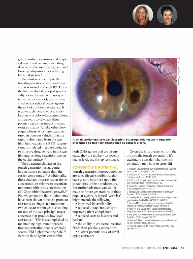
gram-positive organisms and atypi-cal mycobacteria, improved drug delivery in the anterior segment and lower predisposition for selecting bacterial strains.9
The most recent entry to the fourth-generation class, besifl oxa-cin, was introduced in 2009. This is the fi rst product developed specifi -cally for ocular use, with no sys-temic use to speak of; this is often cited as a benefi cial hedge against the risk of antibiotic resistance. It is an entirely new chemical entity known as a chloro-fl uoroquinolone, and appears to offer excellent potency against gram-positive and resistant strains. Unlike other fl uo-roquinolones, which are manufac-tured in aqueous vehicles that are rapidly eliminated from the tear fi lm, besifl oxacin is a 0.6% suspen-sion, formulated in a base designed to improve drug delivery in the tear fi lm and prolong retention time on the ocular surface.10
The structural changes in the fourth-generation drugs confer less resistance potential than the earlier compounds.5,9 Additionally, these changes increase ocular tissue concentrations relative to organism minimum inhibitory concentration (MIC) to inhibit bacterial growth.5,9
Fourth-generation fl uoroquinolones have been shown to be less prone to resistance to single-step mutations (which occur within genes encoding for one of the two principal target enzymes) that produce low-level resistance.11 This is accomplished by maintaining high mutant preven-tion concentration that is generally several fold higher than the MIC.11
Because these agents can inhibit
both DNA gyrase and topoisom-erase, they are unlikely to develop higher level, multi-step resistance.
TOMORROW’S POTENTIALFourth-generation fl uoroquinolones are safe, effective antibiotics that have greatly improved upon the capabilities of their predecessors. But further advances can still be made to future generations of these popular agents. A typical ‘wish list’ might include the following:
• improved bioavailability • reduced dosage frequency to
improve patient compliance• reduced costs to insurers and
patients• the ability to eradicate infection
faster than previous generations• a lower potential risk of devel-
oping resistance
Given the improvements from the third to the fourth generation, it’s exciting to consider what the fi fth generation may have in store! RCCL
1. Andriole V. The quinolones: past, present and future. Clin Infect Dis. 2005 Jul 15.41 Suppl2:S113-9. 2. Applebaum PC, Huber PA, The fluoroquinolone antibacterialss. Int J Antimicrob Agents 16:5-15, 2000.3. Ball P, et al. Therapeutic advances of new fluoroquinolones. Exp Opin On Investig Drugs 1998 May;7(5):761-83.4. Hooper DC. Emerging mechanisms of fluoroquinolone resis-tance. Emerg Inf Dis 2001;7:337-341.5. Blondeau JM. Fluoroquinolones: mechanism of action. Surv Ophthalmol 2004;49(suppl 2):S73-S78.6. Chaudhry NA, et al. Emerging ciprofloxacin resistant pseudomo-nas aeruginosa. AmJ Ophthalmol 1999:128:509-510.7. Goldstein MH, et al. Fluoroquinolone resistance in bacterial keratitis: a 5-year review. Ophthalm 1999: 106:1313-8.8. Asbell PA, et al. Ocular TRUST: Nationwide antimicrobial susceptibility patterns. Am J Ophthalmol 2008;145:951–958. 9. Hwang DG. Fluoroquinolone resistance in ophthalmology. Surv Ophthalmol 2004;49(suppl2):S79-S83.10. Gardner S. Fluoroquinolone is developed expressly for ophthal-mic use. Optometry Times, July 2009.11. Hesje Ck, et al. MICs, MPCs and PK/PDs. Expert Rev Resp Med 2007;1:7-16.
REVIEW OF CORNEA & CONTACT LENSES | APRIL 2014 31
A small, peripheral corneal ulceration. Fluoroquinolones are frequently prescribed to treat conditions such as corneal ulcers.
Ph
oto
: Willia
m T
ow
nsen
d, O
D
030_rccl0414_PSP.indd 31 4/3/14 2:41 PM

F itting patients with diagnostic hard or gas permeable lenses was almost a neces-sity when both of
these modalities fi rst became avail-able on the market. Practitioners relied on diagnostic lens sets and would routinely fi t the patient with several lens options in-offi ce to determine the appropriate fi t and power for each patient. The process was tedious—for those on both sides of the slit lamp.
It has been a great boon to GP lens practice that nowadays most labs can simply create a lens based on the patient’s keratometry values and refraction. Even more complex lenses, such as bitorics and GP multifocals, can be ordered empiri-cally—eliminating the need for a diagnostic fi t.1 Empirical fi tting offers the potential to reduce the chair time of every patient. “Think of it as ordering a custom diagnostic lens for every patient,” says Doug Benoit, OD.1 While empirical fi t-ting works very well in most cases, certain patient scenarios will still require diagnostic lens fi tting.
This technique is particularly helpful in observing the lens-to-cornea relationship and lid interac-tion.2 These fi ts can be especially useful for patients who present with irregular corneal astigmatism due to keratoconus, pellucid marginal degeneration or corneal surgery (e.g., transplant, RK, LASIK). And because lens/eye dynamics are also compromised by ocular surface dis-ease, such patients can benefi t from diagnostic lens fi tting as well. Scleral lenses are almost always fi t using a
diagnostic lens kit, due to factors such as total corneal clearance, scleral contour and lens settling.
MAINTENANCE Diagnostic lenses can be stored either wet or dry. Wet diagnostic lenses have excellent wetting capa-bilities and are ready for immediate use—there’s no need to clean or condition them before use. Keep in mind, however, that when lenses
are stored wet they must have their solution replaced every 30 days to ensure appropriate disinfection.3 Be-cause this can be a time-consuming process, storing diagnostic lenses dry is the standard practice for most doctors.
Such lenses need to be cleaned and conditioned before being ap-plied to a patient’s eye.
The lenses should be cleaned with an approved GP lens cleaner, such as Boston Simplus/Original/Ad-vanced (Bausch + Lomb) or Opti-mum (Lobob). Cleaning the lenses thoroughly ensures a smooth surface free of debris, so spend at least 30 seconds cleaning the lens (Figure 1). Once cleaned, it will need to be conditioned; this will ensure the lens wets well. GP conditioning agents such as Boston Conditioning solu-tion and Boston Simplus work very
well for this purpose. Additionally, an off-label use of Biotrue can aid in wetting. Biotrue contains hyaluro-nan, which binds to the lens surface and can help stubborn, non-wetting GP lenses (Figure 2). Adding Biotrue to the conditioning process may help GP lenses to wet more easily.
Following the trial fi tting, the lenses must be cleaned and disin-fected. The CDC suggests using ophthalmic grade hydrogen per-
oxide 3% to disinfect rigid lenses. According to the CDC, “Contact lenses used in trial fi ttings should be disinfected after each fi tting by using a hydrogen peroxide contact lens disinfecting system for 10 minutes.”3-4
Once the lenses have been suc-cessfully disinfected, they should be stored dry. Be sure to take extra care to ensure all liquid is removed from the lens case, as well as the lens itself. If there is even one drop of liquid, the lens may bind to the case, potentially causing the lens to break as it is being removed from the case when reused at a later date.
TOPICAL ANESTHETICSThe use of a topical anesthetic may be helpful during a diagnostic GP fi tting. If a patient has never worn GP lenses, instill one drop of anes-
Dust Off Those Diagnostic LensesWhile empirical fi tting is becoming the norm, patients presenting with irregular corneas require a more specialized lens fi tting approach.
By Stephanie L. Woo, OD The GP Expert
32 REVIEW OF CORNEA & CONTACT LENSES | APRIL 2014
For lens materials with Dk values over 100, such as Boston XO (Bausch + Lomb), Boston XO2 (Bausch + Lomb) and Menicon Z (Menicon), Boston Advanced may be too abrasive for the lens sur-face. This can cause the material, which is softer and more pliable, to break down faster and scratch more easily. If your patient wears a lens made of a hyper-Dk material, consider recommending other GP care products such as Boston Simplus or Optimum by Lobob.
Hyper-Dk Lens Care
032_rccl0414_gpe.indd 32 4/3/14 10:19 AM
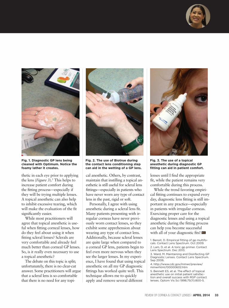
thetic in each eye prior to applying the lens (Figure 3).5 This helps to increase patient comfort during the fi tting process—especially if they will be trying multiple lenses. A topical anesthetic can also help to inhibit excessive tearing, which will make the evaluation of the fi t signifi cantly easier.
While most practitioners will agree that topical anesthetic is use-ful when fi tting corneal lenses, how do they feel about using it when fi tting scleral lenses? Sclerals are very comfortable and already feel much better than corneal GP lenses. So, is it really even necessary to use a topical anesthetic?
The debate on this topic is split; unfortunately, there is no clear-cut answer. Some practitioners will argue that a scleral lens is so comfortable that there is no need for any topi-
cal anesthetic. Others, by contrast, maintain that instilling a topical an-esthetic is still useful for scleral lens fi ttings—especially in patients who have never worn any type of contact lens in the past, rigid or soft.
Personally, I agree with using anesthetic during a scleral lens fi t. Many patients presenting with ir-regular corneas have never previ-ously worn contact lenses, so they exhibit some apprehension about wearing any type of contact lens. Additionally, because scleral lenses are quite large when compared to a corneal GP lens, patients begin to feel even more nervous when they see the larger lenses. In my experi-ence, I have found that using topical anesthetic on all my GP diagnostic fi ttings has worked quite well. This technique allows me to quickly apply and remove several different
lenses until I fi nd the appropriate fi t, while the patient remains very comfortable during this process.
While the trend favoring empiri-cal fi tting continues to expand every day, diagnostic lens fi tting is still im-portant in any practice—especially in patients with irregular corneas. Exercising proper care for the diagnostic lenses and using a topical anesthetic during the fi tting process can help you become successful with all of your diagnostic fi ts! RCCL
1. Benoit, D. Empirical fi tting of gp multifo-cals. Contact Lens Spectrum. Oct 2009. 2. Lam, D, et al. A toric gp primer. Contact Lens Spectrum. Dec 2013.3. Ward, M. Maintaining and Disinfecting GP Diagnostic Lenses. Contact Lens Spectrum. Sep 2006. 4. http://www.cdc.gov/mmwr/preview/mmwrhtml/00000602.htm5. Bennett ES, et al. The effect of topical anesthetic use on initial patient satisfac-tion and overall success with RGP contact lenses. Optom Vis Sci 1998;75(11):800-5.
REVIEW OF CORNEA & CONTACT LENSES | APRIL 2014 33
Fig. 1. Diagnostic GP lens being cleaned with Optimum. Notice the foamy lather it creates.
Fig. 2. The use of Biotrue during the contact lens conditioning step can aid in the wetting of a GP lens.
Fig. 3. The use of a topical anesthetic during diagnostic GP fi tting can aid in patient comfort.
032_rccl0414_gpe.indd 33 4/3/14 10:19 AM
As the old saying goes, if I had a nickel for every time I heard a doctor tell me that I didn’t know his or
her staff, I’d have a lot of nickels. That remark is usually the cul -
mination of a doctor telling me something along these lines:
“At our last offi ce meeting, we discussed that patients need to be called the same day their contact lenses come in. I’ve outlined this protocol to my staff repeatedly, but they only follow it for a few days before reverting to doing it only occasionally. It’s so frustrating.”
After hearing that I’ll ask, “On a scale of one to 10, how important is it that your staff do this consis-tently?”
“It’s extremely important—it’s like a 12! That’s why I continue to ask them to do it!”
“Well, you need to sit your entire staff down at your next meeting and make it clear that it’s a 12. If they can’t get it done, and there isn’t a good reason—not an excuse,
but a legitimate reason—why they can’t, then they’ll have to seek work elsewhere.”
“What? Are you actually telling me that if my staff doesn’t do what they’re supposed to do, I’m sup-posed to fi re them?”
“Yes, I am.”
“But, Gary, you don’t know mystaff. They’ll fi nd 100 reasons for why my idea is a bad one or 200 excuses for why they can’t get the task done.”
And round and round the con-versation goes.
IT’S UP TO YOUThe essence of most “staff” prob-lems isn’t truly a problem with the staff at all. Instead, these problems are often due to a lack of two things repeatedly seen by our con-sultants in the majority of offi ces with which we work.
First, there are no clear systems or processes in place that spell out exactly how and when specifi c tasks must be completed. In this case, calling the patient when their lenses arrive should be connected to opening the box of contact lenses. If this is already the proce-dure and the task is still not being done consistently, then we have ar-rived at the second most common observation—a lack of leadership.Being a leader doesn’t solely mean
being a hatchet man or woman and fi ring people who don’t do what they should. It means that you have to possess the ability to clearly communicate what needs to be done, and why those tasks must be done. In this case, notify-ing patients should be tied to your
practice’s mission of providing exemplary customer service.
If that’s something in your practice’s DNA and a part of every fi ber of everything your staff does, you have a greater likelihood of successfully completing any task. It also forces you to make sure that tasks that you are assigning to your staff are aligned with your prac-tice’s vision and values, and not solely there because you thought to yourself, “Well, I guess that seems like a good idea.”
Next, you have to decide and be-lieve that the task at hand is really a “12.” If so, and it’s consistently not being done, your next job as a leader is to determine whether your staff’s reasons (or, in many cases, excuses) for not completing tasks are valid. If the reasons are in fact legitimate, and the task is really im-portant to you, clear any obstacles that may prevent your staff from succeeding.
Once you’ve done that, if things are still not being completed, then it’s time to wield the hatchet. I know, it’s uncomfortable, but if what you’re asking to be done is highly important and aligned with your values, you are left with two choices: continue stressing and complaining about it or hire some-one else who can get the job done.
I FEEL YOUR PAINI do know your staff. I’ve been there, done that and have the expe-rience and scars to prove it. Lead your staff consistently, fi rmly and fairly—it’ll create a far more enjoy-able and successful environment for everyone involved. RCCL
“You Don’t Know My Staff” Staff problems often begin at the managerial level. Leading in a fair but fi rm manner will create a more successful practice.
“IT’S EXTREMELY IMPORTANT.
THAT’S WHY I CONTINUE TO ASK THEM TO DO IT!”
34 REVIEW OF CORNEA & CONTACT LENSES | APRIL 2014
Out of the Box By Gary Gerber, OD
034_rccl0414_otb.indd 34 4/3/14 10:20 AM

Ask your CooperVision sales representative about current programs and promotions.
» Keeps lenses comfortably hydrated (96% even after 12 hours of wear)
» Reduces protein and lipid deposits
» Sustains oxygen flow » Mimics natural cell membrane chemistry,
reducing chances of rejection and discomfort
*Evaporative Tear Deficiency or Aqueous Tear Deficiency (non-Sjogren’s only). ©2014 CooperVision, Inc. 0991 03/14
No wonder they’re the only daily disposable lenses with the FDA indication, “May provide improved comfort for contact lens wearers
who experience mild discomfort or symptoms relating to dryness during lens wear.”*
Proclear 1 day lenses use exclusive PC TechnologyTM to recreate the phosphorylcholine found naturally in human tears. So, like eyes, they capture a protective film of water.
Comfort all day in an affordable 1-day lens.
Proclear® 1 day Lenses Attract Water.Natural Comfort by Design.
Eyes Need Water.
RCCL0414_Coopervision.indd 1 3/27/14 2:36 PM
PERFORMANCE DRIVEN BY SCIENCETM
PERMANENT PLASMA SURFACE TECHNOLOGY
Ask your sales representative about AIR OPTIX® family of contact lenses or visit MYALCON.COM
THIS IS WHY the smallest details make the biggest difference in patients’ comfort. AIR OPTIX® AQUA contact lenses feature our unique permanent plasma treatment, creating a hydrophilic layer that leaves less than 1% surface silicon1. Less siliconhelps to resist lipid deposits, which can reduce lens comfort. Now your patients can experience consistent comfort from day 1 to day 30.2
*AIR OPTIX® AQUA (lotrafi lcon B) and AIR OPTIX® AQUA Multifocal (lotrafi lcon B) contact lenses: Dk/t = 138 @ -3.00D. AIR OPTIX® NIGHT & DAY® AQUA (lotrafi lcon A) contact lenses: Dk/t = 175 @ -3.00D. AIR OPTIX® for Astigmatism (lotrafi lcon B) contact lenses: Dk/t = 108 @ -3.00D -1.25 x 180. Important information for AIR OPTIX® AQUA (lotrafi lcon B), AIR OPTIX® AQUA Multifocal (lotrafi lcon B) and AIR OPTIX® for Astigmatism (lotrafi lcon B) contact lenses: For daily wear or extended wear up to 6 nights for near/far-sightedness, presbyopia and/or astigmatism. Risk of serious eye problems (i.e., corneal ulcer) is greater for extended wear. In rare cases, loss of vision may result. Side effects like discomfort, mild burning or stinging may occur.Important information for AIR OPTIX® NIGHT & DAY® AQUA (lotrafi lcon A) contact lenses: Indicated for vision correction for daily wear (worn only while awake) or extended wear (worn while awake and asleep) for up to 30 nights. Relevant Warnings: A corneal ulcer may develop rapidly and cause eye pain, redness or blurry vision as it progresses. If left untreated, a scar, and in rare cases loss of vision, may result. The risk of serious problems is greater for extended wear vs. daily wear and smoking increases this risk. A one-year post-market study found 0.18% (18 out of 10,000) of wearers developed a severe corneal infection, with 0.04% (4 out of 10,000) of wearers experiencing a permanent reduction in vision by two or more rows of letters on an eye chart. Relevant Precautions: Not everyone can wear for 30 nights. Approximately 80% of wearers can wear the lenses for extended wear. About two-thirds of wearers achieve the full 30 nights continuous wear. Side Effects: In clinical trials, approximately 3-5% of wearers experience at least one episode of infi ltrative keratitis, a localized infl ammation of the cornea which may be accompanied by mild to severe pain and may require the use of antibiotic eye drops for up to one week. Other less serious side effects were conjunctivitis, lid irritation or lens discomfort including dryness, mild burning or stinging. Contraindications: Contact lenses should not be worn if you have: eye infection or infl ammation (redness and/or swelling); eye disease, injury or dryness that interferes with contact lens wear; systemic disease that may be affected by or impact lens wear; certain allergic conditions or using certain medications (ex. some eye medications). Additional Information: Lenses should be replaced every month. If removed before then,
lenses should be cleaned and disinfected before wearing again. Always follow the eye care professional’s recommended lens wear, care and replacement schedule. Consult package insert for complete information, available without charge by calling (800) 241-5999 or go to myalcon.com.References: 1. Alcon data on fi le, 2011. 2. Eiden SB, Davis R, Bergenske P. Prospective study of lotrafi lcon B lenses comparing 2 week versus 4 weeks of wear for objective and subjective measures of health, comfort and vision. Eye & Contact Lens. 2013; 39(4):290-294.See product instructions for complete wear, care, and safety information. © 2014 Novartis 1/14 AOA14005JAD
Permanent Plasma Surface Technology
Creates a hydrophilic layer of moisture
<1% silicon Resists lipid deposits
RCCL0414_Alcon Air Optix.indd 1 3/27/14 2:41 PM