Embed Size (px)
Citation preview
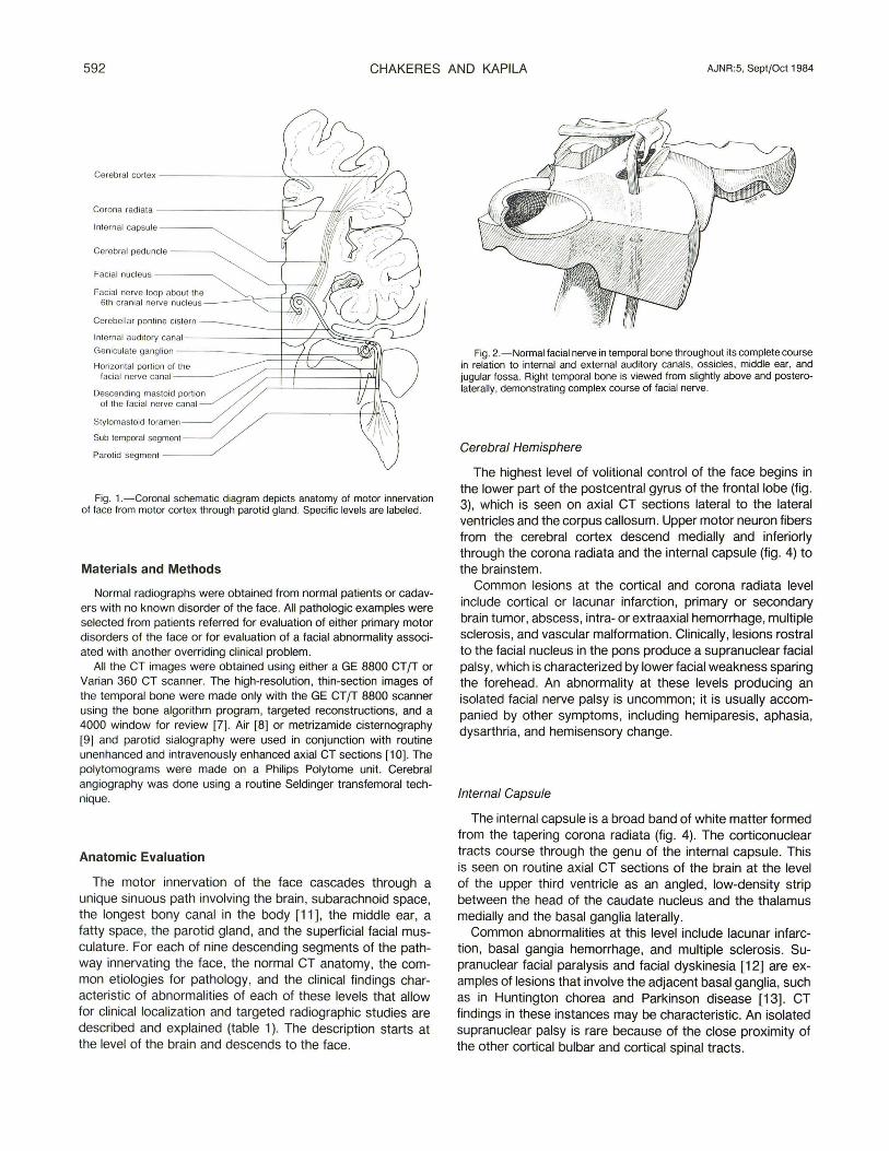
Review
Donald W. Chakeres 1. 2
Ashwani Kapila 1. 2
Received February 8 , 1983; accepted after revision May 15, 1984.
1 Department of Radiology, Neuroradiology Section, University of Texas Health Science Center, 7703 Floyd Curl Dr. , San Antonio , TX 78284. Address reprint requests to D. W. Chakeres .
2 Department of Radiology, Audie Murphy Veterans' Hospital, San Antonio, TX 78284.
AJNR 5:591-597, September/October 1984 .0195-6108/84/0505-0591 © American Roentgen Ray Society
Normal and Pathologic Radiographic Anatomy of the Motor Innervation of the Face
591
Facial motor disorders, including facial paralysis, myokymia, dyskinesia, and hemifacial spasm, are common clinical problems in which radiographic evaluation plays a crucial role. Since every segment of the motor innervation of the face from the brain to the parotid gland can now be seen radiographically, radiologists must understand the normal anatomy, the common pathologic lesions at each level, and the clinical f indings that help localize the abnormality so that the most sensitive and accurate radiographic approach can be planned. Though computed tomograhy alone allows for visualization of every segment, other methods such as cisternography, angiography, poly tomography, sialography, and magnetic resonance imaging are complementary in specific disorders.
Facial motor disorders , including facial paralysis , myokymia, dyskinesia, and hemifacial spasm, are common clinical problems in which a radiographic evaluation plays a crucial role . Though abnormalities of the motor innervation of the face need not be associated with a life-threatening illness, distortion of the face can create serious psychological problems and can significantly interfere with the unique capacity for self-expression reflected by the human countenance. An abnormality of the motor function of the face may also be the presenting symptom of progressive central or peripheral nervous system disease. With the advent of high-resolution, thin-section computed tomography (CT), the complex sinuous anatomy of the innervation of the face, from the cerebral cortex to the parotid gland, can now be routinely visualized radiographically . For simplification of discussion, the cascading pathway of the motor innervation of the face has been divided into nine descending separate anatomic levels (figs. 1 and 2).
Radiographic evaluation of the level of facial motor abnormality now approaches the accuracy of clinical localization [1-4] of the level of abnormality obtainable by a detailed neurologic examination . Since it is not possible to evaluate the innervation of the face throughout its entire course by a single survey radiographic examination , it is necessary to focus the examination on the suspected location and etiology of the abnormality. Because of this , an understanding of the clinical findings that help locate specific abnormalities and of the utility of each of the various radiographic procedures is important.
Patients with supranuclear facial nerve palsy, temporal bone fracture , malignant external otitis , atypical infranuclear facial nerve palsy, myokymia, or hemifacial spasm require localized radiographic evaluation . If surgical intervention is considered , radiographic evaluation is essential. On the other hand, patients with classic Bell palsy (unilateral facial paralysis secondary to herpes simplex infection) [5, 6), Ramsay Hunt syndrome (herpes zoster infection of the facial nerve), or facial myokymia secondary to Guillain-Barre syndrome need not have detailed radiographic workup because they are treated medically and can be diagnosed clinically.
592 CHAKERES AND KAPILA AJNR:5, Sept/Oct 1984
Corona radiata ---------IF=-----t~
tnternal capsule -------...
Cerebral peduncte ------...
Facial nucleus ----------..
Facial nerve toop about th e. __ ~=t--Hi~ 6th cranial nerve nucleus-
Cerebe llar pontine cistern
tnternal auditory cana t--------t-+-;;-~:::::::~
Genicula te ganglion ------=:::=:!=-:if=t;:::s;::::;;:~ Horizontal portion of the
facial nerve canal -----
Descending mastoid portion of the facial nerve canal
Stylomastoid foramen
Sub temporat segment
Parotid segment -----"
Fig. 1.-Coronal schematic diagram depicts anatomy of motor innervation of face from motor cortex through parotid gland. Specific levels are labeled.
Materials and Methods
Normal radiographs were obtained from normal patients or cadavers with no known disorder of the face. All pathologic examples were selected from patients referred for evaluation of either primary motor disorders of the face or for evaluation of a facial abnormality associated with another overriding clinical problem.
All the CT images were obtained using either a GE 8800 CT fT or Varian 360 CT scanner. The high-resolution, thin-section images of the temporal bone were made only with the GE CT fT 8800 scanner using the bone algorithm program, targeted reconstructions, and a 4000 window for review [7]. Air (8) or metrizamide cisternography (9) and parotid sialography were used in conjunction with routine unenhanced and intravenously enhanced axial CT sections (10). The pOlytomograms were made on a Philips Poly tome unit. Cerebral angiography was done using a routine Seldinger transfemoral technique.
Anatomic Evaluation
The motor innervation of the face cascades through a unique sinuous path involving the brain, subarachnoid space, the longest bony canal in the body [11], the middle ear, a fatty space, the parotid gland, and the superficial facial musculature. For each of nine descending segments of the pathway innervating the face, the normal CT anatomy, the common etiologies for pathology, and the clinical findings characteristic of abnormalities of each of these levels that allow for clinical localization and targeted radiographic studies are described and explained (table 1). The description starts at the level of the brain and descends to the face.
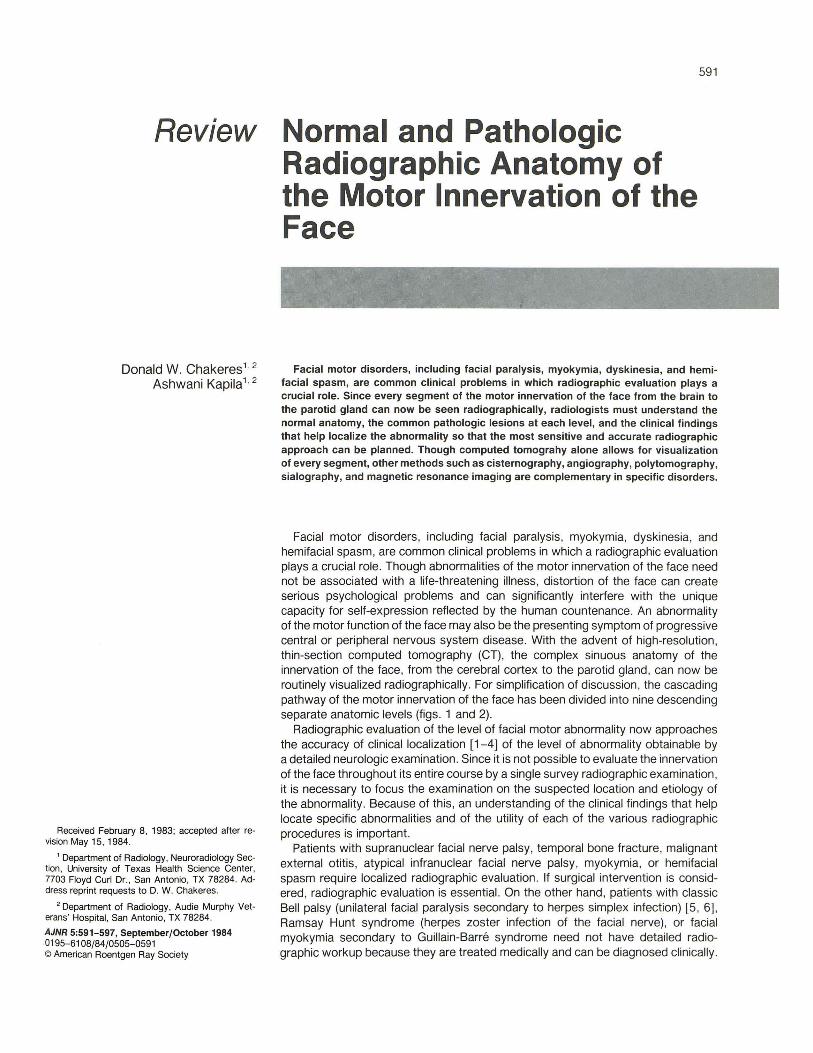
Fig. 2.-Normal facial nerve in temporal bone throughout its complete course in relation to internal and external auditory canals, ossicles, middle ear, and jugular fossa. Right temporal bone is viewed from slightly above and posterolaterally, demonstrating complex course of facial nerve.
Cerebral Hemisphere
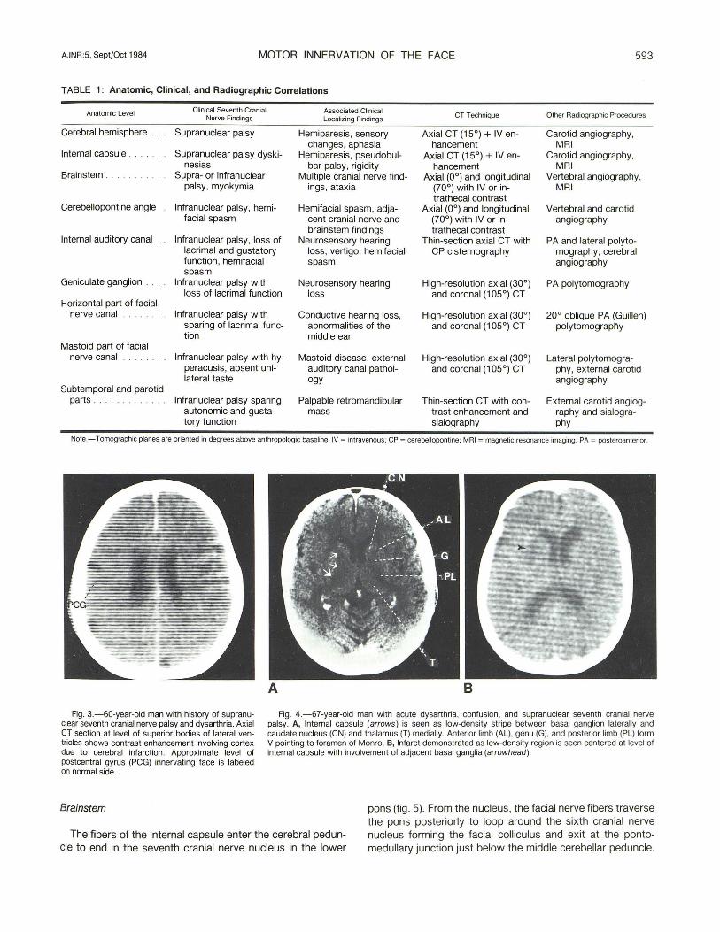
The highest level of volitional control of the face begins in the lower part of the postcentral gyrus of the frontal lobe (fig. 3), which is seen on axial CT sections lateral to the lateral ventricles and the corpus callosum. Upper motor neuron fibers from the cerebral cortex descend medially and inferiorly through the corona radiata and the internal capsule (fig. 4) to the brainstem.
Common lesions at the cortical and corona radiata level include cortical or lacunar infarction, primary or secondary brain tumor, abscess, intra- or extraaxial hemorrhage, multiple sclerosis, and vascular malformation. Clinically, lesions rostral to the facial nucleus in the pons produce a supranuclear facial palsy, which is characterized by lower facial weakness sparing the forehead . An abnormality at these levels producing an isolated facial nerve palsy is uncommon; it is usually accompanied by other symptoms, including hemiparesis, aphasia, dysarthria, and hemisensory change.
Internal Capsule
The internal capsule is a broad band of white matter formed from the tapering corona radiata (fig. 4). The corticonuclear tracts course through the genu of the internal capsule. This is seen on routine axial CT sections of the brain at the level of the upper third ventricle as an angled, low-density strip between the head of the caudate nucleus and the thalamus medially and the basal ganglia laterally.
Common abnormalities at this level include lacunar infarction, basal gangia hemorrhage, and multiple sclerosis. Supranuclear facial paralysis and facial dyskinesia [12] are examples of lesions that involve the adjacent basal ganglia, such as in Huntington chorea and Parkinson disease [13] . CT findings in these instances may be characteristic. An isolated supranuclear palsy is rare because of the close proximity of the other cortical bulbar and cortical spinal tracts.
AJNR:5 , Sept/Oct 1984 MOTOR INNERVATION OF THE FACE 593
TABLE 1: Anatomic, Clinical, and Radiographic Correlations
Anatomic Level Clinical Seventh Cranial Associated Clinical CT Technique Other Radiographic Procedures Nerve Findings Localizing Findings
Cerebral hemisphere Supranuclear palsy Hemiparesis, sensory Axial CT (15°) + IV en- Carotid angiography, changes, aphasia hancement MRI
Internal capsule . Supranuclear palsy dyski- Hemiparesis, pseudobul- Axial CT (15°) + IV en- Carotid angiography, nesias bar palsy, rigidity hancement MRI
Brainstem . . . . . .. . . Supra- or infranuclear Multiple cranial nerve find- Axial (0°) and longitudinal Vertebral angiography, palsy , myokymia ings, ataxia (70°) with IV or in- MRI
trathecal contrast Cerebellopontine angle Infranuclear palsy, hemi- Hemifacial spasm, adja- Axial (0°) and longitudinal Vertebral and carotid
facial spasm cent cranial nerve and (70°) with IV or in- angiography brainstem findings trathecal contrast
Internal auditory canal Infranuclear palsy, loss of Neurosensory hearing Thin-section axial CT with PA and lateral poly to-lacrimal and gustatory loss, vertigo, hemifacial CP cisternography mography, cerebral function, hemifacial spasm angiography spasm
Geniculate ganglion . Infranuclear palsy with Neurosensory hearing High-resolution axial (30°) P A poly tomography loss of lacrimal function loss and coronal (105°) CT
Horizontal part of facial nerve canal Infranuclear palsy with Conductive hearing loss, High-resolution axial (30°) 20° oblique PA (Guillen)
sparing of lacrimal func- abnormalities of the and coronal (105°) CT poly tomography tion middle ear
Mastoid part of facial nerve canal Infranuclear palsy with hy- Mastoid disease, external High-resolution axial (30°) Lateral pOlytomogra-
peracusis, absent un i- auditory canal pathol- and coronal (105°) CT phy, external carotid lateral taste ogy angiography
Subtemporal and parotid parts . Infranuclear palsy sparing Palpable retromandibular Thin-section CT with con- External carotid angiog-
autonomic and gusta- mass trast enhancement and raphy and sialogra-tory function sialography phy
Note.- Tomographic planes are oriented in degrees above anthropologic baseline. IV = intravenous; CP = cerebellopontine; MRI = magnetic resonance imaging. PA = posteroanterior.
Fig . 3.-60-year-old man with history of supranuclear seventh cranial nerve palsy and dysarthria. Axial CT section at level of superior bodies of lateral ventricles shows contrast enhancement involving cortex due to cerebral infarction. Approximate level of postcentral gyrus (PCG) innervating face is labeled on normal side.
Brainstem
Fig. 4.-67-year-old man with acute dysarthria, confusion, and supranuclear seventh cranial nerve palsy. A, Internal capsule (arrows) is seen as low-density stripe between basal ganglion laterally and caudate nucleus (CN) and thalamus (T) medially. Anterior limb (AL), genu (G), and posterior limb (PL) form V pointing to foramen of Monro. B, Infarct demonstrated as low-density region is seen centered at level of internal capsule with involvement of adjacent basal ganglia (arrowhead) .
The fibers of the internal capsule enter the cerebral peduncle to end in the seventh cranial nerve nucleus in the lower
pons (fig . 5). From the nucleus, the facial nerve fibers traverse the pons posteriorly to loop around the sixth cranial nerve nucleus forming the facial colliculus and exit at the pontomedullary junction just below the middle cerebellar peduncle.
594 CHAKERES AND KAPILA AJNR:5, Sept/Oct 1984
A B
8 9
Intraaxial abnormalities commonly seen at this level include glioma, multiple sclerosis [14], viral polyneuritis, infarct, hemorrhage, metastases, contusion , and trauma (fig. 6).
Any lesion at or distal to the facial nerve nucleus produces a peripheral facial nerve palsy characterized by flaccid paralysis affecting both the upper and lower face equally. Alternatively, lesions at this level may cause a unique dyskinesia, such as myochymia [14] . Myochymia is a constant serpentine undulating motion of the face, most often seen in patients with multiple sclerosis (best seen with magnetic resonance imaging) [15] or brainstem glioma. Because many important structures are tightly packed within the brainstem, other associated clinical abnormalities include cranial nerve palsy,
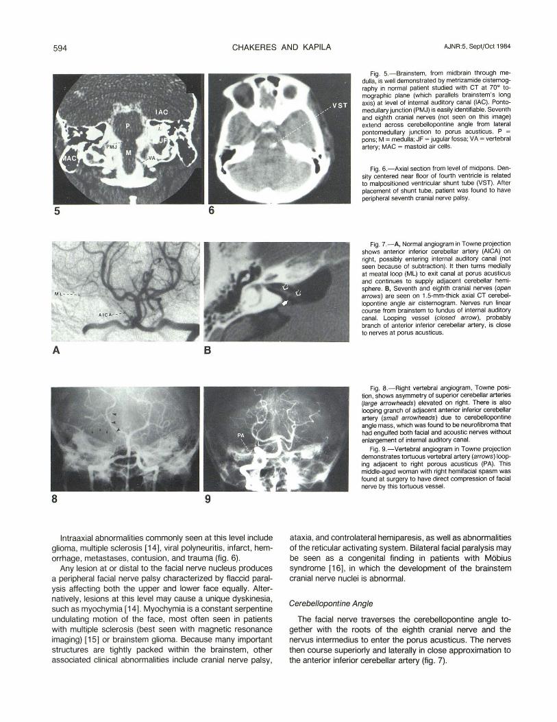
Fig. 5.-Brainstem, from midbrain through medulla, is well demonstrated by metrizamide cisternography in normal patient studied with CT at 70° tomographic plane (which parallels brainstem's long axis) at I",vel of internal auditory canal (lAC). Pontomedullary junction (PMJ) is easily identifiable. Seventh and eighth cranial nerves (not seen on this image) extend across cerebellopontine angle from lateral pontomedullary junction to porus acusticus. P = pons; M = medulla; JF = jugular fossa; VA = vertebral artery; MAC = mastoid air cells.
Fig. 6.-Axial section from level of midpons. Density centered near floor of fourth ventricle is related to mal positioned ventricular shunt tube (VST). After placement of shunt tube, patient was found to have peripheral seventh cranial nerve palsy.
Fig. 7.-A, Normal angiogram in Towne projection shows anterior inferior cerebellar artery (AICA) on right, possibly entering internal auditory canal (not seen because of subtraction). It then turns medially at meatal loop (ML) to exit canal at porus acusticus and continues to supply adjacent cerebellar hemisphere. e, Seventh and eighth cranial nerves (open arrows) are seen on 1.5-mm-thick axial CT cerebellopontine angle air cisternogram. Nerves run linear course from brainstem to fundus of internal auditory canal. Looping vessel (closed arrow), probably branch of anterior inferior cerebellar artery, is close to nerves at porus acusticus.
Fig. 8.-Right vertebral angiogram, Towne position, shows asymmetry of superior cerebellar arteries (large arrowheads) elevated on right. There is also looping granch of adjacent anterior inferior cerebellar artery (small arrowheads) due to cerebellopontine angle mass, which was found to be neurofibroma that had engulfed both facial and acoustic nerves without enlargement of internal auditory canal.
Fig. 9.-Vertebral angiogram in Towne projection demonstrates tortuous vertebral artery (arrows) looping adjacent to right porous acusticus (PA). This middle-aged woman with right hemifacial spasm was found at surgery to have direct compression of facial nerve by this tortuous vessel.
ataxia, and controlateral hemiparesis, as well as abnormalities of the reticular activating system. Bilateral facial paralysis may be seen as a congenital finding in patients with Mobius syndrome [16], in which the development of the brainstem cranial nerve nuclei is abnormal.
Cerebel/opontine Angle
The facial nerve traverses the cerebellopontine angle together with the roots of the eighth cranial nerve and the nervus intermedius to enter the porus acusticus. The nerves then course superiorly and laterally in close approximation to the anterior inferior cerebellar artery (fig. 7).
AJNR:5, Sept/Oct 1984 MOTOR INNERVATION OF THE FACE 595
11 12
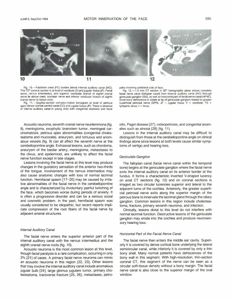
Fig. 1 D.-Falciform crest (FC) divides lateral internal auditory canal (lAC). This 700 coronal section is at level of vestibule (V) and jugular fossa (JF). Facial nerve, nervus intermedius, and superior vestibular branch of eighth cranial nerve lie above crest; cochlear nerve and inferior vestibular branch of eighth cranial nerve lie below crest.
Fig . 11 .-Sagittal-section complex-motion tomogram at level of petrous apex shows normal carotid canal (CC) and jugular fossa (JF). There is absence of internal auditory canal in young child with congenital deafness and facial
Acoustic neuroma, seventh cranial nerve neurilemmona (fig . 8), meningioma, exophytic brainstem tumor, meningeal carcinomatosis, petrous apex abnormalities (congenital cholesteatoma and mucocele), aneurysm, and tortuous and anomalous vessels (fig. 9) can all affect the seventh nerve at the cerebellopontine angle. Extraaxiallesions, such as chordoma, aneurysm of the basilar artery, meningioma, metastases to the clivus, and epidermoid , are unlikely to affect the facial nerve function except in late stages.
Lesions involving the facial nerve at this level may produce changes in the gustatory sensation of the anterior two-thirds of the tongue. Involvement of the nervus intermedius may also cause anatomic changes with loss of normal lacrimal function . Hemifacial spasm [17-20] may be caused by irritative abnormalities of the facial nerve in the cere bello pontine angle and is characterized by involuntary painful twitching of the face , which becomes worse during periods of anxiety. It is often a progressive disorder and can be a serious social and cosmetic problem. In the past, hemifacial spasm was usually considered to be idiopathic, but recent reports implicate compression of the root fibers of the facial nerve by adjacent arterial structures.
Internal Auditory Canal
The facial nerve enters the superior anterior part of the internal auditory canal with the nervus intermedius and the eighth cranial nerve roots (fig . 10).
Acoustic neuroma is the most common lesion at this level, though facial paralysis is a rare complication, occurring in only 2% [21] of cases. A primary facial nerve neuroma can mimic an acoustic neuroma in this region [22, 23]. Other lesions that may involve the internal auditory canal include anomalous jugular bulb [24] , large glomus jugulare tumor, primary cholesteatoma, transverse fracture [25, 26] , metastases, petro-
palsy involving ipsilateral side of face. Fig. 12.-1 .5 mm CT section in 300 tomographic plane shows complete
facial nerve canal (fallopian canal) from internal auditory canal (lAC) through geniculate ganglion (GG), as well as horizontal part of facial nerve canal (HFNC). Small bony dehiscence is visible at tip of geniculate ganglion related to greater superficial petrosal nerve (GPN). JF = jugular fossa ; V = vestibule; TS = tympanic sinus; I = incus.
sitis , Paget disease [27] , osteopetrosis, and congenital anomalies such as atresia [28] (fig. 11).
Lesions in the internal auditory canal may be difficult to distinguish from those at the cerebellopontine angle on clinical findings alone since lesions at both levels cause similar symptoms of vertigo and hearing loss.
Geniculate Ganglion
The fallopian canal (facial nerve canal within the temporal bone) begins at the geniculate ganglion where the facial nerve exits the internal auditory canal on its anterior border at the fundus . It forms a characteristic inverted V-shaped lucency on axial CT sections (fig. 12) and on coronal sections is imaged as two circular lucencies superior and lateral to the adjacent turns of the cochlea. Anteriorly , the greater superficial petrosal nerve exits along the superior margin of the petrous bone to innervate the lacrimal gland through the ciliary ganglion. Common lesions in this region include cholesteatoma, fracture , primary seventh neuroma, and infection.
Clinically, lesions distal to this level do not interfere with normal lacrimal function . Destructive lesions of the geniculate ganglion may erode into the cochlea and produce neurosensory hearing loss.
Horizontal Part of the Facial Nerve Canal
The facial nerve then enters the middle ear cavity. Superiorly it is covered by dense cortical bone underlying the lateral semicircular canal , while inferiorly it is covered by only a thin bony shell. Many normal patients have dehiscences of the bony wall in this segment. With high-resolution, thin-section coronal CT, this segment of the nerve can be seen as a circular soft-tissue density without a bony margin. The facial nerve canal is also close to the superior margin of the oval window.
596 CHAKERES AND KAPILA AJNR:5, Sept/Oct 1984
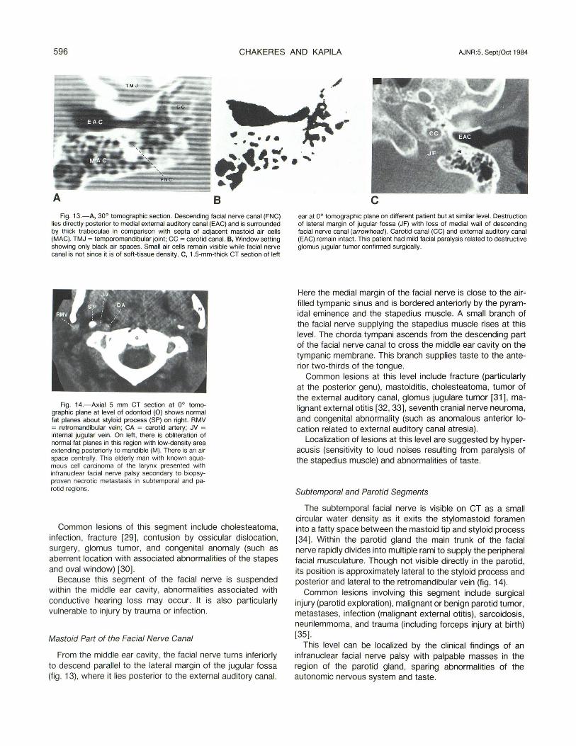
A B Fig. 13.-A, 30° tomographic section. Descending facial nerve canal (FNC)
lies directly posterior to medial external auditory canal (EAC) and is surrounded by thick trabeculae in comparison with septa of adjacent mastoid air cells (MAC). TMJ = temporomandibular joint; CC = carotid canal. S, Window set1ing showing only black air spaces. Small air cells remain visible while facial nerve canal is not since it is of soft-tissue density. C, 1.5-mm-thick CT section of left
Fig . 14.-Axial 5 mm CT section at 0° tomographic plane at level of odontoid (0) shows normal fat planes about styloid process (SP) on right. RMV = retromandibular vein; CA = carotid artery; JV = internal jugular vein. On left , there is obliteration of normal fat planes in this region with low-density area extending posteriorly to mandible (M). There is an air space centrally. This elderly man with known squamous cell carcinoma of the larynx presented with infranuclear facial nerve palsy secondary to biopsyproven necrotic metastasis in subtemporal and parotid regions.
Common lesions of this segment include cholesteatoma, infection , fracture [29], contusion by ossicular dislocation, surgery, glomus tumor, and congenital anomaly (such as aberrent location with associated abnormalities of the stapes and oval window) [30] .
Because this segment of the facial nerve is suspended within the middle ear cavity, abnormalities associated with conductive hearing loss may occur. It is also particularly vulnerable to injury by trauma or infection .
Mastoid Part of the Facial Nerve Canal
From the middle ear cavity , the facial nerve turns inferiorly to descend parallel to the lateral margin of the jugular fossa (fig. 13), where it lies posterior to the external auditory canal.
ear at 0° tomographic plane on different patient but at similar level. Destruction of lateral margin of jugular fossa (JF) with loss of medial wall of descending facial nerve canal (arrowhead) . Carotid canal (CC) and external auditory canal (EAC) remain intact. This patient had mild facial paralysis related to destructive glomus jugular tumor confirmed surgically.
Here the medial margin of the facial nerve is close to the airfilled tympanic sinus and is bordered anteriorly by the pyramidal eminence and the stapedius muscle. A small branch of the facial nerve supplying the stapedius muscle rises at this level. The chorda tympani ascends from the descending part of the facial nerve canal to cross the middle ear cavity on the tympanic membrane. This branch supplies taste to the anterior two-thirds of the tongue.
Common lesions at this level include fracture (particularly at the posterior genu), mastoiditis, cholesteatoma, tumor of the external auditory canal, glomus jugulare tumor [31], malignant external otitis [32, 33], seventh cranial nerve neuroma, and congenital abnormality (such as anomalous anterior location related to external auditory canal atresia).
Localization of lesions at this level are suggested by hyperacusis (sensitivity to loud noises resulting from paralysis of the stapedius muscle) and abnormalities of taste.
Subtemporal and Parotid Segments
The subtemporal facial nerve is visible on CT as a small circular water density as it exits the stylomastoid foramen into a fatty space between the mastoid tip and styloid process [34] . Within the parotid gland the main trunk of the facial nerve rapidly divides into multiple rami to supply the peripheral facial musculature. Though not visible directly in the parotid, its position is approximately lateral to the styloid process and posterior and lateral to the retromandibular vein (fig. 14).
Common lesions involving this segment include surgical injury (parotid exploration), malignant or benign parotid tumor, metastases, infection (malignant external otitis), sarcoidosis, neurilemmoma, and trauma (including forceps injury at birth) [35] .
This level can be localized by the clinical findings of an infranuclear facial nerve palsy with palpable masses in the region of the parotid gland , sparing abnormalities of the autonomic nervous system and taste.
AJNR:5, Sept/Oct 1984 MOTOR INNERVATION OF THE FACE 597
Discussion
The use of appropriate radiographic procedures directed by topographic clinical evaluation of abnormalities involving the facial musculature can result in an accurate and sensitive radiographic evaluation of the causative lesion. While CT is the mainstay in the diagnostic radiographic workup, angiography, complex motion tomography, magnetic resonance imaging , and parotid duct sialography may be necessary to aid in the diagnosis of these common problems.
Not all patients with abnormalities of motor innervation of the face , including most patients with Bell palsy and Ramsay Hunt syndrome, require complete radiographic analysis. However, patients with isolated peripheral facial palsy, supranuclear facial palsy, hemifacial spasm, trauma, myokymia, and allied neurologic symptoms do require a detailed radiographic evaluation .
ACKNOWLEDGMENTS
We thank Robert Hart for manuscript review and Linda Chakeres and Beverly Combs for editorial and secretarial help.
REFERENCES
1. Adams RD, Victor M. Principles of neurology, 2d ed. McGrawHill , 1981;851-856
2. Tonning FM. The reliability of level-diagnostic examinations in acute, peripheral facial palsy. Acta Otolaryngol (Stock h) 1977;84: 414-415
3. Tschiassny K. Eight syndromes of facial paralysis and their significance in locating the lesion. Ann Otol Rhinol Laryngol 1953;62:677-691
4. Alford BR , Jerger JF, Coats AC, Peterson CR, Weber SC. Diagnostic tests of facial nerve function . Otolaryngol Clin North Am 1974;7:331-342
5. Adour KK, Byl FM, Hilsinger RL Jr, Kahn ZM, Sheldon MI. The true nature of Bell' s palsy: analysis of 1 ,000 consecutive patients. Laryngoscope 1978;88: 787 -801
6. Adour KK. Diagnosis and management of facial paralysis. N Engl J Med 1982;302:348-351
7. Chakeres OW, Spiegel PK. A systemic method for comprehensive evaluation of the temporal bone by computed tomography. Radiology 1983;146 :97-106
8. Pinto RS, Kricheff II , Bergeron RT, Cohen N. Small acoustic neuromas: detection by high resolution gas CT cisternography. AJNR 1982;3:283-286, AJR 1982; 139: 129-132
9. Steele JR, Hoffman JC. Brainstem evaluation with CT cisternography. AJNR 1980;1 :521-526, AJR 1981;136:287-292
10. Sone S, Higashihara T, Morimoto S, et al. CT of parotid tumors . AJNR 1982;3:143-147
11 . Shambough GE Jr. Surgery of the ear, 2d ed. Philadelphia: Saunders, 1969:565
12. Tolosa ES. Clinical features of Meige's disease (idiopathic orofacial dystonia)- a report of 17 cases. Arch Neurol 1981 ; 38:147-151
13. Hunker CJ , Abbs JH , Barlow SM . The relationship between parkinsonian rigidity and hypokinesia in the orofacial system: a quantitative analysis . Neurology (NY) 1982;32:749- 754
14. De Silva KL, Pearce J. Facial myokymia: a clue to the diagnosis of multiple sclerosis. Postgrad Med J 1972;48 :657- 661
15. Bydder GM, Steiner RE, Young IR, et al. Clinical NMR imaging of the brain: 140 cases. AJNR 1982;3:459- 480, AJR 1982; 139: 215-236
16. Adams RP, Victor M. Principles of neurology, 2d ed. New York: McGraw-Hili , 1981 : 851
17. Kumagami S. Neuropathological findings of hemifacial spasm and trigeminal neuralgia. Arch Otolaryngo/1974;99 :160-164
18. Iwakuma T, Matsumoto A, Nakamura N. Hemifacial spasm. Comparison of three different operative procedures in 110 patients . J Neurosurg 1982;57 :753-756
19. Kempe GK, Smith DR . Trigeminal neuralgia, facial spasm, intermedius and glossopharyngeal neuralgia with persistent carotid basilar anastomosis . J Neurosurg 1969;31 :445-451
20. Jannetta PJ. Neurovascular compression in cranial nerve and systemic disease. Ann Surg 1980;192:518- 524
21. Hart RG, Gardner DP, Howieson J. Acoustic tumors : atypical features and recent diagnostic tests. Neurology (NY) 1983; 33 :211-221
22. Horn KL, Crumley RL, Schindler RA. Facial neurilemmomas. Laryngoscope 1981 ;91 : 1326-1331
23. Neely JG, Alfrod BR . Facial nerve neuromas. Arch Otolaryngol 1974;100:298-301
24. Stern, J, Goldenberg M. Jugular bulb diverticula in the medial petrous bone. AJNR 1980;1 :153-155, AJR 1980;134 :959-961
25. Potter GO. Trauma to the temporal bone. Semin Roentgenol 1969;4: 143-150
26. McCabe BF. Injuries to the facial nerve. Laryngoscope 1972;82 : 1891-1896
27. Nager GT. Paget's disease of the temporal bone. Ann Otol Rhinol Laryngol [Suppl] 1975;22 : 1-32
28. Cliff MM, Lapayowker MS, Woloshim HJ. Congenital abnormalities of the temporal bone. Semin Roentgenol 1969;4: 122-128
29. Johnson OW, Hasso AN , Stewart CE, Thompson JR, Hinshaw DB. Temporal bone trauma high-resolution computed tomographic evaluation. Radiology 1984;151 :411-415
30. Jahrsdoerfer RA. The facial nerve in congenital middle ear malformations. Laryngoscope 1981;8: 1217 - 1225
31 . Chakeres OW, Lamasters DL. Paragangliomas of the temporal bone: high-resolution CT studies. Radiology 1984;150:749-753
32. Chandler JR. Malignant external otitis: Further considerations . Ann Otol Rhinol Laryngo/1977;86:417- 428
33. Curtin HD, Wolfe P, May M. Malignant external otitis: CT evaluation. Radiology 1982 ;145 :383-388
34. Curtin HD, Wolfe P, Snyderman N. The facial nerve between the stylomastoid foramen and the parotid: computed tomographic imaging. Radiology 1983;149 :165-169
35. Goodhill U. Ear disease, deafness, and dizziness. Hagerstown, MD; Harper & Row, 1979;582