Embed Size (px)
Citation preview
Tendon neuroplastic training: changing the waywe think about tendon rehabilitation: a narrativereviewEbonie Rio,1,2 Dawson Kidgell,3 G Lorimer Moseley,4 Jamie Gaida,1,5,6
Sean Docking,1,2 Craig Purdam,7 Jill Cook1,2
▸ Additional material ispublished online only. To viewplease visit the journal online(http://dx.doi.org/10.1136/bjsports-2015-095215).1Department of Physiotherapy,School of Primary Health Care,Monash University, Melbourne,Victoria, Australia2The Australian Centre forResearch into Injury in Sportand its Prevention, BallaratFederation University, Victoria,Australia3Department of Rehabilitation,Nutrition and Sport, School ofAllied Health, La TrobeUniversity, Melbourne,Victoria, Australia4Sansom Institute for HealthResearch, University of SouthAustralia & Pain, Adelaide,South Australia, Australia5Department of Physiotherapy,University of Canberra, Bruce,Australian Capital Territory,Australia6University of CanberraResearch Institute for Sportand Exercise, Australia7Department of PhysicalTherapies, Australian Instituteof Sport, Bruce, AustralianCapital Territory, Australia
Correspondence toEbonie Rio, 4 Hardy Crescent,Heathmont VIC 3135,Australia:[email protected]
Accepted 5 September 2015Published Online First25 September 2015
To cite: Rio E, Kidgell D,Moseley GL, et al. Br JSports Med 2016;50:209–215.
ABSTRACTTendinopathy can be resistant to treatment and oftenrecurs, implying that current treatment approaches aresuboptimal. Rehabilitation programmes that have beensuccessful in terms of pain reduction and return to sportoutcomes usually include strength training. Muscleactivation can induce analgesia, improving self-efficacyassociated with reducing one’s own pain. Furthermore,strength training is beneficial for tendon matrix structure,muscle properties and limb biomechanics. However,current tendon rehabilitation may not adequately addressthe corticospinal control of the muscle, which may resultin altered control of muscle recruitment and theconsequent tendon load, and this may contribute torecalcitrance or symptom recurrence. Outcomes ofinterest include the effect of strength training on tendonpain, corticospinal excitability and short interval corticalinhibition. The aims of this concept paper are to: (1)review what is known about changes to the primarymotor cortex and motor control in tendinopathy, (2)identify the parameters shown to induce neuroplasticityin strength training and (3) align these principles withtendon rehabilitation loading protocols to introduce acombination approach termed as tendon neuroplastictraining. Strength training is a powerful modulator of thecentral nervous system. In particular, corticospinal inputsare essential for motor unit recruitment and activation;however, specific strength training parameters areimportant for neuroplasticity. Strength training that isexternally paced and akin to a skilled movement taskhas been shown to not only reduce tendon pain, butmodulate excitatory and inhibitory control of the muscleand therefore, potentially tendon load. An improvedunderstanding of the methods that maximise theopportunity for neuroplasticity may be an importantprogression in how we prescribe exercise-basedrehabilitation in tendinopathy for pain modulation andpotentially restoration of the corticospinal control of themuscle-tendon complex.
INTRODUCTIONThe clinical outcomes of treatment of tendinopathyvary—there is currently no single effective treat-ment. Unimodal interventions aimed solely at per-ipheral tissue, in this case the tendon, are unlikelyto address complex corticospinal and neuromuscu-lar adaptations associated with persistent pain. Inthis paper, the term corticospinal control of themuscle will refer to motor unit activation as aresult of excitatory and inhibitory corticospinalinputs onto the spinal motor neuron pool, which
will ultimately affect tendon loading and motorperformance. Therefore, it is logical to explore thecorticospinal pathway in people with tendinopathy.Given the high incidence of bilateral tendinopa-
thy1 and bilateral pathology with predominantlyunilateral load in animal2 and human studies,3 it isalso important to consider the corticospinal controlof the non-painful side in tendinopathy. A frequentand frustrating phenomenon is the clinical presen-tation of the previously unaffected side followingrehabilitation. These observations pose several clin-ical questions: what are the differences in motorcontrol between those people with tendinopathyand those without (that may predispose people totendinopathy or may be adaptations)? Are thereside-to-side differences associated with unilateraltendinopathy? How well does our current rehabili-tation therapy address these issues of motorcontrol?
MOTOR CONTROL IN PEOPLE WITH ANDWITHOUT TENDINOPATHYIn tendon research, muscle strength, usually repre-sented by maximal voluntary contraction (MVC), isfar more often evaluated than is motor control.Interestingly, there is no consistent pattern ofstrength or performance change (either increase ordecrease) in those tendons that have been studied(or the contralateral limb) and this may conflictwith the clinical perception that pain is likely toresult in strength loss due to disuse or willingnessto protect the injured part consciously or subcon-sciously. For example, people with rotator cuff(RC) tendinopathy have been shown to be 15%stronger in measures of abduction on their asymp-tomatic side than controls (and comparable instrength to controls on their symptomatic side),4
and this has also been seen in lateral epicondylal-gia.5 Athletes with patellar tendinopathy (PT, alsotermed jumper’s knee) have been shown to bebetter jumpers than athletes without jumper’sknee6 7; however, several studies report lessstrength on the symptomatic side than in controls(see Heales et al8 for review). It is also reportedthat full strength is not recovered after surgery forAchilles tendinopathy.9 There is no convincingassociation between strength deficit and presence oftendinopathy, as demonstrated by several studies.8
People with PT displayed greater cortical inhib-ition in their quadriceps responses10 than healthyindividuals.11 Increased cortical inhibition has beenshown to be associated with phasic (occurring inphases/intermittent) nociceptive stimuli,12 which
Open accessScan to access more
free content
Rio E, et al. Br J Sports Med 2016;50:209–215. doi:10.1136/bjsports-2015-095215 1 of 8
Review on 27 M
arch 2019 by guest. Protected by copyright.
http://bjsm.bm
j.com/
Br J S
ports Med: first published as 10.1136/bjsports-2015-095215 on 25 S
eptember 2015. D
ownloaded from
describes tendon pain is consistently linked with loading andtherefore, often phasic rather than tonic (sustained) for manytendons. While there was increased cortical inhibition alteringmotor drive in PT, there was also greater corticospinal excitabil-ity (CSE) than in controls inferring differences in the balance ofexcitability and inhibition of motor control as compared withhealthy controls (Rio et al, in press). It may be that strengthchanges still represent a decrease from that individual’s potentialperformance.
A recent systematic review by Heales et al8 on the sensoryand motor deficits of the non-injured side of patients with uni-lateral tendon pain included one study on the patellar tendonand one study on the Achilles tendon; however, there were 18studies on the upper limb tendons. There is clearly a paucity ofliterature investigating lower limb sensory and motor changes intendinopathy. The included lower limb studies measured archheight in PT13 and Achilles tendon structure with imaging.14 Itis not clear how arch height and imaging changes may relate tosensory or motor changes; thus, the sensory or motor changesin lower limb tendinopathy require further investigation. Thisbias towards upper limb studies was paralleled by a recent sys-tematic review on complex regional pain syndrome, which onlyidentified suitable data from upper extremity studies.15 It is pos-sible that differences in functional reorganisation in the primarysomatosensory (and/or primary motor cortex (M1)) corticesfollowing upper limb versus lower limb injury may exist thatlimit the extrapolation of upper limb findings to the lower limb.Given that the predominant role of the upper limb musculaturerelates to position and control of the hand in space, and that thehand is well represented on the somatosensory cortex and M1,injury to upper limb regions may have manifestations associatedwith chronicity that are different from those in the lower limb.There appears to be a bias towards investigations of upper limbconditions.
The cross-sectional design of all studies that have investigatedmotor control in tendinopathy makes it difficult to infer causal-ity and the potential impact of handedness or hand-use profiles(especially in upper limb studies).16–18 That is, there is a morecomplex relationship at play: talented jumpers are more likelyto play in positions that require more jumping (eg, in volleyball,as an outside hitter rather than a setter);19 thus, such partici-pants may be more vulnerable to tendon pain in their dominantlimb, which is likely to have been stronger than their other oneto begin with. These factors will also affect measures of strengthand the stimulus response characteristics of primary motorcortex (M1) cells. Furthermore, maximal strength performancedata are vulnerable to cognitive and motivational factors.20 21
More importantly, maximum strength measures may not reflectthe complex interaction between excitatory and inhibitory influ-ences on the motor command that occur during tasks.
Indices of maximum strength may not provide enough detailabout muscle-tendon loading at submaximal levels, nor have theability to grade muscle recruitment22 or appropriately timedpatterns of activation according to the required task, especiallyin a painful state (eg, see Hodges and Richardson,23 andWadsworth and Bullock-Saxton24). Each of these scenarios havesignificant implications for function and load attenuation.25
Transcranial magnetic stimulation (TMS) studies have shownthat athletes with PT have greater M1 excitability than pain-freejumping athletes, as reflected in larger evoked muscle responsesin the quadriceps (rectus femoris; Rio et al,10 in press). Asstated, athletes with PT also have greater cortical inhibition thanhealthy controls have.11 26 27 That the investigation of theseissues involves identical loading and contextual environment
across patient and control groups clearly points to an imbalancebetween the excitatory and inhibitory influences over muscleactivation around the painful tendon. There may be severalmechanisms that underpin these problems, not least changes inthe response profile of clusters of neurons in M1, but the exactmechanisms remain to be untangled. Nonetheless, the func-tional result seems consistent with a protective adaptation thatreduces the mechanical demands placed on the tendon, that is,an apparently protective adaptation.
Jumping and landing mechanics will be influenced by motorcontrol changes that include both peripheral and central contri-butions to lower limb activation. People with patellar tendonabnormality (pathologically observable on ultrasound) havedemonstrated landing patterns different from those of controlsand importantly, have less variability in movement than healthycontrols.28 Movement variability is thought to minimise loadaccumulation in a specific region and reduced variability may beobserved in, for example, reduced coefficient of variation inknee joint angles during a landing task.29 The invariable motorpattern implies that corticospinal control is altered in some wayand may be due to protective strategies underpinned by evalu-ative processes relating to the consequence or meaning of themotor task.30 In the case of patellar tendon abnormality, invari-able motor patterns may reflect a strategy to avoid pain duringjumping (consequence) as well as the competing desire foroptimal performance (meaning). In fact, better jumping abilityhas been shown to be a risk factor for developing PT31 andindeed, people with PT are better jumpers or at least as good asthose without—termed the ‘jumper’s knee paradox’.6 7 32
Less variability has been observed in asymptomatic controlswho demonstrate a protective postural strategy when theyexpect their back to hurt,33 and people with recurrent back paindemonstrate a protective strategy even when they are painfree.34 Moreover, a reduction in normal variability of posturaladjustments has been observed during experimentally inducedpain, and failure to reinstate normal variability has been pro-posed as a possible risk for recurrence.30 Such findings raise thepossibility that an invariable motor pattern observed in PT mayreflect a system less adaptable to environmental perturbations—an undesirable state.35 Indeed, movement variability has beenproposed as an important feature in actually preventing injury.36
Though the predicted goal of adopting a different motorstrategy is protection, non-resolution of the strategy may in factincrease the likelihood of symptom recurrence.30 Given the highrecurrence rate of tendinopathy, it is pertinent to consider thatthere may be non-resolution of motor strategies even in cur-rently asymptomatic people with a history of tendon pain, ortendon pathology, that predisposes them to symptoms orsymptom recurrence. This outcome may also reflect the compet-ing desires for optimal performance and tissue protection. Thisadds complexity to the design and implementation of rehabilita-tion, as the strategy (movement pattern) adopted will reflectcompeting desires of performance and protection, particularly ifthere has been no change to inputs (of which nociception isonly one). To provide a clinical example, the presence of a posi-tive ‘culture’ of PT in jumping sports, such as volleyball whereplaying with tendon pain is common (and may reinforce aconcept that pain during activity does not equate to tissuedamage), will probably serve to maintain the motor controlstrategy. These contextual factors are likely to further influencethe experience of pain,37 and will vary depending on the envir-onment and one’s own experiences.
Cross-sectional studies do not allow elucidation of whethertendon pathology or the presence of tendon pain precedes
2 of 8 Rio E, et al. Br J Sports Med 2016;50:209–215. doi:10.1136/bjsports-2015-095215
Review on 27 M
arch 2019 by guest. Protected by copyright.
http://bjsm.bm
j.com/
Br J S
ports Med: first published as 10.1136/bjsports-2015-095215 on 25 S
eptember 2015. D
ownloaded from
changes in motor control. That pain will often be accompaniedby altered motor output would be predicted on the basis thatboth pain and motor output can be effective at protecting usfrom bodily threat. A common finding in studies of motorcontrol during pain is altered corticospinal drive to the motorneuron/muscle. However, this phenomenon of greater CSE hasalso been observed during testing that was non-painful, inpeople with PT as compared with activity-matched controls andpeople with other anterior knee pain (Rio et al, accepted).Furthermore, there were no differences between groups foractivity, muscle strength (measured by maximal voluntary iso-metric contraction torque of the quadriceps), muscle activation(represented by surface electromyography) or measures ofmotor neuron activation (maximal femoral nerve stimulation);so it seems plausible that the increase in CSE was associatedwith the PT regardless of the fact that there was no pain duringtesting. It is possible that their net motor drive to the quadricepsmuscle group is unaltered (or even that they may not be as goodas they could be); however, controlled activation and the motorstrategy may be affected. This also implies the motor task stillhas potential ‘consequence’ and ‘meaning’ in the absence ofnociception, which fits with the invariable and abnormallanding strategy observed in people with tendon pathology.
Current tendon rehabilitation may fail to adequately addressthe multitude of contributing factors to altered motor control,which would include not only muscle strength and tendon cap-acity, but corticospinal control encompassing excitability andinhibition as well as belief systems about pain and contextualfactors.
Motor control changes may be bilateralThe lack of consistent abnormalities in muscle strength on thepatient’s affected leg may simply reflect similar abnormalitiesbeing present on the contralateral limb. Heales et al8 reportedthat the contralateral asymptomatic side was weaker than con-trols in upper limb tendinopathy, though abduction in thosewith RC tendinopathy was an exception and no data werereported for the lower limb. Interestingly, data from low backpain studies are also variable with some reporting both hypoac-tivity and others reporting hyperactivity, depending on themuscles and tasks investigated (see Hodges and Moseley38 forreview). Strength deficits persist following surgery and rehabili-tation in unilateral Achilles tendinopathy; however, theside-to-side strength difference was relatively low (7.2–8.8%)and may actually reflect a bilateral motor control deficit.9
However, while the direction of change (increases or decreasesin muscle performance) is not consistent, it does appear that achange in motor control may occur bilaterally.
Regardless of whether motor control changes are a cause ofpain or epiphenomena, changes to motor control may not justbe bilateral but system wide. There is an increasing body of evi-dence that multiple sites are frequently affected due to complexintrinsic and extrinsic factors, and contralateral pathology and/or the development of symptoms is common. That is, oncetendon pain (or pathology) is established at one site, thereappears to be the potential for increased risk of tendinopathyelsewhere. Notwithstanding systemic risk factors that predisposetendinopathy, such as diabetes, elevated cholesterol and geneticfactors (eg, the COL5A1 genotype in isolation39 or in combin-ation),40 there may be other considerations, including anincrease in global sympathetic drive.41 While it is likely thatloading is similar for both legs and bilaterality of pathology(and/or pain) may be, therefore, somewhat expected, data fromthe upper limb with markedly different use profiles (such as
baseball pitchers)3 and unilaterally loaded animal models thatdemonstrate bilateral pathology2 implicate systemic or nervoussystem involvement in tendinopathy. The interhemispheric con-nections and potential for modulation at the spinal cord levelare both potential avenues for bilateral changes outside of bilat-eral loading that have not been as yet investigated.
It is possible that the other side may display differences inresponse to unilateral nociception (or insult) similar to what isobserved in injury such as stroke. The adaptive response seenfollowing stroke includes increased inhibition on the affectedside and increased CSE to the unaffected limb.42 Furthermore,there is actually increased interhemispheric inhibition from thenon-affected hemisphere to the lesioned hemisphere that furtherincreases the inhibition on the affected side during activation ofthe unaffected limb.42 This perhaps has a biological advantageto improve efficiency of the unaffected side; however, thisremains a challenge in rehabilitation (as the affected side has sig-nificant inhibition and is further increased during activation ofthe unaffected limb).
To investigate the unaffected side in people with PT, 13jumping athletes (6 control participants, 7 with unilateral PT)were recruited (Rio, unpublished data available as an onlinesupplementary material). Participants were tested bilaterallywith TMS including their active motor threshold and a stimulusresponse curve as well as short interval cortical inhibition (SICI)using rectus femoris as the target muscle. When compared withhealthy, activity-matched controls, preliminary data in athleteswith PT demonstrate hyperexcitability on their affected side,supporting previous research (Rio et al, unpublished, availableas an online supplementary material) and also displayed mark-edly increased cortical inhibition (n=4 affected side SICI ratio17.12%, n=6 controls 58.78%). This motor control strategymay be likened to a novice driver controlling the speed of theircar with one foot on the brake and one on the accelerator.Interestingly, the contralateral pathway controlling theunaffected side appears less excitable than both the affected side(similar to that observed in stroke) and controls, and also dis-plays increased cortical inhibition (n=4 unaffected side 19.13%;where lower SICI percentage ratio represents greater corticalinhibition). These preliminary findings appear to replicate thefindings of unilateral stroke that affects the M1, in particular theplausibility of greater interhemispheric inhibition and involve-ment of bilateral changes. However, the sample size is small andwarrants further investigation with greater numbers. It does,nevertheless, highlight the danger of using the unaffected side asa control in studies of motor control, excitability or inhibitionin people with tendinopathy. This also requires further evalu-ation as the clinical implications are considerable—strengthtraining in the affected limb may continue to drive inhibition tothe unaffected limb unless we can improve our understanding oftechniques that modulate excess inhibition.
Given the propensity for lower limb activities to be bilateraland symmetrical for efficiency (ambulation, jumping, etc), thechanges in the unaffected side may be an attempt to achievemotor control homoeostasis in a system trying to protect aregion. That is, there are combined and perhaps competingdemands of protecting a vulnerable or potentially vulnerabletendon while still producing torques sufficient for optimal per-formance. This may be argued for many tendinopathies not justin athletes, but in people involved in manual labour or anyoutcome-dependent task.
The contralateral motor control changes may also contributeto or explain the high injury rate of the contralateral tendon fol-lowing rehabilitation. That is, loading patterns in the
Rio E, et al. Br J Sports Med 2016;50:209–215. doi:10.1136/bjsports-2015-095215 3 of 8
Review on 27 M
arch 2019 by guest. Protected by copyright.
http://bjsm.bm
j.com/
Br J S
ports Med: first published as 10.1136/bjsports-2015-095215 on 25 S
eptember 2015. D
ownloaded from
contralateral limb are likely to be different as corticospinal driveof the unaffected side is altered. This provides support for arehabilitative strategy that targets the corticospinal control ofboth sides; however, these strategies may need to be carefullyconsidered. Strategies that modulate inhibition on the affectedside as well as pain, such as isometric muscle contractions inPT,10 should be evaluated for their potential effect on interhe-mispheric inhibition and inhibition of the ipsilateral M1.Rehabilitation that is capable of modulating pain and inhibitionof both M1 may yield important advances in tendonmanagement.
THE KEY QUESTION IS WHAT HAPPENS FIRST?There are two theories about the relationship between pain andchanges in muscle control (see Hodges and Moseley38 forreview), and neither is supported unequivocally by the litera-ture. They propose that either changes in muscle activity causepain (sometimes termed pain-spasm-pain model or vicious cycleof pain in the pain literature) or that changes in muscle activityserve to protect the area by limiting movement (the pain adapta-tion model).43
Experimental pain studies that demonstrate transient changesto motor control after a transient nociceptive stimulus,44–46 aswell as studies on a number of chronic conditions providesupport for the pain adaptation model.43 The definitive studythat studies motor control before the onset of pain remainselusive.
Hyperexcitability of the quadriceps in athletes with PT hasbeen correlated with length of time of symptoms (Rio, unpub-lished data available as an online supplementary material) andchanges in infraspinatus excitability in people with RC tendino-pathy were correlated with chronicity.47 This does not excludethe possibility that aberrant motor control precedes the firstonset of tendinopathy but provides a strong rationale thatchanges in motor control occur in response to nociceptivebarrage. The potential for unilateral nociceptive input to drivemotor control changes bilaterally (or possibly even system wide)underlines the importance of improving our understanding ofnociception-motor interactions,48 particularly in a condition ofchronic nociception where the nociceptive driver is unknown.
The pain adaptation model proposes that agonist activity isreduced and the antagonist activity is increased with pain.43
This may be the case in PT as there were no differences inMVC between athletes with PT and controls (Rio et al,accepted). However, in a separate study, following isometricexercise that reduced their pain, the MVC of the quadriceps ofathletes with PT increased by 18.7%, implying their agonistactivity (the quadriceps) was in fact inhibited (reduced) prior toisometric exercise.10 The hamstrings that serve as the antago-nists have not been measured. Most testing is completed in thelaboratory with a single joint movement for simplicity, forexample, leg extension; however, agonists and antagonistswould change during a more complex movement such as acounter movement jump.
The threat to tissue (as far as the brain is concerned) mayremain even after pain reduction and return to sport, as tendonmay not have ‘healed’ (many consider tendinopathy to be failedhealing; see Cook and Purdam49 for review). In this context,there may be ongoing monitoring and persistent changes to cor-ticospinal control of muscle (to protect the tendon) as the M1and its projections may consider it to be vulnerable.Alternatively, the motor control changes that occur in tendino-pathy may not spontaneously resolve or normalise followingrecovery, thus indicating rehabilitation may not address motor
activation and leaving people vulnerable to recurrence ofsymptoms.
People can and do recover in terms of pain and functiondespite tendon pathology remaining unchanged on clinicalimaging.50 One plausible explanation for this, at a tissue level, isthat the tendon may structurally compensate in the area aroundthe pathological lesion. That is, the volume of structurally intacttissue increases as an adaptive response.51 This tendon may bethicker but potentially amenable to improvements in mechanicalstrength51 through graded rehabilitation that would also altermuscle properties. The reverse is also observed, however, wherepatients can have symptoms without demonstrable structuralpathology and reminds us that there is a disconnect betweenstructure and pain (see Rio et al52). Non-painful motor activa-tion would also improve self-efficacy; so there are a number ofpotential mechanisms linking rehabilitation (in particularstrength training) to improvements in symptoms. However,given the clinical observation that ‘once a tendon, always atendon’ reflects the strong tendency for symptom recurrence,there is clearly room for improvement in our strategic approach.Given the differences in motor control observed across tendino-pathies, how well does current rehabilitation address motorcontrol?
RESISTANCE TRAINING AND MOTOR CONTROLMuscle activation involves activation of broad cortical and sub-cortical regions.53 54 Self-paced resistance training describes aprocess without external timing cues (such as visual or audi-tory). In the clinical setting, an example of self-paced resistance/strength training may be where a patient is advised to completethree sets of 10 contractions lifting a certain weight—it iswithout advice about the timing of the contraction (eccentricand concentric phase) nor are there timing cues, such as ametronome sound, to provide structure to the timing of thecontraction. Studies that have used external pacing, forexample, with a metronome where the individual would con-tract concentrically and eccentrically to the sound of the metro-nome and complete these phases exactly as prescribed or followa visual cue to match the pace of the contraction phases, havedemonstrated increases in excitability in both skill (visuomotortracking)55 or short-term resistance training.56–58 Leung et al58
investigated the effects on excitability and inhibition of a singlebout of visuomotor tracking compared with self-paced resistancetraining and metronome-based resistance training. They foundthat both skilled training and metronome-paced resistance train-ing, but not self-paced resistance training, increased excitabilityand released inhibition in both the trained limb and in theuntrained limb. This has implications for tendinopathy rehabili-tation as both CSE and inhibition have been shown to be differ-ent compared with controls.
CURRENT TENDON REHABILITATION AND PROPOSEDCONCEPTCurrent rehabilitation aims to restore tendon and muscle prop-erties using exercise with a variety of paradigms. There is evi-dence for the efficacy of eccentric-only contractions59–64 andheavy slow resistance (involving both a concentric and eccentriccomponent)65 in improving pain and function in Achilles andPT. Protocols that include strength training (load) appear toprovide the greatest stimulus for the tendon and muscle.
However, deficits in muscle performance have been shown topersist following surgical intervention plus rehabilitation orrehabilitation alone for tendinopathy, despite positive clinicaloutcomes.9 66 Current prescription of tendinopathy
4 of 8 Rio E, et al. Br J Sports Med 2016;50:209–215. doi:10.1136/bjsports-2015-095215
Review on 27 M
arch 2019 by guest. Protected by copyright.
http://bjsm.bm
j.com/
Br J S
ports Med: first published as 10.1136/bjsports-2015-095215 on 25 S
eptember 2015. D
ownloaded from
rehabilitation is best described as self-paced where patients areprovided exercises with guidance of repetitions, sets and load(all of these are variable and no gold standard exists) but,importantly, without external pacing (either visual or auditory).Given the high recurrence rate of tendinopathy and persistentmotor changes following rehabilitation,66 it is possible thatcurrent rehabilitation fails to restore corticospinal control of themuscle-tendon complex(s).
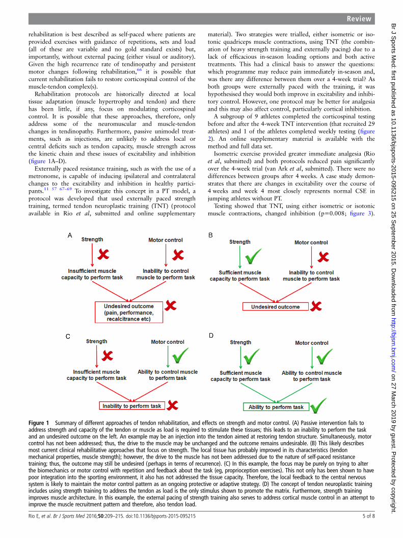
Rehabilitation protocols are historically directed at localtissue adaptation (muscle hypertrophy and tendon) and therehas been little, if any, focus on modulating corticospinalcontrol. It is possible that these approaches, therefore, onlyaddress some of the neuromuscular and muscle-tendonchanges in tendinopathy. Furthermore, passive unimodel treat-ments, such as injections, are unlikely to address local orcentral deficits such as tendon capacity, muscle strength acrossthe kinetic chain and these issues of excitability and inhibition(figure 1A–D).
Externally paced resistance training, such as with the use of ametronome, is capable of inducing ipsilateral and contralateralchanges to the excitability and inhibition in healthy partici-pants.11 57 67–69 To investigate this concept in a PT model, aprotocol was developed that used externally paced strengthtraining, termed tendon neuroplastic training (TNT) (protocolavailable in Rio et al, submitted and online supplementary
material). Two strategies were trialled, either isometric or iso-tonic quadriceps muscle contractions, using TNT (the combin-ation of heavy strength training and externally pacing) due to alack of efficacious in-season loading options and both activetreatments. This had a clinical basis to answer the questions:which programme may reduce pain immediately in-season and,was there any difference between them over a 4-week trial? Asboth groups were externally paced with the training, it washypothesised they would both improve in excitability and inhibi-tory control. However, one protocol may be better for analgesiaand this may also affect control, particularly cortical inhibition.
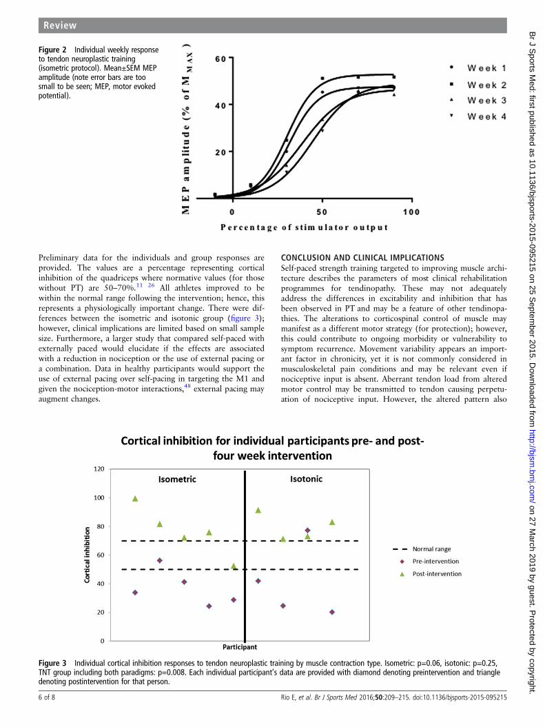
A subgroup of 9 athletes completed the corticospinal testingbefore and after the 4-week TNT intervention (that recruited 29athletes) and 1 of the athletes completed weekly testing (figure2). An online supplementary material is available with themethod and full data set.
Isometric exercise provided greater immediate analgesia (Rioet al, submitted) and both protocols reduced pain significantlyover the 4-week trial (van Ark et al, submitted). There were nodifferences between groups after 4 weeks. A case study demon-strates that there are changes in excitability over the course of4 weeks and week 4 most closely represents normal CSE injumping athletes without PT.
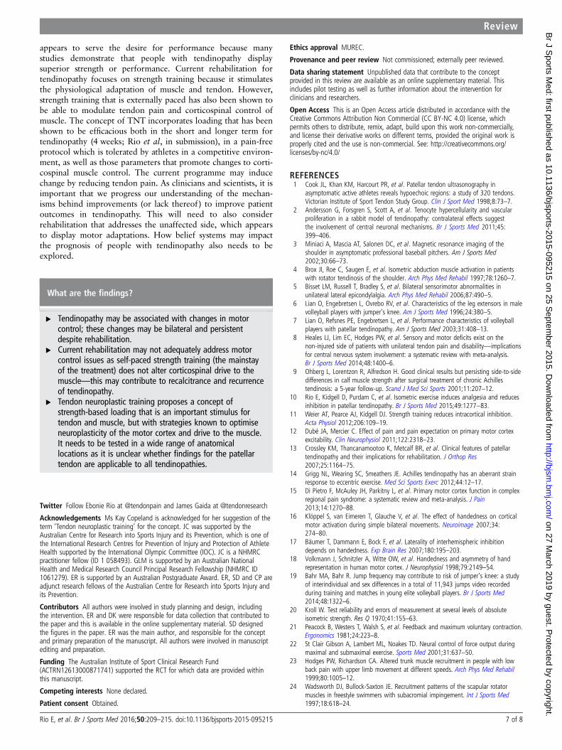
Testing showed that TNT, using either isometric or isotonicmuscle contractions, changed inhibition (p=0.008; figure 3).
Figure 1 Summary of different approaches of tendon rehabilitation, and effects on strength and motor control. (A) Passive intervention fails toaddress strength and capacity of the tendon or muscle as load is required to stimulate these tissues; this leads to an inability to perform the taskand an undesired outcome on the left. An example may be an injection into the tendon aimed at restoring tendon structure. Simultaneously, motorcontrol has not been addressed; thus, the drive to the muscle may be unchanged and the outcome remains undesirable. (B) This likely describesmost current clinical rehabilitative approaches that focus on strength. The local tissue has probably improved in its characteristics (tendonmechanical properties, muscle strength); however, the drive to the muscle has not been addressed due to the nature of self-paced resistancetraining; thus, the outcome may still be undesired (perhaps in terms of recurrence). (C) In this example, the focus may be purely on trying to alterthe biomechanics or motor control with repetition and feedback about the task (eg, proprioception exercises). This not only has been shown to havepoor integration into the sporting environment, it also has not addressed the tissue capacity. Therefore, the local feedback to the central nervoussystem is likely to maintain the motor control pattern as an ongoing protective or adaptive strategy. (D) The concept of tendon neuroplastic trainingincludes using strength training to address the tendon as load is the only stimulus shown to promote the matrix. Furthermore, strength trainingimproves muscle architecture. In this example, the external pacing of strength training also serves to address cortical muscle control in an attempt toimprove the muscle recruitment pattern and therefore, also tendon load.
Rio E, et al. Br J Sports Med 2016;50:209–215. doi:10.1136/bjsports-2015-095215 5 of 8
Review on 27 M
arch 2019 by guest. Protected by copyright.
http://bjsm.bm
j.com/
Br J S
ports Med: first published as 10.1136/bjsports-2015-095215 on 25 S
eptember 2015. D
ownloaded from
Preliminary data for the individuals and group responses areprovided. The values are a percentage representing corticalinhibition of the quadriceps where normative values (for thosewithout PT) are 50–70%.11 26 All athletes improved to bewithin the normal range following the intervention; hence, thisrepresents a physiologically important change. There were dif-ferences between the isometric and isotonic group (figure 3);however, clinical implications are limited based on small samplesize. Furthermore, a larger study that compared self-paced withexternally paced would elucidate if the effects are associatedwith a reduction in nociception or the use of external pacing ora combination. Data in healthy participants would support theuse of external pacing over self-pacing in targeting the M1 andgiven the nociception-motor interactions,48 external pacing mayaugment changes.
CONCLUSION AND CLINICAL IMPLICATIONSSelf-paced strength training targeted to improving muscle archi-tecture describes the parameters of most clinical rehabilitationprogrammes for tendinopathy. These may not adequatelyaddress the differences in excitability and inhibition that hasbeen observed in PT and may be a feature of other tendinopa-thies. The alterations to corticospinal control of muscle maymanifest as a different motor strategy (for protection); however,this could contribute to ongoing morbidity or vulnerability tosymptom recurrence. Movement variability appears an import-ant factor in chronicity, yet it is not commonly considered inmusculoskeletal pain conditions and may be relevant even ifnociceptive input is absent. Aberrant tendon load from alteredmotor control may be transmitted to tendon causing perpetu-ation of nociceptive input. However, the altered pattern also
Figure 2 Individual weekly responseto tendon neuroplastic training(isometric protocol). Mean±SEM MEPamplitude (note error bars are toosmall to be seen; MEP, motor evokedpotential).
Figure 3 Individual cortical inhibition responses to tendon neuroplastic training by muscle contraction type. Isometric: p=0.06, isotonic: p=0.25,TNT group including both paradigms: p=0.008. Each individual participant’s data are provided with diamond denoting preintervention and triangledenoting postintervention for that person.
6 of 8 Rio E, et al. Br J Sports Med 2016;50:209–215. doi:10.1136/bjsports-2015-095215
Review on 27 M
arch 2019 by guest. Protected by copyright.
http://bjsm.bm
j.com/
Br J S
ports Med: first published as 10.1136/bjsports-2015-095215 on 25 S
eptember 2015. D
ownloaded from
appears to serve the desire for performance because manystudies demonstrate that people with tendinopathy displaysuperior strength or performance. Current rehabilitation fortendinopathy focuses on strength training because it stimulatesthe physiological adaptation of muscle and tendon. However,strength training that is externally paced has also been shown tobe able to modulate tendon pain and corticospinal control ofmuscle. The concept of TNT incorporates loading that has beenshown to be efficacious both in the short and longer term fortendinopathy (4 weeks; Rio et al, in submission), in a pain-freeprotocol which is tolerated by athletes in a competitive environ-ment, as well as those parameters that promote changes to corti-cospinal muscle control. The current programme may inducechange by reducing tendon pain. As clinicians and scientists, it isimportant that we progress our understanding of the mechan-isms behind improvements (or lack thereof) to improve patientoutcomes in tendinopathy. This will need to also considerrehabilitation that addresses the unaffected side, which appearsto display motor adaptations. How belief systems may impactthe prognosis of people with tendinopathy also needs to beexplored.
What are the findings?
▸ Tendinopathy may be associated with changes in motorcontrol; these changes may be bilateral and persistentdespite rehabilitation.
▸ Current rehabilitation may not adequately address motorcontrol issues as self-paced strength training (the mainstayof the treatment) does not alter corticospinal drive to themuscle—this may contribute to recalcitrance and recurrenceof tendinopathy.
▸ Tendon neuroplastic training proposes a concept ofstrength-based loading that is an important stimulus fortendon and muscle, but with strategies known to optimiseneuroplasticity of the motor cortex and drive to the muscle.It needs to be tested in a wide range of anatomicallocations as it is unclear whether findings for the patellartendon are applicable to all tendinopathies.
Twitter Follow Ebonie Rio at @tendonpain and James Gaida at @tendonresearch
Acknowledgements Ms Kay Copeland is acknowledged for her suggestion of theterm ‘Tendon neuroplastic training’ for the concept. JC was supported by theAustralian Centre for Research into Sports Injury and its Prevention, which is one ofthe International Research Centres for Prevention of Injury and Protection of AthleteHealth supported by the International Olympic Committee (IOC). JC is a NHMRCpractitioner fellow (ID 1 058493). GLM is supported by an Australian NationalHealth and Medical Research Council Principal Research Fellowship (NHMRC ID1061279). ER is supported by an Australian Postgraduate Award. ER, SD and CP areadjunct research fellows of the Australian Centre for Research into Sports Injury andits Prevention.
Contributors All authors were involved in study planning and design, includingthe intervention. ER and DK were responsible for data collection that contributed tothe paper and this is available in the online supplementary material. SD designedthe figures in the paper. ER was the main author, and responsible for the conceptand primary preparation of the manuscript. All authors were involved in manuscriptediting and preparation.
Funding The Australian Institute of Sport Clinical Research Fund(ACTRN12613000871741) supported the RCT for which data are provided withinthis manuscript.
Competing interests None declared.
Patient consent Obtained.
Ethics approval MUREC.
Provenance and peer review Not commissioned; externally peer reviewed.
Data sharing statement Unpublished data that contribute to the conceptprovided in this review are available as an online supplementary material. Thisincludes pilot testing as well as further information about the intervention forclinicians and researchers.
Open Access This is an Open Access article distributed in accordance with theCreative Commons Attribution Non Commercial (CC BY-NC 4.0) license, whichpermits others to distribute, remix, adapt, build upon this work non-commercially,and license their derivative works on different terms, provided the original work isproperly cited and the use is non-commercial. See: http://creativecommons.org/licenses/by-nc/4.0/
REFERENCES1 Cook JL, Khan KM, Harcourt PR, et al. Patellar tendon ultrasonography in
asymptomatic active athletes reveals hypoechoic regions: a study of 320 tendons.Victorian Institute of Sport Tendon Study Group. Clin J Sport Med 1998;8:73–7.
2 Andersson G, Forsgren S, Scott A, et al. Tenocyte hypercellularity and vascularproliferation in a rabbit model of tendinopathy: contralateral effects suggestthe involvement of central neuronal mechanisms. Br J Sports Med 2011;45:399–406.
3 Miniaci A, Mascia AT, Salonen DC, et al. Magnetic resonance imaging of theshoulder in asymptomatic professional baseball pitchers. Am J Sports Med2002;30:66–73.
4 Brox JI, Roe C, Saugen E, et al. Isometric abduction muscle activation in patientswith rotator tendinosis of the shoulder. Arch Phys Med Rehabil 1997;78:1260–7.
5 Bisset LM, Russell T, Bradley S, et al. Bilateral sensorimotor abnormalities inunilateral lateral epicondylalgia. Arch Phys Med Rehabil 2006;87:490–5.
6 Lian O, Engebretsen L, Ovrebo RV, et al. Characteristics of the leg extensors in malevolleyball players with jumper’s knee. Am J Sports Med 1996;24:380–5.
7 Lian O, Refsnes PE, Engebretsen L, et al. Performance characteristics of volleyballplayers with patellar tendinopathy. Am J Sports Med 2003;31:408–13.
8 Heales LJ, Lim EC, Hodges PW, et al. Sensory and motor deficits exist on thenon-injured side of patients with unilateral tendon pain and disability—implicationsfor central nervous system involvement: a systematic review with meta-analysis.Br J Sports Med 2014;48:1400–6.
9 Ohberg L, Lorentzon R, Alfredson H. Good clinical results but persisting side-to-sidedifferences in calf muscle strength after surgical treatment of chronic Achillestendinosis: a 5-year follow-up. Scand J Med Sci Sports 2001;11:207–12.
10 Rio E, Kidgell D, Purdam C, et al. Isometric exercise induces analgesia and reducesinhibition in patellar tendinopathy. Br J Sports Med 2015;49:1277–83.
11 Weier AT, Pearce AJ, Kidgell DJ. Strength training reduces intracortical inhibition.Acta Physiol 2012;206:109–19.
12 Dubé JA, Mercier C. Effect of pain and pain expectation on primary motor cortexexcitability. Clin Neurophysiol 2011;122:2318–23.
13 Crossley KM, Thancanamootoo K, Metcalf BR, et al. Clinical features of patellartendinopathy and their implications for rehabilitation. J Orthop Res2007;25:1164–75.
14 Grigg NL, Wearing SC, Smeathers JE. Achilles tendinopathy has an aberrant strainresponse to eccentric exercise. Med Sci Sports Exerc 2012;44:12–17.
15 Di Pietro F, McAuley JH, Parkitny L, et al. Primary motor cortex function in complexregional pain syndrome: a systematic review and meta-analysis. J Pain2013;14:1270–88.
16 Klöppel S, van Eimeren T, Glauche V, et al. The effect of handedness on corticalmotor activation during simple bilateral movements. Neuroimage 2007;34:274–80.
17 Bäumer T, Dammann E, Bock F, et al. Laterality of interhemispheric inhibitiondepends on handedness. Exp Brain Res 2007;180:195–203.
18 Volkmann J, Schnitzler A, Witte OW, et al. Handedness and asymmetry of handrepresentation in human motor cortex. J Neurophysiol 1998;79:2149–54.
19 Bahr MA, Bahr R. Jump frequency may contribute to risk of jumper’s knee: a studyof interindividual and sex differences in a total of 11,943 jumps video recordedduring training and matches in young elite volleyball players. Br J Sports Med2014;48:1322–6.
20 Kroll W. Test reliability and errors of measurement at several levels of absoluteisometric strength. Res Q 1970;41:155–63.
21 Peacock B, Westers T, Walsh S, et al. Feedback and maximum voluntary contraction.Ergonomics 1981;24:223–8.
22 St Clair Gibson A, Lambert ML, Noakes TD. Neural control of force output duringmaximal and submaximal exercise. Sports Med 2001;31:637–50.
23 Hodges PW, Richardson CA. Altered trunk muscle recruitment in people with lowback pain with upper limb movement at different speeds. Arch Phys Med Rehabil1999;80:1005–12.
24 Wadsworth DJ, Bullock-Saxton JE. Recruitment patterns of the scapular rotatormuscles in freestyle swimmers with subacromial impingement. Int J Sports Med1997;18:618–24.
Rio E, et al. Br J Sports Med 2016;50:209–215. doi:10.1136/bjsports-2015-095215 7 of 8
Review on 27 M
arch 2019 by guest. Protected by copyright.
http://bjsm.bm
j.com/
Br J S
ports Med: first published as 10.1136/bjsports-2015-095215 on 25 S
eptember 2015. D
ownloaded from
25 Seitz AL, McClure PW, Finucane S, et al. Mechanisms of rotator cuff tendinopathy:intrinsic, extrinsic, or both? Clin Biomech (Bristol, Avon) 2011;26:1–12.
26 Goodwill AM, Pearce AJ, Kidgell DJ. Corticomotor plasticity following unilateralstrength training. Muscle Nerve 2012;46:384–93.
27 Rantalainen T, Weier A, Leung M, et al. Short-interval intracortical inhibition is notaffected by varying visual feedback in an isometric task in biceps brachii muscle.Front Hum Neurosci 2013;7:68.
28 Edwards S, Steele JR, McGhee DE, et al. Landing strategies of athletes with anasymptomatic patellar tendon abnormality. Med Sci Sports Exerc 2010;42:2072–80.
29 James CR, Dufek JS, Bates BT. Effects of injury proneness and task difficulty on jointkinetic variability. Med Sci Sports Exerc 2000;32:1833–44.
30 Moseley GL, Hodges PW. Reduced variability of postural strategy preventsnormalization of motor changes induced by back pain: a risk factor for chronictrouble? Behav Neurosci 2006;120:474–6.
31 Visnes H, Aandahl HA, Bahr R. Jumper’s knee paradox—jumping ability is a riskfactor for developing jumper’s knee: a 5-year prospective study. Br J Sports Med2013;47:503–7.
32 Siegmund JA, Huxel KC, Swanik CB. Compensatory mechanisms in basketballplayers with jumper’s knee. J Sport Rehabil 2008;17:358–71.
33 Moseley GL, Nicholas MK, Hodges PW. Does anticipation of back pain predisposeto back trouble? Brain 2004;127(Pt 10):2339–47.
34 Hodges PW, Richardson CA. Inefficient muscular stabilization of the lumbar spineassociated with low back pain. A motor control evaluation of transversusabdominis. Spine (Phila Pa 1976) 1996;21:2640–50.
35 Stergiou N, Decker LM. Human movement variability, nonlinear dynamics, andpathology: is there a connection? Hum Mov Sci 2011;30:869–88.
36 Bartlett R, Wheat J, Robins M. Is movement variability important for sportsbiomechanists? Sports Biomech 2007;6:224–43.
37 Moseley GL, Arntz A. The context of a noxious stimulus affects the pain it evokes.Pain 2007;133:64–71.
38 Hodges PW, Moseley GL. Pain and motor control of the lumbopelvic region: effectand possible mechanisms. J Electromyogr Kinesiol 2003;13:361–70.
39 Mokone GG, Schwellnus MP, Noakes TD, et al. The COL5A1 gene and Achillestendon pathology. Scand J Med Sci Sports 2006;16:19–26.
40 September AV, Nell EM, O’Connell K, et al. A pathway-based approach investigatingthe genes encoding interleukin-1β, interleukin-6 and the interleukin-1 receptorantagonist provides new insight into the genetic susceptibility of Achillestendinopathy. Br J Sports Med 2011;45:1040–7.
41 Jewson JL, Lambert GW, Storr M, et al. The sympathetic nervous system andtendinopathy: a systematic review. Sports Med 2015;45:727–43.
42 Butefisch CM, Wessling M, Netz J, et al. Relationship between interhemisphericinhibition and motor cortex excitability in subacute stroke patients. NeurorehabilNeural Repair 2008;22:4–21.
43 Lund JP, Donga R, Widmer CG, et al. The pain-adaptation model: a discussion ofthe relationship between chronic musculoskeletal pain and motor activity. Can JPhysiol Pharmacol 1991;69:683–94.
44 Graven-Nielsen T, Svensson P, Arendt-Nielsen L. Effects of experimental muscle painon muscle activity and co-ordination during static and dynamic motor function.Electroencephalogr Clin Neurophysiol 1997;105:156–64.
45 Le Pera D, Graven-Nielsen T, Valeriani M, et al. Inhibition of motor system excitability atcortical and spinal level by tonic muscle pain. Clin Neurophysiol 2001;112:1633–41.
46 Farina S, Valeriani M, Rosso T, et al. Transient inhibition of the human motor cortexby capsaicin-induced pain. A study with transcranial magnetic stimulation. NeurosciLett 2001;314:97–101.
47 Ngomo S, Mercier C, Bouyer LJ, et al. Alterations in central motor representationincrease over time in individuals with rotator cuff tendinopathy. Clin Neurophysiol2015;126:365–71.
48 Nijs J, Daenen L, Cras P, et al. Nociception affects motor output: a review on sensory-motor interaction with focus on clinical implications. Clin J Pain 2012;28:175–81.
49 Cook JL, Purdam CR. Is tendon pathology a continuum? A pathology model toexplain the clinical presentation of load-induced tendinopathy. Br J Sports Med2009;43:409–16.
50 Drew BT, Smith TO, Littlewood C, et al. Do structural changes (eg, collagen/matrix)explain the response to therapeutic exercises in tendinopathy: a systematic review.Br J Sports Med 2014;48:966–72.
51 Docking SI, Cook J. Pathological tendons maintain sufficient aligned fibrillarstructure on ultrasound tissue characterization (UTC). Scand J Med Sci Sports2015.
52 Rio E, Moseley L, Purdam C, et al. The pain of tendinopathy: physiological orpathophysiological? Sports Med 2014;44:9–23.
53 Cohen LG, Ziemann U, Chen R, et al. Studies of neuroplasticity with transcranialmagnetic stimulation. J Clin Neurophysiol 1998;15:305–24.
54 Gerloff C, Cohen LG, Floeter MK, et al. Inhibitory influence of the ipsilateral motorcortex on responses to stimulation of the human cortex and pyramidal tract.J Physiol 1998;510(Pt 1):249–59.
55 Ackerley SJ, Stinear CM, Byblow WD. Promoting use-dependent plasticity withexternally-paced training. Clin Neurophysiol 2011;122:2462–8.
56 Kidgell DJ, Pearce AJ. Corticospinal properties following short-termstrength training of an intrinsic hand muscle. Hum Mov Sci 2010;29:631–41.
57 Kidgell DJ, Stokes MA, Castricum TJ, et al. Neurophysiological responses aftershort-term strength training of the biceps brachii muscle. J Strength Cond Res2010;24:3123–32.
58 Leung M, Rantalainen T, Teo WP, et al. Motor cortex excitability is notdifferentially modulated following skill and strength training. Neuroscience2015;305:99–108.
59 Frohm A, Halvorsen K, Thorstensson A. Patellar tendon load in different types ofeccentric squats. Clin Biomech (Bristol, Avon) 2007;22:704–11.
60 Frohm A, Saartok T, Halvorsen K, et al. Eccentric treatment for patellartendinopathy: a prospective randomised short-term pilot study of two rehabilitationprotocols. Br J Sports Med 2007;41:e7.
61 Jensen K, Di Fabio RP. Evaluation of eccentric exercise in treatment of patellartendinitis. Phys Ther 1989;69:211–16.
62 Jonsson P, Alfredson H, Sunding K, et al. New regimen for eccentric calf-muscletraining in patients with chronic insertional Achilles tendinopathy: results of a pilotstudy. Br J Sports Med 2008;42:746–9.
63 Kongsgaard M, Aagaard P, Roikjaer S, et al. Decline eccentric squats increasespatellar tendon loading compared to standard eccentric squats. Clin Biomech(Bristol, Avon) 2006;21:748–54.
64 Mafi N, Lorentzon R, Alfredson H. Superior short-term results with eccentric calfmuscle training compared to concentric training in a randomized prospectivemulticenter study on patients with chronic Achilles tendinosis. Knee Surg SportsTraumatol Arthrosc 2001;9:42–7.
65 Kongsgaard M, Kovanen V, Aagaard P, et al. Corticosteroid injections, eccentricdecline squat training and heavy slow resistance training in patellar tendinopathy.Scand J Med Sci Sports 2009;19:790–802.
66 Paavola M, Kannus P, Paakkala T, et al. Long-term prognosis of patients withAchilles tendinopathy. An observational 8-year follow-up study. Am J Sports Med2000;28:634–42.
67 Kidgell DJ, Stokes MA, Pearce AJ. Strength training of one limb increasescorticomotor excitability projecting to the contralateral homologous limb. MotorControl 2011;15:247–66.
68 Latella C, Kidgell DJ, Pearce AJ. Reduction in corticospinal inhibition in the trainedand untrained limb following unilateral leg strength training. Eur J Appl Physiol2012;112:3097–107.
69 Pearce AJ, Hendy A, Bowen WA, et al. Corticospinal adaptations and strengthmaintenance in the immobilized arm following 3 weeks unilateral strength training.Scand J Med Sci Sports 2013;23:740–8.
8 of 8 Rio E, et al. Br J Sports Med 2016;50:209–215. doi:10.1136/bjsports-2015-095215
Review on 27 M
arch 2019 by guest. Protected by copyright.
http://bjsm.bm
j.com/
Br J S
ports Med: first published as 10.1136/bjsports-2015-095215 on 25 S
eptember 2015. D
ownloaded from