Embed Size (px)
Citation preview
Rheumatic heart disease
Mitral stenosis
Valvular heart disease
• Rheumatic • Age related • congenital
Mitral valve
• Stenosis• Regurgitation • Prolapse
Mitral stenosis
• 2/3 females • Usually rheumatic• Rarely congenital • 40% of all RHD
Structural defects
• Diffusely thickened –fibrous tissue /calcified deposits
• Mitral commisures fuse • Corde tendinae fuse /shorten• Narrowing of the apex of funnel shaped valves
• Calcification of slender valves immobilises the leaflet and narrows the orifice –thrombus formation –arterial thrombus from calcified Valves
Pathophysiology
• Normal mv –dia -4-6 cm2• <2 cm 2-atrial to ventricular flow is
maintained by increased av pressure gradient –the hallmark of ms
• <1 cm2 –LAP should be atleast 25mm hg is required to maintain normal output .
• Increased Lap --------increased pulm pressure ------increased capillary pressure -----decreased pulm compliance -------exertional dyspnoea.
• Increased heart rate –decreased transvalvular gradient ----increased LAP
• Lv diastolic pressure in normal in ms• Co is normal at rest ---at exercise –decreased co.
$
• Clinical /hemodynamic Features –influenced by• Passive backward transmission of LAP• Pulmonary arteriolar constriction• Intertitial edema• Organic obliterative changes in the pul vascular
bed • Phtn----Tr------rt sided failures---bornheimeffect
symptoms
• Carditis---ms-----2 decades,• Dyspnoea on exertion ----4 th decade—
progressive worsening to death---2-5 yrs• Doe ,orthopnoea ,pnd,arrthmia-premature
atraial complex,paroxysysmal tachycardia,flutter,fibrilation
• Haemoptysis –increased pulm venous pressure
• Recurrant pulm embolism• Pulm infection• Endocarditis• Chest pain -10%• Thrombus formation in the left atrium-af—
appendages of LA• Pedunculated thrombus –ball valve thrombi• -syncope-angina –changing ascultatory signs
On examination
• Malar flush-pinched blue facies• JVP-a wave prominence –af –a wave absent• Palpation-tapping apical impulse ,s1
loud,palpable ,s2 p2 loud• Diastolic thrill• Auscultation-s1 accentuated /snapping –
delayed –mv doesn’t close till LVP>LAP• Qs prolongation ,p2 loud
• A2-p2-os -0.05-0.12• P2-os –severity of ms• Intensity of s1/os –pliability of leAFLET• MDM after os • Duration correlates with ms severity• S1-closure of mitral /tricuspid valve
• Intensity of s1• Pos of mv at onset of vent systole • Rate of increase in LAP• Degree of structural damage of the valve • Amt of tissue bet heart and sthetoscope
• S1 loud –diastole is shortened by tachycardia • S1 split -10-30 msec• S1 –m1t1-----prolonged in rbbb• t1m1 –severe ms ,left atrial myoma lbbb

Mitrl regurgitation
etiology
• Chronic rhd –severe mr- 1/3• Seen in males mostly• Rheumatic process-
rigidity,deformity,retraction of the valve cusps-commisural fusion
• Congenital-endocardial cushion defects• Fibrosis of papillary muscles in MI• Ischeamia –paplillary dysfn

• Lv dilated in DCM• HOCM-ant displace ment of the ant leaflet• Mitral prolapse –MR• Acute MR-inf endocarditis
pathophysiology
• Clinical pic depends on p-v relation ship of LA AND PUL -VENOUS BED
• Increased LAP-Increased pulm edema • Effective forward pressure of lv decreases• Inc-LA volume –due to atrial compliance • Low cardiac out put • Atrial fibrillation
SYMPTOMS
• FATIGUE• Doe• Orthopnea• Pnd• Haemoptysis• Sys embolism• Rh f-jvp inc,tr,phtn,hep congestion
Physical examination
• Sys thrill-left apex• Hyperdynmic apical impulse• Laterally displaced• Palpable p2• Parasternal heave
auscultation
• S1-absent/softor buried in systolic murmur• Decreased co-aorta closes early-a2 early-wide
spliting of s2• Os –indicates ms • Gallop rhythm • Pansystolic murmur
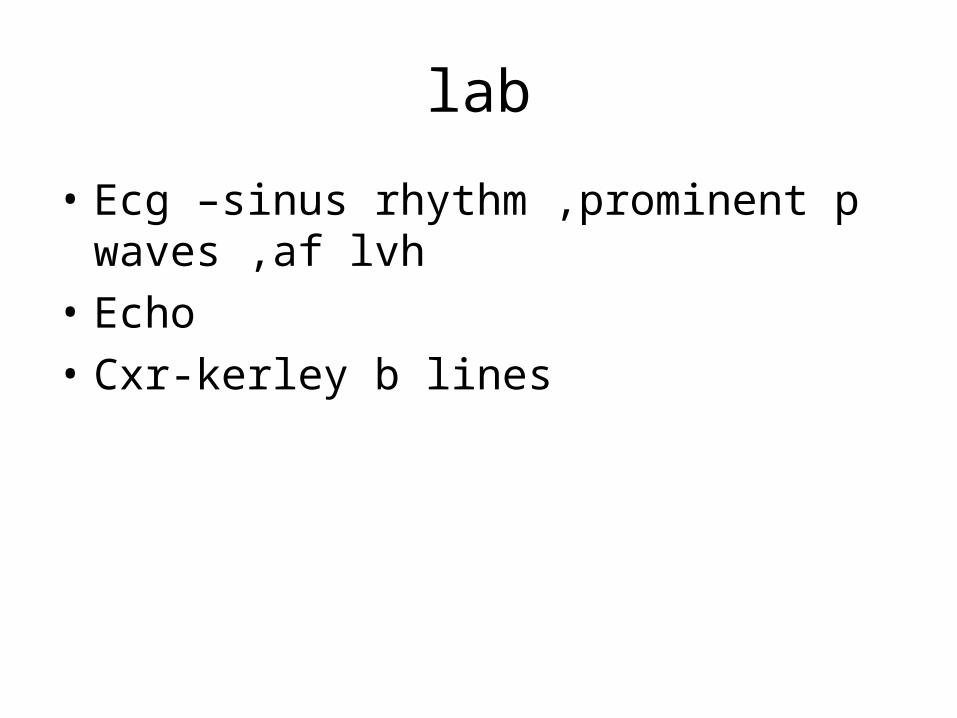
lab
• Ecg –sinus rhythm ,prominent p waves ,af lvh• Echo• Cxr-kerley b lines
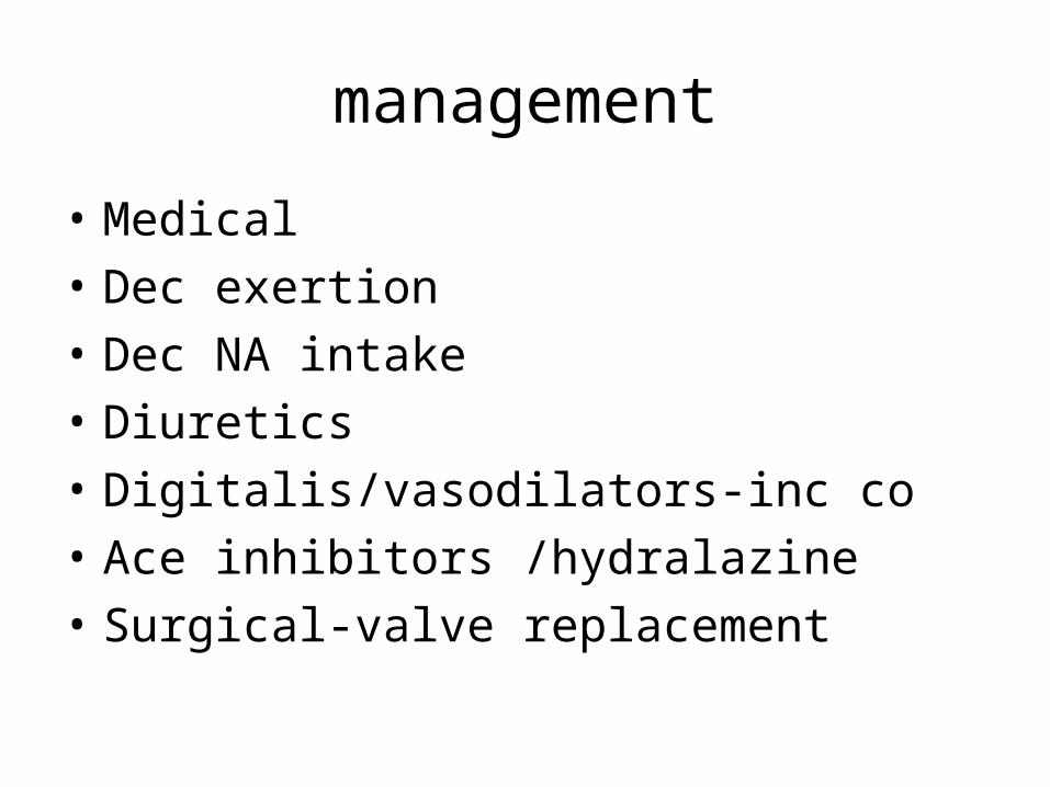
management
• Medical• Dec exertion • Dec NA intake • Diuretics • Digitalis/vasodilators-inc co• Ace inhibitors /hydralazine• Surgical-valve replacement


![[PPT]RHEUMATIC HEART DISEASE · Web viewRHEUMATIC HEART DISEASE Rheumatic Heart Disease is a Notifiable disease Please contact the RHD register Central Australia Ph: 08 895 16909](https://img.pdfslide.net/doc/110x75/5acb00077f8b9acb7c8ea1d8/pptrheumatic-heart-viewrheumatic-heart-disease-rheumatic-heart-disease-is-a-notifiable.jpg)