Embed Size (px)
Citation preview
Rheumatic Heart Disease
Immunology UnitDepartment of Pathology
College of MedicineKing Saud University
Objectives
• To understand basis of rheumatic fever as an immunologically mediated late complication of Streptococcal infection
• To know that autoimmunity results from production of cross reacting antibodies against Streptococcal antigens
• To describe rheumatic heart disease as one of the several manifestations of rheumatic fever
• To know the signs, symptoms, pathogenesis, treatment and prophylaxis of rheumatic heart disease
Rheumatic Fever• Epidemiology of Rheumatic Fever (RF)
• ~3% of persons with untreated group A streptococcal pharyngitis develop rheumatic fever
• 15-20 million new cases a year in developing countries
• Risk factors
– Low standard of living
– Crowding
Rheumatic fever
• Individual (HLA) susceptibility is also important
• Antigen-presenting cells bearing the HLA-DR7molecule from RHD patients preferentially recognize heart-tissue protein (Guilherme L, Kalil J. Ann N Y Acd Sci 2007,1107:426-433)
• Other views in the literature exist, due to
+ The various HLA-typing methods. + Ways of grouping the cases.
Rheumatic fever
• Rheumatic fever is an inflammatory disease which may develop after a Group AStreptococcal infection such as:
– Strep. throat infection or scarlet fever
• Can involve the heart, joints, skin, and brain
• It commonly appears in children ages 5 through 15
Organism
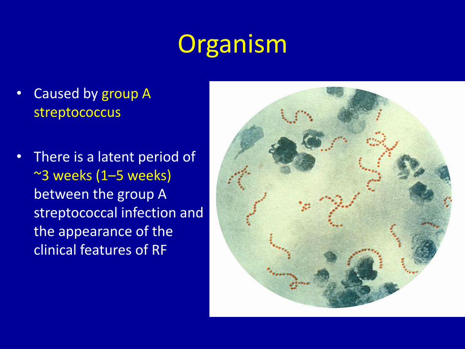
• Caused by group A streptococcus
• There is a latent period of ~3 weeks (1–5 weeks)between the group A streptococcal infection and the appearance of the clinical features of RF
Group A b-haemolytic streptococcus
• All cases associated with recent infection (e.g. pharyngitis)
• Antibody and cellular immune response cross-reacts with human connective tissue
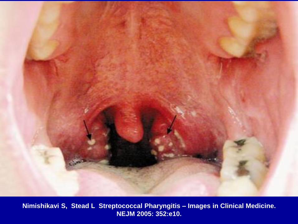
Nimishikavi S, Stead L Streptococcal Pharyngitis – Images in Clinical Medicine.
NEJM 2005: 352:e10.
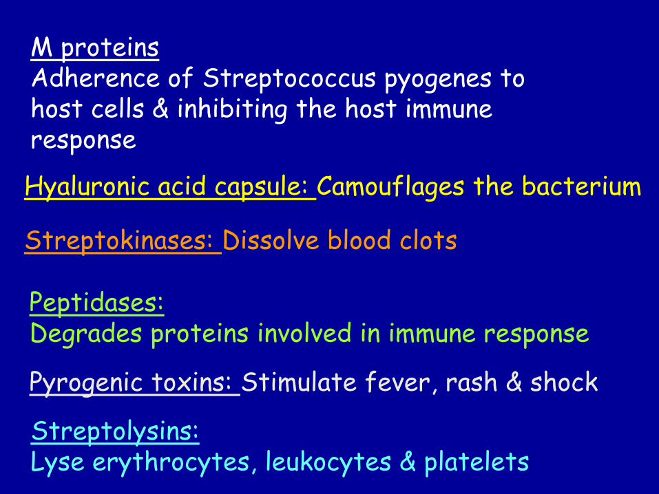
M proteinsAdherence of Streptococcus pyogenes to host cells & inhibiting the host immune response
Hyaluronic acid capsule: Camouflages the bacterium
Streptokinases: Dissolve blood clots
Peptidases:Degrades proteins involved in immune response
Pyrogenic toxins: Stimulate fever, rash & shock
Streptolysins:Lyse erythrocytes, leukocytes & platelets
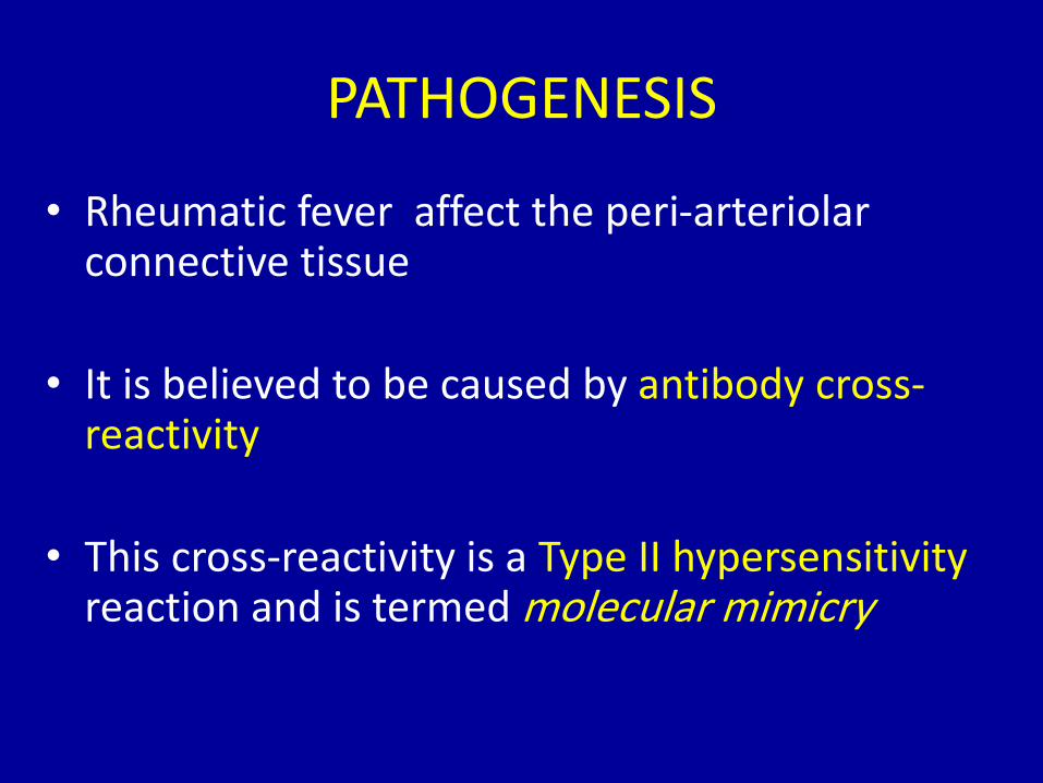
PATHOGENESIS
• Rheumatic fever affect the peri-arteriolar connective tissue
• It is believed to be caused by antibody cross-reactivity
• This cross-reactivity is a Type II hypersensitivityreaction and is termed molecular mimicry
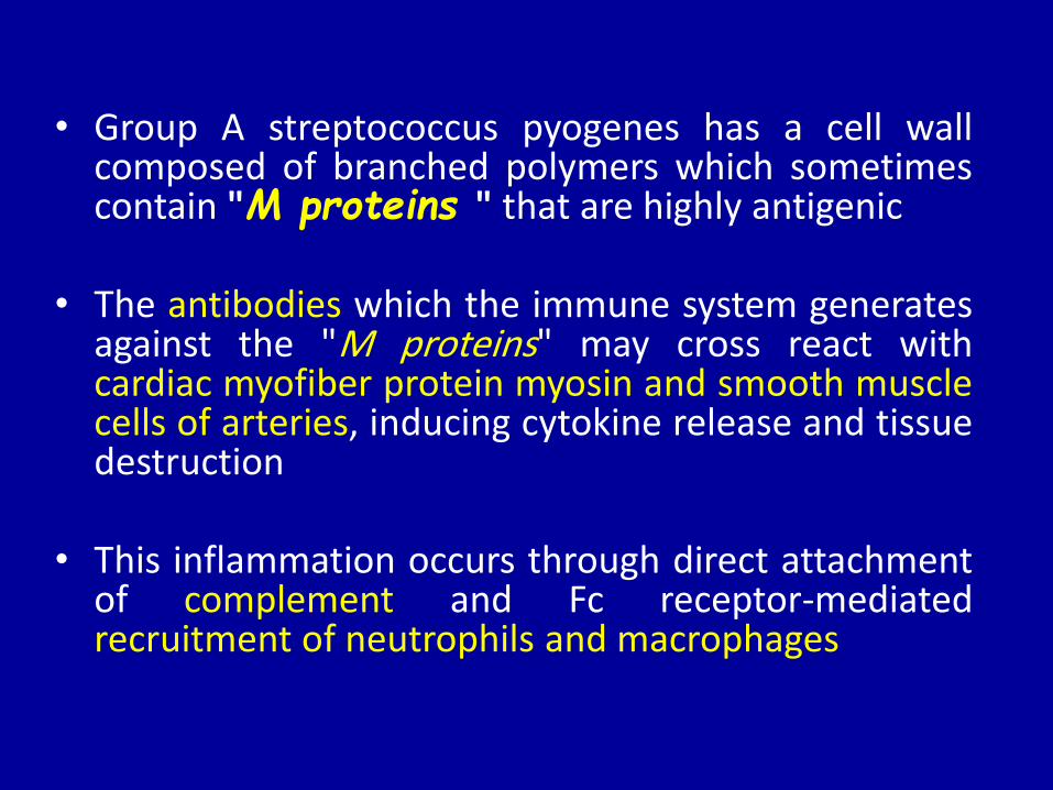
• Group A streptococcus pyogenes has a cell wallcomposed of branched polymers which sometimescontain "M proteins " that are highly antigenic
• The antibodies which the immune system generatesagainst the "M proteins" may cross react withcardiac myofiber protein myosin and smooth musclecells of arteries, inducing cytokine release and tissuedestruction
• This inflammation occurs through direct attachmentof complement and Fc receptor-mediatedrecruitment of neutrophils and macrophages
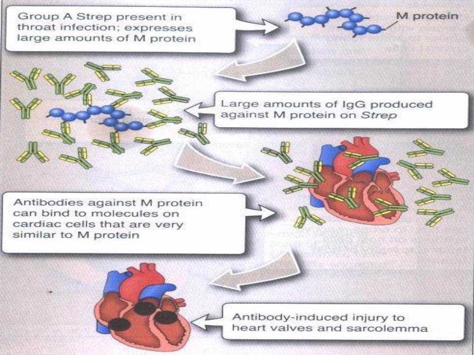
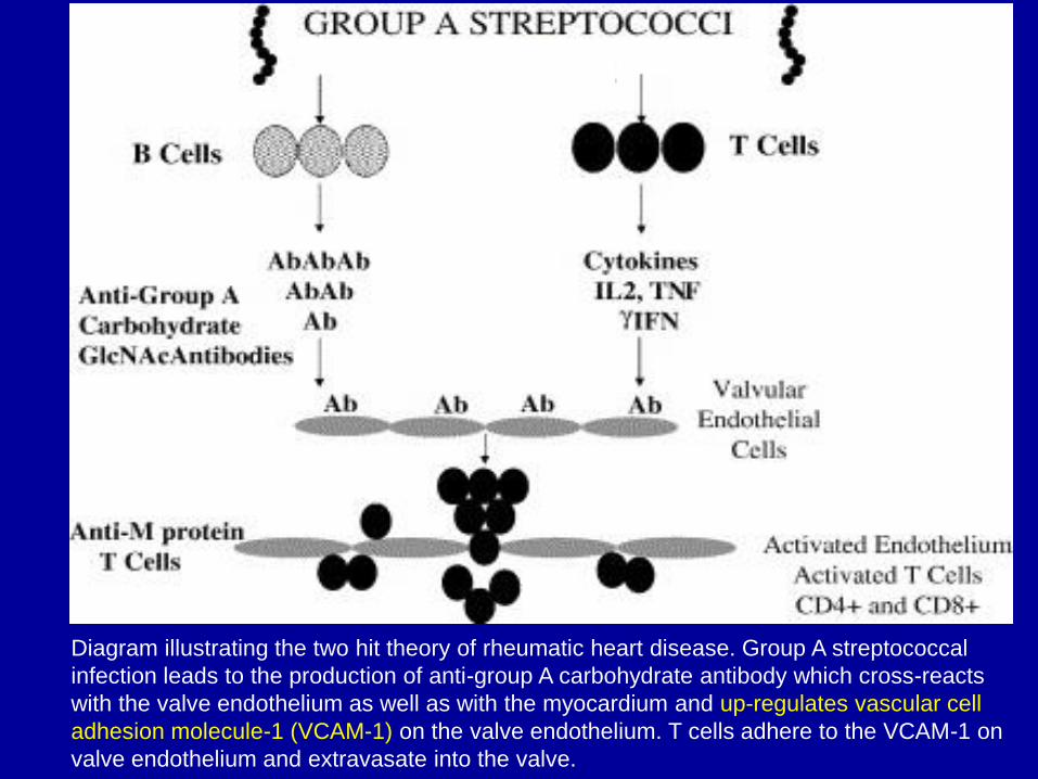
Diagram illustrating the two hit theory of rheumatic heart disease. Group A streptococcal
infection leads to the production of anti-group A carbohydrate antibody which cross-reacts
with the valve endothelium as well as with the myocardium and up-regulates vascular cell
adhesion molecule-1 (VCAM-1) on the valve endothelium. T cells adhere to the VCAM-1 on
valve endothelium and extravasate into the valve.
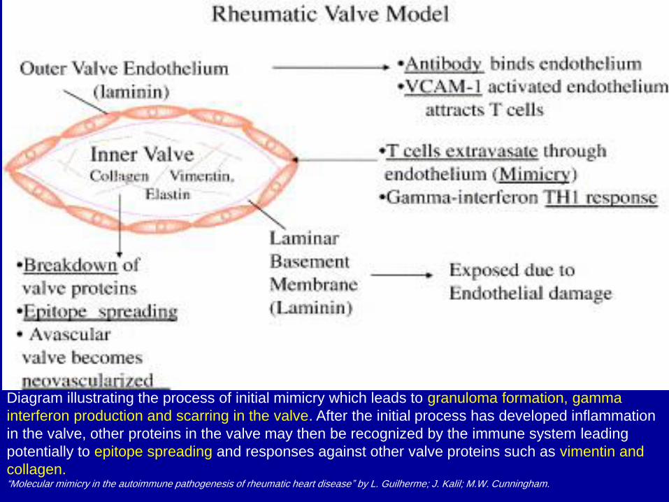
Diagram illustrating the process of initial mimicry which leads to granuloma formation, gamma
interferon production and scarring in the valve. After the initial process has developed inflammation
in the valve, other proteins in the valve may then be recognized by the immune system leading
potentially to epitope spreading and responses against other valve proteins such as vimentin and
collagen.“Molecular mimicry in the autoimmune pathogenesis of rheumatic heart disease” by L. Guilherme; J. Kalil; M.W. Cunningham.
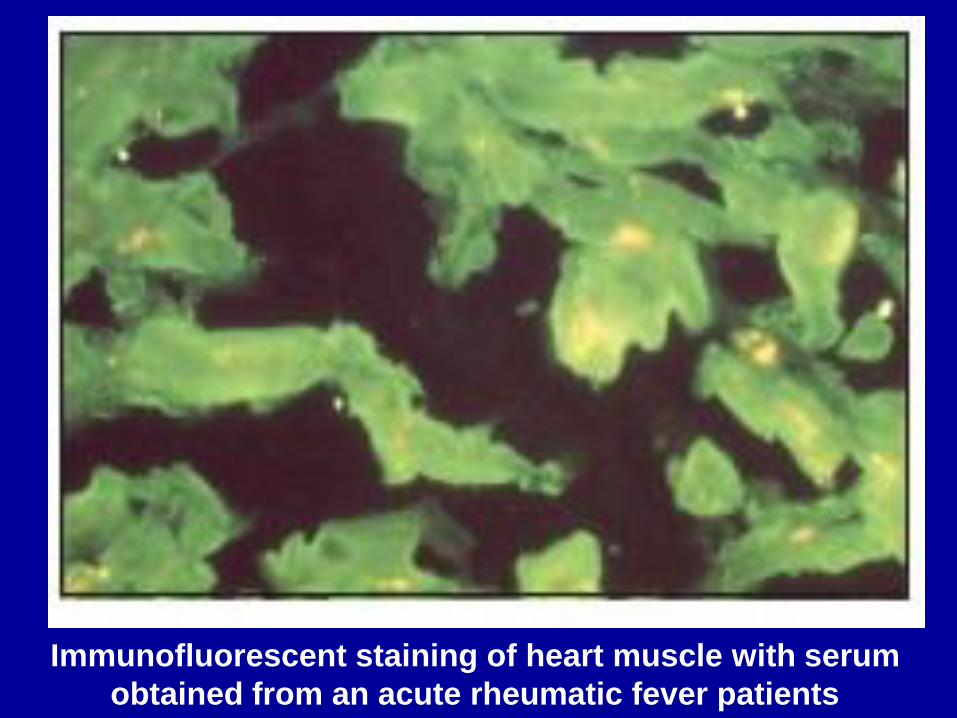
Immunofluorescent staining of heart muscle with serum
obtained from an acute rheumatic fever patients
Pathophysiology
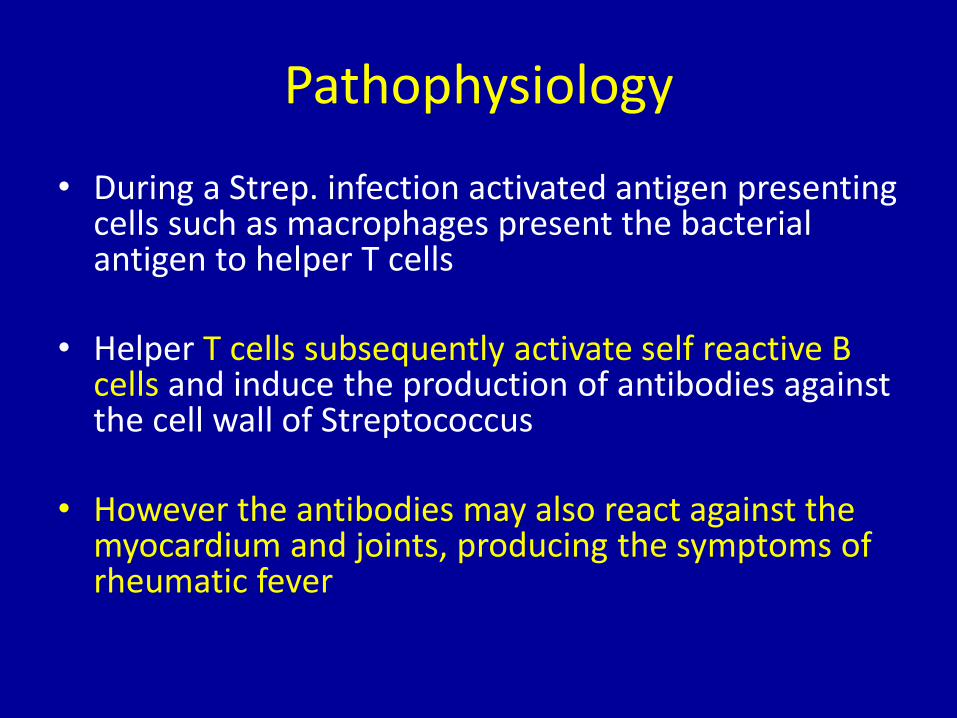
• During a Strep. infection activated antigen presenting cells such as macrophages present the bacterial antigen to helper T cells
• Helper T cells subsequently activate self reactive B cells and induce the production of antibodies against the cell wall of Streptococcus
• However the antibodies may also react against the myocardium and joints, producing the symptoms of rheumatic fever
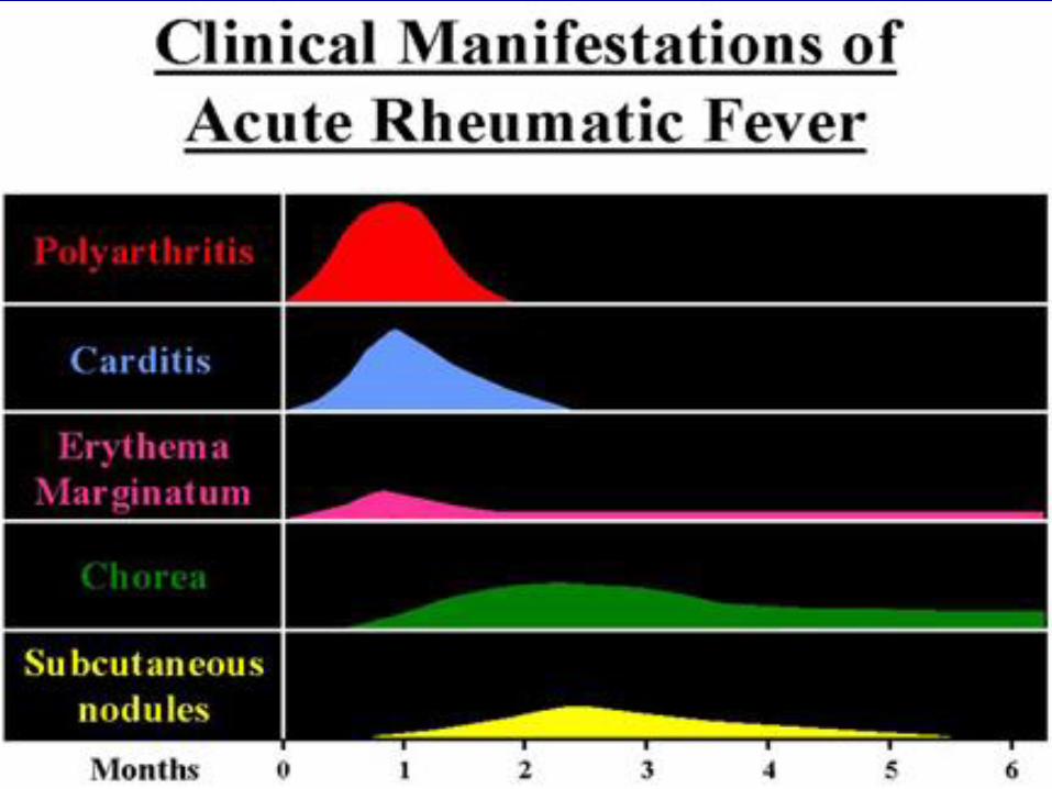
Clinical Presentation
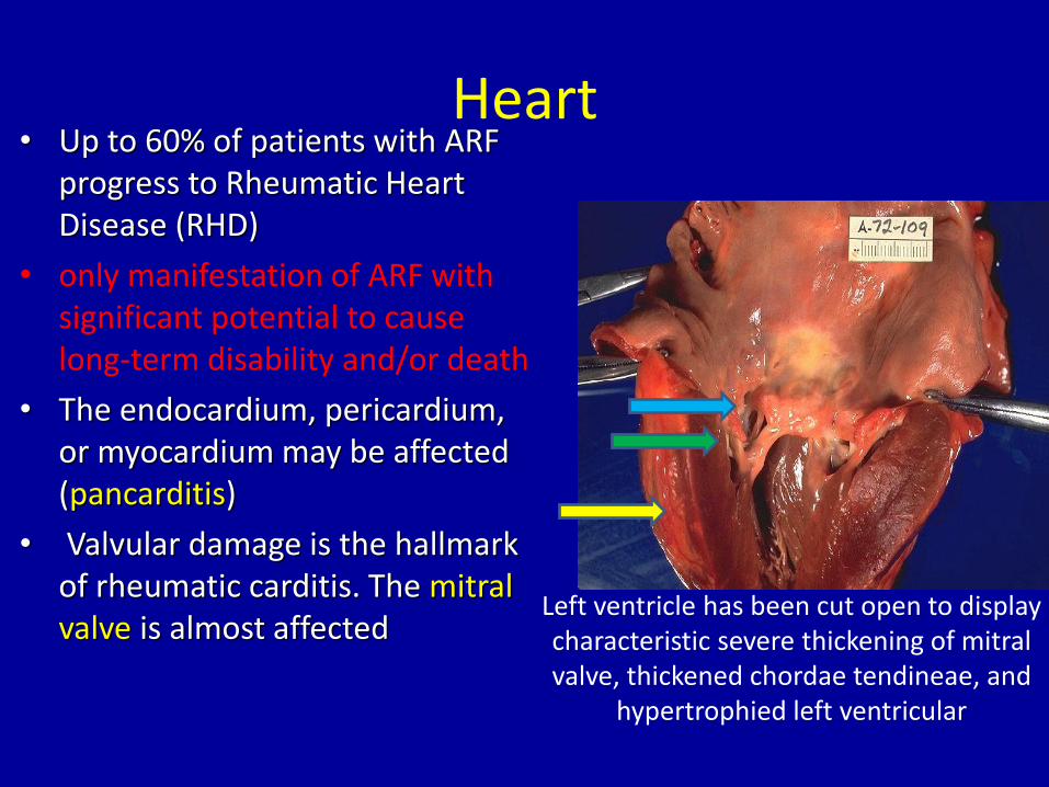
Heart • Up to 60% of patients with ARF
progress to Rheumatic Heart Disease (RHD)
• only manifestation of ARF with significant potential to cause long-term disability and/or death
• The endocardium, pericardium, or myocardium may be affected (pancarditis)
• Valvular damage is the hallmark of rheumatic carditis. The mitral valve is almost affected
Left ventricle has been cut open to display characteristic severe thickening of mitral valve, thickened chordae tendineae, and
hypertrophied left ventricular
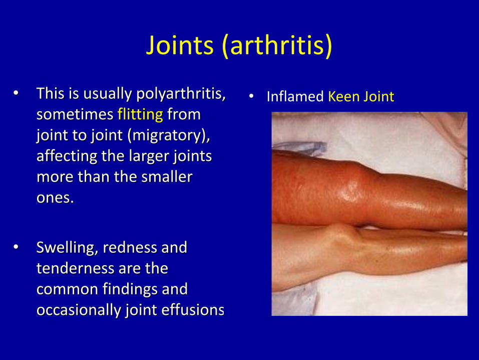
Joints (arthritis)
• This is usually polyarthritis, sometimes flitting from joint to joint (migratory), affecting the larger joints more than the smaller ones.
• Swelling, redness and tenderness are the common findings and occasionally joint effusions.
• Inflamed Keen Joint
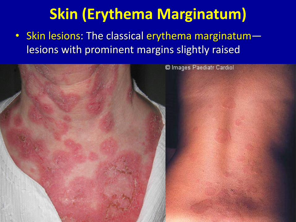
Skin (Erythema Marginatum)• Skin lesions: The classical erythema marginatum—
lesions with prominent margins slightly raised
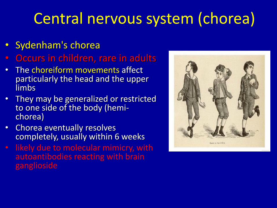
Central nervous system (chorea)
• Sydenham's chorea• Occurs in children, rare in adults• The choreiform movements affect
particularly the head and the upper limbs
• They may be generalized or restricted to one side of the body (hemi-chorea)
• Chorea eventually resolves completely, usually within 6 weeks
• likely due to molecular mimicry, with autoantibodies reacting with brain ganglioside
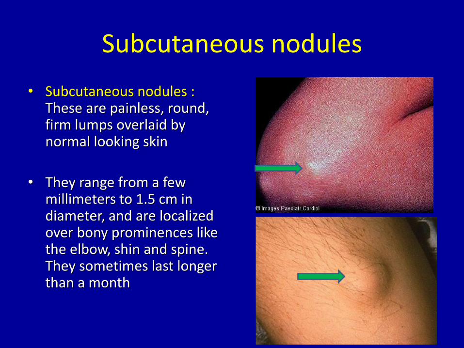
Subcutaneous nodules
• Subcutaneous nodules :These are painless, round, firm lumps overlaid by normal looking skin
• They range from a few millimeters to 1.5 cm in diameter, and are localized over bony prominences like the elbow, shin and spine. They sometimes last longer than a month
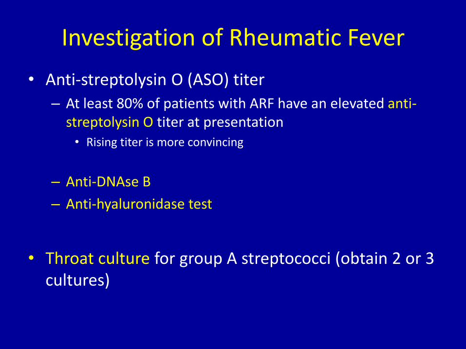
Investigation of Rheumatic Fever
• Anti-streptolysin O (ASO) titer
– At least 80% of patients with ARF have an elevated anti-streptolysin O titer at presentation
• Rising titer is more convincing
– Anti-DNAse B
– Anti-hyaluronidase test
• Throat culture for group A streptococci (obtain 2 or 3 cultures)
Rheumatic Fever – Clinical Course
• Subsequent attacks
– Increased vulnerability to reactivation of disease with subsequent strep infections
– Same symptoms with each attack
– Carditis worsens with each attack
– Heart valves are frequently deformed (mitral)
– Heart failure develops after decades
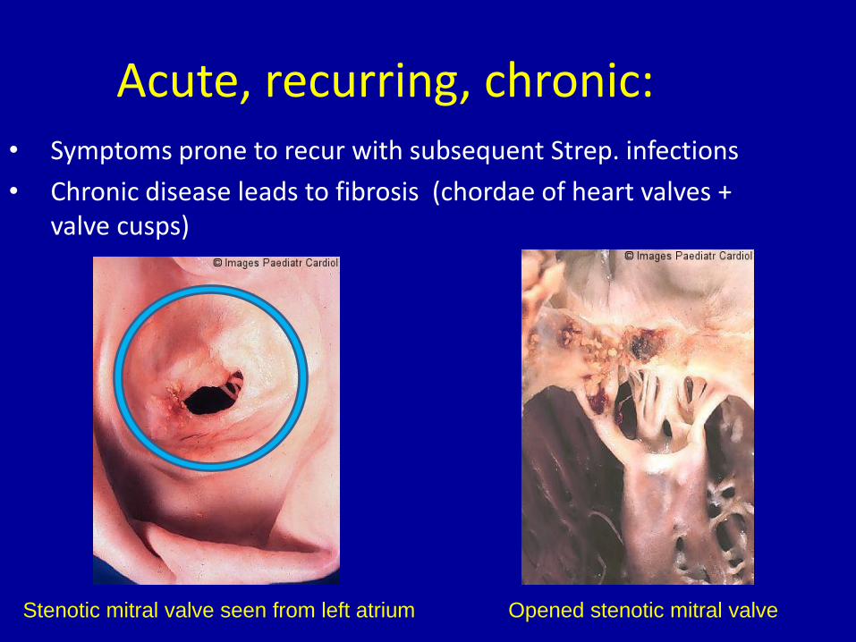
Acute, recurring, chronic:• Symptoms prone to recur with subsequent Strep. infections
• Chronic disease leads to fibrosis (chordae of heart valves + valve cusps)
Stenotic mitral valve seen from left atrium Opened stenotic mitral valve
Treatment of Rheumatic Fever
• Treat first strep throat infection with penicillin
• Treat other manifestations symptomatically
• Prophylactic long term anti-strep therapy given to anyone who has had rheumatic fever
Take home message
• Rheumatic heart disease results from cross reacting antibodies binding the heart valves
• Repeated attacks of Streptococcal throat infection over the years damage heart valves resulting in either stenotic or incompetent heart valves
• Treatment involves surgical replacement of the damaged heart valves
• In patients with rheumatic fever long term administration of penicillin is recommended for prevention of future infections by group A Streptococcus

Thank you